DIAGNOSTIC CRITERIA OF PNEUMOCONIOSIS Sartorelli P, Paolucci V
Occupational Medicine, Department of Medical Surgical and Neurological Sciences, University of Siena, Italy
Abstract
Asbestosis, silicosis and CBD are the most common pneumoconiosis. Being characterized by the presence of interstitial pulmonary fibrosis, a relevant issue is represented by the differential diagnosis with non-occupational interstitial pulmonary diseases. Epidemiological data are scarce due to the lack of standardized diagnostic criteria, varied physician awareness and training, the limitations of the available data sources (death certificates, hospital records, medical surveillance, notification to the public insurance system) and the long latency period between exposure and onset of the disease. Diagnosis of pneumoconiosis requires the recognition of occupational exposure, the existence of an adequate latency period, the exclusion of extraprofessional causal factors, and the presence of compatible clinical, radiological and functional respiratory aspects. The CT scan performed with high-resolution technique (High Resolution Computed Tomography - HRCT) allows to confirm radiological signs of pneumoconiosis highlighting the early stages that cannot be diagnosed with the standard chest radiography. Indeed, this technique is much more sensitive than standard Rx even if it is difficult to determine in which extent, given the variability of data reported in the literature. The main limitation of this method is the huge variability intra and inter-operator. Therefore, in the last ten years several groups have tried to create an interpretation model for the classification of pneumoconiosis. ICOERD is a classification scheme for digital HRCT built on a model similar to the ILO classification for the standard chest radiogram. It consists of several elements for classification: small opacity with regular or irregular shape/linear to which must also be defined the location and the profusion, large opacities, ground glass, honeycombing and emphysema.
Pathological diagnosis of asbestosis is placed in the presence of a diffuse pulmonary fibrosis with a particular pattern of asbestos bodies and/or fibers attesting a relevant exposure. In asbestosis the interstitial fibrosis is located in the basilar and subpleural regions as in idiopathic pulmonary fibrosis (IPF) which is the main differential diagnosis. In addition to the presence of asbestos bodies and fibers, histologically asbestosis differs from IPF in the poor inflammatory component and the lesser presence of fibroblastic foci. The very early stages, characterized by bronchiolitis, pose a difficult diagnostic problem because lesions are very similar to those smoked-related. In any case, peribronchiolar fibrosis does not represent asbestosis.
The most common form of silicosis (chronic simple silicosis) occurs after a latency period of at least 10 years and can be as long as 40 years. A more rapid onset is caused by intense exposure: the clinical appearance is similar, but the latency is shorter (5-10 years). In asbestosis and silicosis the most important factor in determining fibrosis is the cumulative dose.
The CBD is a granulomatous disease similar to sarcoidosis, caused by cell-mediated sensitization to the metal.
The management of cases of pneunoconiosis does not differ from that of other pulmonary fibrosis if not for two things: the removal from exposure and the need of primary and secondary prevention in exposed co-workers.
Introduction
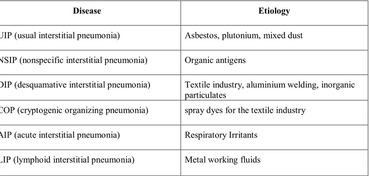
Occupational interstitial lung diseases (OILD), historically defined as pneumoconiosis, represent a clinically heterogeneous group of diseases whose causative agents are continuously being studied because of the wide industrial use of innovative materials. As the other occupational diseases also pneumoconiosis do not differ clinically from non-professional forms, making their early diagnosis of great importance for the implementation of primary and secondary prevention measures. Epidemiological data are scarce due to the lack of standardized diagnostic criteria, varied physician awareness and training, the limitations of the available data sources (death certificates, hospital records, medical surveillance, notification to the public insurance system) and the long latency period between exposure and onset of the disease (1). Occupational exposures may not only cause pneumoconiosis, but they are also able to increase the risk of idiopathic interstitial lung diseases (table 1).
Table 1: Idiopathic interstitial lung diseases and relative occupational etiology (from Glazer and Newman 2004, modified).
Disease Etiology
UIP (usual interstitial pneumonia) Asbestos, plutonium, mixed dust NSIP (nonspecific interstitial pneumonia) Organic antigens
DIP (desquamative interstitial pneumonia) Textile industry, aluminium welding, inorganic particulates
COP (cryptogenic organizing pneumonia) spray dyes for the textile industry AIP (acute interstitial pneumonia) Respiratory Irritants
In the past, several authors investigated the relationship between occupational exposure and interstitial lung diseases (2, 3). From the data of the European Register of interstitial lung diseases the prevalence of pneumoconiosis is between 4 and 18% and the incidence between 13 and 19% (4). Epidemiological data obtained from the General Practice Research Database in the period 1997-2008 showed an incidence of pneumoconiosis in the general population of 2.7 per 100,000 person-years with an increasing trend for asbestosis until 2005 and decreasing for the other OILD since 2003 (5). Examining a new case of interstitial lung disease (ILD) without known cause, it is necessary to consider in the first instance the professional medical history and only in the case of absence of occupational exposures to define the disease as idiopathic. In this sense, there are elements of the medical history particularly useful in directing the diagnosis towards pneumoconiosis. Among these we list the presence of a number of ILD cases in a population of workers, the young age, an adequate period of latency, the work-related exacerbation of symptoms and a slower progression than expected (1). Several studies confirm the importance of occupational history for the purposes of a correct diagnosis of pneumoconiosis. In a series of unselected patients with ILD the professional exposure to causative agents was not identified in 25% of cases for the lack of a proper occupational history, the occupational disease being diagnosed by mineralogical analysis (6). Other studies have highlighted the significant underestimation of berylliosis (chronic beryllium disease - CBD), often misdiagnosed as sarcoidosis. A retrospective study conducted in Germany on 84 patients suffering from sarcoidosis and not investigated under the professional point of view showed that among them 34 had some exposure to beryllium and an appropriate latency period for the development of CBD (7). For some exposures, in particular those that involve the immune system with a process of sensitization as CBD, the latency period may be shorter (weeks or months). For these agents the temporal association between symptoms and exposure can provide an important clue to the diagnosis (8). For other exposures such as asbestos or silica the latency period is measured in decades. Finally, it should be considered that non-occupational or indirect exposures could play an important role and require targeted surveys. Examples of this are cases of CBD in housewives and mesothelioma in the wives of workers exposed to asbestos.
Following the correct occupational history the evaluation of pneumoconiosis continues according to that of non-professional forms with laboratory tests, pulmonary function tests and radiological examinations. In particular, the laboratory tests are intended to exclude other possible causes as infections and connective tissue diseases. Results of pulmonary function tests may vary from predominantly obstructive forms to restrictive and mixed forms. Similarly the radiological changes vary with the exposure (9). Finally, for atypical forms or occupational exposures to agents new or poorly characterized the lung biopsy with mineralogical analysis of lung tissue should be considered (10).
Important pathogenetic factors include the individual anatomical and physiological characteristics that affect the deposition and the lung clearance of dust particles (e.g. the efficiency of nasal filtering and mucociliary clearance, the overall length of the respiratory tree, the cigarette smoking and genetic factors) (11). Other factors are dependent on the characteristics of the causative agent, such as size, solubility and biopersistence (12). Depending on the physico-chemical characteristics the agent can behave both as an immune antigen and as hapten and therefore cause different immunological reactions. In general the pathogenic agent triggers a chronic inflammatory process that leads to fibrosis. In the case of fibers this is particularly linked to the length/diameter ratio (12). From the therapeutic point of view non-professional interstitial lung fibrosis are treated with supportive pharmacological therapies and rehabilitation. However, for some forms such as silicosis and asbestosis there is a clear association between the progression and the cumulative exposure (13) and the removal of the pathogenic agent is essential to limit the parenchymal damage. Furthermore, the diagnosis of pneumoconiosis represents a sentinel event and needs the implementation of primary and secondary prevention programs within the cohort of exposed workers (14, 15). The classification of pneumoconiosis distinguishes forms caused by inorganic fibers, non-fibrous dust and metals.
Actually the diagnosis of pneumoconiosis requires the recognition of occupational exposure, the existence of an adequate latency period, the exclusion of extraprofessional causal factors, and the presence of compatible clinical, radiological and functional respiratory aspects (1). Table 2 shows the main causative agents of pneumoconiosis with the relevant exposure scenario.
Table 2: Pneumoconiosis induced by inorganic fibers (fibrous inorganic dust - IFD), non-fibrous inorganic powders (inorganic non-fibrous dust - INFD) and metals (M).
Agent Occupational exposure
Asbestos (IFD) Mining, asbestos cement production, manufactured articles, thermo-electro-acoustic insulation, contaminant in talc or vermiculite, rail, maritime and automotive sectors
Crystalline silica (INFD) Mining, building materials, tunnel excavation, road maintenance, production and processing of ceramic and glass
Coal dust (INFD) Mining
Berillyum (M) Nuclear weapons, electronics, ceramics, metal recycling, dental prosthesis, alloy processing, aerospace and defense industry Cobalt (M) Production of hard metals, diamond polishing, grinding, use and
Imaging
The classification scheme of radiographic images proposed by International Labour Office (16) was initially designed to codify the radiographic abnormalities of pneumoconiosis in a simple and reproducible way for epidemiological purposes. Subsequently, the scope has been extended to the clinical field (screening and health surveillance of exposed to mineral dust). The classification does not define pathological entities or estimate the suitability of the work for medico-legal purposes. Sometimes the interpretation of a radiogram may vary between two different readers (inter-observer variation, the minimum number of readers being two) or the single reader at two different times (intra-observer variation). The main factors that lead to this inconsistency are the poor quality of the radiograph and the lack of experience in the use of the ILO classification (17). The National Institute for Occupational Safety and Health (NIOSH) has therefore helped to reduce this problem conducting training programs for the use of ILO classification with acquisition, after an examination of the title of NIOSH Certified B Reader. The CT scan performed with high-resolution technique (High Resolution Computed Tomography - HRCT) allows to confirm radiological signs of pneumoconiosis highlighting the early stages that cannot be diagnosed with the standard chest radiography. Indeed, this technique is much more sensitive than standard Rx even if it is difficult to determine to which extent, given the variability of data reported in the literature. The main limitation of this method is the huge variability intra and inter-operator. Therefore, in the last ten years several groups have tried to create an interpretation model for the classification of pneumoconiosis. In 2001 a Finnish group proposed a new HRCT score for the classification of asbestos-related parenchymal opacities (18). The interobserver variability was quantified by submitting CT independently to three experienced radiologists who knew only the patient's age, at time zero and after more than a month. The asbestos-induced fibrotic changes (septal thickening, pleural sub lines, parenchymal bands and honeycombing) were divided semiquantitatively into 6 classes on a scale from 0 to 5. If the radiologist was not able to classify the lesions in one of the predefined classes, he should use 5 subcategories (0.5, 1.5, etc.). The class 2 (mild fibrosis) was regarded as the threshold for the diagnosis of asbestosis in HRCT. Signs of emphysema were classified in both lungs separately, using a similar scale from 0 to 5 without subcategories. Once classified the CT with this score index a NIOSH B reader read standard radiographs of the same patients classifying them with ILO 2000. Between CT and Rx ILO score for the grading of fibrosis a good correlation was observed, suggesting that the use of a semiquantitative score in CT could represent a reliable method. A similar semiquantitative HRCT score for the classification of pleural plaques was developed few years later by a group of Brazilian scientists with an excellent inter-observer agreement (19). Three radiologists examining a Japanese population consisting of 21 miners and 6 non-occupationally exposed to mineral dust, compared independently the radiograms of the chest with the respective CT by applying the ILO 1980 to the standard Rx and the model Hosoda-Shida on CT. The concordance of results in at least two of the three readers (both in terms of profusion and morphology of opacity) was 96% (20). In 2004, German Hering et al. following the ILO model selected a set of CT images still to date reference for insurance purposes for the classification of pneumoconiosis (The German standardized HRCT classification of occupational and environmental thoracic diseases). The following year, an international group of experts coordinating the various models published the International Classification of high resolution CT for Occupational and Environmental Respiratory Diseases (ICOERD). This international working group has provided over the years the validation of the method, in particular with regard to the inter-individual variability of radiologists.
The ICOERD classification
The CT scan performed with high-resolution technique (High Resolution Computed Tomography - HRCT) allows to confirm radiological signs of pneumoconiosis highlighting the early stages that cannot be diagnosed with the standard chest radiography. Indeed, this technique is much more sensitive than standard Rx even if it is difficult to determine to which extent, given the variability of
data reported in the literature. The main limitation of this method is the huge variability intra and inter-operator. Therefore, in the last ten years several groups have tried to create an interpretation model for the classification of pneumoconiosis. ICOERD is a classification scheme for digital HRCT built on a model similar to the ILO classification for the standard chest radiogram. It consists of several elements for classification: small opacity with regular or irregular shape/linear to which the location and the profusion must also be defined, large opacities, ground glass, honeycombing and emphysema. The severity of pneumoconiosis is layered in four grades (0-3) to be assigned to each region of each lung (upper, middle, bottom left and right). Pleural lesions must be distinguished between parietal and visceral pleura and must be characterized by extension, thickness and presence of calcifications with relative locations. The evaluation of the use of CT images of reference has been documented by Suganuma and other members of the 'ICOERD Study Group (21). The study pointed out a good inter-observer agreement (8 NIOSH B reader) for reading the parenchymal opacities, emphysema and areas of honeycombing and moderate areas of groundglass. On the other hand the concordance for pleural lesions was not satisfactory. All this suggests a better applicability of the classification method to the parenchymal lesions than the pleural ones.
Asbestosis
All types of fibers can cause asbestosis as well as other asbestos-related diseases, provided that the exposure was sufficient. The dose that can potentially cause asbestosis is higher than those necessary to determine other asbestos-related diseases and it is classically the dose of at least 25 fibers/ml/year, even if it is generally accepted that in some cases the disease appears even at lower exposures (1). Albeit very slow compared to what happens in other interstitial lung diseases. The latency period is long, going from 15 to 40 years, while in many cases the disease progresses over the time even after the end of the exposure.
The definition of asbestosis in pathological terms is that of an interstitial pulmonary fibrosis associated with the presence of asbestos bodies (AB) and fibers in the lung (22). The College of American Pathologists (CAP) and the Pulmonary Pathology Society (PPS) defined the pathological features necessary to make a differential diagnosis between idiopathic pulmonary fibrosis (Usual Interstitial Pneumonia - UIP) and asbestos-related pulmonary fibrosis, since both have a bilateral basal subpleural distribution (23). However asbestosis, unlike the UIP, is associated with minimal inflammation, which justifies the slow progression and poor fibroblast activation which always involves the visceral pleura.
However, especially at an early stage it is difficult to make a diagnosis of asbestosis in smokers or in workers with simultaneous exposure to other mineral dusts (silica, iron or aluminum hydroxide) Actually asbestos exposure can be associated with fibrosis of the bronchiolar walls and the alveolar ducts, sometimes incorrectly defined peribronchiolar fibrosis and therefore likely to be confused with the fibrosis of the alveolar walls of the bronchioles or "true peribronchiolar." The American Thoracic Society (ATS) and the European Respiratory Society (ERS) suggested the term respiratory bronchiolitis-associated interstitial lung disease (RB-ILD) to identify a group of diseases histologically characterized by fibrosis of the bronchiolar walls (24). These lesions were observed for the first time in experimental animals exposed to asbestos fibers; in many cases they showed an evolution toward the diffuse fibrosis. Afterwards cases of this type were observed at first in heavy smokers and subsequently also in workers exposed to asbestos or to non-coniotic mineral dusts. Bellis et al. (25) looked for small airways lesions the results of post mortem in a group of 199 dead subjects. Following the CAP and NIOSH standards they defined the association of small airways lesions with fiber concentrations and AB in lung tissue compatible with occupational exposure as an early stage of asbestosis called asbestosis grade 1 (AG 1). In this way they identified AG 1 in 15% of the studied subjects. The presence of AG 1 was significantly correlated with bilateral pleural plaques, large numbers of AB and history of exposure to asbestos. The study concluded that AG 1 should be considered an asbestos-related disease and fundamental for the
diagnosis of professional lung cancer. However more recently CAP rejected the definition of AG 1 because in asbestosis the fibrosis tends to move towards the outside of the pulmonary acinus and to involve the pulmonary parenchyma with connective bridges, while in these diseases, especially in the smoking-related forms, the progression to diffuse fibrosis like the classic asbestosis, is not demonstrated, but a complete remission after cessation of exposure to the pathogenic agent was observed. Therefore today, as suggested by CAP and PPS, it is preferable to define the bronchiolar walls asbestos-induced fibrosis as asbestos airways disease (23). In 2004 ATS revised the criteria for the diagnosis of asbestosis and other non-neoplastic asbestos-related diseases summarized as follows (26):
- Evidence of structural changes demonstrated by Imaging
Histology
- Evidence of plausible causation demonstrated by Occupational history
Markers of exposure (e.g. pleural plaques) Recovery of AB
- Exclusion of alternative diagnoses
- Evidence of functional impairment demonstrated by Signs and symptoms (e.g. crackles)
Restrictive or obstructive patterns in ventilatory function Impaired gas exchange (1)
Inflammation (e.g. by BAL) Exercise testing (1)
(1) diffusion capacity is generally reduced late so in the early stages oximetry during the exercise test represents a more sensitive method.
Regarding the radiological diagnosis of asbestosis a numbert of studies demonstrated the higher sensitivity of HRCT in detecting asbestos-related pleural and parenchymal lesions compared to the standard chest radiography (18).
The most common signs of asbestosis detectable on HRCT include (27) - findings of fibrosis,
- honeycombing in advanced disease,
- subpleural dotlike opacities in early disease, - subpleural lines,
- ab) parietal thickening or plaques,
- ab) parenchymal bands particularly in association oleural thickening,
- earliest abnormalities posterior and basal, - ground-glass opacities.
a) most common signs
b) signs more useful in the differential diagnosis
For the diagnosis of asbestosis HRCT should normally be performed in the prone position so that the posterior regions of the lung, typically affected early, are not within the signs of stasis. None of the detectable signs on HRCT can be considered specific, so the diagnosis is easier when the lesions are not isolated or even better if they are bilateral and symmetrical. The association with pleural plaques, though not required, is helpful to the diagnosis. Punctate opacities and subpleural lines (which are curved and parallel to the pleura) reflect the presence of peribronchiolar fibrosis and are
characteristic of the initial forms . The lines represent subpleural intralobular lesions, sometimes reflecting the presence of atelectasis especially in proximity of the pleural plaques.
The problems related to the diagnosis of early lesions of asbestos by HRCT was presented in a study on 72 asymptomatic workers of a shipbuilding industry exposed to asbestos with negative standard Rx (ILO 0/0 or 0/1) and a control group consisting of 20 patients not exposed (28). In 28% of cases HRCT showed parenchymal lesions consisting in subpleural lines and thickened inter- and intralobular lines. Pleural and parenchymal lesions were significantly less frequent in the control group, while the duration of exposure was significantly longer in the workers with parenchymal involvement. These results show that HRCT is able to detect pleural-parenchymal lesions before clinical symptoms and signs in the standard Rx. However such lesions are not specific, allowing the diagnosis on a probabilistic basis only when the exposure is proved. On the other hand the negativity of HRCT cannot exclude the presence of early signs of asbestosis. In a study on 25 cases of asbestosis diagnosed histologically, 9 patients (36 % of the population) had negative HRCT (29). Quantitative and qualitative indicators of exposure to asbestos used in the diagnosis of asbestosis Asbestosis is proved by the presence of more than one criterion associated with exposure to asbestos significant because none of the clinical and radiological criteria is specific. For the diagnosis the undisputed key role of biomarkers of past exposure (AB and asbestos fibers) is no longer limited to the lung tissue (30) but also to the bronchoalveolar lavage fluid (BALF). Moreover, even the pathology of asbestosis is based on the combination of interstitial fibrosis and a specific concentration of AB in the lung tissue. In this sense as a demonstration of past exposure to asbestos for diagnostic purposes a dry tissue concentration of AB exceeding 1000/g was established (31). According to diagnostic criteria for asbestosis set by CAP and PPS (23) 2 or more AB per square centimeter of a section of lung of 5 microns thick are needed for diagnostic confirmation together with a precise fibrotic pattern. Conversely a lower number of AB does not exclude the diagnosis. Actually AB represent a specific, but not very sensitive exposure marker. Furthermore a relationship between concentration of AB and amphibole exposure, but less with chrysotile exposure was showed (32). Moreover, the clearance of AB is probably faster than amphibole fibers (33). Mollo et al. (34) determined the AB concentration on lung tissue in 924 unselected surgical cases of lung cancer and carried out the pathological diagnosis of the tissue not infiltrated by tumor. This resulted in a diagnosis of asbestosis in 6% of the population examined with an estimate of asbestos-related lung cancer in Italy of 2000 cases/year. In a similar study (35) the AB concentration in lung tissue was determined by optical microscopy in 87 cases of lung cancer, after definition of exposure history. In 5 cases out of 6 occupational exposure was confirmed by a high AB concentration, while in one case a high AB concentration corresponded to an anamnestically doubtful exposure. This diagnostic procedure, implying the availability of a sufficient amount of lung tissue obtained in vivo with the open biopsy is generally not justified, and it would be substantially limited to cases in which lung resection for carcinoma is carried out.
The determination of fibers by electron microscopy may allow in vivo both a qualitative and semiquantitative exposure characterization, and to facilitate the differential diagnosis between UIP and asbestosis, although there is evidence that in cases with presence of honeycombing the method is invalidated by a high number of false positives and false negatives (36). In addition, electron microscopy measurement of fiber concentration in the lung tissue and BALF is not sufficiently reproducible, so it is difficult to compare results obtained in different laboratories. However, using standardized methods, even in the presence of different results, fiber concentrations that are high or low in a laboratory are equally high or low in another laboratory. In practice this means that for a more reliable examination each laboratory should analyze a number of samples in order to create a range of values. Sartorelli et al. (37) measured fiber concentrations by transmission electron microscopy (TEM) in BALF of 108 subjects occupationally exposed to asbestos and 57 controls finding a statistically significant difference between the two groups with a 100% concordance between exposure and the fiber concentration. All subjects with occupational exposure were
positive with the confidence interval lower limit of exposed workers 10 times higher than the confidence interval upper limit of the controls, while the concentration of AB in BALF was below the detection limit in 17.8% of exposed subjects. Similar conclusions have emerged from a subsequent study (33), which expanded considerably the number of cases previously considered. In a study involving the repetition of BAL with the mineralogical analysis in 22 occupationally exposed subjects, differences in the fibers concentration in the liquid of the first and second BAL was not statistically significant, confirming the good reproducibility of the method (38). The uncertainties related to the mineralogical analysis of BALF (mainly due to the high coefficient of variation for low fiber concentrations) suggested, however, that in exposure assessment this biomarker is more appropriate to a qualitative/categorical approach rather than a quantitative one. More recently the reliability of dose and effect markers in the health monitoring of workers previously exposed to asbestos was studied in a population of 158 workers of which 49 (31%) with asbestos-related disease (39). Using non-parametric statistical methods 6 random variables were analyzed with respect to the presence or not of asbestos-related diseases and the different working sectors, demonstrating a significative greater amphibole concentration in patients with asbestosis and in railway rolling stock insulation removal (p <0.01). There was not a correlation between mesothelin and amphiboles, chrysotile and total fibers concentration. A comparison of two analysis methods, by TEM and scanning electron microscopy (SEM), carried out on 15 samples demonstrated the significance of the linear regression between the measurements obtained with the two different techniques, but the results were different, for which it was necessary to introduce a correction factor to compare TEM/SEM data (40).
Silicosis
Silicosis occurs after inhalation exposure to crystalline silica contained in various minerals (such as quartz, cristobalite , tridymite) or to silicate powder contained in various compounds (ceramic paste, bricks) (41). From the epidemiological point of view silicosis is particularly prevalent in countries with low and middle income. China has the largest number of patients with silicosis with more than 500 000 cases recorded between 1991 and 1995 and more than 6000 new cases and 24,000 deaths reported annually (42). In the Brazilian gold mine in Minas Gerais they reported more than 4,500 workers with silicosis between 1978 and 1998 (43). In South Africa among the gold miners dead for accidents and autopsied between 1975 and 2007, the percentage of silicosis increased from 3 % to 32 % for blacks miners and from 18% to 22% for white miners (44). Silicosis is still a concern for the health at work even in developed countries. In this regard between 1990 and 1993 about 600,000 workers in UK and more than 3 million in Europe were exposed to crystalline silica (45). In UK between 1996 and 2009 at least 100 new cases per year were reported (46). However, the incidence of mortality has been significantly reduced. The mortality rate in the United States fell from 8.9 per million in 1968 to 0.7 in 2004 (41).
The pathogenic mechanism of silica is represented by direct cytotoxic damage on alveolar macrophages with subsequent release of inflammatory cytokines that induce a fibroblastic proliferation. These cells give rise to the formation of nodules of concentric collagen and silica particles incorporated in fibrous capsules (47). The most common form is the simple chronic silicosis that occurs after intense and prolonged exposures and with a latency period of at least 10 years and up to 40 years (48). The accelerated silicosis, a more rapid course, occurs at higher exposures with a the latency period of 5-10 years. Clinically this form is similar to the chronic one, but the disease is often more severe. In complicated silicosis nodules are larger than one centimeter. In such a case areas of massive predominantly apical and rear fibrosis can be observed. A third clinical form that may develop after an exposure period of a few months or however less than two years takes the name of silicoproteinosis, similar to alveolar proteinosis. In this case, the radiologic appearance is that of a widespread groundglass, while inside the alveoli a PAS positive proteinaceous material can be observed (47, 49). The BALF of non-smoker patients with chronic silicosis is characterized by an excess of macrophages. With a more rapid progression an increase of
lymphocytes and neutrophils is observed (50). In BALF of exposed workers it is also possible to observe a number of macrophages containing birefringent particles that would represent quartz crystals, but which can actually be constituted by other silicates; because of this, they are not always indicative of the pulmonary burden of silica.
Exposure to silica can increase the risk of lung disease and cancer (51). Patients with silicosis have a greatly increased risk of developing active forms of tuberculosis, with a mortality that only today is decreasing thanks to the implementation of prevention and control measures (52). With regard to the relationship between silicosis and chronic obstructive pulmonary disease (COPD), increased incidence of COPD is demonstrated in subjects exposed to silica independently from tobacco smoke and from the evidence of radiological silicosis. In particular, longitudinal studies suggest that lung function loss occurs with exposure to silica at concentrations between 0.1 and 0.2 mg/m3 and that
the effect of cumulative exposure to silica on obstructive disease is independent of the silicosis. In fact, even in the absence of silicosis, with intense exposure (average of at least 30-40 years), an important functional reduction can develop (53). Exposure to silica also increases the risk of developing chronic renal failure and autoimmune diseases, particularly scleroderma, rheumatoid arthritis and Wegener's granulomatosis (54, 55). In 1997 a monograph of the International Agency for Research on Cancer (IARC) classified occupational exposure to crystalline silica as carcinogenic to humans (56). At the time a metanalysis of 16 of the largest studies conducted in populations with well-documented exposure to silica and low probability of confounding with other occupational exposures, showed a relative risk (RR) of 1.3 (95% confidence interval [CI] = 1.2-1.4). The risk of lung cancer also appeared to be significantly higher in patients with silicosis exposed to higher doses (RR = 2.3 [CI] = 2.2-2.4 in 19 studies) (57). Afterwards a large amounts of epidemiological data has been published. Pelucchi et al. (58) conducted a systematic review of epidemiological studies on exposure to silica and risk of lung cancer published after the IARC monograph arguing for a clear carcinogenic role of silica in patients with silicosis, while in the absence of silicosis the pathogenetic mechanism is still not clear. The radiological picture is generally represented by nodular opacities mainly localized in the upper and in the posterior lobes with centrilobular distribution. In about 10% of cases a hilar adenopathy is seen with characteristic peripheral calcifications (eggshell) of the lymph nodes (1). Sometimes the lymph nodes can be calcified and this pattern has been correlated from a functional point of view with reduced values of diffusion of carbon monoxide value (59). The nodules may be single, with a diameter usually less than 5 mm, or merge into an aspect of massive fibrosis. In complicated silicosis radiographically nodules are large opacities generally symmetrical, often with irregular margins and unusually calcified. Pleura adjacent to these areas may have fibrotic thickening or invaginations. By the time in the massive forms opacities tend to flow to the pulmonary hilum causing in the appearance of large areas of emphysema in the context of which it is not uncommon to observe cavitated areas of necrosis due to ischemia or tuberculosis (60, 61, 62). The HRCT is more sensitive than standard Rx in the diagnosis of silicosis that generally does not require a lung biopsy.
With regard to prevention in 1995 the World Health Organization (WHO) and International Labour Organization (ILO) established a program for the Elimination of Silicosis.
Berylliosis
The chronic beryllium disease (CBD) is a granulomatous disease similar to sarcoidosis that occurs after exposure and sensitization to beryllium. As in sarcoidosis the lung is the main organ involved, but also other organs (such as skin, the muscle- skeletal system and salivary glands) may be affected. The disease can be caused by exposure to dust or metal fumes containing pure beryllium, beryllium in low percentage or oxides of beryllium (1, 63, 64). Regarding the association between beryllium exposure and risk of lung cancer a recent review has asluded that any evidence is available because in studies it was observed that the excess mortality found depends on the other variables such as smoking of tobacco, and on the exposure period or the cumulative dose (65). The pathogenetic mechanism of the CBD lies in the ability of some molecules human histocompatibility antigen (HLA) to tie the beryllium and present it to T cells (CD4+). In particular in subjects
suffering from CBD a subregion HLA- DPB1, changed for the presence of a glutamic acid residue in position 69, was identified (66). The presence of this mutation in homozygosity was subsequently associated with an increased risk of sensitization and development of disease among the exposed (67). Given the pathogenetic mechanism it is possibile to diagnose CBD by measuring the in vitro antigen-specific cell-mediated immune response to Beryllium (blood beryllium lymphocyte proliferation test - BeLPT). Important issues on the use of the test are known (68): - BeLPT identifies beryllium sensitization and CBD before and better than any other currently
available clinical trial;
- cases of CBD identified with the BeLPT are clinically significant;
-- a subset of persons identified by BeLPT, who have not yet clinical disease, are at risk and therefore require careful monitoring and further treatment with corticosteroids;
- BeLPT can be used to improve the clinical diagnostic accuracy and correct misdiagnosis;
- BeLPT can be used for the screening of a large number of workers exposed because it is sensitive and specific and has a high positive predictive value, negative for CBD;
- in any working population studied to date BeLPT has identified beryllium sensitizations and CBD escaped to conventional screening;
- the worker populations studied using the BeLPT can help to clarify the role of genetics, exposure and dysregulation of the inflammatory cascade in the genesis of occupational lung disease.
Regarding the standardization of BeLPT on peripheral blood, two algorithms were developed with the achievement of an adequate sensitivity (66-86%) and excellent specificity (99%) (69). The BALF profiles in the CBD are characterized by a moderate increase in the number of cells that is attributable to the dramatic increase of the lymphocytes present in lung tissue as interstitial infiltrates and granulomas (50, 70). A positive correlation was found between the presence of ground-glass areas and the extension of the nodules on HRCT scan and the number of lymphocytes (71). All studies that analyze subsets of T cells show an increase in the CD4/CD8 ratio that varies from 3.7 to 7.2. Furthermore, comparing the results of BeLPT on BALF with those on the peripheral blood through the stimulation index of the peak, the first are much higher confirming a better sensitivity (50). Many studies have also demonstrated in BALF of patients with CBD a significant increase of TNF and IL-6 compared with controls (unexposed, exposed non-sensitized and sensitized) (72). Given the immune-mediated mechanism, unlike what happens in other OILDs such as silicosis and asbestosis, the trend seems less linear dependent on the dose so it is possible to find a clinically significant disease in subjects with low exposure and with considerable variability of the latency period that can last from 2 months to 40 years (63, 64). Actually the current exposure limit set by OSHA in 2 mg/m3 does not seem protective either by raising or by the development of the pathology (72).
The radiological picture of the CBD is similar to that of sarcoidosis and includes peribronchovascular small nodules, nodular thickening of interlobular septa, ground-glass abnormality and in 25% of patients mediastinal and hilar lymphadenopathy (9, 62, 73). In the presence of a beryllium exposure history, the golden standard for the diagnosis of CBD is represented by the association between the positivity of BeLPT in blood or BALF and histology. However, in the absence of lung biopsy, the diagnosis is based on the positivity of BeLPT, the presence of lymphocytosis in BALF and a consistent radiological picture and pulmonary function.
Hard metal disease (HMD)
The hard metals are alloys composed mainly of cobalt and tungsten carbide particles. Other metals such as chromium, molybdenum and titanium may be present in traces. In addition to conventional uses of cobalt in the electronics and mechanics, it is important the exposure derived from the use of metal tools for drilling and cutting and diamond polishing. The clinical spectrum of respiratory diseases from exposure to hard metals is extremely complex including hypersensitivity (asthma)
and parenchymal disease (bronchiolitis obliterans, interstitial fibrosis, hypersensitivity pneumonitis) (63). The first evidence of parenchymal disease due to exposure to hard metals dates back to 1940 (74). Cross-sectional studies conducted on workers engaged in the production of hard metals alloys have shown a prevalence of parenchymal disease from 0.7 to 13% (75, 76). A Swedish cross-sectional study reported that 16% of dental technicians exposed to the dust of cobalt, chromium and molybdenum for at least 5 years showed radiological evidence of pneumoconiosis (77). In a retrospective study they re-evaluated a population exposed to hard metals recruited with a thirty years screening in which no correlation between the intensity/duration of exposure and the onset of pulmonary fibrosis was found. In 45% of cases pathological disease progression was observed despite the employment cessation. Steroid therapy in some cases contributed to the decrease of symptoms, but it did not prove effective in stopping the progression of the pathology (78). The pathogenesis of the disease is not well defined. The immune-mediated mechanism is suggested by the inverted CD4/CD8 ratio in the BALF of some patients with HMD and the known ability of sensitization of the metal. In vivo studies in sensitized individuals (positive patch test) showed a positive lymphocyte proliferation test (79, 80). Genetics also appears to play an important pathogenic role in causing the disease. Potolicchio et al. (81) demonstrated an association between the HMD and the presence of HLA-DPB1 modified for the presence of a glutamic acid residue in position 69 as already known for the CBD. Other authors suggest a role of metals in alloys with cobalt in causing interstitial lung disease (82). Experimental tests shown that the toxicity of cobalt is increased in the presence of metal carbides such as tungsten carbide (82, 83, 84). In rats instillation of tungsten carbide-cobalt powder acutely induces a severe alveolitis and a fatal pulmonary edema similar to that observed with crystalline silica (83). Alveolitis persists for at least a month and repeated exposure provides histopathologic evidence of fibrosis (85). On the contrary instillation of cobalt or tungsten alone produces a moderate acute inflammation and minimal delayed reaction. In vitro cobalt (86) and metal carbides (87) interact with oxygen to produce activated toxic species. Since cobalt is inefficient in the transfer of electrons to oxygen, it has been suggested that the tungsten carbide may act as a carrier of electrons or is capable of moving electrons from cobalt to oxygen (87). Consequently the production of reactive oxygen species and free radicals may be responsible for lung damage. The observation that few exposed workers develop interstitial disease can be explained by individual variability in antioxidant defenses, although this theory requires confirmation. The need for an interaction between cobalt and other components in the pathogenesis of HMD was challenged for the existence of cases of interstitial lung disease in workers, who used grinding wheels with diamond discs, strongly exposed to fine powder without cobalt tungsten carbide (88, 89). Also in vitro cobalt ions are capable of causing oxidative changes even in the absence of tungsten carbide (90). Industrial Hygiene Surveys suggest that the levels of cobalt are often underestimated (91, 92). From the diagnostic point of view a typical characteristic of the HMD is the presence, in BALF and in biopsy specimens, of multinucleated strange looking giant cells. These, considered a hallmark of the disease, are common in giant cell interstitial pneumonia (63). Available data on BALF belong to 30 patients with HMD and 19 exposed subjects reported as clinical cases (93, 94). BALF in patients with HMD shows a marked increase in the number of cells. The cell model is mixed (lymphocytes, neutrophils and eosinophils) with varying percentages of these three cell types. The different cell models in BALF were attributed to the different stages of the disease. In the studies cited the CD4/CD8 ratio was available for 7 patients and ranged from 0.1 to 1.6 (95, 96). Giant cell counts in 20 subjects showed a range from 0.1 to 10% (93, 97, 98). The inflammatory cells are increased and the lymphocyte count may be normal or increased with helper/suppressor inverted ratio (97, 99). The persistence of alveolitis and a high number of eosinophils in BALF despite the cessation of exposure may suggest a worse prognosis (99). Although the BALF may be useful in the diagnostic phase for the presence of characteristic elements such as a high cellularity and the presence of giant cells, currently there is not sufficient evidence to support its diagnostic utility in a clinical setting (50). The radiologic findings are nonspecific and characterized by ground-glass, reticular opacities, traction bronchiectasis and areas
of parenchymal consolidation. A prevalence in the lower lobes was described. The honeycombing is a late and unusual finding (63, 100).
References
1. Glazer CS, Newman LS. Occupational interstitial lung disease. Clin Chest Med 2004; 25: 467-478.
2. Baumgartner KB, Samet JM, Coultas DB, et al. Occupational and environmental risk factors for idiopathic pulmonary fibrosis: a multicenter case-control study. Am J Epidemiol 2000; 152: 307-315.
3. Scott J, Johnston I, Britton J. What causes cryptogenic fibrosing alveolitis? A case-control study of environmental exposure to dust. BMJ 1990; 301: 1015-1117.
4. Thomeer MJ, Costabe U, Rizzato G, et al. Comparison of registries of interstitial lung diseases in three European countries. Eur Respir J Suppl 2001; 32: 114s-118s.
5. Amar RK, Jick SS, Rosenberg D, et al. Incidence of the Pneumoconioses in the United Kingdom General Population between 1997 and 2008. Respiration 2012; 84: 200-206.
6. Monso E, Tura JM, Marsal M, et al. Mineralogical microanalysis of idiopathic pulmonary fibrosis. Arch Environ Health 1990; 45: 185-188.
7. Müller-Quernheim J, Gaede KI, Fireman E, Zissel G. Diagnoses of chronic beryllium disease within cohorts of sarcoidosis patients. Eur Respir J 2006; 27: 1190-1195.
8. Burge P. How to take an occupational exposure history relevant to lung disease. In: Hendrick D, Burge P, Beckett W, et al. Occupational disorders of the lung, WB Saunders Editore, Philadelphia, 2002.
9. Flors L, Domingo ML, Leiva-Salinas C, et al. Uncommon occupational lung diseases: high-resolution CT findings. Am J Roentgenol 2010; 194: W20-W26.
10. Churg A. Mineralogic analysis of lung tissue. In: Hendrick D, Burge P, Beckett W, et al. Occupational disorders of the lung, WB Saunders Editore, Philadelphia, 2002.
11. Nemery B, Bast A, Behr J, et al. Interstitial lung disease induced by exogenous agents: factors governing susceptibility. Eur Respir J Suppl 2001; 32: 30s-42s.
12. Mossman BT, Lippmann M, Hesterberg TW, et al. Pulmonary endpoints (lung carcinomas and asbestosis) following inhalation exposure to asbestos. J Toxicol Environ Health B Crit Rev 2011; 14: 76-121.
13. Becklake MR. Asbestos and other fiber-related diseases of the lungs and pleura: distribution and determinants in exposed populations. Chest 1991; 100: 248-254.
14. Aldrich TE, Leaverton PE. Sentinel event strategies in environmental health. Annu Rev Public Health 1993; 14: 205-217.
15. Rutstein DD. The principle of the sentinel health event and its application to the occupational diseases. Arch Environ Health 1984; 39: 158.
16. ILO, International Labour Office. (2002). Guidelines for the use of ILO international classification of radiographs of pneumoconioses. Available from:
http://www.ilo.org/wcmsp5/groups/public/---ed_protect/---protrav/---safework/documents/publication/wcms_108568.pdf
17. Parker JE. Radiological criteria: The use of chest imaging techniques in asbestos related diseases. In: Parker JE. Asbestos, asbestosis and cancer, proceedings of an international expert meeting, Finnish Institute of Occupational Healh (FIOH), Helsinki, 1997.
18. Huuskonen O, Kivisaari L, Zitting A, et al. High-resolution computed tomography classification of lung fibrosis for patients with asbestos-related disease. Scand J Work Environ Health 2001; 27: 106-112.
19. Meirelles GS, Kavakama JI, Jasinowodolinski D, et al. Pleural plaques in asbestos-exposed workers: reproducibility of a new high-resolution CT visual semiquantitative measurement method. J Thorac Imaging 2006; 21: 8-13.
20. Suganuma N, Kusaka Y, Hosoda Y, et al. The Japanese Classification of Computed Tomography for Pneumoconioses with Standards films: Comparison with the ILO International Classification of Radiographs for Pneumoconioses. J Occup Health 2001; 43: 24-31.
21. Suganuma N, Kusaka Y, Hering KG, et al. International CT Classification Study Group. Int Arch Occup Environ Health 2006; 79: 472-476.
22. Aberle DR, Gamsu G, Ray CS, Fenerstein IM. Asbestos-related pleural and parenchymal fibrosis: detection with high-resolution CT. Radiology 1988; 166: 729-734.
23. Roggli VL, Gibbs AR, Attanoos R, et al. Pathology of asbestosis - An update of the diagnostic criteria: Report of the asbestosis committee of the College of American Pathologists and Pulmonary Pathology Society. Arch Pathol Lab Med 2010; 134: 462-480.
24. American Thoracic Society/European Respiratory Society International Multidisciplinary Consensus. Classification of the Idiopathic Interstitial Pneumonias. Am J Resp Crit Care Med 2002; 165: 277-304.
25. Bellis D, Andrion A, Delsedime L, Mollo F. Minimal pathologic changes of the lung and asbestos exposure. Hum Pathol 1989; 20: 102-106.
26. American Thoracic Society. Official Statement: Diagnosis and initial management of non malignant diseases related to asbestos. Am J Resp Crit Care Med 2004; 170: 691-715.
27. Webb WR, Muller NL, Naidich DP. High-Resolution CT of the lung, 3rd Edition, Lippincot-Raven Editore, Philadelphia-New York, 2001.
28. Falaschi F, Boraschi P, Antonelli A, et al. Diagnosis with high resolution computerized tomography of early asbestos-induced diseases. Radiol Med 1993; 86: 220-226.
29. Gamsu G, Murray KA, Webb WR, et al. High-resolution computed tomography sampling for detection of asbestos-related lung disease. Acad Radiol 1995; 2: 111-115.
30. American Thoracic Society. Medical Selection of the American Lung Association: The diagnosis of nonmalignant diseases related to asbestos. Am Rev Respir Dis 1986; 134: 363-368. 31. Churg A: Current issues in the pathologic and mineralogic diagnosis of asbestos-induced
diseases. Chest 1983; 84: 275-280.
32. Case BW: Biological indicators of chrysotile exposure. Ann Occup Hyg 1994; 38: 503-518. 33. Romeo R, Scancarello G, Cassano P, et al. Assessment of asbestos exposure via mineralogical
analysis of bronchoalveolar lavage fluid. Med Lav 2004; 95: 17-32.
34. Mollo F, Magnani C, Bo P, et al. A pathologic study of 924 unselected cases. Am J Clin Pathol 2002; 117: 90-95.
35. Sartorelli P, Muzzupappa C, Romeo R, et al. Attribution of lung cancer to occupational asbestos exposure. G Ital Med Lav Erg 2006; 28: 428-429.
36. Schneider F, Sporn TA, Roggli VL. Asbestos fiber content of lungs with diffuse interstitial fibrosis: An analytical scanning electron microscopic analysis of 249 cases. Arch Pathol Lab Med 2010; 134: 457-461.
37. Sartorelli P, Scancarello G, Romeo R, et al. Asbestos exposure assessment by mineralogical analysis of bronchoalveolar lavage fluid. J Occup Environ Med 2001; 43: 872-881.
38. Sartorelli P, Romeo R, Scancarello G, et al. Measurement of asbestos fibre concentrations in fluid of repeated bro-choalveolar lavages ok exposed workers. Ann. Occup.Hyg 2007; 51: 495-500.
39. Montomoli L, Romeo R, Sisinni AG, et al. Dose and effects biomarkers in the diagnosis of asbestos-related diseases and health surveillance of exposed workers. G It Med Lav Erg 2012; 34 (Suppl.3): 577-580.
40. Sartorelli P, Romeo R, Pescaglini M, et al. Determination of asbestos fiber concentration in brochoalveolar lavage fluid: comparison between trasmission and scansion electron microscopy. G Ital Med Lav Erg 2010; 32 (Suppl.2): 290-291.
41. Bang KM, Attfield MD, Wood JM, Syamlal G. National trends in silicosis mortality in the United States, 1981-2004. Am J Ind Med. 2008; 51: 633-639.
42. WHO, Word Health Organization. (2000). Silicosis. Available from: http://web.archive.org/web/20070510005843/http://www.who.int/mediacentre/factsheets/fs238/ en/ (accessed Sept 1, 2011).
43. Carneiro APS, Barreto SM, Siqueira AL, et al. Continued exposure to silica after diagnosis of silicosis in Brazilian gold miners. Am J Ind Med 2006; 49: 811–818.
44. Nelson G, Girdler-Brown B, Ndlovu N, Murray J. Three decades of silicosis: disease trends at autopsy in South African gold miners. Environ Health Perspect 2010; 118: 421-426.
45. Kauppinen T, Toikkanen J, Pedersen D, et al. Occupational exposure to carcinogens in the European Union. Occup Environ Med 2000; 57: 10-18.
46. HSE, Health and Safety Executive. (2013). Pneumoconiosis and silicosis. Available from: http://www.hse.gov.uk/statistics/causdis/pneumoconiosis/index.htm
47. Castranova V, Vallyathan V. Silicosis and coal workers' pneumoconiosis. Environ Health Perspect 2000; 108 (Suppl.4): 675-684.
48. De Vuyst P, Camus P. The past and present of pneumoconioses. Curr Opin Pulm Med 2000; 6: 151-156.
49. Marchiori E, Ferreira A, Müller NL. Silicoproteinosis: high-resolution CT and histologic findings. J Thorac Imaging. 2001; 16: 127-129.
50. Meyer KC, Raghu G, Baughman RP, et al. An official ATS clinical practice guideline: The clinical utility of bronchoalveolar lavage cellular analysis in interstitial lung disease. Am J Respir Crit Care Med 2012; 185: 1004-1014.
51. Leung CC, Tak Sun Yu I, Chen W. Silicosis. Lancet 2012; 379: 2008-2018.
52. Nasrullah M, Mazurek JM, Wood JM, et al. Silicosis mortality with respiratory tuberculosis in the United States, 1968-2006. Am J Epidemiol 2011; 174: 839-848.
53. Rushton L. Chronic obstructive pulmonary disease and occupational exposure to silica. Rev Environ Health 2007; 22: 255-272.
54. NIOSH, National Institute Occupational Safety and Health. (2002). Health effects of occupational exposure to respirable crystalline silica. Available from: http://www.cdc.gov/niosh/docs/2002-129/pdfs/2002-129.pdf
55. Parks CG, Conrad K, Cooper GS. Occupational exposure to crystalline silica and autoimmune disease. Environ Health Perspect 1999; 107 (Suppl.5): 793-802.
56. IARC, International Agency for Research on Cancer. (1997). IARC monographs on the evaluation of carcinogenic risks to humans: silica, some silicates, coal dust and para-aramid fibrils. Available from: http://monographs.iarc.fr/ENG/Monographs/vol68/mono68.pdf
57. Steenland K, Stayner L. Silica, asbestos, man-made mineral fibers, and cancer. Cancer Causes Control 1997; 8: 491-503.
58. Pelucchi C, Pira E, Piolatto G, et al. Occupational silica exposure and lung cancer risk: a review of epidemiological studies 1996-2005. Ann Oncol 2006; 17: 1039-1050.
59. Ooi CG, Khong PL, Cheng RS, et al. The relationship between mediastinal lymphnode attenuation with parenchymal lung parameters in silicosis. Int J Tuberc Lung Dis 2003; 7: 1199-1206.
60. Akira M. High-resolution CT in the evaluation of occupational and environmental disease. Radiol Clin North Am 2002; 40: 43-59.
61. Arakawa H, Honma K, Saito Y, et al. Pleural disease in silicosis: pleural thickening, effusion, and invagination. Radiology. 2005; 236: 685-693.
62. Chong S, Lee KS, Chung MJ, et al. Pneumoconiosis: comparison of imaging and pathologic findings. Radiographics 2006; 26: 59-77.
63. Kelleher P, Pacheco K, Newman LS. Inorganic dust pneumonias: the metal-related parenchymal disorders. Environ Health Perspect 2000; 108 (Suppl.4): 685-696.
64. Maier LA. Clinical approach to chronic beryllium disease and other nonpneumoconiotic interstitial lung diseases. J Thorac Imaging 2002; 17: 273–284.
65. Boffetta P, Fryzek JP, Mandel JS. Occupational exposure to beryllium and cancer risk: a review of the epidemiologic evidence. Crit Rev Toxicol 2012; 42: 107-118.
66. McCanlies EC, Kreiss K, Andrew M, Weston A. HLA-DPB1 and chronic beryllium disease: a HuGE. Am J Epidemiol. 2003; 157: 388-398.
67. McCanlies EC, Ensey JS, Schuler CR, et al. The association between HLA DPB1Glu69 and chronic beryllium disease and beryllium sensitization. Am J Ind Med. 2004; 46: 95-103.
68. Newman LS. Significance of the blood beryllium lymphocyte proliferation test. Environ Health Perspect 1996; 104 (Suppl.5): 953-956.
69. Middleton DC, Lewin MD, Kowalski PJ, et al. The BeLPT: algorithms and implications. Am J Ind Med. 2006; 49: 36-44.
70. Fontenot AP, Amicosante M. Metal-induced diffuse lung disease. Semin Respir Crit Care Med 2008; 29: 662-669.
71. Daniloff EM, Lynch DA, Bartelson BB, et al. Observer variation and relationship of computed tomography to severity of beryllium disease. Am J Respir Crit Care Med 1997; 155: 2047-2056. 72. McCleskey TM, Buchner V, Field RW, Scott BL. Recent advances in understanding the biomolecular basis of chronic beryllium disease: a review. Rev Environ Health 2009; 24: 75-115.
73. Lynch DA. Imaging of beryllium-related diseases. In: Genevois A, De Vust P. Imaging of occupational and environmental disorders of the chest, Springer-Verlag Editore, Berlin, 2006. 74. Jobs H, Ballhausen C. The medical and technical points of view of metal ceramics as a source
of dust. Vertravensarzt Krankkasse 1940; 8: 142-148.
75. Sprince NL, Chamberlin RI, Hales CA, et al. Respiratory disease in tungsten carbide production workers. Chest 1984; 86: 549-557.
76. Zou S, Zou T, Ma F. An epidemiologic study on pulmonary fibrosiscaused by hard alloy dust. Chung Hua Yu Fang Hsueh Tsa Chih 1995; 29: 70-72.
77. Seldén AI, Persson B, Bornberger-Dankvardt SI, et al. Exposure to cobalt chromium dust and lung disorders in dental technicians. Thorax 1995; 50: 769-772.
78. Posgay M, Németh L, Mester A. Radiological aspects of hard metal disease. Rofo 1993; 159: 439-443.
79. Kusaka Y, Nakano Y, Shirakawa T, Morimoto K. Lymphocyte transformation with cobalt in hard metal asthma. Ind Health 1989; 27: 155-163.
80. Veien NK, Svejgaard E. Lymphocyte transformation in patients with cobalt dermatitis. Br J Dermatol 1978; 99: 191-196.
81. Potolicchio I, Mosconi G, Forni A, et al. Susceptibility to hard metal lung disease is strongly associated with the presence of glutamate 69 in HLADP beta chain. Eur J Immunol 1997; 27: 2741-2743.
82. Lison D, Lauwerys R, Demedts M, Nemery B. Experimental research into the pathogenesis of cobalt/hard metal lung disease. Eur Respir J 1996; 9: 1024-1028.
83. Lasfargues G, Lison D, Maldague P, Lauwerys R. Comparative study of the acute lung toxicity of pure cobalt powder and cobalt-tungsten carbide mixture in rat. Toxicol AppI Pharmacol 1992; 112: 41-50.
84. Lison D, Lauwerys R. In vitro cytotoxic effects of cobalt-containing dusts on mouse peritoneal and rat alveolar macrophages. Environ Res 1990; 52: 187-198.
85. Lasfargues G, Lardot C, Delos M, et al. The delayed lung responses to single and repeated intratracheal administration of pure cobalt and hard metal powder in the rat. Environ Res 1995; 69: 108-121.
86. Lewis CP, Demedts M, Nemery B. Indices of oxidative stress in hamster lung following exposure to cobalt(ll) ions: in vivo and in vitro studies. Am J Respir Cell Mol Biol 1991; 5: 163-169.
87. Lison D, Carbonnelle P, Mollo L, et al. Physicochemical mechanism of the interaction between cobalt metal and carbide particles to generate toxic activated oxygen species. Chem Res Toxicol 1995; 8: 600-606.
88. Demedts M, Gheysens B, Nagels J, et al. Cobalt lung in diamond polishers. Am Rev Respir Dis 1984; 130: 130-135.
89. Nemery B, Lewis CP, Demedts M. Cobalt and possible oxidant-mediated toxicity. Sci Total Environ 1994; 150: 57-64.
90. Nemery B, Nagels J, Verbeken E, et al. Rapidly fatal progression of cobalt lung in a diamond polisher. Am Rev Respir Dis 1990; 141: 1373-1378.
91. Mosconi G, Bacis M, Leghissa P, et al. Occupational exposure to metallic cobalt in the Province of Bergamo. Results of a 1991 survey. Sci Total Environ 1994; 150: 121-128.
92. Sala C, Mosconi G, Bacis M, et al. Cobalt exposure in “hard metal” and diamonds grinding tools manufacturing and in grinding processes. Sci Total Environ 1994; 150: 111-116.
93. Cugell DW, Morgan WK, Perkins DG, Rubin A. The respiratory effects of cobalt. Arch Intern Med 1990; 150: 177-183.
94. Schwarz Y, Kivity S, Fischbein A, et al. Evaluation of workers exposed to dust containing hard metals and aluminum oxide. Am J Ind Med 1998; 34: 177-182.
95. Michetti G, Mosconi G, Zanelli R, et al. Bronchoalveolar lavage and its role in diagnosing cobalt lung disease. Sci Total Environ 1994; 150: 173-178.
96. Mosconi G, Zanelli R, Migliori M, et al. Study of lung reactions in six asymptomatic workers occupationally exposed to hard metal dusts. Med Lav 1991; 82: 131-136.
97. Della Torre F, Cassani M, Segale M, et al. Trace metal lung diseases: a new fatal case of hard metal pneumoconiosis. Respiration 1990; 57: 248-253.
98. Migliori M, Mosconi G, Michetti G, et al. Hard metal disease: eight workers with interstitial lung fibrosis due to cobalt exposure. Sci Total Environ 1994; 150: 187-196.
99. Fomi A. Bronchoalveolar lavage in the diagnosis of hard metal disease. Sci Total Environ 1994; 150: 69-76.
100. Enriquez LS, Mohammed TL, Johnson GL, et al. Hard metal pneumoconiosis: a case of giant-cell interstitial pneumonitis in a machinist. Respir Care 2007; 52: 196-199.