Advance Access publication September 16, 2017
© The Author(s) 2017. Published by Oxford University Press on behalf of the Maryland Psychiatric Research Center. All rights reserved. For permissions, please email: [email protected]
Familial Risk and a Genome-Wide Supported DRD2 Variant for Schizophrenia
Predict Lateral Prefrontal-Amygdala Effective Connectivity During Emotion
Processing
Tiziana Quarto1,2, Isabella Paparella1, Davide De Tullio1, Giovanna Viscanti1, Leonardo Fazio1, Paolo Taurisano1, Raffaella Romano1, Antonio Rampino1, Rita Masellis1, Teresa Popolizio3, Pierluigi Selvaggi1,4, Giulio Pergola1, Alessandro Bertolino1, and Giuseppe Blasi*,1
1Psychiatric Neuroscience Group, Department of Basic Medical Sciences, Neuroscience and Sense Organs, University of Bari “Aldo
Moro”, Bari, Italy; 2Cognitive Brain Research Unit, Department of Psychology and Logopedics, Faculty of Medicine, University of
Helsinki, Helsinki, Finland; 3IRCCS “Casa Sollievo della Sofferenza”, San Giovanni Rotondo, Italy; 4Department of Neuroimaging,
Institute of Psychiatry, Psychology and Neuroscience, King’s College London, London, UK
*To whom correspondence should be addressed; tel: +390 8055 93629; fax: +390 8055 93204; e-mail: [email protected]
The brain functional mechanisms translating genetic risk into emotional symptoms in schizophrenia (SCZ) may include abnormal functional integration between areas key for emotion processing, such as the amygdala and the lat-eral prefrontal cortex (LPFC). Indeed, investigation of these mechanisms is also complicated by emotion process-ing comprisprocess-ing different subcomponents and by disease-associated state variables. Here, our aim was to investigate the relationship between risk for SCZ and effective connec-tivity between the amygdala and the LPFC during different subcomponents of emotion processing. Thus, we first char-acterized with dynamic causal modeling (DCM) physiolog-ical patterns of LPFC–amygdala effective connectivity in healthy controls (HC) during implicit and explicit emotion processing. Then, we compared DCM patterns in a subsam-ple of HC, in patients with SCZ and in healthy siblings of patients (SIB), matched for demographics. Finally, we inves-tigated in HC association of LPFC–amygdala effective connectivity with a genome-wide supported variant increas-ing genetic risk for SCZ and possibly relevant to emotion processing (DRD2 rs2514218). In HC, we found that a “bottom-up” amygdala-to-LPFC pattern during implicit processing and a “top-down” LPFC-to-amygdala pattern during explicit processing were the most likely directional models of effective connectivity. Differently, implicit emo-tion processing in SIB, SCZ, and HC homozygous for the SCZ risk rs2514218 C allele was associated with decreased probability for the “bottom-up” as well as with increased probability for the “top-down” model. These findings sug-gest that task-specific anomaly in the directional flow of information or disconnection between the amygdala and the LPFC is a good candidate endophenotype of SCZ.
Key words: endophenotype/DRD2 rs2514218/dynamic causal model/implicit emotion processing/explicit emotion processing
Introduction
Notwithstanding the well-established link between emotional anomalies and SCZ,1 the brain functional
mechanisms translating genetic risk for this brain dis-order into these clinical symptoms have not been fully characterized yet.2–4 For example, a number of studies
investigating regional brain activity in patients revealed reduced recruitment of the amygdala in response to emo-tional stimuli,5–7 while others indicated intact or even
greater recruitment of this brain region.8–10 Furthermore,
reduced11,12 or greater13 prefrontal cortex activation in
patients with SCZ compared with healthy controls (HC) have been reported during different emotional tasks.
A factor possibly contributing to such inconsistencies is that the physiology of emotion processing includes different components, which may elicit different and complex brain patterns.14,15 For example, a more
auto-matic and intuitive (implicit) processing of emotional stimuli appears to be mainly mediated by the amygdala, while explicit emotional evaluation prominently engages the prefrontal cortex, including its lateral portion.16–19
Importantly, these brain regions are functionally con-nected, as indicated previously.20–23 In this regard, animal
and human studies suggest that different patterns of brain functional connectivity as a function of specific subcom-ponents of emotional processing are likely.24,25 Previous
areas during automatic processing of emotional stimuli, whereas the direction of the modulation is inverted during explicit emotional evaluation or regulation.26 Overall, this
earlier body of work suggests that it is crucial to investi-gate the reciprocal functional influence of limbic and cor-tical regions and how it relates to different components of emotion processing. This investigation is key to identify putative anomalies in the functional relationship between brain regions in SCZ. Consistent with this perspective, well credited models have suggested that anomalies in the influence of dopamine receptors on NMDAR-mediated changes in synaptic efficacy27,28 may subtend altered brain
functional integration, or “disconnection,” in SCZ.28–34
Accordingly, previous studies in SCZ reported altered functional connectivity between the amygdala and pre-frontal regions during emotion processing.4,35–39
Another factor possibly leading to the lack of identi-fication of reproducible and key brain correlates of the processing of emotions in SCZ is that brain activity in patients may be confounded by state variables, including pharmacological treatment and levels of symptoms.40–42
A strategy to overcome this issue is the study of healthy siblings of patients (SIB) with schizophrenia, who share on average 50% of genetic variation with probands and their brain activity is not affected by state variables. Thus, investigation of these individuals is a first step in disam-biguating if and how anomalous processing of emotions is linked with risk for SCZ. However, only few studies have been performed to date using this approach and they have reported inconsistent results.40,43–45 In
particu-lar, previous findings in SIB suggest that the functional coupling between the amygdala and the cingulate cor-tex during implicit processing of emotional faces is not a trait phenotype of SCZ.40 On the other hand, other
results obtained with effective connectivity approaches in unaffected first-degree SCZ relatives provide opposite evidence. For example, a study in adolescent offspring of SCZ indicated reduced effective connectivity between the amygdala and the prefrontal cortex as measured with dynamic causal modeling (DCM).46 Similarly, a more
recent study38 revealed reduced graph-based connectivity
during emotional face processing in a subnetwork includ-ing the limbic and visual cortex as well as the pallidum and the thalamus in relatives of SCZ compared with con-trols. Thus, different functional connectivity approaches may lead to inconsistent findings across studies. Overall, the paucity and the inconsistency of the results in this field call for further investigation.
Another important point is that the investigation of anomalies in emotion processing in SIB is only a first step in order to identify emotion-related brain endopheno-types for the disorder. To further support the utility of such phenotypes for genetic investigations, another step is the study of their relationship with genetic variants increasing risk for SCZ. In this regard, it is well known that dopamine and the dopaminergic D2 receptor are
crucial for emotion processing,47–49 modulate physiology
of the amygdala and the prefrontal cortex,50–52 and have
been strongly implicated in SCZ.53 Furthermore,
varia-tion within the D2 gene (DRD2) has been associated with emotional phenotypes.54 Moreover, the largest
genome-wide association study to date indicated that the C allele of a single nucleotide polymorphism (SNP) in close prox-imity to the D2 coding gene DRD2 (rs2514218 C/T) is associated with diagnosis of SCZ,55 and with SCZ-related
phenotypes.56,57
The aim of this study is to investigate the association of familial risk and of a genome-wide supported variant increasing risk for SCZ with patterns of brain functional effective connectivity during emotional processing. Given that inconsistencies of previous reports in SCZ7,10,38,40,46
may be explained in part by the different components involved in emotion processing, we separately investi-gated implicit and explicit emotional processes. Moreover, unlike other recent studies,38 we focused on effective
con-nectivity between the amygdala and the lateral prefron-tal cortex (LPFC), which are brain regions functionally coupled,20–23 modulated by dopamine,50–52 previously
associated with SCZ,5–7,9–13 and strongly involved in
emo-tion processing.19,58,59 With this aim, we used DCM, which
is an effective connectivity approach describing how the present state of one neuronal population causes dynamics in another neuronal population and how this interaction changes under the influence of external perturbations (eg, experimental manipulations) or brain activity.60 Thus,
this approach is not affected by the limitations of other methodologies addressing brain functional connectivity which do not account for directionality, influence, or cau-sality between interacting regions.61 Therefore, it is well
suited for unveiling directionality of cortico-limbic con-nectivity during implicit and explicit emotional processes. Here, we first investigated patterns of LPFC–amyg-dala effective connectivity during emotion processing in a sample of HC to establish the physiology of these net-works. Then, we studied the putative modifying effect of familial risk for SCZ on effective connectivity patterns. Finally, to further support the utility of these pheno-types for genetic studies, we investigated whether DRD2 rs2514218 affects dynamics of effective connectivity. We hypothesized that effective connectivity between the amygdala and the LPFC during emotion processing may be modulated by risk for SCZ. In particular, we hypoth-esized that SCZ and SIB may exhibit a similar alteration of physiological models of amygdala–LPFC effective connectivity and that DRD2 variation might be associ-ated with such anomaly.
Materials and Methods Participants
Two hundred seventeen HC were included in the study in order to address physiological patterns of effective
connectivity during implicit and explicit processing of facial emotions (table 1). Furthermore, 56 of these HC, 36 SIB, and 40 SCZ were included to address the rela-tionship between LPFC–amygdala effective connec-tivity and familial risk for SCZ in groups with similar N and matched demographics. In greater detail, sub-samples of HC were iteratively and randomly gener-ated using Excel from the larger group. Furthermore, iterative t-tests were performed on such subsamples using Excel in order to compare their demographics (ie, handedness, gender, age, socioeconomic status, and intelligence quotient) with those of SCZ and SIB. Such iterations were stopped when they provided a subsample of HC in which demographics were matched with those of the other diagnostic groups (all P > .05) (table 1). DNA samples were also available for 151 individuals of the total sample of HC. These subjects were genotyped for DRD2 rs2514218. After genotyping, there were
39 subjects with the TT genotype for this SNP, whose handedness, gender, age, and socioeconomic status were used to randomly identify with the procedures described above matched 39 CT and 39 CC individuals (all P > .05) (table 1).
All participants were white Caucasians from the region of Puglia, Italy. The Structured Clinical Interview for Diagnostic and Statistical Manual of Mental Disorders IV was used to confirm diagnosis of SCZ for patients and to exclude any psychiatric disorder for SIB and HC. All SCZ had been on stable pharmacological treatment with first or second generation antipsychotics for at least 8 weeks before entering the study. Exclusion criteria and other details are specified in the supplementary mate-rial. All subjects provided written informed consent to the study after the procedure had been fully explained to them. The present study was approved by the local Institutional Review Board.
Table 1. Characteristics (Mean ± Standard Error) of the Samples Used in This Study Characterization of LPFC–amygdala effective connectivity in HC
HC (n = 217) Age, years 26.1 (6.6) Gender, n Male 105 Female 112 Handedness 0.7 (0.5) Hollingshead index 37 (17.3) IQ 107.7 (16.1)
Effect of familial risk for schizophrenia on LPFC–amygdala effective connectivity
HC (n = 56) SIB (n = 36) SCZ (n = 40) Yates-corrected χ2 P Age, years 31.4 (10.4) 35.4 (10.1) 33.2 (8.5) NS Gender, n Male 30 13 24 3.42 NS Female 26 23 16 Handedness 0.8 (0.4) 0.8 (0.5) 0.8 (0.5) NS Hollingshead index 29.6 (12.9) 28.6 (15.3) 28.2 (14.4) NS Premorbid IQ 111 (5.3) 108.8 (8.9) 108.1 (7.8) NS Chlorpromazine equivalents 536 (249)
PANSS total score 72.7 (15.5)
Association of DRD2 rs2514218 with LPFC–amygdala effective connectivity
CC (n = 39) TC (n = 39) TT (n = 39) Yates-corrected χ2 P Age, years 27.23 (5.87) 27.18 (8.27) 27.44 (7.84) NS Gender, n Male 20 17 18 <0.001 NS Female 19 22 21 Handedness 0.87 (0.41) 0.77 (0.58) 0.87 (0.41) NS Hollingshead index 39.88 (15.28) 42.63 (16.15) 40.59 (17.61) NS IQ 108.51 (11.21) 109.95 (10.75) 108.2 (8.24) NS
Note: IQ, Intelligence quotient; HC, healthy controls; SCZ, schizophrenia patients; SIB, unaffected siblings of schizophrenia patients;
Genotyping
Subjects were genotyped for DRD2 rs2514218 (see supple-mentary material). Based on this polymorphism, 39 sub-jects were TT, 60 CT, and 52 CC. The CC and CT groups were downsized to match the TT group as above reported. Functional Magnetic Resonance Imaging Task
The event-related functional magnetic resonance imaging (fMRI) task15,45,62–64 consisted of 2 runs, each presenting
angry, fearful, happy and neutral facial expressions from a validated set of facial pictures (NimStim, http://www. macbrain.org/resources.htm).65 During one run
(emo-tional perceptual processing—implicit processing), sub-jects identified the gender of each face. In the other run (explicit emotional evaluation—explicit processing), they had to decide if they would like to “approach” or “avoid” the face (supplementary material).
Demographic, Clinical, and Behavioral Data Analysis ANOVAs and χ2 were used to assess demographic
dif-ferences between groups and to investigate the effect of diagnosis and genotype on behavioral data. t-Tests for independent samples were used for post hoc analyses. Functional Magnetic Resonance Imaging Data Acquisition and Analysis
Blood oxygen level dependent (BOLD) fMRI was acquired on a GE Signa 3T scanner while participants performed the task (supplementary material). Analysis of the fMRI data was completed using Statistical Parametric Mapping 8 (SPM8, Wellcome Department of Cognitive Neurology) (supplementary material). Voxels associated with a main effect of emotion (all emotions vs fixation crosshair) in the large sample of HC (family wise error voxel-wise corrected P < .05) (see supplementary table 1 for detailed statistics) were used for time series extraction in the DCM analysis (see below).
Effective Connectivity
We used DCM (version 10) as implemented in SPM8 to investigate LPFC–amygdala effective connectivity.66 In
DCM, regional time series derived from a GLM analysis are used to analyze connectivity and its modulation by experi-mental conditions. DCM models hidden neuronal dynam-ics and the influence that one neuronal system exerts over another.66 It allows modeling of the endogenous coupling
between 2 regions, which is context independent (“intrin-sic connections”). The impact of experimental stimuli can be modeled directly on specific regions (“driving input”) or on the strength of coupling between 2 regions (“modu-latory input”). In DCM, the modeled neuronal dynamics are transformed into a measured response—the BOLD signal—using a hemodynamic forward model.66
Time Series Extraction. Three brain regions cru-cially involved in processing of emotional faces20–23 were
included in the model: the primary visual cortex (V1) (as the region of access of visual stimuli), the amyg-dala, and the LPFC. Regional time series were extracted at the single-subject level using a combination of func-tional and anatomical criteria. In particular, the voxel-wise FWE corrected results of the GLM, investigating the main effect of emotion in the large sample of HC, were masked with anatomical regions of interest (ROIs) (Brodmann Area 17, amygdala, lateral superior, mid-dle and inferior frontal gyrus) as defined with the Wake Forest University PickAtlas (http://fmri.wfubmc.edu/ cms/software#PickAtlas). Then, the peak of activity resulting from this procedure was used as the center of an 8-mm radius sphere from which the first eigenvariate was extracted. Given that all peaks were located in the right hemisphere, we focused our analysis in this hemisphere only.
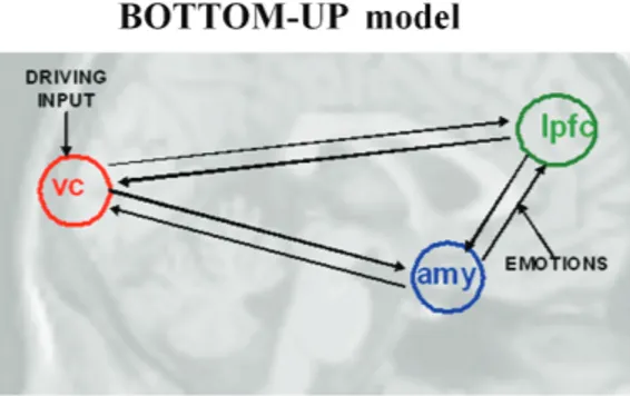
Model Space and Selection. The BMS analysis focused on the modulation of LPFC–amygdala effective connec-tivity by contextual stimuli (eg, facial expressions pre-sented during implicit vs explicit emotional faces task). Two models were built assuming bilateral intrinsic con-nections between V1, the amygdala and the LPFC, as well as V1 as the driving input region. These 2 models dif-fered from each other for the influence of the modulatory effects of facial expressions during implicit or explicit processing on the connections between the amygdala and the LPFC. In our first model (“bottom-up”), the mod-ulatory input (all faces vs crosshair) impacted the con-nection from the amygdala to the LPFC. In the second model (“top-down”), the modulatory input impacted the connection from the LPFC to the amygdala (figure 1).
After model set up, random-effects Bayesian model selection (BMS) analyses were performed in order to calculate exceedance probabilities (EP) (ie, the probabil-ity that one model is more likely than another model) in each comparison of interest (ie, implicit vs explicit proc-essing of facial expressions in (1) the total sample of HC; (2) the matched samples of HC, SCZ, SIB; and (3) DRD2 rs2514218 TT, CT and CC of HC). All the BMS analyses were performed for the explicit and implicit run separately. In addition, we performed Bayesian model averaging (BMA) on the winning model, an alterna-tive approach that allows for statistical comparison of parameters between groups (see supplementary material for details of this analysis and results).
Results
Characterization of LPFC–Amygdala Effective Connectivity in HC
BMS on the whole sample of 217 HC indicated that the “bottom-up” was the winning model during implicit
emotion processing. In this model, the modulatory effects were set from the amygdala to the LPFC. In particular, EP (0.90) of this model in HC was “pos-itive” according to a widely used classification,67 while
EP of the top-down model was “not positive” (0.10).67
Furthermore, BMS on the explicit run indicated that the “top-down” was the winning model in the same sam-ple of HC. Here, the modulatory effects were set from the LPFC to the amygdala. EP (0.96) of this model was “strong”,67 while EP of the bottom-up model was not
positive (0.04) (figure 2).
Effect of Familial Risk for SCZ on LPFC–Amygdala Effective Connectivity
Further BMS was performed in the matched samples of HC, SIB, and SCZ. Results of BMS during implicit and explicit processing in HC were consistent with those performed in the larger sample. In particular, winning models were the “bottom-up” during implicit processing, (“bottom-up” EP = 0.73; “top-down” EP = 0.27), and the “top-down” during explicit processing (“bottom-up” EP = 0.33; “top-down” EP = 0.67) (figure 3A).
Differently, BMS results in SCZ revealed that the “top-down” was the winning model during implicit process-ing (“bottom-up” EP = 0.24; “top-down” EP = 0.76). Furthermore, the “top-down” was the winning model also during explicit processing (“bottom-up” EP = 0.31; “top-down” EP = 0.69) (figure 3A). As in SCZ, findings in SIB indicated that the “top-down” was the winning model during implicit (“bottom-up” EP = 0.39; “top-down” EP = 0.61) and explicit (“bottom-up” EP = 0.39; “top-down” EP = 0.61) processing (figure 3A). Overall, in both SCZ and SIB during implicit processing there was a lower probability for the “bottom-up” compared with the “top-down” model.
Behavioral Data
Behavioral data indicated a main effect of diagnosis on reaction time during implicit processing of facial expres-sions (mean ± standard deviation: HC = 532.3 ± 122.5; SIB = 664.7 ± 127; SCZ = 645.2 ± 115.2) (F2,124 = 16.06, P < .001). Post hoc analysis indicated greater reaction time in SCZ and SIB compared with HC (P < .001), whereas no significant difference was present between SCZ and SIB (P > .05). Moreover, there was a diagno-sis by emotion interaction (F6,378 = 6.12; P < .001) on the number of avoided faces during the explicit task. Post hoc analysis revealed that SCZ avoided more happy faces and less fearful and angry faces compared with both SIB and HC (P < .04). Further details of behavioral data are included in the supplementary material.
Association of DRD2 rs2514218 With LPFC– Amygdala Effective Connectivity
BMS results in TT and CT HC revealed that the “bot-tom-up” was the winning model during the implicit proc-essing run (TT = “bottom-up”: 0.83; “top-down”: 0.17; CT = “bottom-up”: 0.98; “top-down”: 0.02), while the “top-down” was the winning model during the explicit processing run (TT = “bottom-up”: 0.07; “top-down”: 0.93; CT = “top-down”: 0.62; “bottom-up”: 0.38) (figure 3B). BMS results in CC HC (who are homozygous Fig. 1. Models of effective connectivity tested in the study.
Fig. 2. Graph showing exceedance probability (EP) of the
“bottom-up” and “top-down” models of effective connectivity between the amygdala and the LPFC in HC during implicit emotion processing and explicit emotional evaluation.
for the SCZ risk allele) indicated that the “top-down” was the winning model during explicit processing (“bot-tom-up”: 0.35; “top-down”: 0.65) (figure 3B). On the other hand, during implicit processing there was no clear winning model, but a slightly lower probability for the “bottom-up” compared with the “top-down” model in individuals homozygous for the risk C allele (“bot-tom-up”: 0.47; “top-down”: 0.53) (figure 3B). Finally, there was no effect of genotype on behavior during the task (all P > .05).
Given these effects of rs2514214, we also explored the LPFC–amygdala effective connectivity in the whole sam-ple of HC, and in a samsam-ple of HC matched with SIB and SCZ, excluding individuals homozygous for the C allele. This investigation did not relevantly change the results (supplementary material).
Discussion
Our results indicate abnormal patterns of effective con-nectivity between the amygdala and the LPFC during different subcomponents of emotion processing in SCZ and in SIB. Furthermore, HC homozygous for the C allele of DRD2 rs2514218, a genome-wide supported variant increasing genetic risk for SCZ,55 display
simi-lar functional brain abnormalities. Overall, these find-ings suggest that familial risk for SCZ is associated with anomalous effective connectivity between the LPFC and
the amygdala especially during implicit processing of emotional stimuli. Furthermore, they suggest that a risk allele for SCZ confers liability for this brain functional abnormality.
The results that we found in SCZ, SIB, and HC homo-zygous for the C allele of DRD2 rs2514218 should be read in light of our findings in the large sample of HC that we investigated. Here, the emotional task at hand differentially modulated the physiological effective con-nectivity between the amygdala and the LPFC during emotion processing. More in detail, there was a greater probability for an amygdala-to-LPFC effective connec-tivity during implicit, perceptual processing of emotional stimuli. On the other hand, explicit emotional evaluation was sustained by greater probability for the opposite pat-tern of effective connectivity between these brain regions. A possible interpretation of these findings may be based on the known primary role of the amygdala in percep-tual processing of emotional stimuli57 and of the LPFC
in the explicit evaluation of emotional stimuli and emo-tional regulation.19,59 Also, several findings suggest that
emotional stimuli imply a faster amygdala response com-pared to those of associative cortices, such as the LPFC.68
Thus, it is possible that the amygdala acts as a first func-tional node in the processing of emofunc-tional information conveyed by our task involving implicit, perceptual proc-essing. Then, the amygdala may send relevant inputs to the LPFC that exerts a role in integrating emotional Fig. 3. Graphs showing (A) exceedance probability (EP) of the “bottom-up” and “top-down” models of effective connectivity between
the amygdala and the LPFC in matched samples of HC, SCZ, and SIB. (B) Exceedance probability (EP) of the “bottom-up” and “top-down” models of effective connectivity between the amygdala and the LPFC as a function of DRD2 rs2514218 in HC.
information in a more general context of information processing, as previously suggested.69 Differently, the
explicit emotional part of our task needs integration of emotional regulation, social and cognitive functions, which are more supported by the LPFC according to previous literature.17,19,59,69 Thus, a task-mediated flow of
information from the LPFC to the amygdala may con-tribute to modulate more automatic responses sustained by the latter brain region when regulatory processes based on social and/or cognitive evaluation of emotional stimuli are required.
Our results indicate that effective connectivity is dys-functional in both SCZ and in SIB. In greater detail, in both these groups of individuals the “top-down” model is more likely than the “bottom-up” model during both implicit and explicit emotion processing. Given that HC had the “bottom-up” as the winning model during implicit processing and the “top-down” as the winning model during explicit processing, these results indicate that the probability for a physiological model of effective connectivity in SCZ and SIB during implicit processing of emotional stimuli is not preserved. These findings sug-gest a task-specific breakdown of the physiological flow of information between the amygdala and the LPFC in SCZ. Importantly, they also suggest that the abnormal pattern of effective connectivity during implicit emo-tion processing is a crucial phenotype of SCZ and that it is associated with familial risk for the disorder, rather than with specific state variables, including pharmaco-logical treatment and levels of symptoms. In this regard, these results are in line with models proposing altered brain functional integration as a key pathophysiologi-cal mechanism for SCZ,28,30–34 and consistent with
stud-ies reporting anomalous functional coupling between the amygdala and prefrontal regions in SCZ and in subjects at greater risk for this disorder.4,35–38,46,70,71 Furthermore,
they converge with recent evidence indicating a decrease of the amygdala-to-LPFC effective connectivity during implicit emotion processing in SCZ compared to con-trols,39 and are consistent with the view that this anomaly
may subtend an inability to properly evaluate and/or reg-ulate emotional inputs at the frontal level.39 Interestingly,
a well-credited model of SCZ proposes that symptoms of this brain disorder may be explained by the abnormal attribution of emotional salience to irrelevant stimuli,70,71
which is also consistent with our findings.
Intriguingly, we also found in HC a similar between-group modulation of LPFC–amygdala effective connec-tivity by DRD2 rs2514218, which has been associated with diagnosis of SCZ in the largest genome wide asso-ciation study to date.55 Here, the alteration of patterns
of effective connectivity in HC homozygous for the SCZ risk C allele was similar to those present in SIB and in SCZ. As in SCZ and SIB, we found that the physiological pattern of amygdala/LPFC effective connectivity in CC HC is altered during implicit emotion processing, such
that there was no winning model in this genotype group. On the other hand, the T allele predicted the physiolog-ical model of effective connectivity between these brain regions. Thus, these results are consistent with the likely involvement of D2 signaling in SCZ and with its rele-vance for emotion processing.54 Moreover, they are also
consistent with the above-mentioned model postulating abnormal attribution of emotional salience to irrelevant stimuli in SCZ,72 for which a dopaminergic dysregulation
is key.72 More in general, they support the use of
effec-tive connectivity during emotion processing for genetic investigations aimed at identifying true endophenotypes for the disease.
Importantly, the effect of rs2514218 on molecular phenotypes related to D2 receptor is still not clear. For instance, a recent study57 did not find any detectable
rela-tionship between this polymorphism and DRD2 expres-sion levels, suggesting that the molecular mechanisms of the present findings should be further investigated. On the other hand, recent work has found association of rs2514218 with striatal function in SIB,57 as well as with
response to antipsychotic treatment in patients,56
sup-porting the relevance of this SNP for correlates of SCZ. However, in these previous studies heterozygous and homozygous individuals were collapsed in one group of C or T carriers to investigate genotype effects. This strat-egy does not allow to verify consistency of these previous findings with ours when considering the model of genetic association of rs2514218 with SCZ-related phenotypes.
Some potential limitations should be considered in this study. First, the moderately high level of premorbid functioning of the SCZ patients could limit the general-izability of the findings to other SCZ patients. Second, we used a relatively slow repetition time during fMRI acquisition compared with those attainable with multi-band methods. This limitation prevented us to obtain a lower temporal resolution. Third, DCM analysis did not allow us to consider in the statistical design physiologi-cal covariates. This limitation prevented us to completely over-ride these possible sources of noise. However, our investigation of temporal signal-to-noise ratio (see sup-plementary material) suggests that these physiological parameters do not strongly affect our results. Fourth, the design of the experiment did not allow us to directly com-pare implicit vs explicit models of effective connectivity. However, this comparison was not the major aim of our study, while our purpose was to investigate modulation of effective connectivity during different emotional pro-cesses as a function of risk for SCZ and of rs2514218 genotype. Fifth, the fixation crosshair used as a baseline in this study does not allow us to control for the activ-ity or connectivactiv-ity related to visual complexactiv-ity, cognitive demand, and motor response of the emotional stimuli. However, it controls for activity or connectivity related to basic body functions (eg, breath, basic vision, space perception, body temperature regulation, etc.) while
preserving detection of brain connectivity during emo-tion processing. This approach may overcome some of the limitations associated with other baseline strategies and is consistent with the method used in several previous studies.14 Sixth, our model specification is a simplification
of the complex brain network sustaining emotion proc-essing and we may not exclude the putative relevance of other brain nodes or other possible model specifications in the context of our work. However, we focused our analyses on brain regions involved in emotion process-ing, consistently associated with SCZ and modulated by dopamine signaling. Finally, we focused on association of effective connectivity during emotion processing with a single genetic variation when SCZ is very likely associ-ated with polygenic risk. This strategy does not allow a full understanding of the relationship between effective connectivity during emotion processing and the genetics of SCZ. On the other hand, we investigated association of rs2514218 with LPFC–amygdala effective connectiv-ity to further support the utilconnectiv-ity of this phenotype for genetic studies relevant to SCZ.
In conclusion, our results suggest that altered LPFC– amygdala effective connectivity during emotion process-ing is a crucial correlate of SCZ, which may be further investigated as a promising candidate endophenotype for this brain disorder. In this regard, further studies should address the relationship between effective connectivity during emotion processing and polygenic scores indexing genetic risk for SCZ. Such investigation might shed more light on the pathophysiological underpinnings of emo-tional dysfunction of this brain disorder.
Supplementary Material
Supplementary data are available at Schizophrenia Bulletin online.
Funding
The present study has been financially supported by the European Community’s Seventh Framework Programme (project number 602450; URL: http://ec.europa.eu/ research/health/medical-research/brain-research/projects/ imagemend_en.html). The funders had no role in study design, data collection and analysis, decision to publish, or preparation of the manuscript.
Acknowledgm ents
We wish to thank Dr Mariana Nair Castro for useful comments and great assistance during the revision proc-ess and Dr Linda Antonucci for many helpful discussions on the field of connectivity. We also would like to express our thanks to all the volunteers for having participated to the study.
References
1. Tandon R, Nasrallah HA, Keshavan MS. Schizophrenia, “just the facts” 4. Clinical features and conceptualization.
Schizophr Res. 2009;110:1–23.
2. Aleman A, Kahn RS. Strange feelings: do amygdala abnor-malities dysregulate the emotional brain in schizophrenia?
Prog Neurobiol. 2005;77:283–298.
3. Li H, Chan RC, McAlonan GM, Gong QY. Facial emotion processing in schizophrenia: a meta-analysis of functional neuroimaging data. Schizophr Bull. 2010;36:1029–1039. 4. Anticevic A, Van Snellenberg JX, Cohen RE, Repovs G,
Dowd EC, Barch DM. Amygdala recruitment in schizophre-nia in response to aversive emotional material: a meta-analy-sis of neuroimaging studies. Schizophr Bull. 2012;38:608–621. 5. Phillips ML, Williams L, Senior C, et al. A differential neural
response to threatening and non-threatening negative facial expressions in paranoid and non-paranoid schizophrenics.
Psychiatry Res. 1999;92:11–31.
6. Gur RE, McGrath C, Chan RM, et al. An fMRI study of facial emotion processing in patients with schizophrenia. Am
J Psychiatry. 2002;159:1992–1999.
7. Paradiso S, Andreasen NC, Crespo-Facorro B, et al. Emotions in unmedicated patients with schizophrenia during evalu-ation with positron emission tomography. Am J Psychiatry. 2003;160:1775–1783.
8. Crespo-Facorro B, Paradiso S, Andreasen NC, et al. Neural mechanisms of anhedonia in schizophrenia: a PET study of response to unpleasant and pleasant odors. JAMA. 2001;286:427–435.
9. Kosaka H, Omori M, Murata T, et al. Differential amygdala response during facial recognition in patients with schizo-phrenia: an fMRI study. Schizophr Res. 2002;57:87–95. 10. Holt DJ, Kunkel L, Weiss AP, et al. Increased medial
tem-poral lobe activation during the passive viewing of emotional and neutral facial expressions in schizophrenia. Schizophr
Res. 2006;82:153–162.
11. Takahashi H, Koeda M, Oda K, et al. An fMRI study of dif-ferential neural response to affective pictures in schizophre-nia. Neuroimage. 2004;22:1247–1254.
12. Williams LM, Das P, Liddell BJ, et al. Fronto-limbic and autonomic disjunctions to negative emotion distinguish schizophrenia subtypes. Psychiatry Res. 2007;155:29–44. 13. Taylor SF, Liberzon I, Decker LR, Koeppe RA. A functional
anatomic study of emotion in schizophrenia. Schizophr Res. 2002;58:159–172.
14. Fusar-Poli P, Placentino A, Carletti F, et al. Functional atlas of emotional faces processing: a voxel-based meta-analysis of 105 functional magnetic resonance imaging studies. J
Psychiatry Neurosci. 2009;34:418–432.
15. Blasi G, Hariri AR, Alce G, et al. Preferential amygdala reactivity to the negative assessment of neutral faces. Biol
Psychiatry. 2009;66:847–853.
16. Gusnard DA, Akbudak E, Shulman GL, Raichle ME. Medial prefrontal cortex and self-referential mental activity: relation to a default mode of brain function. Proc Natl Acad Sci U S
A. 2001;98:4259–4264.
17. Phillips ML, Drevets WC, Rauch SL, Lane R. Neurobiology of emotion perception II: Implications for major psychiatric disorders. Biol Psychiatry. 2003;54:515–528.
18. Ochsner KN, Knierim K, Ludlow DH, et al. Reflecting upon feelings: an fMRI study of neural systems supporting the
attribution of emotion to self and other. J Cogn Neurosci. 2004;16:1746–1772.
19. Ochsner KN, Ray RD, Cooper JC, et al. For better or for worse: neural systems supporting the cognitive down- and up-regulation of negative emotion. Neuroimage. 2004;23:483–499. 20. Ochsner KN, Bunge SA, Gross JJ, Gabrieli JD. Rethinking
feelings: an FMRI study of the cognitive regulation of emo-tion. J Cogn Neurosci. 2002;14:1215–1229.
21. Stein JL, Wiedholz LM, Bassett DS, et al. A validated net-work of effective amygdala connectivity. Neuroimage. 2007;36:736–745.
22. Delgado MR, Nearing KI, Ledoux JE, Phelps EA. Neural circuitry underlying the regulation of conditioned fear and its relation to extinction. Neuron. 2008;59:829–838.
23. Erk S, Mikschl A, Stier S, et al. Acute and sustained effects of cognitive emotion regulation in major depression. J Neurosci. 2010;30:15726–15734.
24. Sotres-Bayon F, Cain CK, LeDoux JE. Brain mechanisms of fear extinction: historical perspectives on the contribution of prefrontal cortex. Biol Psychiatry. 2006;60:329–336.
25. Phillips ML, Ladouceur CD, Drevets WC. A neural model of voluntary and automatic emotion regulation: implica-tions for understanding the pathophysiology and neurode-velopment of bipolar disorder. Mol Psychiatry. 2008;13:829, 833–857.
26. Davis JI, Gross JJ, Ochsner KN. Psychological distance and emotional experience: what you see is what you get. Emotion. 2011;11:438–444.
27. Stephan KE, Friston KJ, Frith CD. Dysconnection in schizo-phrenia: from abnormal synaptic plasticity to failures of self-monitoring. Schizophr Bull. 2009;35:509–527.
28. Friston K, Brown HR, Siemerkus J, Stephan KE. The dys-connection hypothesis (2016). Schizophr Res. 2016;176:83–94. 29. Weinberger DR. A connectionist approach to the prefrontal
cortex. J Neuropsychiatry Clin Neurosci. 1993;5:241–253. 30. Andreasen NC, Paradiso S, O’Leary DS. “Cognitive
dysme-tria” as an integrative theory of schizophrenia: a dysfunction in cortical-subcortical-cerebellar circuitry? Schizophr Bull. 1998;24:203–218.
31. Friston KJ. The disconnection hypothesis. Schizophr Res. 1998;30:115–125.
32. Friston K. Disconnection and cognitive dysmetria in schizo-phrenia. Am J Psychiatry. 2005;162:429–432.
33. Stephan KE, Baldeweg T, Friston KJ. Synaptic plasti-city and dysconnection in schizophrenia. Biol Psychiatry. 2006;59:929–939.
34. Pettersson-Yeo W, Allen P, Benetti S, McGuire P, Mechelli A. Dysconnectivity in schizophrenia: where are we now?
Neurosci Biobehav Rev. 2011;35:1110–1124.
35. Das P, Kemp AH, Flynn G, et al. Functional disconnections in the direct and indirect amygdala pathways for fear process-ing in schizophrenia. Schizophr Res. 2007;90:284–294. 36. Leitman DI, Loughead J, Wolf DH, et al. Abnormal superior
temporal connectivity during fear perception in schizophre-nia. Schizophr Bull. 2008;34:673–678.
37. Vai B, Sferrazza Papa G, Poletti S, et al. Abnormal cortico-limbic connectivity during emotional processing correlates with symptom severity in schizophrenia. Eur Psychiatry. 2015;30:590–597.
38. Cao H, Bertolino A, Walter H, et al. Altered functional sub-network during emotional face processing: a potential inter-mediate phenotype for schizophrenia. JAMA Psychiatry. 2016;73:598–605.
39. Potvin S, Lungu O, Tikàsz A, Mendrek A. Abnormal effect-ive fronto-limbic connectivity during emotion processing in schizophrenia. Prog Neuropsychopharmacol Biol Psychiatry. 2017;72:1–8.
40. Rasetti R, Mattay VS, Wiedholz LM, et al. Evidence that altered amygdala activity in schizophrenia is related to clinical state and not genetic risk. Am J Psychiatry. 2009;166:216–225. 41. Blasi G, Popolizio T, Taurisano P, et al. Changes in prefrontal
and amygdala activity during olanzapine treatment in schizo-phrenia. Psychiatry Res. 2009;173:31–38.
42. Rigoni D, Pellegrini S, Mariotti V, et al. How neuroscience and behavioral genetics improve psychiatric assessment: report on a violent murder case. Front Behav Neurosci. 2010;4:160.
43. Habel U, Klein M, Shah NJ, et al. Genetic load on amyg-dala hypofunction during sadness in nonaffected brothers of schizophrenia patients. Am J Psychiatry. 2004;161:1806–1813. 44. van Buuren M, Vink M, Rapcencu AE, Kahn RS. Exaggerated brain activation during emotion processing in unaffected siblings of patients with schizophrenia. Biol Psychiatry. 2011;70:81–87.
45. Lo Bianco L, Blasi G, Taurisano P, et al. Interaction between catechol-O-methyltransferase (COMT) Val158Met geno-type and genetic vulnerability to schizophrenia during explicit processing of aversive facial stimuli. Psychol Med. 2013;43:279–292.
46. Diwadkar VA, Wadehra S, Pruitt P, et al. Disordered corti-colimbic interactions during affective processing in children and adolescents at risk for schizophrenia revealed by func-tional magnetic resonance imaging and dynamic causal mod-eling. Arch Gen Psychiatry. 2012;69:231–242.
47. LeDoux J. Fear and the brain: where have we been, and where are we going? Biol Psychiatry. 1998;44:1229–1238.
48. Pezze MA, Feldon J. Mesolimbic dopaminergic pathways in fear conditioning. Prog Neurobiol. 2004;74:301–320.
49. Mariotti V, Melissari E, Amar S, et al. Effect of prolonged phenytoin administration on rat brain gene expression assessed by DNA microarrays. Exp Biol Med (Maywood). 2010;235:300–310.
50. Levey AI, Hersch SM, Rye DB, et al. Localization of D1 and D2 dopamine receptors in brain with subtype-specific anti-bodies. Proc Natl Acad Sci U S A. 1993;90:8861–8865. 51. Rosenkranz JA, Grace AA. Dopamine attenuates prefrontal
cortical suppression of sensory inputs to the basolateral amygdala of rats. J Neurosci. 2001;21:4090–4103.
52. Seamans JK, Yang CR. The principal features and mecha-nisms of dopamine modulation in the prefrontal cortex. Prog
Neurobiol. 2004;74:1–58.
53. Howes OD, Kapur S. The dopamine hypothesis of schizo-phrenia: version III–the final common pathway. Schizophr
Bull. 2009;35:549–562.
54. Blasi G, Lo Bianco L, Taurisano P, et al. Functional vari-ation of the dopamine D2 receptor gene is associated with emotional control as well as brain activity and connect-ivity during emotion processing in humans. J Neurosci. 2009;29:14812–14819.
55. Schizophrenia Working Group of the Psychiatric Genomics Consortium. Biological insights from 108 schizophrenia-associated genetic loci. Nature. 2014;511:421–427.
56. Zhang JP, Robinson DG, Gallego JA, et al. Association of a schizophrenia risk variant at the DRD2 locus with anti-psychotic treatment response in first-episode psychosis.
57. Vink M, de Leeuw M, Luykx JJ, et al. DRD2 Schizophrenia-risk allele is associated with impaired striatal functioning in unaffected siblings of schizophrenia patients. Schizophr Bull. 2016;42:843–850.
58. Hariri AR, Bookheimer SY, Mazziotta JC. Modulating emo-tional responses: effects of a neocortical network on the lim-bic system. Neuroreport. 2000;11:43–48.
59. Ochsner KN, Gross JJ. The cognitive control of emotion.
Trends Cogn Sci. 2005;9:242–249.
60. Stephan KE, Penny WD, Moran RJ, den Ouden HE, Daunizeau J, Friston KJ. Ten simple rules for dynamic causal modeling. Neuroimage. 2010;49:3099–3109.
61. Birnbaum R, Weinberger DR. Functional neuroimag-ing and schizophrenia: a view towards effective connectiv-ity modeling and polygenic risk. Dialogues Clin Neurosci. 2013;15:279–289.
62. Taurisano P, Blasi G, Romano R, et al. DAT by perceived MC interaction on human prefrontal activity and connect-ivity during emotion processing. Soc Cogn Affect Neurosci. 2013;8:855–862.
63. Del’Guidice T, Latapy C, Rampino A, et al. FXR1P is a GSK3β substrate regulating mood and emotion processing.
Proc Natl Acad Sci U S A. 2015;112:E4610–E4619.
64. Quarto T, Blasi G, Maddalena C, et al. Association between ability emotional intelligence and left insula during social judgment of facial emotions. PLoS One. 2016;11:e0148621.
65. Tottenham N, Tanaka JW, Leon AC, et al. The NimStim set of facial expressions: judgments from untrained research par-ticipants. Psychiatry Res. 2009;168:242–249.
66. Friston KJ, Harrison L, Penny W. Dynamic causal modelling.
Neuroimage. 2003;19:1273–1302.
67. Kass RE, Raftery AE. Bayes factors. J Am Stat Assoc. 1995;90:773–795.
68. Quirk GJ, Armony JL, LeDoux JE. Fear conditioning enhances different temporal components of tone-evoked spike trains in auditory cortex and lateral amygdala. Neuron. 1997;19:613–624. 69. Pessoa L. On the relationship between emotion and
cogni-tion. Nat Rev Neurosci. 2008;9:148–158.
70. Modinos G, Pettersson-Yeo W, Allen P, McGuire PK, Aleman A, Mechelli A. Multivariate pattern classification reveals differential brain activation during emotional pro-cessing in individuals with psychosis proneness. Neuroimage. 2012;59:3033–3041.
71. Pulkkinen J, Nikkinen J, Kiviniemi V, et al. Functional map-ping of dynamic happy and fearful facial expressions in young adults with familial risk for psychosis—Oulu Brain and Mind Study. Schizophr Res. 2015;164:242–249.
72. Kapur S. Psychosis as a state of aberrant salience: a frame-work linking biology, phenomenology, and pharmacology in schizophrenia. Am J Psychiatry. 2003;160:13–23.
73. Winton-Brown TT, Fusar-Poli P, Ungless MA, Howes OD. Dopaminergic basis of salience dysregulation in psychosis.