“Sapienza” University
Faculty of Medicine and Dentistry
D
OCTORALT
HESISMyocardial Metabolism Evaluation and Ketones Utilization
in the Failing Human Heart
Ph.D. fellow: Supervisors:
Dr. Luca MONZO Prof. Carlo GAUDIO
Prof. Josef KAUTZNER
Dr. Vojtech MELENOVSKY
A thesis submitted in fulfillment of the requirements for the degree of Doctor of Philosophy
in
Biomedical Technologies in Clinical Medicine XXXI cycle
Index
1. INTRODUCTION 2
1.1 A Metabolic Approach to Heart Failure 2
1.2 Overview of Normal Myocardial Substrate Metabolism 3
1.3 Derangement of Energy Metabolism in Heart Failure 5
1.4 Ketone Body Metabolism 6
2. RATIONALE AND AIM OF THE STUDY 8
3. METHODS 10
3.1 Patients Selection 10
3.2 Study Protocol 10
3.3 Investigation Procedure and Substrates Analysis 11
3.4 Statistical Analysis 12
4. RESULTS 13
4.1 Clinical Characteristics 13
4.2 Procedural Characteristics 13
4.3 Myocardial Metabolism and Correlates 14
5. DISCUSSION 16 6. STUDY LIMITATIONS 19 7. CONCLUSIONS 20 8. ACKNOWLEDGEMENTS 20 9. REFERENCES 21 10. TABLES 30 11. FIGURES 35
1. Introduction
1.1 A Metabolic Approach to Heart Failure
During the past half-century, the advances in the prevention, diagnosis, and management of cardiovascular disease (CVD) have been nothing short of spectacular. Age-adjusted CVD-related deaths have declined by about two-thirds in industrialized nations (1). Mortality rates associated with the acute coronary syndromes, valvular and congenital heart disease, uncontrolled hypertension, and many arrhythmias all have fallen dramatically.
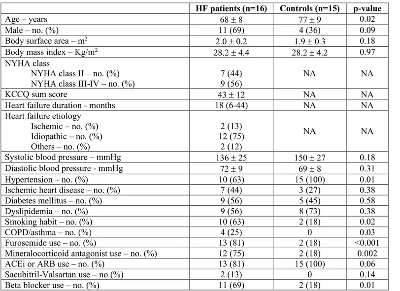
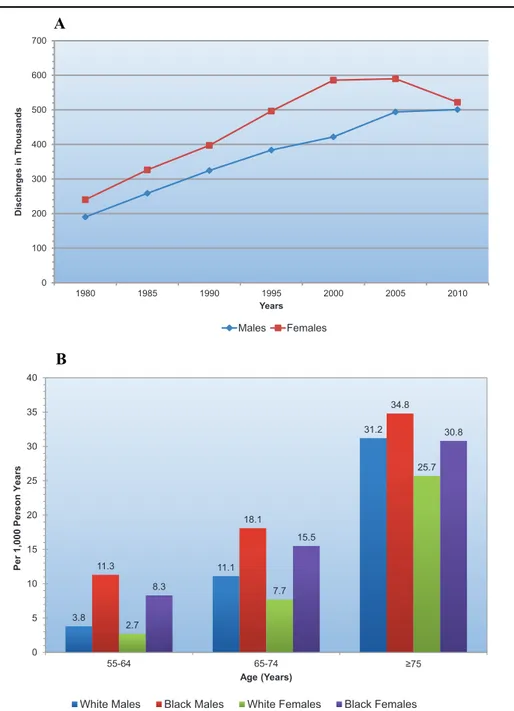
Heart failure (HF) is a notable exception to these encouraging trends. Indeed, after normal delivery, it is the most common cause of hospitalization. Annual hospital discharges in patients with a primary diagnosis of HF have risen steadily since 1975, and now exceed 1 million discharges per year in the United States, although they may at last be levelling off (2) (Figure 1A), or actually decreasing (3, 4). In Europe, hospitalizations for HF are clearly declining (5, 6). HF is primarily a disease of the elderly that affects about 10% of men and 8% of women over the age of 60 years, and its prevalence rises with age (Figure 1B) and has risen overall. In the United States, patients with a primary diagnosis of HF now make >3 million physician visits per year (2).
Survival after a diagnosis of HF has improved during the past 30 years; the age-adjusted death rate has declined (3, 4, 7), and the mean age at death from HF has risen (5, 8). However, despite these modest improvements, the 5-year mortality is still approximately 50% worse than that of many cancers (9).
There are many reasons why a human heart can fail (ischemic heart disease, myocarditis, valvular diseases, etc) (10), but the available evidence suggests that the failing heart is an engine out of fuel — that is, altered energetics play an important role in the mechanisms of HF (11). The heart consumes more energy than any other organ. It cycles about 6 kg of ATP every day — 20 to 30 times its own weight. Each day, it beats about 100,000 times and pumps approximately 10 tons of blood through the body. To acquire the energy that is necessary to carry out its function, the heart converts
chemical energy stored in fatty acids and glucose into the mechanical energy of the actin–myosin interaction of myofibrils. Failure to produce an adequate amount of energy causes mechanical failure of the heart (12). The concept that the failing heart is an energy-starved engine that has run out of fuel is decades old. It was proposed in 1939 by Herrmann and Decherd (13), who, in their article entitled “The Chemical Nature of Heart Failure,” described a significantly reduced creatine content in failing myocardium. It is today well known that, in the end-stages of HF, the myocardium has low ATP content due to a decreased ability to generate ATP by oxidative metabolism, and thus is unable to effectively transfer the chemical energy from the metabolism of carbon fuels to contractile work (14, 15). The consequences of metabolic dysfunction in HF are poorly understood, but there is growing evidence to support the concept that the alterations in substrate metabolism seen in HF contribute to contractile dysfunction and to the progression of left ventricular (LV) remodelling that are characteristic of the HF state. Current medical therapies for HF are aimed at suppressing neurohormonal activation (e.g., angiotensin converting enzyme inhibitors, angiotensin II receptor antagonists, ß-adrenergic receptor antagonists, and aldosterone receptor antagonists), and treating fluid volume overload and hemodynamic symptoms (diuretics, digoxin, inotropic agents) (16-21). These pharmacotherapies for HF can improve clinical symptoms and slow the progression of contractile dysfunction and expansion of LV chamber volume; nevertheless, there is still progression, and the prognosis for even the optimally treated patient remains poor (2). Moreover, there is recent evidence that intense suppression of the neurohormonal systems does not provide further benefit compared with more modest therapy (22, 23). Thus, there is a need for novel therapies for HF, independent of the neurohormonal axis, that can improve cardiac performance and prevent or reverse the progression of LV dysfunction and remodelling.
1.2 Overview of Normal Myocardial Substrate Metabolism
The heart has a very high energy demand and essentially no energy reserves. So, it must continually generate ATP at a high rate to sustain contractile function, basal metabolic processes, and ionic
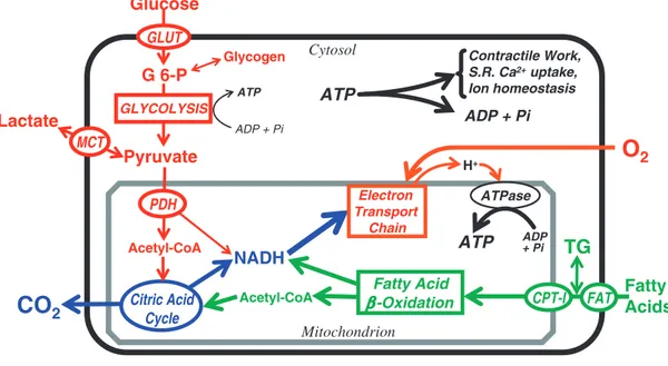
homeostasis (24). In the normal adult heart, almost all (>95%) of ATP production is derived from mitochondrial oxidative phosphorylation, with the remainder being derived from glycolysis and GTP formation in the tricarboxylic acid (TCA) cycle. The heart has a relatively low ATP content (5 µmol/g wet wt) and high rate of ATP hydrolysis (» 30 µmol × g wet wt-1 × min-1 at rest); thus, under normal conditions, there is complete turnover of the myocardial ATP pool approximately every 10 s (25-27). To sustain sufficient ATP generation, the heart acts as an “omnivore” and can use a variety of different carbon substrates as energy sources if available (28, 29) (Figure 2). However, the adult heart normally obtains 50– 70% of its ATP from fatty acid b-oxidation (25, 27, 30). The b-oxidation of fatty acids is under complex control and is dependent on a number of factors, including 1) fatty acid supply to the heart; 2) the presence of competing energy substrates (glucose, lactate, ketones, amino acids); 3) energy demand of the heart; 4) oxygen supply to the heart; 5) allosteric control of fatty acid uptake, esterification, and mitochondrial transport; and 6) the control of mitochondrial function, including direct control of fatty acid b-oxidation, TCA cycle activity, and electron transport chain activity (28, 31, 32). The transcriptional control of enzymes involved in fatty acid metabolism and mitochondrial biogenesis are also important determinants of fatty acid b-oxidation rates (33). Of importance, is that the normal heart has a substantial amount of “metabolic flexibility” that allows it to switch back and forth between fatty acids and carbohydrates oxidation, depending on the workload of the heart, the energy substrate supply to the heart, and the hormonal and nutritional state. For instance, an increase in fatty acid oxidation rates in the heart, is accompanied by a dramatic decrease in the oxidation of pyruvate originating from glucose (glucose oxidation), and vice versa (24). This metabolic flexibility allows the heart to produce the necessary amount of ATP needed by the heart to sustain cardiac contractility under a wide variety of conditions.
In the fasting state, ketones are taken up by the human heart, along with glucose, lactate, pyruvate, glycerol, and fatty acids, with the highest avidity per unit mass among body tissues and with a fractional extraction (~ 40%) comparable to that of pyruvate and far higher than that of glucose (~ 2%) or fatty acids (15–20%) (34). By combining mass uptake (~ 10 mmol/min) with the heat of
combustion, it can be calculated that in the overnight fasted state the ketone body ß-hydroxybutyrate contributes 15% of resting cardiac energy expenditure compared with 45% of fatty acids and 8% of glucose/lactate/pyruvate.
1.3 Derangement of Energy Metabolism in Heart Failure
Heart failure has a complex pathophysiology, which is attributed to differences in aetiology, severity, disease duration, and comorbidities such as diabetes and obesity. Furthermore, these factors themselves can greatly affect myocardial energy metabolism (24).
Regardless of the type of heart failure, cardiac energy metabolism can be compromised (Figure 3). This is evidenced by in vivo measurements of ATP and phosphocreatine (PCr) content (using 31P-magnetic resonance spectroscopy) in the human failing heart, that can be approximately 30-40% lower than in the normal heart (35, 36). Cardiac PCr/ATP ratios are decreased in HF and correlate with New York Heart Association (NYHA) functional class, predicting adverse outcomes of these patients (35, 36). This energy deficit in the failing heart is partly attributable to defects in mitochondrial function and electron transport chain activity (37, 38), but also to alterations in energy substrate use by the heart that can decrease cardiac efficiency (39, 40).
The changes in energy substrate preferences that occur in HF are controversial and not completely understood. It is generally assumed that the failing heart switches from fatty acid to glucose metabolism in the failing heart (12, 41, 42). Studies of substrate utilization in HF have yielded conflicting results, but most experimental data indicate that fatty acid utilization, which is unchanged or slightly increased in early HF (43, 44), is substantially decreased in advanced heart failure (45). However, it is probably more accurate to suggest that the heart switches from mitochondrial oxidative metabolism towards glycolysis as a source of ATP production (39, 46). A decrease in fatty acid oxidation in HF likely occurs both due a decrease in overall mitochondrial function and oxidative capacity, as well as a decrease in the transcription of a number of enzymes involved in fatty acid oxidation (12, 37, 41). However, mitochondrial glucose oxidation can also decrease dramatically in
the failing heart, that occurs due both to a decrease in overall mitochondrial oxidative capacity, and a decrease in the activity of the key rate-limiting enzyme of glucose oxidation, pyruvate dehydrogenase (39, 46). This decrease in glucose oxidation coupled to an increase in glycolytic rates, results in a mismatch between glycolysis and glucose oxidation. This mismatch between glycolysis and glucose oxidation results in an increase in lactate and proton production by the heart, similar to what is seen in ischemic heart disease (39), that in turn can lead to intracellular Na+ and Ca2+ overload, and finally to an impaired cardiac power and performance (47-49).
Clinical studies demonstrate that at a late stage HF results in an increase in plasma ketone body concentration that appears to be secondary to elevated fatty acid levels (50-52). Ketones, and in particular ß-hydroxybutyrate, demonstrated in experimental observations to have a highly beneficial energetic profile (53). The energetics of mitochondrial ß-hydroxybutyrate oxidation compares favourably with the oxidation of pyruvate; in fact, when ß-hydroxybutyrate is added to the perfusion medium of working rat hearts, the heat of combustion per unit of carbon has been calculated to be 31% increased and the oxygen cost of this energy output to be 27% decreased (54). This advantage is granted by a more efficient oxidation of the mitochondrial coenzyme Q couple and an increase in the free energy of cytosolic ATP hydrolysis. As a consequence, in the isolated working heart ß-hydroxybutyrate increases external cardiac work at the same time as it reduces oxygen consumption, thereby improving cardiac efficiency by 24%
1.4 Ketone Body Metabolism
Ketone bodies, that are β-hydroxybutyrate, acetoacetate and acetone (in low abundance), are produced in the liver and used in peripheral tissues as an energy source when glucose is not readily available because of either a limited exogenous supply or impaired insulin signalling or when fatty acids are in surplus, which occurs in marked activation of lipolysis (55). The heart extracts and oxidizes ketone bodies in a concentration-dependent manner (56-59). Plasma ketone bodies are formed from fatty acids in the liver, and the arterial plasma concentration is normally very low, thus
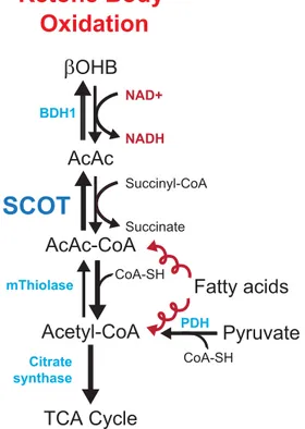
they are normally a minor substrate for the myocardium. During starvation and poorly controlled diabetes, plasma ketone body concentrations are elevated secondary to low insulin and high fatty acids, and they become a major substrate for the myocardium (60, 61). On entering the cell, they rapidly form acetyl-CoA via a series of reactions catalysed by ß-hydroxybutyrate dehydrogenase, succinyl-CoA:3-oxoacid-CoA transferase (SCOT), and mitochondrial acetyl-CoA acetyltransferase (62) and enter in the TCA cycle (Figure 4). All enzymes are present in the heart, and the reactions are driven primarily by substrate availability, except for the SCOT reaction, which is a succinyl CoA– dependent process.
As with fatty acids, the uptake and oxidation of glucose and lactate are inhibited by elevated plasma ketone bodies (30, 63), with the inhibitory effect presumably mediated through product inhibition on pyruvate dehydrogenase. Oxidation of ketone bodies inhibits also myocardial fatty acid oxidation (56-58, 63, 64).
As a function of oxygen consumption, ketone bodies are the most efficient substrate for ATP production and therefore could be a particularly useful fuel source under conditions in which oxygen delivery to tissues is reduced, such as in advanced HF (54, 65). Furthermore, manifest and latent deficiencies of ketone bodies utilization are associated with cardiomyopathy. The OXCT1 gene encodes a mitochondrial matrix enzyme that is essential for ketone body oxidation, SCOT (65, 66). Some individuals with recessive mutations of OXCT1 present with dilated cardiomyopathy (67). In addition, ketogenic diets cause biochemical disturbances and cardiac dysfunction in certain vulnerable patients, possibly due to latent defects in ketone body metabolism (68). These findings support the hypothesis that ketone body can play a role in the HF myocardial metabolism.
2. Rationale and Aim of the Study
Despite advances in the treatment of HF, our understanding of the energy metabolic mechanisms limiting cardiac pump function remains incomplete. It is increasingly recognized that metabolic remodelling is integral to HF development and progression (12, 42) and that the overall reduction in the myocardial oxidative capacity is purported to be the root cause of energy deficiency in the failing heart.
An elevation in serum KBs levels has been observed by several studies in patients with HF (51, 69). Interestingly, a new glucose-lowering agent, the sodium–glucose cotransporter 2 (SGLT2) inhibitor empaglifozin, was demonstrated to increase ketogenesis in type 2 diabetes (T2D) (70). In the EMPA-REG OUTCOME trial, that tested empagliflozin against placebo in 7,020 patients with T2D who were at increased cardiovascular risk a striking relative risk reduction in cardiovascular outcomes was found (71). These beneficial effects of empagliflozin has been proposed to be due an increase in ketone use by the heart, and it has been proposed that ketones are a “superfuel” for the heart that can increase cardiac efficiency and, in turn, cardiovascular protection beyond the classical mechanisms described for glucose lowering agents (i.e. improvement in glycaemic control, decrease in body weight, reductions in blood pressure and uric acid level) (72). Moreover, experimental observations showed that KBs improve work efficiency at the mitochondrial level (54), suppress oxidative stress by inhibiting histone deacetylases (73), upregulate mitochondrial biogenesis and, by stabilizing cell membrane potential, exhibit antiarrhythmic potential (55).
Finally, recent proteomics and metabolomics studies demonstrated strong and concordant evidence of increased ketone bodies oxidation in HF providing new insights into the reliance of the failing heart on ketone bodies for energy supply (Figure 5). In particular, Aubert et al. (74) examined longitudinal changes in cardiac energy metabolism during the development of HF murine models of compensated hypertrophy and HF. Using a proteomic approach, this group identified significant downregulation of proteins involved in fatty acid utilization in both compensated hypertrophy and HF hearts with a concurrent 2- to 3-fold increase in b-hydroxybutyrate dehydrogenase (BDH1), the
enzyme that catalyzes the initial step in the ketone oxidation pathway. Moreover, the authors found increases in hydroxybutyrylcarnitine and acetylcarnitine in the HF group, a known metabolic signature of ketone metabolism. Bedi et al. (75) described an independent study of end-stage human HF in which metabolomics analysis showed increased ketogenic b-hydroxybutyrate-CoA and evidence for enhanced myocardial utilization of b-hydroxybutyrate. These changes were accompanied by an upregulation of BDH1, BHD2 (cytosolic isoform), and succinyl-CoA:3-oxoacid-CoA transferase (SCOT), key enzymes in the ketone oxidation pathway.
Data described in literature provide just clues on HF metabolism in humans (animal models, end-stage HF, small populations). The main aim of our study was to identify and quantify myocardial substrates utilization in mild to moderate human HF. We hypothesized to that failing hearts would display signs of reduced fatty acid uptake in favor of ketone bodies.
3. Methods
3.1 Patients Selection
Patients who satisfying criteria of HF with reduced ejection fraction (HFrEF) according to the current guidelines by European Society of Cardiology (76) and an indication to implant a cardiac device (cardiac resynchronization therapy/implantable cardiac defibrillator/single or dual-chamber pacemaker) were enrolled. In particular, chronic stable patients, who presented with signs or symptoms of HF (dyspnoea, fatigue, and exercise intolerance) consistent with a New York Heart Association (NYHA) functional class II to III and reduced left ventricle ejection fraction (<40%) were considered suitable for screening.
Exclusion criteria included: severe obesity (body mass index>40); acute heart illness (i.e. myocarditis, acute coronary syndrome, atrial permanent arrhythmias); infiltrative or hypertrophic cardiomyopathy; cancer in the active phase; hepatitis B or C; human immunodeficiency virus; severe renal disease (glomerular filtration rate <15 ml/m).
All patients received standard medical therapy per discretion of the attending cardiologists and must provide informed consent. The control group was represented by non-HF patients scheduled for single or dual chambers pacemaker implantation. The study was approved by the local ethical committee and all patients signed written informed consent.
3.2 Study Protocol
Study population and controls were enrolled in the investigational Centre (Institute for Clinical and Experimental Medicine, Prague, Czech Republic). Patients with HFrEF and clinical indication to implant a cardiac device were screened to be part of the study population, while patients without HF and scheduled to implant a cardiac device were screened to be part of the control cohort.
All patients underwent to a complete cardiac evaluation, including clinical history, the Kansas City Cardiomyopathy Questionnaire (KCCQ), physical examination, and echocardiogram (Vivid7,
General Electric Healthcare, Wauwatosa, Wisconsin). Left ventricular (LV) function and dimensions were measured according to contemporary recommendations (77). Mitral and tricuspid regurgitation was assessed semi-quantitatively and expressed in 4 grades (absent/mild, moderate, moderate to severe and severe). Right ventricular (RV) systolic pressure was estimated from the tricuspid regurgitation velocity and right atrial pressure estimate, based on inferior vena cava (IVC) diameter. RV function was quantified in an apical 4-chamber view by using tricuspid annular systolic excursion (M-mode TAPSE). Glomerular filtration rate was estimated using the Modification of Diet in the Renal Disease equation (78). Plasma sodium, albumin, glucose and creatinine were measured by using an automated Abbott Architect ci1600 analyzer. The B-type natriuretic peptide (BNP) concentrations were measured by using microparticle immunoassay (Architect BNP, Abbott Laboratories, Chicago, Illinois; long-term analytical CV 4.5%).
3.3 Investigation Procedure and Substrates Analysis
All investigations were done after at least 6-hour or overnight fast. In every patient enrolled in the study we collected three blood samples within few minutes of each other. The arterial and venous samples were collected by the fluoroscopy-guided axillary vein puncture technique (79), while the coronary sinus (CS) sample was harvested by a dedicated sheet introduced via axillary vein approach in the CS under fluoroscopy guidance. To allow the correct sampling of all CS branches the sheet was placed no deeper than 1-1.5 cm into the CS body. All enrolled participants were under mild sedation during the procedure. Blood samples were collected after at least 10 minutes from the last administration of sedative drugs and without any on-going infusion to avoid sample dilution. No antiarrhythmic drugs or isoproterenol were administered before sampling.
All samples were immediately placed on ice, and within 20 minutes, they were centrifugally separated at a centrifugal force of 800 × g at 4 °C. The plasma was collected, distributed into aliquots, and frozen at –80 °C. β-hydroxybutyrate concentration was analyzed instead of total ketones because it is the most abundant in the bloodstream (80), accounts for the larger part of the circulating ketone
bodies during fasting (81) and showed a significant role in cellular signaling (82). β-hydroxybutyrate, fatty acids and glucose concentrations were measured with use of commercially available enzymatic assays coupled to chromogenic substrates (Wako Chemicals USA, Inc.; Richmond, VA). Arterial and CS oximetric measurements (A-VOX Systems, Inc.; San Antonio, Texas) were used to calculate the myocardial arteriovenous difference in partial oxygen pressure. The fractional extraction (FE) of each substrate was computed according to the formula: FE = [(SA – SCS)/SA] x 100, where SA and SCS are respectively the substrates concentration in the axillary artery and in the coronary sinus. Net myocardial exchange (uptake or release) of substrates was computed as: SA – SCS.
3.4 Statistical Analysis
Data are presented as means ± standard deviation using conventional methods except for variables without normal distribution where median and interquartile range are shown. Variables not normally distributed had been log-transformed before the analysis. Skewness was tested using the Shapiro-Wilk normality test. Pearson coefficient was calculated to express correlation. Statistical significances between groups were performed using Students unpaired t-test or Mann-Whitney U-test. The level of statistical significance was set at a 2-sided p-value <0.05.
All analyses were performed using JMP 9.0 statistical software (SAS Institute, Inc., Cary, North Carolina).
4. Results
4.1 Clinical Characteristics
The mean age of patients with HF (n=16) was 68 ± 8 years, 69% were male, 44% had ischemic heart disease and 56% had diabetes. The main etiology of HF was idiopathic (75%) and the mean duration of the disease was 18 (6-44) months. Approximately 75% and 80% of patients were treated with mineralocorticoid antagonist (MRA) and angiotensin converting enzhyme inhibitors (ACEi)/angiotensin receptor blockers (ARB), respectively, 69% with beta-blockers (BB) and more than 80% were taking loop diuretics (Table 1). Mean LVEF was 28.2 ± 4.9% and mean QRS duration was 165 ± 19 msec (Table 2).
Among controls (n=15), 45% had diabetes and 27% ischemic heart disease, and all of them had hypertension. Their mean age was 77 ± 9 years, 36% males (Table 1); mean LVEF was 57.1 ± 3.7% and mean QRS duration was 118 ± 24 msec.
Blood biochemistry profile showed no relevant differences between groups except, as expected, for a higher BNP plasma concentration in HF patients compared to controls (279 [217-932] mmol/L vs 74 [40-150], p=0.008) (Table 2).
4.2 Procedural Characteristics
In the HF group 13 patients (81%) received a cardiac resynchronization therapy (CRT) defibrillator and 3 (19%) a CRT pacemaker; among these, 50% were upgrades from a single or dual-chamber pacemaker or defibrillator. The mean total procedural time was 90 ± 25 min. All controls were implanted with a dual-chamber pacemaker with a mean total procedural time of 64 ± 24 min (Table 3).
All patients received intraprocedural sedation, without any significative difference between groups. No differences in fasting time before the procedure were noted (HF 16 ± 6 hours vs controls 16 ± 5 hours, p=0.97) (Table 3).
Two patients, one in the HF group and one in the control group had pericardial effusion during the procedure after CS cannulation. In the HF patient the effusion was complicated by cardiac tamponade that needed an urgent pericardiocentesis. Both patients completely recovered after few days of hospitalization and no chronic sequalae were registered.
4.3 Myocardial Metabolism and Correlates
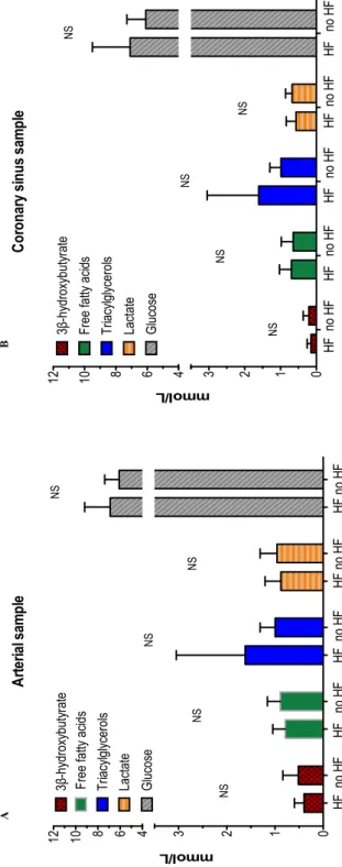
We analyzed both the net myocardial exchange (arterio-venous difference) and fractional myocardial extraction of energetic substrates, to investigate the craving of the heart for specific substances, and the difference in concentration of these substrates between patients with and without HF (Table 4).
Substrates concentrations and partial pressure of oxygen were similar in HF and non-HF patient both in the arterial and in the CS samples (Table 4 and Figure 6). Both net myocardial substrates exchange and fractional extraction showed a significant reduction in the mean myocardial fatty acids uptake in HF patients compared to controls (net myocardial substrates exchange: HF 0.07 ± 0.23 mmol/L vs non-HF 0.25 ± 0.16 mmol/L, p=0.03; fractional myocardial extraction: HF 4 ± 44% vs non-HF 31 ± 19%, p=0.05). Opposite, β-hydroxybutyrate, lactate, partial pressure of oxygen, triacyclglycerols and glucose extractions were relatively unchanged between groups (Table 4 and Figure 7). When HF metabolism was tested for differences in diabetic and non-diabetic patients, no difference in net and fractional myocardial substrates extraction was found (Figure 8).
In the linear regression analysis, larger end-diastolic (p<0.001) and end-systolic (p=0.001) LV diameters, higher aldosterone (p=0.02) and renin (p=0.05) plasma concentrations and increased right ventricular systolic pressure (p=0.05) and LV mass (p=0.04) were inversely correlated with the net
fatty acids myocardial exchange. In contrast, left ventricular ejection fraction was directly correlated (p=0.01) (Table 6; Figure 9 and Figure 10).
The only variable statistically, but marginally, associated with the net β-hydroxybutyrate myocardial exchange was HF duration (Table 5 and Figure 11). Interestingly, β-hydroxybutyrate plasma concentration was associated neither with HF duration (Figure 11) nor with triacilgliceroles concentration, both in HF and in controls (Figure 12).
5. Discussion
The main finding of our study is that there was no difference in the net and fractional extraction of β-hydroxybutyrate in mild to moderate HFrEF compared to patients who had structurally normal hearts. Interestingly, the net cardiac extraction of β-hydroxybutyrate was directly associated to HF duration. We also demonstrated a reduced myocardial fatty acids uptake in HF patients and its negative association with neurohormonal and echocardiographic HF hallmarks.
Previous human and experimental studies showed an increased reliance of the failing heart on ketone bodies for energy supply in severe or end-stage HF (51, 74, 75). This metabolic change was mirrored by the upregulation of the key enzymes in the ketone oxidation pathway such as BDH1, BHD2 (cytosolic isoform), and SCOT (75). Whether the switch in myocardial substrate oxidation away from fatty acids toward ketones in advanced HF is a positive compensatory adaptation or a pathological maladaptation is a matter of considerable debate, but it has been proposed that this switch is energetic beneficial and can maintain cardiac energy supply under situation of limited energy production (55). An interesting insight in this field came from the EMPA-REG OUTCOME trial (71) where the striking reduction of cardiovascular outcomes and mortality were partly adduced to an empaglifozin-mediated increase in plasma concentration of ketone bodies (72, 83).
In our study we found that plasma concentration of β-hydroxybutyrate in the artery and CS and its net and fractional myocardial extraction were unchanged compared to controls. These differences with previous studies could be explained in first instance by different population characteristics. Aubert et al. (74) evidenced the increase of key enzymes in the ketone oxidation pathway in a murine model of compensated hypertrophy and HF. Lommi et al. demonstrated an increase in ketone bodies plasma concentrations in 45 highly symptomatic patients with chronic moderate to severe HF (29 patients in NYHA class III or IV) not on betablockers therapy. Recently, Bedi et al. (75) showed increased ketogenic β-hydroxybutyrate-CoA and evidence for enhanced myocardial utilization of β-hydroxybutyrate in end-stage human HF. Opposite, we enrolled patients with mild to moderate HF (NYHA class II-III), in a stable phase and well medically treated.
We also found a direct correlation between β-hydroxybutyrate myocardial extraction and HF duration. This finding is in according to previous experimental observations in dogs where changes in substrate use were a late-stage phenomenon and a consistent myocardial metabolic switch occurred only in more severe or decompensated HF (43). Interestingly, β-hydroxybutyrate plasma concentration was associated neither with HF duration nor with fatty acids concentration, both in HF and in controls. While the uncoupling between β-hydroxybutyrate plasma concentration and HF duration may be due to the observation of an early phase of metabolic remodeling, and so to the scarce rely of the heart on ketone bodies for its energetic supply, the poor relation between ketones and fatty acids concentration is probably multifactorial. One possible cause may be the high level of neurohormonal blockade in our HF patients that, reducing the sympathetic overdrive, can lead to a low rate of lipolysis (50). Another possible explanation may be that in our population insulin was still effective in the heart metabolism control (low level of tissues insulin-resistance), and therefore in suppressing ketogenesis (55).
We studied both diabetics and non-diabetics patients. It is well-known that diabetic hearts cannot use glucose fully due to shortage of insulin effects and may therefore be forced to switch to almost exclusive use of fatty acids for energy sources (84). This metabolic signature may change the utilization of energy substrates between diabetic and non-diabetic failing heart. In our study we didn’t find any difference in substrate utilization between patients in HF with and without diabetes. This was probably due to the well-controlled glycemic status of all enrolled diabetic patients.
As outlined in the Introduction, studies in animals provide a consistent view that failing heart have reduced fatty acid uptake whereas the few studies in humans are largely conflicting (24). Paolisso et al. (85) measured the net extraction of fatty acids by the myocardium using simultaneous arterial and CS sampling in patients with mild to moderate HF (NYHA class II-III) and in age-matched healthy individuals. HF patients had elevated plasma norepinephrine and insulin as well as a 50% increase in fatty acids concentrations. Fatty acids uptake and the estimated fatty acid β-oxidation rates were » 40% higher in HF patients than in controls, despite no difference in coronary
blood flow or the rate of cardiac energy expenditure. More recently, direct measurements of fatty acids oxidation were made in patients with dilated cardiomyopathy using an infusion of [3H]-oleate tracer and arterial and CS sampling to assess oxidation to 3H
2O (86). Arterial fatty acids concentration was not different between groups. Compared with controls, HF patients have reduced uptake and oxidation of fatty acids both in absolute terms and when normalized to myocardial oxygen consumption.
We showed a slight reduction in the fatty acids net and fractional myocardial extraction in patients with HF compared to controls. Moreover, we demonstrated an inverse association between fatty acids myocardial uptake and neurohormonal and echocardiographic HF hallmarks. These findings are consistent with an initial down-regulation of β-oxidation in our HF population, with a reduction in myocardial fatty acids uptake concordant with the worsening of HF. This suggests that the basic pattern of substrate utilization may function on a continuum, with only slight modifications in the early stages (i.e. mild down-regulation of β-oxidation but still no evidence of increased ketones or glucose utilization) and a stronger compensatory switch in end-stage HF as an attempt to “rescue” the myocardium when the function has become severely compromised (28, 43, 51, 75).
In light of our results we believe that the degree of cardiac metabolic derangement could give insight into the severity of HF, and that a reduced extraction of fatty acids after an overnight fast may be considered as a warning of incipient problems with the maintenance of the body’s energy stores. Whether these metabolic changes may have a role in the prognosis, such as neurohormonal activation or BNP plasma concentration, deserves attention in future studies.
6. Study Limitations
This study presents some limitations that should be acknowledged. Highly optimized and managed treatment of our HF cohort may have influenced by some degree cardiac metabolic changes, that could be mitigated (87). We observed an excess of dilatative cardiomyopathy (probably due to a referral bias) in our HF group, so the extension to other HF categories of our results should be cautious. The number of patients enrolled in each group was low, so the respective differences between the groups need to be confirmed in larger cohorts. A potential confounding variable between the groups was the difference in age distribution (proportionately patients in HF group were younger). In fact, clinical observations showed an altered myocardial metabolism and substrate flexibility (88) with ageing and a consequent decreased capacity to oxidize fatty acids (89). All patients underwent to mild sedation with low doses of midazolam and fentanyl during implanting procedure. Although fentanyl might alter circulating metabolites at high doses (90) (no clear data on midazolam), no data are available for low doses. Anyway, the mean dose of anesthetic drugs in our study was very low and similar between groups (so if any effects was present, it should be equally distributed in the groups). We enrolled as controls patients who had structurally normal hearts and with the indication to implant a cardiac pacemaker for sick sinus syndrome or atrio-ventricular block (AVB). Among these, approximately 19% had a high-degree AVB. We don’t know if this condition could have induced any degree of cardiac metabolic changes in substrates uptake and utilization, in turn impacting on our final results. Finally, in our study we didn’t measure blood flow as well as substrate oxidation, that could provide a better understanding of the heart ability in substrates extraction and utilization.
7. Conclusions
In patients with HF and systolic disfunction the cardiac extraction of β-hydroxybutyrate was similar to patients who had structurally normal hearts. The net cardiac extraction of β-hydroxybutyrate was slightly and directly associated to HF duration. A reduced myocardial fatty acids uptake in HF patients, consistent with a downregulation of β-oxidation, and its association to neurohormonal and imaging hallmarks of HF was also showed.
Further studies are needed to fully assess myocardial metabolic phenotype changes over the progression from mild to advanced and end-stage HF and if these metabolic changes may have a role in the prognosis and in future therapeutic approaches.
8. Acknowledgements
I express my sincere gratitude to all nurses, technicians and physicians of the IKEM pacing lab for their assistance in recruiting patients and collecting samples.
9. References
1. Nabel EG, Braunwald E. A tale of coronary artery disease and myocardial infarction. N Engl J Med. 2012;366(1):54-63.
2. Benjamin EJ, Blaha MJ, Chiuve SE, Cushman M, Das SR, Deo R, et al. Heart Disease and Stroke Statistics-2017 Update: A Report From the American Heart Association. Circulation. 2017;135(10):e146-e603.
3. Chen J, Normand SL, Wang Y, Krumholz HM. National and regional trends in heart failure hospitalization and mortality rates for Medicare beneficiaries, 1998-2008. JAMA. 2011;306(15):1669-78.
4. Levy D, Kenchaiah S, Larson MG, Benjamin EJ, Kupka MJ, Ho KK, et al. Long-term trends in the incidence of and survival with heart failure. N Engl J Med. 2002;347(18):1397-402.
5. Laribi S, Aouba A, Nikolaou M, Lassus J, Cohen-Solal A, Plaisance P, et al. Trends in death attributed to heart failure over the past two decades in Europe. Eur J Heart Fail. 2012;14(3):234-9. 6. Shah R, Wang Y, Foody JM. Effect of statins, angiotensin-converting enzyme inhibitors, and beta blockers on survival in patients >or=65 years of age with heart failure and preserved left ventricular systolic function. Am J Cardiol. 2008;101(2):217-22.
7. Jhund PS, Macintyre K, Simpson CR, Lewsey JD, Stewart S, Redpath A, et al. Long-term trends in first hospitalization for heart failure and subsequent survival between 1986 and 2003: a population study of 5.1 million people. Circulation. 2009;119(4):515-23.
8. Roger VL. The heart failure epidemic. Int J Environ Res Public Health. 2010;7(4):1807-30. 9. Askoxylakis V, Thieke C, Pleger ST, Most P, Tanner J, Lindel K, et al. Long-term survival of cancer patients compared to heart failure and stroke: a systematic review. BMC Cancer. 2010;10:105.
10. Mann DL, Bristow MR. Mechanisms and models in heart failure: the biomechanical model and beyond. Circulation. 2005;111(21):2837-49.
11. Taegtmeyer H. Cardiac metabolism as a target for the treatment of heart failure. Circulation. 2004;110(8):894-6.
12. Neubauer S. The failing heart--an engine out of fuel. N Engl J Med. 2007;356(11):1140-51. 13. Herrmann G, Decherd GM. The chemical nature of heart failure. Ann Intern Med. 1939;12:1233-44.
14. Ashrafian H. Cardiac energetics in congestive heart failure. Circulation. 2002;105(6):e44-5. 15. Dzeja PP, Redfield MM, Burnett JC, Terzic A. Failing energetics in failing hearts. Curr Cardiol Rep. 2000;2(3):212-7.
16. Committees CIa. A randomized trial of beta-blockade in heart failure. The Cardiac Insufficiency Bisoprolol Study (CIBIS). Circulation. 1994;90(4):1765-73.
17. Packer M, Bristow MR, Cohn JN, Colucci WS, Fowler MB, Gilbert EM, et al. The effect of carvedilol on morbidity and mortality in patients with chronic heart failure. U.S. Carvedilol Heart Failure Study Group. N Engl J Med. 1996;334(21):1349-55.
18. Pfeffer MA, Braunwald E, Moye LA, Basta L, Brown EJ, Jr., Cuddy TE, et al. Effect of captopril on mortality and morbidity in patients with left ventricular dysfunction after myocardial infarction. Results of the survival and ventricular enlargement trial. The SAVE Investigators. N Engl J Med. 1992;327(10):669-77.
19. Group CTS. Effects of enalapril on mortality in severe congestive heart failure. Results of the Cooperative North Scandinavian Enalapril Survival Study (CONSENSUS). N Engl J Med. 1987;316(23):1429-35.
20. Cohn JN, Tognoni G, Valsartan Heart Failure Trial I. A randomized trial of the angiotensin-receptor blocker valsartan in chronic heart failure. N Engl J Med. 2001;345(23):1667-75.
21. Pfeffer MA, Swedberg K, Granger CB, Held P, McMurray JJ, Michelson EL, et al. Effects of candesartan on mortality and morbidity in patients with chronic heart failure: the CHARM-Overall programme. Lancet. 2003;362(9386):759-66.
22. Swedberg K, Bristow MR, Cohn JN, Dargie H, Straub M, Wiltse C, et al. Effects of sustained-release moxonidine, an imidazoline agonist, on plasma norepinephrine in patients with chronic heart failure. Circulation. 2002;105(15):1797-803.
23. Sabbah HN, Stanley WC, Sharov VG, Mishima T, Tanimura M, Benedict CR, et al. Effects of dopamine beta-hydroxylase inhibition with nepicastat on the progression of left ventricular dysfunction and remodeling in dogs with chronic heart failure. Circulation. 2000;102(16):1990-5. 24. Lopaschuk GD, Ussher JR, Folmes CD, Jaswal JS, Stanley WC. Myocardial fatty acid metabolism in health and disease. Physiol Rev. 2010;90(1):207-58.
25. Opie LH. Metabolism of the heart in health and disease. I. Am Heart J. 1968;76(5):685-98. 26. Opie LH. Metabolism of the heart in health and disease. 3. Am Heart J. 1969;77(3):383-410. 27. Opie LH. Metabolism of the heart in health and disease. II. Am Heart J. 1969;77(1):100-22 contd.
28. Lopaschuk GD, Belke DD, Gamble J, Itoi T, Schonekess BO. Regulation of fatty acid oxidation in the mammalian heart in health and disease. Biochim Biophys Acta. 1994;1213(3):263-76.
29. Stanley WC, Lopaschuk GD, Hall JL, McCormack JG. Regulation of myocardial carbohydrate metabolism under normal and ischaemic conditions. Potential for pharmacological interventions. Cardiovasc Res. 1997;33(2):243-57.
30. Bing RJ, Siegel A, Ungar I, Gilbert M. Metabolism of the human heart. II. Studies on fat, ketone and amino acid metabolism. Am J Med. 1954;16(4):504-15.
31. Kudo N, Barr AJ, Barr RL, Desai S, Lopaschuk GD. High rates of fatty acid oxidation during reperfusion of ischemic hearts are associated with a decrease in malonyl-CoA levels due to an increase in 5'-AMP-activated protein kinase inhibition of acetyl-CoA carboxylase. J Biol Chem. 1995;270(29):17513-20.
32. Neely JR, Rovetto MJ, Oram JF. Myocardial utilization of carbohydrate and lipids. Prog Cardiovasc Dis. 1972;15(3):289-329.
33. Desvergne B, Michalik L, Wahli W. Transcriptional regulation of metabolism. Physiol Rev. 2006;86(2):465-514.
34. Ferrannini E, Santoro D, Bonadonna R, Natali A, Parodi O, Camici PG. Metabolic and hemodynamic effects of insulin on human hearts. Am J Physiol. 1993;264(2 Pt 1):E308-15.
35. Krahe T, Schindler R, Neubauer S, Ertl G, Horn M, Lackner K. [31P-cardio-MR-spectroscopy in myocardial insufficiency]. Rofo. 1993;159(1):64-70.
36. Neubauer S, Horn M, Cramer M, Harre K, Newell JB, Peters W, et al. Myocardial phosphocreatine-to-ATP ratio is a predictor of mortality in patients with dilated cardiomyopathy. Circulation. 1997;96(7):2190-6.
37. Casademont J, Miro O. Electron transport chain defects in heart failure. Heart Fail Rev. 2002;7(2):131-9.
38. Weiss RG, Gerstenblith G, Bottomley PA. ATP flux through creatine kinase in the normal, stressed, and failing human heart. Proc Natl Acad Sci U S A. 2005;102(3):808-13.
39. Masoud WG, Ussher JR, Wang W, Jaswal JS, Wagg CS, Dyck JR, et al. Failing mouse hearts utilize energy inefficiently and benefit from improved coupling of glycolysis and glucose oxidation. Cardiovasc Res. 2014;101(1):30-8.
40. Fukushima A, Milner K, Gupta A, Lopaschuk GD. Myocardial Energy Substrate Metabolism in Heart Failure : from Pathways to Therapeutic Targets. Curr Pharm Des. 2015;21(25):3654-64. 41. Aubert G, Vega RB, Kelly DP. Perturbations in the gene regulatory pathways controlling mitochondrial energy production in the failing heart. Biochim Biophys Acta. 2013;1833(4):840-7. 42. Ingwall JS, Weiss RG. Is the failing heart energy starved? On using chemical energy to support cardiac function. Circ Res. 2004;95(2):135-45.
43. Chandler MP, Kerner J, Huang H, Vazquez E, Reszko A, Martini WZ, et al. Moderate severity heart failure does not involve a downregulation of myocardial fatty acid oxidation. Am J Physiol Heart Circ Physiol. 2004;287(4):H1538-43.
44. Stanley WC, Recchia FA, Lopaschuk GD. Myocardial substrate metabolism in the normal and failing heart. Physiol Rev. 2005;85(3):1093-129.
45. Osorio JC, Stanley WC, Linke A, Castellari M, Diep QN, Panchal AR, et al. Impaired myocardial fatty acid oxidation and reduced protein expression of retinoid X receptor-alpha in pacing-induced heart failure. Circulation. 2002;106(5):606-12.
46. Zhabyeyev P, Gandhi M, Mori J, Basu R, Kassiri Z, Clanachan A, et al. Pressure-overload-induced heart failure induces a selective reduction in glucose oxidation at physiological afterload. Cardiovasc Res. 2013;97(4):676-85.
47. Liu T, Takimoto E, Dimaano VL, DeMazumder D, Kettlewell S, Smith G, et al. Inhibiting mitochondrial Na+/Ca2+ exchange prevents sudden death in a Guinea pig model of heart failure. Circ Res. 2014;115(1):44-54.
48. Nakayama H, Chen X, Baines CP, Klevitsky R, Zhang X, Zhang H, et al. Ca2+- and mitochondrial-dependent cardiomyocyte necrosis as a primary mediator of heart failure. J Clin Invest. 2007;117(9):2431-44.
49. Nakamura TY, Iwata Y, Arai Y, Komamura K, Wakabayashi S. Activation of Na+/H+ exchanger 1 is sufficient to generate Ca2+ signals that induce cardiac hypertrophy and heart failure. Circ Res. 2008;103(8):891-9.
50. Lommi J, Koskinen P, Naveri H, Harkonen M, Kupari M. Heart failure ketosis. J Intern Med. 1997;242(3):231-8.
51. Lommi J, Kupari M, Koskinen P, Naveri H, Leinonen H, Pulkki K, et al. Blood ketone bodies in congestive heart failure. J Am Coll Cardiol. 1996;28(3):665-72.
52. Lommi J, Kupari M, Yki-Jarvinen H. Free fatty acid kinetics and oxidation in congestive heart failure. Am J Cardiol. 1998;81(1):45-50.
53. Cahill GF, Jr., Veech RL. Ketoacids? Good medicine? Trans Am Clin Climatol Assoc. 2003;114:149-61; discussion 62-3.
54. Sato K, Kashiwaya Y, Keon CA, Tsuchiya N, King MT, Radda GK, et al. Insulin, ketone bodies, and mitochondrial energy transduction. FASEB J. 1995;9(8):651-8.
55. Cotter DG, Schugar RC, Crawford PA. Ketone body metabolism and cardiovascular disease. Am J Physiol Heart Circ Physiol. 2013;304(8):H1060-76.
56. Chen V, Wagner G, Spitzer JJ. Regulation of substrate oxidation in isolated myocardial cells by beta-hydroxybutyrate. Horm Metab Res. 1984;16(5):243-7.
57. Forsey RG, Reid K, Brosnan JT. Competition between fatty acids and carbohydrate or ketone bodies as metabolic fuels for the isolated perfused heart. Can J Physiol Pharmacol. 1987;65(3):401-6.
58. Hasselbaink DM, Glatz JF, Luiken JJ, Roemen TH, Van der Vusse GJ. Ketone bodies disturb fatty acid handling in isolated cardiomyocytes derived from control and diabetic rats. Biochem J. 2003;371(Pt 3):753-60.
59. Sultan AM. Effects of diabetes and insulin on ketone bodies metabolism in heart. Mol Cell Biochem. 1992;110(1):17-23.
60. Avogaro A, Nosadini R, Doria A, Fioretto P, Velussi M, Vigorito C, et al. Myocardial metabolism in insulin-deficient diabetic humans without coronary artery disease. Am J Physiol. 1990;258(4 Pt 1):E606-18.
61. Hall JL, Stanley WC, Lopaschuk GD, Wisneski JA, Pizzurro RD, Hamilton CD, et al. Impaired pyruvate oxidation but normal glucose uptake in diabetic pig heart during dobutamine-induced work. Am J Physiol. 1996;271(6 Pt 2):H2320-9.
62. Ashrafian H, Frenneaux MP, Opie LH. Metabolic mechanisms in heart failure. Circulation. 2007;116(4):434-48.
63. Stanley WC, Meadows SR, Kivilo KM, Roth BA, Lopaschuk GD. beta-Hydroxybutyrate inhibits myocardial fatty acid oxidation in vivo independent of changes in malonyl-CoA content. Am J Physiol Heart Circ Physiol. 2003;285(4):H1626-31.
64. Lammerant J, Huynh-Thu T, Kolanowski J. Inhibitory effects of the D(-)isomer of 3-hydroxybutyrate on cardiac non-esterified fatty acid uptake and oxygen demand induced by norepinephrine in the intact dog. J Mol Cell Cardiol. 1985;17(4):421-33.
65. Veech RL. The therapeutic implications of ketone bodies: the effects of ketone bodies in pathological conditions: ketosis, ketogenic diet, redox states, insulin resistance, and mitochondrial metabolism. Prostaglandins Leukot Essent Fatty Acids. 2004;70(3):309-19.
66. Cotter DG, d'Avignon DA, Wentz AE, Weber ML, Crawford PA. Obligate role for ketone body oxidation in neonatal metabolic homeostasis. J Biol Chem. 2011;286(9):6902-10.
67. Tildon JT, Cornblath M. Succinyl-CoA: 3-ketoacid CoA-transferase deficiency. A cause for ketoacidosis in infancy. J Clin Invest. 1972;51(3):493-8.
68. Best TH, Franz DN, Gilbert DL, Nelson DP, Epstein MR. Cardiac complications in pediatric patients on the ketogenic diet. Neurology. 2000;54(12):2328-30.
69. Du Z, Shen A, Huang Y, Su L, Lai W, Wang P, et al. 1H-NMR-based metabolic analysis of human serum reveals novel markers of myocardial energy expenditure in heart failure patients. PLoS One. 2014;9(2):e88102.
70. Ferrannini E, Baldi S, Frascerra S, Astiarraga B, Heise T, Bizzotto R, et al. Shift to Fatty Substrate Utilization in Response to Sodium-Glucose Cotransporter 2 Inhibition in Subjects Without Diabetes and Patients With Type 2 Diabetes. Diabetes. 2016;65(5):1190-5.
71. Zinman B, Wanner C, Lachin JM, Fitchett D, Bluhmki E, Hantel S, et al. Empagliflozin, Cardiovascular Outcomes, and Mortality in Type 2 Diabetes. N Engl J Med. 2015;373(22):2117-28. 72. Ferrannini E, Mark M, Mayoux E. CV Protection in the EMPA-REG OUTCOME Trial: A "Thrifty Substrate" Hypothesis. Diabetes Care. 2016;39(7):1108-14.
73. Shimazu T, Hirschey MD, Newman J, He W, Shirakawa K, Le Moan N, et al. Suppression of oxidative stress by beta-hydroxybutyrate, an endogenous histone deacetylase inhibitor. Science. 2013;339(6116):211-4.
74. Aubert G, Martin OJ, Horton JL, Lai L, Vega RB, Leone TC, et al. The Failing Heart Relies on Ketone Bodies as a Fuel. Circulation. 2016;133(8):698-705.
75. Bedi KC, Jr., Snyder NW, Brandimarto J, Aziz M, Mesaros C, Worth AJ, et al. Evidence for Intramyocardial Disruption of Lipid Metabolism and Increased Myocardial Ketone Utilization in Advanced Human Heart Failure. Circulation. 2016;133(8):706-16.
76. Ponikowski P, Voors AA, Anker SD, Bueno H, Cleland JG, Coats AJ, et al. 2016 ESC Guidelines for the diagnosis and treatment of acute and chronic heart failure: The Task Force for the diagnosis and treatment of acute and chronic heart failure of the European Society of Cardiology (ESC). Developed with the special contribution of the Heart Failure Association (HFA) of the ESC. Eur J Heart Fail. 2016;18(8):891-975.
77. Lang RM, Badano LP, Mor-Avi V, Afilalo J, Armstrong A, Ernande L, et al. Recommendations for cardiac chamber quantification by echocardiography in adults: an update from the American Society of Echocardiography and the European Association of Cardiovascular Imaging. J Am Soc Echocardiogr. 2015;28(1):1-39 e14.
78. Levey AS, Bosch JP, Lewis JB, Greene T, Rogers N, Roth D. A more accurate method to estimate glomerular filtration rate from serum creatinine: a new prediction equation. Modification of Diet in Renal Disease Study Group. Ann Intern Med. 1999;130(6):461-70.
79. Belott P. How to access the axillary vein. Heart Rhythm. 2006;3(3):366-9.
80. Grabacka M, Pierzchalska M, Dean M, Reiss K. Regulation of Ketone Body Metabolism and the Role of PPARalpha. Int J Mol Sci. 2016;17(12).
81. Pan JW, Rothman TL, Behar KL, Stein DT, Hetherington HP. Human brain beta-hydroxybutyrate and lactate increase in fasting-induced ketosis. J Cereb Blood Flow Metab. 2000;20(10):1502-7.
82. Newman JC, Verdin E. beta-hydroxybutyrate: much more than a metabolite. Diabetes Res Clin Pract. 2014;106(2):173-81.
83. Pham D, Albuquerque Rocha N, McGuire DK, Neeland IJ. Impact of empagliflozin in patients with diabetes and heart failure. Trends Cardiovasc Med. 2017;27(2):144-51.
84. Lehrke M, Marx N. Diabetes Mellitus and Heart Failure. Am J Cardiol. 2017;120(1S):S37-S47.
85. Paolisso G, Gambardella A, Galzerano D, D'Amore A, Rubino P, Verza M, et al. Total-body and myocardial substrate oxidation in congestive heart failure. Metabolism. 1994;43(2):174-9. 86. Neglia D, De Caterina A, Marraccini P, Natali A, Ciardetti M, Vecoli C, et al. Impaired myocardial metabolic reserve and substrate selection flexibility during stress in patients with idiopathic dilated cardiomyopathy. Am J Physiol Heart Circ Physiol. 2007;293(6):H3270-8.
87. Ushijima K, Maekawa T, Ishikawa-Kobayashi E, Ando H, Shiga T, Fujimura A. Influence of beta-blockers on the myocardial mRNA expressions of circadian clock- and metabolism-related genes. J Am Soc Hypertens. 2013;7(2):107-17.
88. Soto PF, Herrero P, Kates AM, Dence CS, Ehsani AA, Davila-Roman V, et al. Impact of aging on myocardial metabolic response to dobutamine. Am J Physiol Heart Circ Physiol. 2003;285(5):H2158-64.
89. Lesnefsky EJ, Chen Q, Hoppel CL. Mitochondrial Metabolism in Aging Heart. Circ Res. 2016;118(10):1593-611.
90. Reneman RS, Van der Vusse GJ. Effect of fentanyl on myocardial metabolism during ischemia. Angiology. 1982;33(1):51-63.
10. Tables
Table 1. Clinical characteristics of HF patients and controls
List of abbreviations: HF, heart failure; NA, not applicable; NYHA, New York Heart Association; COPD, Chronic obstructive pulmonary disease; ACEi, angiotensin converting enzyme inhibitor; ARB, angiotensin receptor blocker; KCCQ, Kansas City Cardiomyopathy Questionnaire.
HF patients (n=16) Controls (n=15) p-value
Age – years 68 ± 8 77 ± 9 0.02
Male – no. (%) 11 (69) 4 (36) 0.09
Body surface area – m2 2.0 ± 0.2 1.9 ± 0.3 0.18
Body mass index – Kg/m2 28.2 ± 4.4 28.2 ± 4.2 0.97
NYHA class
NYHA class II – no. (%) NYHA class III-IV – no. (%)
7 (44) 9 (56)
NA NA
KCCQ sum score 43 ± 12 NA NA
Heart failure duration - months 18 (6-44) NA NA
Heart failure etiology Ischemic – no. (%) Idiopathic – no. (%) Others – no. (%) 2 (13) 12 (75) 2 (12) NA NA
Systolic blood pressure – mmHg 136 ± 25 150 ± 27 0.18
Diastolic blood pressure - mmHg 72 ± 9 69 ± 8 0.31
Hypertension – no. (%) 10 (63) 15 (100) 0.01
Ischemic heart disease – no. (%) 7 (44) 3 (27) 0.38
Diabetes mellitus – no. (%) 9 (56) 5 (45) 0.58
Dyslipidemia – no. (%) 9 (56) 8 (73) 0.38
Smoking habit – no. (%) 10 (63) 2 (18) 0.02
COPD/asthma – no. (%) 4 (25) 0 0.03
Furosemide use – no. (%) 13 (81) 2 (18) <0.001
Mineralocorticoid antagonist use – no. (%) 12 (75) 2 (18) 0.002
ACEi or ARB use – no. (%) 13 (81) 15 (100) 0.06
Sacubitril-Valsartan use – no (%) 2 (13) 0 0.14
Table 2. Biochemical, electrocardiographic and echocardiographic characteristic of the study population and controls
HF patients (n=16) Controls (n=15) p-value Blood biochemistry Sodium – mmol/L 139.2 ± 2.0 137.8 ± 3.1 0.20 Potassium – mmol/L 4.2 ± 0.3 4.2 ± 0.3 0.47 Chloride – mmol/L 106.1 ± 5.1 106.5 ± 3.2 0.94 Total proteins – g/L 62.9 ± 5.5 62.8 ± 2.9 0.93 Albumin – g/L 37.1 ± 2.0 37.6 ± 1.1 0.49 C-reactive protein – mg/L 2.1 (1.0; 10.0) 1.5 (0.7; 2.4) 0.09 GFR-MDRD – (mL/min/1.73 m2) 71.2 ± 25.3 76.4 ± 27.7 0.63 Urea – mmol/L 7.7 (5.7; 9.4) 5.9 (4.9; 8.5) 0.20
Total Cholesterol – mmol/L 4.7 ± 1.3 3.9 ± 0.8 0.08
Triacylglycerol – mmol/L 1.6 ± 1.4 1.0 ± 0.3 0.11
Glucose – mmol/L 6.6 ± 2.4 5.7 ± 1.3 0.21
Glycated hemoglobin – mmol/mol 46.3 ± 11.2 44.9 ± 2.4 0.71
Brain natriuretic peptide – mmol/L 379 (217; 932) 74 (40; 150) 0.008
Aldosterone – pmol/L 209 (123; 385) 180 (73; 273) 0.25
Renin – ng/L 15 (6; 89) 57 (20; 88) 0.33
Hemoglobin – g/L 124 (116; 131) 125 (120; 130) 0.83
Electrocardiography
Heart rate – bpm 69 ± 13 59 ± 12 0.05
Left bundle branch block – no. (%) 16 (100) 1 (7) <0.001
Right bundle branch block – no. (%) 0 4 (36) 0.004
QRS duration - msec 165 ± 19 118 ± 24 <0.001
Echocardiography
Left atrium volume index – ml/m2 50.2 ± 15.9 44.7 ± 10.2 0.30
Left ventricular end diastolic volume - ml 208.3 ± 96.5 105.3 ± 30.7 <0.001
Left ventricular end systolic volume - ml 154.9 ± 82.5 42.2 ± 15.4 <0.001
Left ventricular ejection fraction - % 28.2 ± 4.9 57.1 ± 3.7 <0.001
TAPSE - mm 19.3 ± 4.4 21.2 ± 3.2 0.29
Left ventricular mass index – gr/m2 138.1 ± 28.9 84.4 ± 23.3 <0.001
E/e’ ratio 13.9 ± 8.2 9.3 ± 3.5 0.08 Mitral regurgitation Grade 1-2 – no. (%) Grade 3-4 – no. (%) 10 (62) 6 (38) 15 (100) 0 0.02 Tricuspid regurgitation Grade 1-2 – no. (%) Grade 3-4 – no. (%) 15 (93) 1 (7) 13 (91) 2 (9) 0.05
Right ventricular systolic pressure - mmHg 36.0 ± 13.8 22.4 ± 6.4 0.006
Inferior vena cava diameter - mm 19 ± 6 17 ± 4 0.36
Table 3. Intraprocedural data for HF patients and controls
HF patients (n=16) Controls (n=15) p-value Type of device Dual-chamber pacemaker – no (%) CRT-D – no. (%) CRT-P – no. (%) 0 13 (81) 3 (19) 15 (100) 0 0 <0.001 Type of procedure First implant – no (%) Upgrade – no (%) 8 (50) 8 (50) 15 (100) 0 0.005
Fasting before procedure - hours 16 ± 6 16 ± 5 0.97
Intraprocedural sedation – no. % 16 (100) 15 (100) 1.00
Midazolam average dose - mg 2.6 ± 1.5 2.2 ± 1.0 0.41
Fentanyl average dose – mcg 262 ± 96 236 ± 81 0.45
Oxygen saturation - % 97.2 ± 1.9 98.5 ± 1.1 0.06
Oxygen administration# – L/m 3.3 ± 0.8 3.5 ± 0.7 0.62
Total procedural time - min 90 ± 25 64 ± 24 0.01
Total x-ray time - min 12.5 ± 9.8 5.3 ± 2.7 0.01
Complications – no. (%) 1 (7) 1 (8) 0.79
Table 4. Heart metabolism in HF patients and controls
HF patients (n=16) Controls (n=15) p-value Arterial blood
3β-hydroxybutyrate – mmol/L 0.40 ± 0.20 0.52 ± 0.32 0.31
Free fatty acids – mmol/L 0.79 ± 0.26 0.89 ± 0.27 0.31
Triacylglycerols – mmol/L 1.62 ± 1.43 1.00 ± 0.31 0.11
Partial pressure of oxygen - kPa 16.4 ± 5.6 18.5 ± 5.5 0.36
Lactate – mmol/L 0.88 ± 0.33 0.96 ± 0.35 0.58
Glucose – mmol/L 6.9 ± 2.3 6.1 ± 1.3 0.21
Coronary sinus blood
3β-hydroxybutyrate – mmol/L 0.15 ± 0.11 0.21 ± 0.16 0.26
Free fatty acids – mmol/L 0.70 ± 0.33 0.65 ± 0.33 0.68
Triacylglycerols – mmol/L 1.61 ± 1.44 0.99 ± 0.31 0.11
Partial pressure of oxygen - kPa 3.4 ± 0.4 3.6 ± 0.5 0.28
Lactate – mmol/L 0.57 ± 0.27 0.68 ± 0.18 0.21
Glucose – mmol/L 7.1 ± 2.4 6.1 ± 1.2 0.18
Net myocardial substrates exchange
β-hydroxybutyrate – mmol/L 0.25 ± 0.18 0.31 ± 0.23 0.52
Free fatty acids – mmol/L 0.07 ± 0.23 0.25 ± 0.16 0.03
Triacylglycerols – mmol/L 0.009 ± 0.05 0.007 ± 0.02 0.88
Partial pressure of oxygen - kPa 12.96 ± 5.56 14.89 ± 5.36 0.39
Lactate – mmol/L 0.31 ± 0.16 0.26 ± 0.25 0.56
Glucose – mmol/L -0.12 ± 0.44 -0.17 ± 0.29 0.76
Fractional myocardial substrates extraction
β-hydroxybutyrate – % 59 ± 35 59 ± 27 0.97
Free fatty acids – % 4 ± 44 31 ± 19 0.05
Triacylglycerols – % 1.1 ± 5.3 0.7 ± 2.1 0.83
Partial pressure of oxygen - % 76 ± 8 78 ± 9 0.66
Lactate – % 36 ± 13 24 ± 15 0.07
Table 5. Variables associated with β-hydroxybutyrate (β-OHB) and fatty acids (FFAs) myocardial uptake in patients with heart failure by univariable linear regression model
Net myocardial β-OHB extraction
Net myocardial FFAs extraction Regression
coefficient p-value
Regression
coefficient p-value
Left ventricular end-diastolic diameter - mm -0.005 0.107 -0.011 <0.001
Left ventricular end-systolic diameter - mm -0.002 0.328 -0.007 0.001
Left ventricular ejection fraction - % 0.002 0.395 1.228 0.010
Heart failure duration - months 0.001 0.050 0.001 0.708
Left ventricular mass index – gr/m2 -0.001 0.069 -0.002 0.038
Left atrial volume index - ml/m2 -0.001 0.673 -0.001 0.597
QRS - msec -0.001 0.163 -0.002 0.094
TAPSE - mm 0.002 0.818 0.006 0.597
E/E’ -0.007 0.281 -0.005 0.504
Right ventricular systolic pressure - mmHg -0.002 0.441 -0.007 0.047
Inferior vena cava diameter - mm -0.008 0.335 -0.016 0.062
KCCQ - points 0.002 0.373 0.003 0.223
Log [BNP] -0.033 0.284 -0.026 0.489
Log [Renin] -1.656 0.202 -1.988 0.050
Log [Aldosterone] -1.132 0.067 -1.285 0.022
Glucose - mmol/L -0.013 0.490 -0.027 0.218
List of abbreviations: BNP, brain natriuretic peptide; KCCQ, Kansas City Cardiomyopathy Questionnaire; TAPSE, tricuspid annular systolic excursion.
A
e532 March 7, 2017 Circulation. 2017;135:e146–e603. DOI: 10.1161/CIR.0000000000000485
Benjamin et al
Chart 21-3. First acute decompen-sated heart failure annual event rates per 1000 from ARIC Commu-nity Surveillance (2005–2013).
ARIC indicates Atherosclerosis Risk in Communities Study.
Source: ARIC and National Heart, Lung, and Blood Institute.
3.8 11.1 31.2 11.3 18.1 34.8 2.7 7.7 25.7 8.3 15.5 30.8 0 5 10 15 20 25 30 35 40 55-64 65-74 ≥75
Per 1,000 Person Years
Age (Years)
White Males Black Males White Females Black Females
Chart 21-4. Hospital discharges for heart failure by sex (United States: 1980–2010).
Hospital discharges include people discharged alive, dead, and status unknown.
Source: National Hospital Discharge Survey/National Center for Health Statistics and National Heart, Lung, and Blood Institute.
0 100 200 300 400 500 600 700 1980 1985 1990 1995 2000 2005 2010 Discharges in Thousands Years Males Females
Downloaded from http://ahajournals.org by on December 8, 2018
B
Figure 1. Heart failure epidemiology A) Hospital discharges for heart failure by sex
(United States: 1980–2010). B) First acute decompensated heart failure annual event rates per 1000 from ARIC (Atherosclerosis Risk in Communities Study) Community Surveillance (2005–2013). From: Benjamin EJ, et al. Heart Disease and Stroke Statistics-2017 Update: A Report From the American Heart Association. Circulation. 2017 Mar 7;135(10):e146-e603.
11. Figures
concentration of inhibitory or stimulatory metabolites, or translocation of metabolic proteins to their site of func-tion. These mechanisms allow for the rapid adaptation to acute stresses such as exercise, ischemia, or fasting.
B. Carbohydrate Metabolism
In the well-perfused heart, !60 –90% of the acetyl-CoA comes from !-oxidation of fatty acids, and 10 – 40% comes from the oxidation of pyruvate (126, 433, 492, 493, 495) that is derived in approximately equal amounts from glycolysis and lactate oxidation (126, 433, 492, 493, 495). The glycolytic pathway converts
glucose 6-phosphate and NAD"to pyruvate and NADH
and generates two ATP for each molecule of glucose. The NADH and pyruvate formed in glycolysis are either
shuttled into the mitochondrial matrix to generate CO2
and NAD"and complete the process of aerobic
oxida-tive glycolysis or converted to lactate and NAD"in the
cytosol (nonoxidative glycolysis).
The healthy nonischemic heart is a net consumer of lactate even under conditions of near-maximal cardiac power (204, 292, 426). The myocardium becomes a net lactate producer only when there is accelerated glycolysis in the face of impaired oxidation of pyruvate, such as occurs with ischemia (85, 331, 433) or poorly controlled diabetes (15, 145, 434). There is a high rate of bidirec-tional lactate transmembrane flux and conversion to pyru-vate (125, 141, 204, 293, 492, 493). Lactate transport across the cardiac sarcolemma is facilitated by the monocar-boxylic acid transporter-1 (MCT-1) (Fig. 2; Refs. 118, 203). Glycolytic substrate is derived from exogenous glu-cose and glycogen stores. Gluglu-cose transport into cardio-myocytes is regulated by the transmembrane glucose gra-dient and the content of glucose transporters in the sar-colemma (mainly GLUT-4, and to a lesser extent GLUT-1)
(Fig. 2). There is a translocation of glucose transporters from intracellular vesicles to the sarcolemmal membrane in response to insulin stimulation, increased work de-mand, or ischemia (433, 506, 507), which increases the membrane capacitance for glucose transport and the rate of glucose uptake. Translocation of GLUT-4 into the sar-colemma is also stimulated by activation of AMP-acti-vated protein kinase (AMPK) (379, 506), which occurs during exercise stress in the rat heart (71). Mice with cardiac-specific overexpression of a dominant negative mutant of AMPK have depressed rates of glucose uptake (499), suggesting a critical role for AMPK in regulating basal glucose uptake in the heart. Russell et al. (380) recently demonstrated that transgenic mice expressing inactive AMPK have normal GLUT4 expression as well as baseline and insulin-stimulated cardiac glucose up-take, but fail to increase glucose uptake and glycolysis during ischemia (380), illustrating a key role for AMPK in mediating insulin-independent ischemia-induced glu-cose uptake.
An additional source of glucose 6-phosphate for the heart is intracellular glycogen stores. The glycogen pool in the heart is relatively small (!30 "mol/g wet wt com-pared with !150 "mol/g wet wt in skeletal muscle) (35, 331, 429) and has a relatively rapid turnover despite stable tissue concentrations (155). Glycogen concentrations are increased by an elevated supply of exogenous substrate and/or hyperinsulinemia (235, 246, 433), and glycogenol-ysis is activated by adrenergic stimulation (e.g., increases
in cAMP and Ca2"), a fall in the tissue content of ATP, and
a rise in inorganic phosphate such as occur with ischemia or intense exercise (133, 176, 433). Recently, there has been considerable interest focused on the role of AMPK in regulating glycogen content in the heart (9, 71, 499). Con-stitutively active AMPK due to a mutation in a regulatory subunit of the enzyme was recently shown to be
associ-FIG. 2. The pathways and regulatory
points of myocardial substrate metabolism. CPT-I, carnitine palmitoyltransferase-I; FAT, fatty acid transporter/CD36; G 6-P, glucose 6-phosphate; GLUT, glucose transporters; MCT, monocarboxylic acid transporters; PDH, pyruvate dehydrogenase.
1096 STANLEY, RECCHIA, AND LOPASCHUK
by 10.220.33.4 on September 28, 2017
http://physrev.physiology.org/
Downloaded from
Figure 2. Substrates and related pathways of myocardial energetic metabolism. List
of abbreviations: CPT-I, carnitine palmitoyltransferase-I; FAT, fatty acid transporter/CD36; G6-P, glucose 6-phosphate; GLUT, glucose transporters; MCT, monocarboxylic acid transporters; PDH, pyruvate dehydrogenase. From: Stanley WC, et al. Myocardial substrate metabolism in the normal and failing heart. Physiol Rev. 2005;85(3):1093-129.