Address for correspondence: Dr. Marcello Albanesi, Department of Emergency and Organ Transplantation, Section of Allergology, School and Chair of Allergology and Clinical Immunology, University of Bari-Aldo Moro, Piazza Giulio Cesare, Policlinico, 70124 Bari, Italy, phone: +39 080 5478817, fax: +39 080 5593576, e-mail: [email protected]
Received: 19.10.2017, accepted: 28.12.2017.
Immunological characterization of onion (Allium cepa)
allergy
Marcello Albanesi1, Carlo Pasculli1, Lucia Giliberti1, Maria Pia Rossi1, Danilo Di Bona1, Maria Filomena Caiaffa2, Luigi Macchia1
1 Department of Emergency and Organ Transplantation, School and Chair of Allergology and Clinical Immunology,
University of Bari-Aldo Moro, Bari, Italy
2 Department of Medical and Surgical Sciences, School and Chair of Allergology and Clinical Immunology, University of Foggia, Foggia, Italy
Adv Dermatol Allergol 2019; XXXVI (1): 98–103 DOI: https://doi.org/10.5114/ada.2019.82829
A b s t r a c t
Introduction: Onion (Allium cepa) handling can induce contact dermatitis, rhinoconjunctivitis and asthma. However, only sporadic reports exist on allergic reactions to onion consumption.
Aim: We describe herein a case of a 35-year-old man who had an episode of anaphylaxis following cooked onion ingestion. We evaluated onion-specific IgE, the possible cross-reactivity between onion and peach and lymphocyte proliferation in response to onion.
Material and methods: Specific IgE was evaluated using two techniques: skin test and ImmunoCAP technology. Cross-reactivity between onion and peach was evaluated by IgE-ELISA inhibition test. As for lymphocyte prolifera-tion, blood mononuclear cells were stained with CFSE dye and cultured with an in-house onion extract. Proliferation and phenotype was assessed by flow-cytometry.
Results: The skin test and ImmunoCAP confirmed the IgE-dependent response towards onion. The incubation of the patient serum with increasing concentrations of the peach extract reduced only scarcely (~30%) onion-specific IgE. Interestingly, B cells but not T cells showed proliferation in response to onion extract.
Conclusions: In conclusion, our report shows that cooked onion can induce severe allergic reactions, suggesting the presence of thermostable components. Moreover, we applied for the first time a B-cell-based approach to the diagnosis of food allergy. This latter approach might also be applied to other allergic conditions.
Key words: food allergy, onion allergy, lymphocyte proliferation, IgE-ELISA inhibition, lipid transfer proteins.
Introduction
Onion belongs to the Allioideae, a sub-family in the family of Amaryllidaceae that includes onion (Allium cepa), garlic (A. sativum), chive (A. schoenoprasum), leek (A.
am-peloprasum) and rakkyo (Allium chinense) [1]. These plants are largely used for culinary purposes worldwide and, par-ticularly, in the Mediterranean diet. Onion can induce both immediate and delayed hypersensitivity reactions. In fact, in sensitized subjects, onion handling can induce not only rhinoconjunctivitis and asthma but also contact dermatitis [2]. However, despite its wide dietary use, only sporadic reports exist on allergic reactions to onion ingestion [3–5]. Moreover, there have been no reports yet on anaphylactic reactions following consumption of cooked onion.
In clinical practice, both skin reactivity towards food allergens and food-specific immunoglobulins E (IgE)
lev-els in serum are used for the diagnosis and management of food allergy [6]. Taken together, these two parameters define the global levels of allergen-specific IgE as skin re-activity is quantitatively related to mast cell bound IgE. Indeed, IgE interact via the Fc fragment with FceRI, ex-pressed on different cell types, in particular mast cells. FceRI is a high affinity receptor that has a dissociation con-stant (Kd) of 10–9 [6]. As a consequence, the vast majority of IgE (i.e. the bound pool) is linked to tissue resident mast cells. This larger bound pool of IgE can be assessed only by the skin testing technique. The remaining IgE molecules (i.e. the much lesser unbound pool) circulate in plasma. In allergic subjects, the amount of unbound allergen-specific IgE may be evaluated by ImmunoCAP technology [7, 8].
Three main allergens have been characterized for on-ion: i) All c 3, a 12 kDa lipid transfer protein (LTP); ii) All c 4, a profilin; and iii) All c, an alliin lyase. Allergenic
cross-re-activity has been demonstrated between members of the
Allioideae family [9, 10]. Moreover, cross-reactivity with other food allergens such as peach was postulated [3].
Aim
We describe herein a case of a 35-year-old man who had an episode of severe systemic reaction to cooked onion inges-tion, which required treatment at the emergency department. We evaluated both mast cell bound and unbound onion-spe-cific IgE levels (along with other food and respiratory aller-gens) and assessed the possible cross-reactivity with peach. Moreover, in allergic subjects, it has been recently dem-onstrated that a subset of circulating allergen-specific B cells, with a memory phenotype, exists. Importantly, these cells are able to proliferate in response to the cognate allergen [10].
Based on this observation, to further characterize the on-ion sensitizaton-ion, we developed a cytofluorimetric approach seeking demonstration of proliferation of circulating B cell in response to onion extract.
Material and methods
Patient
We studied a 35-year-old man who reported an epi-sode of anaphylaxis/severe systemic reaction (intense itching, urticaria, dyspnoea, dysphonia and malaise) a few minutes after cooked onion consumption.
Quantitative skin testing
Skin testing was carried out in a strictly quantitative fashion, in a single session, on the volar side of the fore-arm. A collection of 34 commercial food allergen extracts (Lofarma, Milan, Italy), 15 respiratory allergen extracts (ALK-Abellò, Milan, Italy; on the back of the patient) and an in-house semi-purified extract of onion were used (see below for extract preparation). Both commercial and in-house semi-purified onion extracts were tested in duplicate.
As for skin reactivity quantitative assessment, the area of the wheals generated was calculated as described. In order to achieve normalization, results (the duplicate for onion) were expressed in terms of the ratio between the wheal area and the histamine area, referred to as Skin Index [7–9].
The quantitative skin prick testing procedure was also carried out with both raw and microwaved cooked onion and with garlic and Muscari comosum, a locally consumed edible bulb [11].
As for the latter, we subjected 50 g of onion to heating treatment in a Philips M305 microwave oven at 2540 MHz for 7’.
Preparation of onion and peach “in-house” extracts A suitable amount (50 g) of the edible part of fresh
A. cepa was washed properly, minced and homogenated
for 15’ by a Heidolph DIAX 900 homogenizer with a Hei-dolph 10 F probe, at 25,000 rpm, on ice, in the presence of 5 ml PBS 10X w/o Ca++, 200 µl ethylenediaminetet-raacetic acid (EDTA) 0.5 M and 350 µl of plant cell-spe-cific protease inhibitor cocktail (Sigma, Milan, Italy). The inhibitor cocktail contained 4-(2-aminoethyl) benzene-sulfonyl fluoride hydrochloride (AEBSF), 1,10-phenan-troline, pepstatin A, bestatin and trans-epoxysuccinyl-L-leucylamido-(4-guanidino)butane (E64), at unknown concentrations. The homogenate was then centrifuged at 12,000 × g, for 20’ at 4°C and the supernatant was re-covered. This step was repeated two more times. Succes-sively, the sample was ultracentrifuged at 100,000 × g, for 2 h, at 4°C. Thus, 4 ml of 100,000 × g supernatant were obtained from 50 g of fresh A. cepa. The extract was kept at –80°C, until used.
The protein content was determined according to the colorimetric Bradford method [12].
The same procedure was used for the generation of the in-house peach extract.
Serum IgE measurement
Circulating IgE levels were measured by ImmunoCAP technology (ImmunoCAP Thermo Fischer, Milan, Italy) in serum collected from the patient.
Amino acid sequence identity assessment
The amino acid sequences of All c 3 (Q41258) and Pru p3 (AAV40850) are available in GenBank. The percent-age of sequence identity was calculated using “Needle” program 6.0 version.
IgE-ELISA inhibition test
IgE-ELISA inhibition experiments were performed to evaluate the possible cross-reactivity between onion and peach extracts. To perform the IgE-ELISA inhibition experiments, 80 µl of serum from the patient were pre-incubated for 24 h at 4°C, while gently shaking, with a series of 3 samples of 20 µl, containing either onion or peach proteins in PBS w/o Ca++ and Mg++, 2.5 mM EDTA. For both allergens, samples with an increasing protein content were used. Thus, 0.4, 2 and 8 µg, respectively for onion (auto-inhibition) and 0.23, 1.14 and 4.56 µg, respec-tively, for peach (cross-inhibition) were loaded.
Thereafter, specific-IgE levels were assessed using an ELISA-capture assay marketed by RADIM, Pomezia, Italy. To this aim, an anti-IgE coated 96-well plate was used. Fifty µl of the patient serum were incubated together with 100 µl of either biotinylated onion allergen or biotinylated peach allergen (both obtained from RADIM, Pomezia, Italy). After incubation (1 h, at 37°C) and extensive washing, 150 µl of Conjugate E2 was added to all wells and incubated for 30 min, at 37°C, in the dark. Upon washing, the addition of 100 µl of Substrate Solution revealed the presence of spe-cific IgE. Optical density (O.D.) measurement was carried
out at l 450 nm. To this aim, a microplate reader (Biorad, model 450, Milan, Italy) was used.
Peripheral blood mononuclear cells (PBMCs) preparation, carboxyfluorescein diacetate
succinimidyl ester labelling (CFSE) and flow cytometry The PMCs were obtained from the patient and 2 non- allergic healthy donors buffy coats by dextran sedimen-tation followed by centrifugation on Lymphoprep (Ny-comed Pharma, Oslo, Norway) and hypotonic lysis of contaminating erythrocytes.
Peripheral blood mononuclear cells (PBMCs) were la-belled with 1 µM of the cytoplasmic dye carboxyfluores-cein diacetate succinimidyl ester (CFSE) (Sigma). CFSE-stained PBMCs (2 × 10⁶ cells/ml) were cultured in RPMI media, with streptomycin (100 U/ml), and supplemented with 5% autologous plasma. Antigen titration studies of cultures incubated for 3 and 5 days, respectively, were performed to optimize culture conditions. Thus, 2 × 10⁵ cell samples were distributed to triplicate wells of a
96-well flat-bottom plate and cultured (200 µl final volume) in the presence of an in-house onion extract (10 µg/ml). After 3-day culture, lymphocyte subsets were anal-ysed by flow cytometry using: anti-CD3-Allophycocyanin (eBioscience) or anti-CD19-Pacific Blue (eBioscience). Ac-tively proliferating cells were distinguished by excluding cells with high CFSE.
Ethics approval and consent to participate This study was approved by the ethical committee of “Ospedali Riuniti-Foggia”, Via Luigi Pinto 1, 70100, Foggia.
Oral informed consent was obtained and recorded in the clinical file of the patient.
Results
Onion-specific IgE response
As shown in Figure 1 A, the skin prick testing tech-nique revealed multiple allergenic sensitizations. In par-Figure 1. Evaluation of onion-specific IgE. A – Quantitative skin prick tests for a commercially available array of food allergens. B – Quantitative skin prick tests for a commercially available onion extract and semi-purified in-house onion extract; prick tests with raw and cooked onion, raw garlic and M. comosum; C – circulating food allergen-specific IgE, as measured by ImmunoCAP. The Skin Index represents the ratio between the area of the allergen wheal and the area of the exogenous histamine reference wheal
10 8 6 4 2 0 Skin Index Commer cial Raw onionIn-ho use Cook ed onionGarlic M. comosum 10 8 6 4 2 0 Skin Index WheatFenne l Bean Haz elnut
Tomato Orange Garlic
Pean ut
Almond Pea
Celery Peach Onion
A
B
9 8 7 6 5 4 3 2 1 0 IgE [kU/l] WheatFenne l Bean Haz elnutTomato Orange Garlic
Pean ut
Almond Pea
Celery Peach Onion
ticular, the Skin Index in response to the commercial onion extract was 3.66 (Figures 1 A, B). Interestingly, the Skin Index obtained in response to raw onion was 2-fold higher compared to the commercial extract (i.e. 7.25). Likewise, the Skin Index obtained after the skin test per-formed with the in-house extract was 8.15 (Figure 1 B).
As mentioned before, the patient developed an ana-phylactic reaction following the ingestion of cooked on-ion. In order to test the allergenic activity of cooked onion we also performed skin testing with cooked onion. Im-portantly, although substantially reduced, cooked onion was still able to induce a valid cutaneous response with a Skin Index of 0.91 (Figure 1 B). Garlic (Figures 1 A, B) and M. comosum (Figure 1 B) were also positive.
Finally, we evaluated the presence of soluble specific IgE towards the same food allergens deemed positive with skin testing, in the patient’s serum. Thus, circulating IgE levels for all these allergens were detected (Figure 1 C). The results obtained confirmed the poly-sensitization status revealed with skin testing and confirmed the pres-ence of onion-specific IgE (3.99 kU/l). Notably, the peach-specific IgE level was 7.71 kU/l, as opposed to more mod-est skin reactivity.
Skin tests for an array of 15 respiratory allergens were also performed, revealing sensitizations for:
Der-matophagoides pteronyssinus (Skin Index: 2.5); D.
fari-nae (3.5); Olea europaea (2.2); Parietaria judaica (0.3);
Artemisia vulgaris (0.35); Cupressus arizonica (1.3). The
relevant RAST values were (in kU/L) 20.2; 11.5; 13.8; 0.42; 0.67; 0.30, respectively. Total IgE was 153 kU/l.
Onion and peach cross-reactivity assessment Three allergens have been characterized for onion. In particular, All c 3, one of the main onion allergens is a LTP that might show cross-reactivity with other LTPs. Thus, in the serum of the patient we evaluated the presence of specific IgE for LTPs of the allergens that were deemed positive in both skin tests and ImmunoCAP. In particu-lar, as shown in Figure 2 A, we found IgE for Ara h 9 (11.2 kU/l; LTP from peanuts), Cor a 8 (1.07 kU/l; LTP from hazelnut), Pru p 3 (12.3 kU/l; LTP from peach), Tri a 14 (3.4 kU/l; LTP from wheat), Art v 3 (2.18 kU/l; LTP from A.
absinthium) and Ole e 7 (14.1 kU/l; LTP from O. europaea). In order to investigate the possible cross-reactivity between onion and peach we performed IgE-ELISA inhibi-tion tests with in-house semi-purified onion and peach ex-tracts. Pre-incubation of the patient serum with increasing concentrations of onion proteins abolished the anti-onion IgE titre already after pre-incubation with 0.4 mg of pro-teins (Figure 2 B). In contrast, the anti-onion IgE titre was only scarcely reduced upon pre-incubation with an excess of peach proteins (4.56 µg; Figure 2 C). Accordingly, inhibi-tion of IgE-ELISA for peach with onion extract led to no decrease in the peach-specific IgE titres (data not shown). In order to corroborate these findings, we assessed the amino acid sequence homology of All c 3 and Figure 2. Evaluation of onion cross-reactivity. A – Circulating LTP-specific IgE as measured by ImmunoCAP. B – IgE-ELISA inhibition by onion proteins. Serum of the patient was incubated for 24 h at 4°C with the indicated amount of onion protein from an in-house extract. C – IgE-ELISA inhibition by peach proteins. Serum of the patient was incubated for 24 h at 4°C with the indicated amount of peach protein from an in-house extract. D – All c 3 and Pru p 3 amino acid sequence identity. Vertical lines between the two sequences indicate the identical amino acid residues
mvrvvsllaastfillimiisspyansqnicprvnrivtpcvayg lgrapiapccralndlrfvntrnl rraacrclvgvvnrnpglrr nprfqniprdcrntfvrpfwwrpriqcgrinltdkliyldaee maysamtklalvva lcmvvsvpiaqait-cgqassslapcipyv rgggavppac cngirnvn nlarttpdrqaacnclkqlsasvpgvnp n-naaalpgkcgvsi pykisastncatvk
15 10 5 0 8 6 4 2 0 8 6 4 2 0 IgE [kU/l]
Onion-specific IgE [kU/l] Onion-specific IgE [kU/l]
Ara h 9 Cor a 8 Tri a 14 Ole e 7 Pru p 3 Art v 3 0 0.4 4 8 0 0.23 1.14 4.56
Onion extract [µg] Peach extract [µg]
A
B
C
D
All c 3Pru p 3. Interestingly, these two proteins share only 23% of sequence identity (Figure 2 D).
B cell specifically proliferate in response to onion extract
The existence of circulating allergen-specific B cells was previously demonstrated in the allergic subject. These cells have a memory phenotype and are able to proliferate upon the cognate allergen encounter [10].
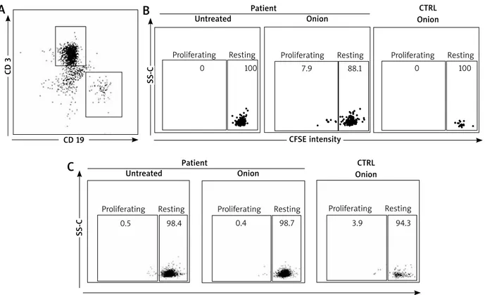
In order to investigate this possibility, we sought for onion-specific B cells in the patient blood. Patient lym-phocytes were stained with CFSE and assessed for their capacity to proliferate in response to the semi-purified in-house onion extract (10 µg). B cells were identified on the basis of the expression of the B cell marker CD19, whereas T cells were identified by the CD3 marker (Figure 3 A). Importantly, CD19+ (Figure 3 B) but not CD3+ cells (Figure 3 C) of the patient proliferated when incubated with the semi-purified in-house onion extract (Figures 3 A, B). Proliferation was not observed in healthy controls (either CD19+ or CD3+ cells).
Discusssion
Onion and other edible plants belonging to the
Al-lioideae family are widely used in the Mediterranean
diet, as well as in other diets, either raw or cooked. So far, despite its large consumption, allergic reactions due to onion ingestion remain anecdotal and, to our knowl-edge, severe allergic reactions following cooked onion ingestion have never been described. Moreover, it was reported that patients that experienced an allergic reac-tion to raw onion ingesreac-tion tolerated cooked onion [2–5].
In our work we describe a case of a severe allergic reaction after cooked onion ingestion.
We studied the allergic response towards onion us-ing different techniques. At first we performed skin prick tests in order to assess the mast-cell bound pool of on-ion-specific IgE. However, although a limited number of well-standardized food allergen extracts exist, most food allergen extracts are little or non-standardized. Thus, the skin reactivity and thereby the evaluation of the amount of allergen specific IgE might be influenced by the qual-ity of the extract (e.g. protein concentration, protein de-naturation etc.). Moreover, the composition of non-stan-dardized, commercially available extracts may vary greatly between manufacturers. With respect to onion, we tested both a commercial onion extract and an in-house onion extract and, in order to achieve normalization, we then expressed the results obtained as a Skin Index, previously defined as the ratio between the area of the wheal gener-ated by the allergen and the area of the wheal genergener-ated by exogenous histamine [7–9]. Importantly, we found that
CD 3
SS-C
Figure 3. Onion-specific B-cell proliferation. A – Gating strategy for CD 19 and CD 3 cells. Representative CD 19+ (B) and CD 3+ (C) cell proliferation assessed by reduction of CFSE intensity for the patient compared to healthy controls. Experi-ments were done in triplicates
A
B
PatientUntreated Onion
Proliferating Resting Proliferating Resting Proliferating Resting
CTRL Onion CD 19 Patient Untreated Onion Proliferating Resting Proliferating Resting Proliferating Resting
CTRL Onion SS-C
C
CFSE intensity 0 0.5 0.4 3.9 7.9 0 100 98.4 98.7 94.3 88.1 100the Skin Index obtained with the in-house onion extract was higher compared to the one obtained with the com-mercial extract. Our results confirm that extract quality matters in food allergy diagnosis.
In order to test the allergenic activity of cooked onion we then performed skin tests with both raw and cooked onion. It is worth noticing that the allergenic activity of onion after heating treatment was reduced but not lost, suggesting the existence of one or more thermostable components (Figure 1 B). Thus, among the characterized onion allergens (All c 3, All c 4, All c), All c 3 belongs to the LTP superfamily and should thereby be considered thermostable [13–16]. In the case reported, this protein could perhaps be responsible for the clinical symptoms experienced by the patient.
As for the postulated cross-reactivity between onion and peach, although some reports seem to convincingly suggest that [3], our findings indicate that this cross reac-tivity is marginal since an excess of peach proteins inhib-ited the IgE-ELISA for onion by approximately 30% only (Figure 2 C). Amino acid sequence identity data (~23%) seem to support this interpretation.
Diagnosis in food allergy may sometimes be diffi-cult as pointed out by the skin test results (see above), thereby new investigational tools may be helpful. It is well known that production of IgE involves B cell and, in particular, antibody-producing plasma-cells. Aside plasma-cells, in pollen-allergic subjects, it has been re-cently demonstrated that the circulating allergen-specific B cell population exists [10]. Importantly, these cells have a memory phenotype (i.e. CD19+, CD20+ and CD27+). Thus, upon encounter with the cognate allergen, they can pro-liferate and eventually give rise to antibody-producing plasma cells. Taken into account these observations, in order to confirm the onion sensitization in our patient, we developed a lymphocyte proliferation test using flow cytometry technique. In fact, we found for the first time an onion-specific CD19+ cell population, able to prolifer-ate in response to onion extract (Figure 3 B). This latter cell population is not present in healthy controls. These findings are further validated by the fact that similar ob-servations could not be done with CD 3+ cells.
Conclusions
Our report shows that cooked onion can induce se-vere allergic reactions that are likely due to the LTP All c 3, a thermostable allergen. Moreover, we applied for the first time a B-cell-based approach to the diagnosis of food allergy. Importantly, this latter approach might also be applied to other allergic conditions such as hymenop-tera venom allergy or drug allergy.
Conflict of interest
The authors declare no conflict of interest.
References
1. Sanchez-Hernandez MC, Hernandez M, Delgado J, et al. Al-lergenic cross reactivity in the Liliaceae family. Allergy 2000; 55: 297-308.
2. Valdivieso R, Subiza J,Varela-Losada S, et al. Bronchial asth-ma, rhynoconjunctivitis and contact dermatitis caused by onion. J Allergy Clin Immunol 1994; 94: 928-30.
3. Asero R, Mistrello G, Roncarolo D, Amato S. A case of onion allergy. J Allegy Clin Immunol 2001; 108: 309-10.
4. Arena A, Cislaghi C, Falagiani P. Anaphylactic reaction to the ingestion of raw onion: a case report. Allergol Immunopathol (Madrid) 2000; 28: 287-9.
5. Antolin-Amerigo D, Gandolfo-Cano M, Gonzalez-de Olano D, et al. Occupational allergy to Spanish omelette. J Investig Allergol Clin Immunol 2012; 22: 522-3.
6. Adkinson N, Bochner B, Burks A, et al. (eds.). Middleton’s Allergy. 8th edn. 2014.
7. Corallino M, Nico A, Kourtis G, et al. Skin testing technique and precision in stinging insect allergy. J Clin Nurs 2007; 16: 1256-64.
8. Macchia L, Caiaffa MF, Di Felice G, et al. Changes in skin reactivity, specific IgE and IgG levels after one year of im-munotherapy in olive pollinosis. Allergy 1991; 46: 410-8. 9. Enrique E, Malek T, De Mateo JA, et al. Involvement of lipid
transfer protein in onion allergy. Ann Allergy Asthma Immu-nol 2007; 98: 202.
10. Wong KJ, Timbrell V, Xi Y, et al. IgE+ B cells are scarce, but allergen-specific B cells with a memory phenotype circulate in patients with allergic rhinitis. Allergy 2015; 70: 420-8. 11. Foti C, Nettis E, Cassano N, et al. Allergy to Muscari
como-sum bulb. Allergy 2007; 62: 1217-8.
12. Bradford MM. A rapid and sensitive method for the quanti-tation of microgram quantities of protein utilizing the prin-ciple of protein-dye binding. Anal Biochem 1976; 72: 248-54. 13. Kao SH, Hsu CH, Su SN, et al. Identification and immunology
characterization of an allergen Alyin lyase from garlic (Alium Sativum). J Allergy Clin Immunol 2004; 113: 161-8.
14. Matricardi PM, Kleine-Tebbe J, Hoffmann HJ, et al. EAACI mo-lecular allergology user’s guide. 2016; Part C. Pediatr. Allergy Immunol 2016; 27 Suppl 23: 1-250.
15. Salcedo G, Sanchez-Monge R, Diaz-Perales A, et al. Plant non-specific lipid transfer proteins as food and pollen al-lergens. Clin Exp Allergy 2004; 34: 1336-41.
16. Cavatorta V, Sforza S, Aquino G, et al. In vitro gastrointes-tinal digestion of the major peach allergen Pru p3, a lipid transfer protein: molecular characterization of the products and assessment of their IgE binding abilities. Mol Nut Food Res 2010; 54: 1452-7.