ABSTRACT
Aim: To assess whether performing routinely
2-deoxy-2-[18F]fluoro-D-glucose PET/CT (18FDG PET/CT) scan from the upper thigh to the vertex of skull is clinically relevant.
Materials and Methods: 3502 (1634 female; mean-age
60+16) consecutive patients undergoing 18FDG PET/CT were retrospectively analyzed. Patients were divided in 10 groups according to primary malignancy. Chi-square analysis was used to assess differences among proportions. A p value < 0.05 was considered significant.
Results: 18FDG PET/CT was positive in head district in 130/3502 (3,7%) patients. In all patients lesions were unknown before PET/CT examination. PET/CT showed 158 positive brain/head uptake in the 130 patients. The 158 lesions were localized in: brain (43/158; 27%), bone (52/158; 33%), lymph node (1/158; 0,6%), soft tissue (55/158; 35%) and other sites (7/158; 4,4%). According to each group, patients were positive in the head district in 1.0% for Gastrointestinal Cancer (7/690), 3.0 % for Genitourinary Cancer (3/101), 3.7 % for Haemathologic Cancer (59/1590), 2.7 % for Gynaecologic Cancer (3/112), 7.8% for Head-Neck-Thyroid and Parathyroid Cancer (26/331), 3.5% for Breast Cancer (7/200), 2.6% for Lung Cancer (7/271), 3.4% for Melanoma (2/59), 7.4% for Sarcoma (2/27), 11.6% for Unknown Primary Tumour (14/121).
Conclusion: Our data show a relatively high incidence of
brain/head lesion in patients with Unknown Primary Tumour.
Keywords: PET/CT; whole-body acquisition; head; brain.
I. INTRODUCTION
2-deoxy-2-[18F]fluoro-D-glucose Positron emission tomography-computed tomography (18FDG PET/CT) is an established whole-body imaging technique used in clinical oncology practice. Whole-body 18FDG PET/CT (FDG-wb-PET/CT) has been used in differentiating and characterizing indeterminate lesions, in differentiating recurrent disease from therapeutic effects, in staging and evaluating the extent of disease, and in monitoring therapy in a variety of cancers [1-3].
Usually oncological 18FDG PET/CT scan is performed from the upper thigh to the base of skull, taking approximately 24-28 minutes, because most of FDG avid lesions are expected within this field of view. However, the detection of head region metastases may change staging and prognosis and thus the management of patients. Brain metastases are a relatively common complication of cancer and can affect up to 40% of patients, depending upon the primary histology, while skull metastases are less frequent [4-6]. The efficacy of FDG PET in describing cerebral metastases is controversial. The sensitivity of FDG PET in revealing cerebral metastases in patients with malignancy has been reported to vary from 68% to 82% when compared with anatomic imaging [4, 7]. This large range of sensitivity likely reflects two factors: first, the high level of physiologic cerebral FDG uptake which may interfere with the identification of them; and second, the inability of PET scanner to show lesions smaller than 6-10 mm. Therefore, many studies suggest that 18FDG PET/CT is suboptimal for the detection of cerebral and skull metastases and many centers do not routinely perform scanning of the head in whole-body FDG PET/CT scan [8-12]. However, 18FDG PET/CT scan including the head can discover unknown lesions in this anatomic district [4, 13-15], infact FDG-wb-PET/CT is the method of choice also for malignant head and neck tumors in both primary staging and restaging and its use, as a part of the therapeutic algorithm, results in a reduced number of unnecessary surgical procedures [16]. 18FDG PET/CT scan including the head may be relevant since all the main cross-sectional anatomical imaging techniques, such as CT and MR, are frequently performed not as total body examination, lacking the ability to detect and delineate intracranial and skull metastases.
Therefore, the aim of this study was to assess whether performing routinely 18FDG PET/CT scan from the upper thigh to the vertex of skull (FDG-wb-PET/CT) be clinically relevant, considering the length of scan time and exposure radiations.
II. METHODOLOGY
Patient population
A total of 3502 consecutive patients (1634 female; mean-age 60+16), undergoing FDG-wb-PET/CT scan for oncological purpose from May 2012 to
Is 2-deoxy-2-[
F]fluoro-D-glucose PET/CT acquisition from the upper thigh to
the vertex of skull useful in oncological patients?
Salvatore B. (1), Caprio M.G. (3), Fonti R. (1), D’Amico D. (4), Fraioli F. (3),
Salvatore M. (3), Pace L. (2).
1Consiglio Nazionale delle Ricerche– Istituto di Biostrutture e Bioimmagini, Napoli, Italy 2 Dipartimento di Medicina e Chirurgia, Università degli Studi di Salerno, Italy
3Dipartimento ad attività integrate di Diagnostica Morfologica e Funzionale, Radioterapia, Medicina Legale. Università degli Studi di Napoli Federico II, Italy
4Fondazione SDN-IRCCS, Napoli, Italy Corresponding author: [email protected]
November 2013, were retrospectively analyzed. All patients were divided according to their primary malignancy localization in 10 groups: Gastrointestinal Cancer, Genitourinary Cancer, Haemathologic Cancer, Gynaecologic Cancer, Head-Neck-Thyroid and Parathyroid Cancer, Breast Cancer, Lung Cancer, Sarcoma, Melanoma, Unknown Primary Tumor.
The present study was performed according to the declaration of Helsinki. All patients gave their informed consent for PET/CT examination. The present retrospective analysis did not require Institutional Review Board approval nor informed consent by national institution.
FDG
FDG was synthesized using a Mini Trace GE cyclotron and CPCU automated chemistry module (Fx-FDG GE). 18Fluorine as fluoride was produced using a proton-neutron reaction on 95% enriched 18oxygen water. 18Fluorine-FDG was synthesized in the CPCU using the modified Hamacher synthesis. The product was delivered pure, sterile and in an injectable form.
Scan acquisition. All patients fasted 8 hours and
underwent a blood glucose test examination before the injection of an average of 370 MBq of 18FDG via a cubital vein. None of the patients included in the study had serum glucose levels higher than 140 mG/dL. Patients were kept quiet and confortable for 60 min after the injection. No muscle relaxants were administered. Immediately before PET/CT scan acquisition, all patients were asked to empty their bladder.
Images were obtained on a Discovery LS (GE Medical Systems, Milwaukee, USA), an integrated system consisting in NXi PET scanner and Light Speed Plus four rows MDCT system.
PET/CT acquisition included a whole body acquisition: in supine position, vertex to upper thigh four-slice spiral CT scan with acquisition parameters of 140 kv, 80 mAs, 0,5 s/CT rotation, a pitch of 1.5, performed during normal breathing. This was followed by a PET acquisition of the same axial range for 4 min per cradle position. Patient’s head was locked to minimize head movement during imaging. 18FDG PET images were reconstructed with a 4.5 mm effective thickness. The CT data were used for attenuation correction and images were reconstructed using a standard iterative algorithm.
Image interpretation. 18FDG-wb-PET/CT images were analyzed on a dedicated workstation (XELERIS, GEMS) in the coronal, sagittal and transaxial planes as well as 3-dimensional projections. Interpretation was based on evaluation of 18FDG uptake at the tumor site and surrounding sites. In particular, distinctly focal uptake higher than surrounding non diseased tissues and with a SUV max > 2.5 was considered as malignant, otherwise it was considered negative. Two experienced nuclear medicine physician, unaware of clinical as well as all other pertinent data, analyzed all patient studies.
Statistical analysis. A Chi-square analysis was used to
assess differences among proportions. A p value < 0.05 was considered significant.
III. RESULTS
Of the 3502 patients included in the study, 19.7% (690/3502) had Gastrointestinal Cancer, 2.8% (101/3502) Genitourinary Cancer, 45.4% (1590/3502) Hematologic Cancer, 3.2% (112/3502) Gynaecologic Cancer, 9.5% (331/3502) Head-Neck-Thyroid-Parathyroid Cancer, 5.7% (200/3502) Breast Cancer, 7.7% (271/3502) Lung Cancer, 1.7% (59/3502) Melanoma, 0.8% (27/3502) Sarcoma and 3.5% (121/3502) Unknown Primary Tumor.
18
FDG-wb-PET/CT was positive in head district in 130/3502 (3.7%) of our population. None of the patients showed symptoms and/or clinical findings suggestive of such lesions. In particular, pathological uptake of 18FDG in the head district was found in 1.0% of Gastrointestinal Cancer patients (7/690), 3.0 % of Genitourinary Cancer patients (3/101), 3.7 % of Haemathologic Cancer patients (59/1590), 2.7 % of Gynaecologic Cancer patients (3/112), 7.8% of Head-Neck-Thyroid and Parathyroid Cancer patients (26/331), 3.5% of Breast Cancer patients (7/200), 2.6% of Lung Cancer patients (7/271), 3.4% of Melanoma patients (2/59), 7.4% of Sarcoma patients (2/27), 11.6% of Unknown Primary Tumour patients (14/121) (p< 0.0001) (Fig.1).
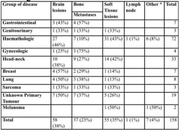
A total of 158 18FDG-wb-PET/CT brain/head lesions were observed in the 130 patients. Of these 158 lesions, 27% (43/158) were cerebral, 33% (52/158) involved bone structures, 0.6% (1/158) involved lymph nodes, 35% (55/158) were localized in soft tissue and 4.4% (7/158) had other head location. Table 1 shows head lesions according to primary cancer disease. Brain lesions were more frequent in patients with lung and breast cancer while bone lesions and soft tissue localizations were more frequent in gastrointestinal and gynaecologic cancer and in haematological disease and melanoma respectively.
Table 1. Number and type of lesions for each cancer
IV. DISCUSSION
In this study a relatively low frequency (3.7%) of head abnormalities at 18FDG-wb-PET/CT was found. This finding is in agreement with that of a previous study [4], performed using only a PET scanner, suggesting that 18
FDG-PET has a little impact in the investigation of cerebral metastases, with 3,9% of abnormal findings in head district.
However, in a particular subsets of disease, Unknown Primary Tumour a somewhat different percentage of head lesions (11.5%) was observed. Thus it seems to be appropriate to perform PET/CT including the whole head in this condition. On the other hand, it has been demonstrated that FDG-PET represents an accurate and useful test for the work-up of patients with suspected or proven intracranial metastases [17].
In a previous study [18] it has been recognized that among common malignant tumours that metastasize to the brain, lung cancer (41,6%), melanoma and breast cancer (8,3%) are more frequent. We found that brain lesions are more frequent in patients with breast and lung cancer as compared with the other neoplastic diseases studied. In particular in patients with lung cancer it has been demonstrated that there is a difference of metabolism in brain metastases from non-small-cell-lung cancer (NSCLC) and in those from small cell lung cancer (SCLC). One third of brain metastases from lung cancer are hypometabolic, but NSCLC was more frequently associated with hypermetabolic metastatic brain lesions than SCLC [19], in our group of patients we found only
hypermetabolic lesions but we haven’t the histological correlation for each lesion.
Two studies performed in patients with melanoma showed that routine skull base to upper thigh acquisition is adequate for this group of patients and in particular PET is an extremely useful tool in the detection of metastatic melanoma, playing a primary role also in the staging of this disease to detect metastic lesions in particular in soft tissues [13, 20] as showed also by our study.
Kole A.C. et al. [15] showed that 18FDG-PET was able to identify the site of unknown primary tumor in 24% of patients unsuccessful studied by Conventional Imaging and in the present study PET was able to identify 1 brain lesion and 13 cervical lymph nodes, according to our results that showed among all types of cancer, PET/CT was positive in head district in 11,5% of patients with UPT, demonstrating the utility to perform a whole-body PET/CT including head in this subsets of patients. These data are also in agreement with two large meta-analysis of the performance of 18FDG-PET in the detection of Unknown Primary Tumor and they have demonstrated that 18FDG-PET could be useful in the staging of these patients [21, 22]. In the same way a recent study by Sebro et al. demonstrated that although the lesions found using a true whole-body FDG PET/CT scan, rarely change the stage, might have impact on clinical management [23].
Our findings demonstrated that soft tissue metastases detected with 18FDG-wb-PET/CT are in a higher percentage (35%) among all detected lesions. These results are quite similar to those of Nguyen NC. et al. [24] that concluded showing the detection of soft tissue metastases, higher in 18FDG-wb-PET/CT, may have a prognostic implications, providing more accessible biopsy sites and avoiding invasive procedures. Moreover Pfannenberg C. et al. [25] have demonstrated 18FDG PET/CT has higher sensitivity than MRI in detecting skin and soft tissue metastases, supporting the increasing role of 18FDG PET/CT in cancer patient management.
We acknowledge the limitations of our retrospective study. First of all we have not a histological correlation for each lesion. There are undoubtedly false positives findings, because 18FDG uptake and resulting increased tracer activity is not limited to neoplastic tissue and in the same way 18FDG PET/CT is suboptimal in detecting brain metastases due to the intense physiologic background uptake in the brain and the hypometabolic nature of some brain metastases. In addition 18FDG PET/CT from the vertex of skull to the upper thigh requires additional several minutes of image acquisition and also an additional radiation dose that can be useless for the all categories of patients.
V. CONCLUSION
In conclusion our data support the importance of the inclusion of head district in the whole body 18FDG PET/CT scan only for patients with Unknown Primary
Tumor where a faster diagnosis could avoid unnecessary extensive procedures at staging or follow-up.
REFERENCES
1. Jeong HJ, Chung JK, Kim YK, Kim CY, Kim DG, Jeong JM et al. Usefulness of whole-body (18)F-FDG PET in patients with suspected metastatic brain tumors. J Nucl Med. 2002 Nov;43(11):1432-7.
2. Facey K, Bradbury I, Laking G and Payne E. Overview of the clinical effectiveness of positron emission tomography imaging in selected cancers. Health Technology Assessment 2007; Vol.11: No.44:iii-iv, xi-267. Review.
3. Mohr P, Eggermont AM, Hauschild A and Buzaid A. Staging of cutaneous melanoma. Ann Oncol. 2009 Aug; 20 Suppl 6 vi14-vi21. Review. 4. Ludwig V, Komori T, Kolb D, Martin WH,
Sandler MP, Delbeke D. Cerebral lesions incidentally detected on 2-deoxy-2-[18F]fluoro-D-glucose positron emission tomography images of patients evaluated for body malignancies. Mol Imaging Biol. 2002 Oct; 4(5):359-62.
5. Yasuda S, Ide M, Fujii H, Nakahara T, Mochizuki Y, Takahashi W et al. Application of positron emission tomography imaging to cancer screening. Br J Cancer. 2000 Dec;83(12):1607-11.
6. Even-Sapir E, Lerman H, Gutman M, Lievshitz G, Zuriel L, Polliack A et al. The presentation of malignant tumours and pre-malignant lesions incidentally found on PET-CT. Eur J Nucl Med Mol Imaging. 2006 May;33(5):541-52.
7. Stubbs E, Kraas J, Morton KA, Clark PB. Brain Abnormalities Detected on Whole-Body 18F-FDG PET in cancer patients: spectrum of findings. AJR 2007; Mar 188 (3):866-73.
8. Kitajima K, Nakamoto Y, Okizuka H, Onishi Y, Senda M, Suganuma N et al. Accuracy of whole-body FDG-PET/CT for detecting brain metastases from non-central nervous system tumors. Ann Nucl Med 2008 Aug 22 (7):595-602.
9. Griffeth LK, Rich KM, Dehdashti F, Simpson JR, Fusselman MJ, McGuire AH et al. Brain metastases from non-central nervous system tumors: evaluation with PET. Radiology 1993;186:37-44.
10. Larcos G, Maisey MN. FDG-PET screening for cerebral metastases in patients with suspected malignancy. Nucl Med Commun 1996;17:197-8. 11. Marom EM, McAdams HP, Erasmus JJ,
Goodman PC, Cluhane DK, Coleman RE et al.
Staging non-small cell lung cancer with whole-body PET. Radiology 1999;212:803-9.
12. Rohren EM, Provenzale JM, Barboriak DP, Coleman RE. Screening for cerebral metastases with FDG PET in patients undergoing whole-body staging of non-central nervous system malignancy. Radiology 2003;226:181-7.
13. Niederkohr RD, Rosenberg J, Shabo G, Quon A. Clinical value of including the head and lower extremities in 18F-FDG PET/CT imaging for patients with malignant melanoma. Nucl Med Commun. 2007 Sep;28(9):688-95.
14. Basu S, Ramani SK, Lad S. Unusual involvement of scalp and bilateral kidneys in an aggressive mediastinal diffuse large B cell lymphoma: documentation by FDG-PET imaging. Clin Nucl Med. 2009 Sep;34(9):638-41.
15. Kole AC, Nieweg OE, Pruim J, Hoekstra HJ, Koops HS, Roodenburg JL et al. Detection of unknown occult primary tumors using Positron Emission Tomography. Cancer 1998: 1160-1166. 16. Ferda J, Ferdovà E, Zàhlava J, Walter J,
Mukenshabi P, Daum O et al. (18)F-FDG-PET/CT of orofacial tumors, a value of whole-body imaging approach. Eur J Radiol. 2010 Feb;73(2):241-8.
17. Gupta N.C., Nicholson P. and Bloomfield S.M. FDG-PET in the staging work-up of patients with Suspected Intracranial Metastatic Tumors. Ann of Surg 1999; 230(2):202-206.
18. Go KG, Pruim J, Que TH, Vaalburg W, Haaxma-Reiche H. Evaluation of dissemination studies with FDG whole-body positron emission tomography in patients with suspected metastatic tumors of brain and spine. Acta Neurochir 2000; 142:627-631.
19. Lee HY, Chung JK, Jeong JM, Lee DS, Kim DG, Jung HW et al. Comparison of FDG-PET findings of brain metastasis from non-small-cell lung cancer and small-cell lung cancer. Ann Nucl Med 2008 (22): 281-286.
20. Holder WD, White RL, Zuger JH, Easton EJ, Greene FL. Effectiveness of positron emission tomography for detection of melanoma metastases. Ann Surg. 1998 May; 227(5):764-71. 21. Delgado-Bolton RC, Fernández-Pérez C,
González-Maté A, Carreras JL. Meta-analysis of the performance of 18F-FDG PET in primary tumor detection in unknown primary tumors. J Nucl Med. 2003 Aug;44(8):1301-14.
22. Kwee TC, Basu S, Cheng G, Alavi A. FDG PET/CT in carcinoma of unknown primary. Eur J Nucl Med Mol Imaging. 2010 Mar;37(3):635-44.
23. Sebro R, Mari-Aparici C, Hernandez-Pampaloni M. Value of true whole-body FDG-PET/CT scanning protocol in oncology: optimization of its use based on primary diagnosis. Acta Radiol. 2013 Mar 5.
24. Nguyen NC, Chaar BT, Osman MM. Prevalence and patterns of soft tissue metastasis: detection with true whole-body F-18 FDG PET/CT. BMC Med Imaging. 2007 Dec 12;7:8.
25. Pfannenberg C, Aschoff P, Schanz S, Eschmann SM, Plathow C, Eigentler TK et al. Prospective comparison of 18F-fluorodeoxyglucose positron emission tomography/computed tomography and whole-body magnetic resonance imaging in staging of advanced malignant melanoma. Eur J Cancer. 2007 Feb;43(3):557-64.