0014-2980/01/0303-812$17.50 + .50/0 © WILEY-VCH Verlag GmbH, D-69451 Weinheim, 2001
Dendritic cells as a major source of
macrophage-derived chemokine/CCL22 in vitro and in vivo
Marisa Vulcano1 , Cristina Albanesi2 , Antonella Stoppacciaro3 , Renzo Bagnati1 , Giovanna D’Amico1 , Sofie Struyf1 , Pietro Transidico1 , Raffaella Bonecchi1 , Annalisa Del Prete1 , Paola Allavena1 , Luigi P. Ruco3 , Chiara Chiabrando1 , Giampiero Girolomoni2 , Alberto Mantovani1,4and Silvano Sozzani1 1
Istituto di Ricerche Farmacologiche “Mario Negri”, Milano, Italy 2
Istituto Dermopatico dell’Immacolata, IRCCS, Roma, Italy 3
Dipt. Medicina Sperimentale e Patologia, Universit `a La Sapienza, Roma, Italy 4
Universit `a di Milano, Milano, Italy
Macrophage-derived chemokine (MDC)/CCL22 is a CC chemokine active on dendritic cells (DC), NK cells and Th2 lymphocytes. The present study was aimed at comprehensively investigating MDC production in vitro and in vivo. DC were the most potent producers of MDC among leukocytes tested. Endothelial cells did not produce MDC under a variety of conditions. Signals that induce maturation (lipopolysaccharide, IL-1, TNF, CD40 ligand, rec-ognition of bacteria and yeast) dramatically augmented MDC production, and dexametha-sone and vitamin D3 blocked it. Prostaglandin E2, which blocked the acquisition of IL-12 production and the capacity to promote Th1 generation, did not affect MDC production. Using mass spectrometry-based techniques, DC supernatants were found to contain N-terminally truncated forms of MDC [MDC(3–69), MDC(5–69) and MD(C7–69)] as well as the full-length molecule. In vivo, CD1a+, CD83+, MDC+DC were found in reactive lymph nodes, and in Langerhans’ cell histiocytosis. Skin lesions of atopic dermatitis patients showed that CD1a+
or CD1b+
DC, and DC with a CD83+
phenotype were responsible for MDC production in this Th2-oriented disorder. Thus, DC are the predominant source of MDC in vitro and in vivo under a variety of experimental and clinical conditions. Processing of MDC to MDC(3–69) and shorter forms which do not recognize CCR4 is likely to represent a feedback mechanism of negative regulation.
Key words: Chemokine / Dendritic cell / Cell trafficking / Allergy
Received 17/10/00 Revised 12/12/00 Accepted 18/12/00
[I 21453]
Abbreviations: DC: Dendritic cell MDC: Macrophage-derived chemokine TARC: Thymus- and activation-regulated chemokine CLA: Cutaneous lymphocyte-associated antigen HUVEC: Human umbilical cord vascular endothelial cell MCP: Monocyte chemotactic protein MIP: Macrophage inflammatory protein CD40L: CD40 ligand
RANTES: Regulated upon activation, normal T cell expressed and secreted DL: Dermatopathic lymphadeno-pathy LCH: Langerhans’ cell histiocytosis
1 Introduction
Dendritic cells (DC) are professional APC that play a piv-otal role in the initiation of specific immunity [1, 2]. Imma-ture DC patrol nonlymphoid tissues and are very efficient in taking up incoming antigens. In response to inflamma-tory signals DC undergo maturation, a process during
which DC increase membrane expression of MHC and co-stimulatory molecules and augment their ability to activate T lymphocytes, including naive T cells [1–5]. A necessary step in the induction of immunity is the tion of DC to lymphoid organs and spleen. In vivo migra-tion of DC is controlled by a rapid and coordinated regu-lation of chemokines and chemokine receptors during DC maturation [3–5].
Macrophage-derived chemokine (MDC)/CCL22 [6] is a CC chemokine produced in a constitutive way by macro-phages and DC, and by activated B lymphocytes in vitro [7–11]. MDC is chemotactic for DC, IL-2-activated NK cells and chronically activated T lymphocytes [7–10]. It binds to and activates CCR4, a chemokine receptor shared with thymus- and activation-regulated chemo-kine (TARC)/CCL17 [6,12], and preferentially expressed by CD4+and CD8+lymphocytes with a Th2/Tc2 pheno-type, and cutaneous lymphocyte-associated antigen (CLA)+
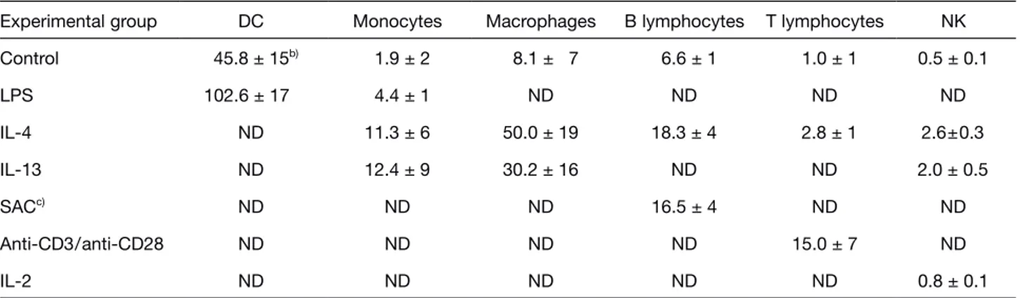
Table 1. In vitro MDC production by different leukocyte subsetsa)
Experimental group DC Monocytes Macrophages B lymphocytes T lymphocytes NK
Control 45.8 ± 15b) 1.9 ± 2 8.1 ± 7 6.6 ± 1 1.0 ± 1 0.5 ± 0.1 LPS 102.6 ± 17 4.4 ± 1 ND ND ND ND IL-4 ND 11.3 ± 6 50.0 ± 19 18.3 ± 4 2.8 ± 1 2.6±0.3 IL-13 ND 12.4 ± 9 30.2 ± 16 ND ND 2.0 ± 0.5 SACc) ND ND ND 16.5 ± 4 ND ND Anti-CD3/anti-CD28 ND ND ND ND 15.0 ± 7 ND IL-2 ND ND ND ND ND 0.8 ± 0.1
a) Cells were incubated for 48 h in the presence of the agonists. b) MDC levels (ng/106
cells), evaluated by ELISA, are expressed as mean values ± SD of three to eight independent experiments. c) Staphylococcus aureus Cowan strain.
MDC production is not constitutive but it can be induced by Th2 cytokines such as IL-4 and IL-13, and down-regulated by IFN-+ , a prototypic Th1 cytokine [14, 16]. Chemokines, including MDC, can be processed by CD26, a dipeptidylpeptidase expressed by endothelial and epithelial cells, and activated T lymphocytes [17]. Chemokine truncation by CD26 results in no change, loss or increase of their biological activity according to the target chemokine [17]. MDC truncation by CD26 results in the production of proteins lacking two or four N-terminal amino acids that have no ability to interact with CCR4 [18].
Because of its regulation by IL-4 and IL-13, and the pref-erential expression of CD26 by Th1 vs. Th2 cells, MDC is believed to be part of a Th2 amplification loop and to play a role in the selective migration of Th2 lymphocytes in conditions such as atopic disorders and allergies, that are largely mediated by these cells [19–21].
In vivo, MDC message expression is found in the thymus and in lymph nodes [7, 22–24]. In mouse lymph nodes MDC mRNA expression is confined to DC present in the T cell areas [23]. CD11c+
cells purified from mouse lymph nodes were also found to express MDC mRNA, with the expression increased after in vitro maturation [9, 23]. No data about in vivo MDC protein production are so far available, and information about MDC expression in human DC is scanty. The goal of the present study was to comprehensively characterize the regulation of MDC production in immature and mature human DC, and to examine the expression of MDC protein in human patho-logical conditions. Furthermore, a new analytical method was developed to identify the presence of MDC pro-cessed forms.
2 Results
2.1 MDC production by leukocytes
Table 1 shows that, as previously reported [7], DC and monocyte-derived macrophages secrete MDC in the absence of deliberate stimulation. LPS strongly up-regulated the production in DC, while it was only a mod-est activator in monocytes. IL-4 and IL-13 induced MDC production in monocytes, monocyte-derived macro-phages, in B lymphocytes and in NK cells. In T lympho-cytes anti-CD3/anti-CD28 stimulation was the most effective stimulation for MDC production. Neither resting endothelial cells [human umbilical cord vascular endo-thelial cells (HUVEC) and human microvascular endothe-lial cell line (HMEC)] nor IL-4-, IL-13-, or IFN+ -activated HUVEC produced MDC (data not shown). These results indicated that DC and macrophages are the most potent MDC-producing cells in vitro.
2.2 MDC production during DC maturation DC generated from monocytes secreted MDC with a rate of production that declined by the end of the culture and resulted in 95.5±50 ng/106
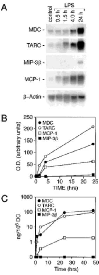
cells MDC (average ± SD; range 24–170; n=10) on day 6. At this time, DC were washed and incubated in the presence of 10 ng/ml LPS. As shown in Fig. 1A and B, basal expression was strongly up-regulated starting 90 min after stimulation and increased up to 24 h. Accumulation of MDC RNA was followed by protein secretion that peaked at 24 h (Fig. 1C). At this time point the average concentration of MDC present in the medium was 130±30 ng/106
cells (range 40–300; n=13; Fig. 2B). Under the same experi-mental conditions, TARC/CCL17, the CC chemokine that
Fig. 1. Up-regulation of MDC, TARC, MIP-3g and MCP-1 in
human monocyte-derived DC. (A) Northern blot analysis of total RNA (10 ? g/lane) purified from DC stimulated with 10 ng/ml LPS. (B) Densitometric analysis of the autoradio-graphs (24 h exposure) shown in (A). (C) ELISA determina-tion of chemokines in the supernatants of DC shown in (A).
Fig. 2. MDC expression in mature DC. DC were stimulated with 20 ng/ml IL-1; 20 ng/ml TNF; 10 ng/ml LPS; CD40L (4:1) for 24 h.
(A) Monocyte-derived DC; (B) CD34+
-derived DC. Cells were analyzed in Northern blot assays (top panels) as described above. Supernatants were tested for MDC production by ELISA (bottom panels). Results are presented as average values of three (A) or two (B) experiments.
shares the same receptor with MDC and is similarly regu-lated in other cell types, was induced by LPS with com-parable kinetics (Fig. 1A and B) and a mean protein level of 204±50 ng/106
cells (n=3) at 24 h. On the other hand, monocyte chemotactic protein (MCP)-1/CCL2 and mac-rophage inflammatory protein (MIP)-3g /CCL19, two additional CC chemokines, were induced to a much lesser extent at the message levels (Fig. 1A and B) and MIP-3g protein production was below the sensitivity of the detection system used (1 ng/ml; Fig. 1C).
DC can be induced to acquire a mature phenotype by stimulation with pathogen-derived agonists (LPS), pro-inflammatory cytokines (IL-1 and TNF), and T cell-derived signals (e.g. CD40 ligation) [1, 2]. Fig. 2A shows that engagement of CD40, or incubation with LPS, TNF or IL-1 had a comparable ability in inducing MDC synthe-sis and release by maturing DC. Similarly, DC derived from CD34+
cells showed a basal expression of MDC that was also up-regulated by LPS, IL-1 or CD40 ligand (CD40L; Fig. 2B, and data not shown). In addition, DC readily produced very high levels of MDC and TARC, moderate to high levels of regulated upon activation, normal T cell expressed and secreted (RANTES), MIP-1§ and IL-8, and low or negligible amounts of MCP-1 (Fig. 3). It is interesting to note that CD40 ligation, an immune signal, is more effective in the induction of con-stitutive chemokines than inflammatory chemokines. Conversely, inflammatory signals, like LPS, are strong inducers of both “classes” of chemokines.
Fig. 3. Induction of constitutive and inflammatory
chemoki-nes by LPS and CD40 ligation in DC. DC were incubated in the presence of 10 ng/ml LPS or CD40L (4:1, DC:transfect-ants) for 24 h. Supernatants were evaluated for the presence of chemokines by ELISA. Results are the average determi-nations (± SD) of three independent experiments.
2.3 Regulation of MDC production
The ability of DC to induce naive T lymphocyte activation may be altered by agents that interfere with cell matura-tion or cytokine producmatura-tion. Corticosteroids, vitamin D3 and PGE2 exert multiple effects on DC functions and share the ability to inhibit IL-12 production by these cells [25]. Interaction of DC with T cells in the absence of IL-12 may support the generation of a Th2-skewed response
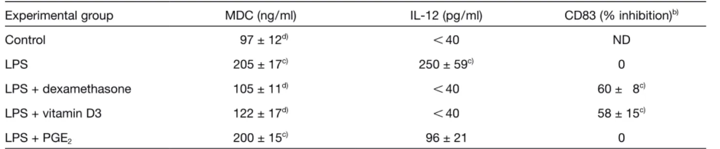
Table 2. Modulation of MDC production by inhibitors of IL-12 production in DCa)
Experimental group MDC (ng/ml) IL-12 (pg/ml) CD83 (% inhibition)b)
Control 97 ± 12d) X 40 ND LPS 205 ± 17c) 250 ± 59c) 0 LPS + dexamethasone 105 ± 11d) X 40 60 ± 8c) LPS + vitamin D3 122 ± 17d) X 40 58 ± 15c) LPS + PGE2 200 ± 15c) 96 ± 21 0 a) Immature DC (106
/ml) were exposed to 10 ? M dexamethasone, 10 ? M PGE2, 1 ? M vitamin D3 for 6 h before stimulation with 100 ng/ml LPS for 48 h. Results are the average numbers ± SD of three to five independent experiments. MDC levels were evaluated by ELISA.
b) % of inhibition of mean fluoresence channels with respect to LPS values. c) p X 0.05 vs. control by paired Student’s t-test.
c) p X 0.05 vs. LPS by paired Student’s t-test.
[26]. Table 2 shows that dexamethasone and vitamin D3 decreased MDC production induced by LPS. On the contrary, PGE2 did not have a statistically significant effect. As expected dexamethasone and vitamin D3, but not PGE2, inhibited DC maturation as assessed by CD83 induction (Table 2). Therefore, MDC production by maturing DC is apparently correlated with cell maturation but not with IL-12 production.
Immature DC take up antigens by multiple mechanisms [1, 27]. To evaluate whether these pathways could induce MDC production, DC were incubated in the pres-ence of dextran (mannose receptor mediated), albumin (fluid phase pinocytosis) or opsonized zymosan (FcR mediated). Fig. 4 reports that neither the uptake of dex-tran nor that of albumin induced per se MDC secretion. In contrast, phagocytosis of opsonized zymosan, a strong cell activator signal [28], was as effective as LPS in inducing MDC production (Fig. 4). Additionally, two prototypic pathogens, OK432 (gram-positive bacteria), and Candida albicans, were as strong as LPS in inducing MDC release (Fig. 4).
2.4 Presence of MDC and its N-terminally truncated forms by HPLC-MS/MS
The biological activity of MDC is strictly dependent on an intact N terminus. Truncation of the protein by proteolytic enzymes, including CD26, hampers the interaction of the protein with CCR4, the only known MDC receptor [12]. Since all available ELISA cannot discriminate the differ-ent MDC forms, we devised a novel HPLC-MS/MS assays for such measurements. As reported in Fig. 5A, this technique allowed to discriminate among the differ-ent molecular masses of full-length and truncated pro-teins in a quantitative way. Different amounts of
full-Fig. 4. Role of endocytosis in MDC production by DC. DC
were cultured in the presence of 1 mg/ml dextran; 1 mg/ml albumin; opsonized zymosan (OZA; 200 ? g/ml); OK432 (100 ? g/ml); C. albicans (100 ? g/ml) for 24 h and then tested by ELISA for their ability to release MDC. Average numbers (±SD) of three to seven independent experiments are reported.
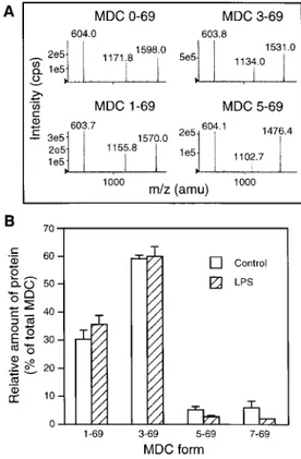
Fig. 5. Detection of MDC and its truncated forms by
HPLC-MS/MS with ion spray ionization analysis in immature and mature DC supernatants. (A) MS/MS spectra of MDC pro-teins. The spectra were obtained by fragmentation of the parent [M + 7H]7+
ions (m/z 1172, 1156, 1134, 1103) of the different MDC forms. The m/z 604 product ion is common to all forms and corresponds to a triply-charged y fragment (y153+
). The other product ions (m/z 1598, 1570, 1531, 1476) are the complementary quadruply-charged b fragments (b554+
, b544+ , b524+
, b504+
). (B) Relative presence of intact and truncated forms of MDC in the supernatants of immature and LPS-maturated DC (n=4). Total MDC concentration (100%), as assessed by ELISA, was 64±24 ng/ml and 168±39 ng/ml (n=4) for immature and mature DC, respec-tively. The equivalent figures obtained by HPLC-MS/MS (sum of the different forms) were 54±14 ng/ml and 189±28 ng/ml, respectively.
length and truncated proteins were present in DC super-natants at 48 h (Fig. 5B). MDC(1–69) and MDC(3–69) were the two predominant forms, representing 30 and 59% of the total protein, respectively. Limited amounts of MDC(5–69) and MDC(7–69) were also detected, mak-ing up 2.3 and 5.9% of total MDC, respectively (Fig. 5B). MDC levels (ng/ml) measured by ELISA (y) were strongly correlated with the sum of all MDC forms measured by HPLC-MS/MS (x) (r=0.88, pX 0.004; n=8; y=10+0.9x). This result was expected, given the 100% cross-reactivity of the ELISA antibody against N-terminally truncated forms of MDC.
2.5 Expression of MDC in human tissues
In all sections from normal human tissues (skin, lung, intestine, thyroid, salivary glands, thymus and lymph nodes) but thymus and lymph nodes, MDC+cells were absent. In the thymus, immunostaining for MDC was detected in medullary epithelial cells and in DC as previ-ously reported [24] (data not shown).
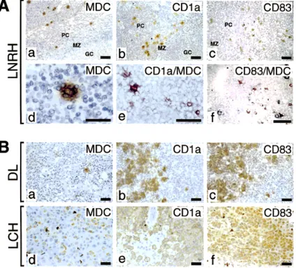
In human lymph node reactive hyperplasia, MDC+ cells were confined to the T cell-dependent paracortical area, the same area where also CD1a+Langerhans’ cells and CD83+
DC localize (Fig. 6A, top panels a, b, c). Similar to Langerhans’ cells, they show dendritic morphology and form rosettes with the nearby lymphocytes (Fig. 6A, panel d). Double staining experiments showed that MDC+cells constituted a subset of CD1a+cells (mean: 57%; range: 23–68%) and of CD83+
cells (mean: 21%; range: 18–37%; Fig. 6A, panels e and f). No CD68+
cells, a marker for macrophages, were found to express MDC
(data not shown). Therefore, MDC production in tissues is confined to a mature subset of Langerhans’ cells. Subsequent experiments evaluated MDC production in two pathological conditions characterized by accumula-tion of Langerhans’ cells, namely dermatopathic lymph-adenopathy (DL), and Langerhans’ cell histiocytosis (LCH). DL is a pathological condition characterized by the massive accumulation of CD1a+/CD83+Langerhans’ cells in the lymph node paracortex (Fig 6B, panels b and c). LCH is a pathological condition characterized by tissue localization of CD1a+
/CD83+
Fig. 6. Expression of MDC in human lymph node reactive hyperplasia (LNRH), dermopathic lymphadenopathy (DL), and
Langer-hans’ cell histiocytosis (LCH). (A) LNRH: MDC immunostaining shows scattered cells located in the paracortex (a) that can form rosettes with lymphocytes (d). MDC+
cells localize in the same area of CD1a Langherans’ cells (b) and CD83+
DC (c). Section of LNRH were double stained for MDC (red color) and CD1a (e) or CD83 (f) (black color). MDC co-localizes with CD1a, and CD83 in DC of the paracortex. (B) DL: MDC+
cells are few and scattered in the residual normal paracortex (a). The paracortex is partially effaced by the accumulation of CD1a+
- (b), and CD83+
- (c) MDC negative Langerhans’ cells (a). LCH: The majority of the Langer-hans’ cells express MDC (d) as shown by CD1a (e) and CD83 (f) staining of contiguous sections. Avidin-biotin-horseradish perox-idase complex developed with 3–3’diaminobenzidine, counterstained with haematoxylin. 400x, or 1000x (panels e and f) enlarg-ments. PC = paracortex; MZ = mantle zone; GC = germinal center.
(Fig. 6B, panels e and f). Fig. 6B shows that Langerhans’ cells present in LHC, but not those present in DL, were positive for MDC expression (panels d and a, respec-tively).
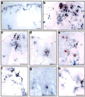
Tissue sections from normal human skin, chronic atopic dermatitis lesional skin, positive patch test reaction to nickel and chronic psoriasis lesions were also evaluated. MDC expression was absent in normal skin (Fig. 7A). Conversely, MDC-positive cells were observed in sec-tions from atopic dermatitis (Fig. 7B-E), allergic contact dermatitis skin (Fig. 7F-H) and psoriasis, in both the epi-dermis and epi-dermis. Double staining showed that the cells positive for MDC were mostly a subset of CD1a+ (4.4–10.1%) or CD1b+
(9.1–30.5%) cells, and a higher percentage of CD83+
DC, especially in atopic dermatitis skin (35.2%; Table 3). Only a minority (3.2–4.4%) of CD14+ cells were stained by anti-MDC mAb. Very few MDC-expressing cells were T cells, with less than 2% of CD3+
being also positive for MDC in all the skin diseases studied (data not shown). No B cells and scanty CD56+
cells (NK cells) could be detected in skin sections, and they were not investigated for MDC co-expression. Thus, MDC production in lesional skin appears to be confined to DC, and particularly to DC with a mature phenotype (CD83+
), with only a minor contribution of CD14+ mono-cytes and even less of T lymphomono-cytes. It should be noted, however, that only a portion (18.6–35.2%) of mature DC expressed MDC, a finding possibly related to the different maturational stages of infiltrating DC. Atopic dermatitis presented a higher number of CD1a+
and CD1b+
DC, and of mature CD83+
DC, as well as a higher percentage of MDC+
cells compared to allergic contact dermatitis and psoriasis (Table 3).
3 Discussion
This study investigates the regulation of MDC/CCL22 production by leukocytes in vitro and in vivo, and pro-vides three main new findings. First, DC are the major producers in vitro, and probably the unique
leuko-Fig. 7. Expression of MDC in atopic dermatitis and allergic contact dermatitis. Sections from normal human skin (a), atopic
der-matitis (b-e) and allergic contact derder-matitis (f-h) were double stained for MDC (red color) and CD1a (a, b, f), CD83 (c and g), CD1b (d and h) or CD14 (e) (blue color). In panels c, e, f and h arrows indicate the double positive cells. Bars, 25 ? m.
cyte subsets to produce MDC in vivo (Table 1, 3, and Fig. 6, 7). Second, MDC production is mostly associated with the mature CD83+
DC phenotype both in vitro and in vivo. However, in vivo, also CD83–
DC can express MDC protein (Fig. 6B, D-F). Third, DC supernatants contain MDC both as intact protein and as truncated forms, as assessed by a new analytical method.
Table 3. MDC expression in cells of the DC or monocyte lineage in Th2- and Th2-mediated skin diseasesa) MDC+ /CD1a+ MDC+ /CD83+ MDC+ /CD1b+ MDC+ /CD14+ Normal skin (n = 3) 1 ± 1/176 ± 20 1 ± 1/12 ± 4 0/70 ± 10 0/70 ± 12 Atopic dermatitis (n = 5) 42 ± 6/418 ± 32 44 ± 5/125 ± 15 39 ± 6/128 ± 10 7 ± 2/220 ± 16 (10.1 %) (35.2 %) (30.5 %) (3.2 %)
Allergic contact dermatitis (n = 4) 12 ± 3/272 ± 18 8 ± 2/42 ± 4 6 ± 3/76 ± 8 5 ± 2/138 ± 11
(4.4 %) (19 %) (9.1) 3.6 %)
Psoriasis vulgaris (n = 3) 18 ± 4/330 ± 32 11 ± 3/59 ± 7 ND 7 ± 3/160 ± 21
(5.4 %) (18.6 %) (4.4 %)
a) Skin sections were double stained for MDC and the indicated membrane markers. Slides were analyzed blind by two observers and positive cells were counted with an eyepiece graticule at a magnification of 200 ×. For each biopsy, two sections were stained for each mAb and positive cells were evaluated in ten adjacent fields. Results are expressed as the mean number ± SD (and percentage) of double-positive on surface marker positive cells.
In DC, production of MDC was biphasic, the first phase being present during the differentiation of monocytes to immature DC, and the second phase being induced by signals able to drive DC maturation. This finding con-firms previous reports in which MDC expression was evaluated at the mRNA level [7, 22, 23], and extend them showing that MDC is, together with TARC, the most
abundant chemokine produced by DC with over 100 ng/ 106
cells released in 24 h. In fact, other inflammatory chemokines, such as RANTES, MIP-1§ and IL-8 are pro-duced in lower amounts, while MCP-1 and MIP-3g were released at very low levels. When different leukocyte populations were compared for their ability to release MDC in vitro, DC appeared the most relevant producers, followed by monocyte-derived macrophages G mono-cytes G B lymphocytes G T lymphocytes G Th2 and Th1 polarized cells (Table 1 and [29]). CCR4 is also up-regulated in mature DC [30], suggesting a role for MDC and TARC in co-localization and interaction of DC and T lymphocytes. Another chemokine produced by immature DC and up-regulated during maturation is fractalkine. Unlike MDC, in DC fractalkine is not induced by inflam-matory stimuli (e.g. LPS and TNF) and it is almost exclu-sively up-regulated by CD40L [31] in migratory murine skin DC. Since fractalkine is active on activated T lym-phocytes, but not on DC, it is tempting to speculate that the expression of fractalkine and MDC by mature DC is not redundant and might promote different cell-to-cell interactions.
All the maturation signals tested in vitro, including LPS, C. albicans, gram-positive bacteria (OK432), IL-1 and TNF, induced MDC production. Similarly, phagocytosis of opsonized zymosan, an event known to induce pro-duction of pro-inflammatory cytokines (e.g. IL-1 and TNF) [28] induced levels of MDC comparable to those observed with LPS. On the contrary, endocytosis of dex-tran and albumin per se were not effective in inducing MDC production. Inhibition of LPS-induced DC matura-tion by dexamethasone and vitamin D3 resulted in a reduced production of MDC. On the contrary, PGE2 treatment, which does not inhibit DC maturation, had no effect on the release of this chemokine.
HPLC-MS/MS analysis of MDC in the supernatants of immature and mature DC revealed that the protein is present in different molecular forms. Full-length (1–69) MDC as well as its truncated forms, devoid of two (3–69), four (5–69) or six (7–69) amino acids were detected, with MDC(3–69) and MDC(1–69) being the predominant molecular species. Maturation of DC with LPS did not change the relative proportion of the truncated forms. The membrane dipeptidylpeptidase CD26 can generate truncated forms of MDC (3–69 and 5–69) in vitro. A fur-ther processed form of MDC (9–69) was purified from the supernatant of a CD8+
T cell clone, suggesting the involvement of additional proteases in MDC processing [32]. MDC(3–69) and MDC(5–69) lack the ability to bind and activate CCR4, therefore they are inactive on T lym-phocytes and DC but surprisingly retain chemotactic activity for human monocytes [18]. If the data presented in this study reflect an in vivo situation, it means that
some of the MDC produced by DC may be directed to monocytes (effectors of a Th1 response) rather than to Th2 cells. The assays currently available (e.g. ELISA, immunohistochemistry or mRNA measurements) do not allow to discriminate between intact and truncated forms of MDC. Therefore, the availability of this new technique to detect N-terminally processed proteins provides a new tool to elucidate the role of chemokines in vivo. Immunohistochemical analysis showed that under nor-mal conditions, MDC expression is confined only to lymph nodes and thymus. In reactive lymph node hyper-plasia, as well as in skin affected by atopic dermatitis, allergic contact dermatitis or psoriasis, MDC-positive cells were mostly CD1a+
or CD1b+
DC, and many of them expressed a CD83+
mature phenotype. In contrast, only a minority of CD14+
or CD68+
cells and very few CD3+ T lymphocytes were stained by anti-MDC mAb. Thus, also in vivo, DC seem to be the major MDC-producing cell population. CD1a+
and CD83+
mature DC are found increased in LCH and DL [33]. However, MDC-producing cells were observed only in the former situation. LCH, but not DL, is characterized by a massive local produc-tion of inflammatory cytokines (GM-CSF, TNF, IL-1, IL-2, IL-4, IL-5 and IFN) [34,35]. This finding strongly suggests that also in vivo, MDC production is not exclusively asso-ciated with a mature DC phenotype but may be regu-lated by the local cytokine context.
Atopic dermatitis is a pathology characterized by an abundant infiltrate of Langerhans’ cells and DC with the features of monocyte-derived DC [36]. Although in lower numbers, MDC-positive DC were also detected in lesio-nal biopsies of psoriasis and allergic contact dermatitis to nickel. Atopic dermatitis is associated with a predomi-nant expansion and activation of Th2 cells whereas aller-gic contact dermatitis and psoriasis are primarily Th1-mediated diseases [11, 37]. These data indicated that MDC production, without being an exclusive character-istic of atopic disorders, is preferentially associated with a Th2 rather than a Th1 lymphocyte infiltrate. Recently it was shown that atopic dermatitis patients have increased levels of circulating MDC compared to patients suffering from Th1-dominated disorders or con-trol subjects [38]. Moreover, in the atopic dermatitis-like lesions of NC/Nga mice MDC is expressed by dermal DC [39]. MDC is chemotactic for T lymphocytes that express the skin homing receptor, CLA [15]. Thus, MDC expressed by CD83+
DC in the skin may function to recruit CCR4+
lymphocytes in chronically inflamed skin. Collectively, these results indicate that CD1a+and CD83+ DC are the main cells that produce MDC both in vitro and in vivo when exposed to an appropriate cytokine mic-roenviroment. The possibility to detect cleaved forms of
the protein in crude supernatants, and possibly in bio-logical fluids, will be instrumental to investigate the regu-lation of this chemokine by proteases and in particularly by CD26, an enzyme preferentially expressed by Th1 lymphocytes.
4 Materials and methods
4.1 Cytokines and reagents
GM-CSF was a gift from Novartis (Milan, Italy). Human IL-13 was a gift from Dr. A. Minty, Sanofi Elf Bio Recherches (Lab `ege, France). Human TNF-§ and IL-1 g were from BASF/ Knoll (Germany) and Chiron (Milan, Italy), respectively. All the other reagents were from Sigma (St. Louis, MO) and were endotoxin free. OK432, a lyophilized preparation of attenu-ated Su strain (group A, type 3) of Streptococcus pyogenes was a gift from Chugai Pharmaceutical Co. Ltd. (Tokyo, Japan). Heat-inactivated C. albicans was from ATCC (Rock-ville, MD).
4.2 Leukocyte preparation
DC were generated in vitro as previously described [40] by incubating blood monocytes with 50 ng/ml GM-CSF and 20 ng/ml IL-13. DC were also prepared from purified cord blood CD34+
cells [41]. DC maturation was achieved in the presence of 10 ng/ml LPS, 20 ng/ml TNF or 20 ng/ml IL-1 for 24 h, or as otherwise specified. CD40L-transfected J558L cells or mock-transfected control cells were cultured with DC at a 1:4 ratio. Macrophages were derived from fresh monocytes cultured with 1000 U/ml M-CSF for 8 days. Total T cells were separated from buffy coats by Ficoll gradient. T cells (106/well) were cultured in 24-well flat-bottom plates pre-coated with anti-human CD3 mAb (5 ? g/ml, OKT3) plus anti-human CD28 mAb (1 ? g/ml, PharMingen, San Diego, CA), overnight. Supernatants were collected after 48 h. NK cells were obtained as previously described [42].
4.3 Northern blot analysis
Total RNA was extracted by the guanidinium thiocyanate method, blotted and hybridized as described [40]. The MDC- and MCP-1/CCL2-specific probes were obtained as described [7]. The human full-length MIP-3g /CCL19-specific probe was excised from EST clone W05519 (IMAGE Consortium, Research Genetics, Huntsville, AL) with NotI and EcoRI. The TARC/CCL17-specific probe was obtained by reverse transcription-PCR amplifying the full-length cDNA reported sequence (AA175762) with specific primers (5’-ATGGCCCCACTGAAGATGCTGGCC-3’ and 5’-TCAA-GACCTCTCAAGGCTTTGCAG-3’), and confirmed by se-quencing.
4.4 ELISA
The MDC ELISA based on antibodies generously provided by Dr. P. A. Gray (ICOS, Bothell, WA) was previously described [16]. The antibodies cross-reacted 100% with truncated forms of MDC, MD(C3–69) and MDC(5–69). Sand-wich ELISA for TARC and MIP-3g (R<ISO>D Systems, Min-neapolis, MN), RANTES/CCL5 (Amersham) and MIP-1§ / CCL3 (Endogen) were used. ELISA for IL-8 and MCP-1 were previously described [43].
4.5 Analysis of MDC and its truncated forms by HPLC-MS/MS
DC supernatants were spiked with internal MDC(0–69) stan-dard, lyophilized, reconstituted with water/acetonitrile 1:1, and acidified to pH 6 with 1 N formic acid. Samples were then analyzed by HPLC-MS/MS. Control samples run in par-allel showed that the presence of 10% FCS in the incubation medium did not change the relative amount of truncated forms of MDC recovered at the end of the incubation time (48 h; data not shown). Analysis of MDC forms was per-formed with a newly developed method using a PE Sciex API 3000 triple quadrupole instrument, interfaced with Perkin-Elmer Series 200 micro LC pumps and a TurboIon Spray source operated in the positive ionization mode (Bag-nati et al., in preparation). MS source conditions were set to maximize analyte signals and a collision energy of -46 eV was used for fragmentation by collision-activated dissocia-tion (CAD). For each compound, two MS/MS transidissocia-tions from the precursor ion [M + 7H]7+
to its two major product ions (Fig. 5A) were simultaneously monitored: m/z 1172 - G 1598 and 1172 - G 604 for MDC(0–69), m/z 1156 - G 1570 and 1156 - G 604 for MDC(1–69), m/z 1134 - G 1531 and 1134 - G 604 for MDC(3–69), m/z 1103 - G 1476 and 1103 - G 604 for MDC(5–69).
4.6 Immunohistochemistry
Normal lung (n=3), intestine (n=2), thyroid (n=5), salivary glands (n=2), thymus (n=2) and lymph node reactive hyper-plasia (n=4) specimens were obtained from routine surgery (University of Brescia, Italy). Punch biopsies of normal skin (n=3), chronic atopic dermatitis lesional skin (n=5), 48-h patch test reactions to nickel (n=4) and psoriasis lesions (n=3) were obtained at IDI (Rome, Italy). Single stainings were performed with mAb against: MDC mAb clones 272D and 272Z [24], CD1a, CD86 (PharMingen), CD83 (Immuno-tex, Marseille, France), CD68 and CD14 (Becton and Dickin-son, Mountain View, CA), developed with ABC kit (Vector Laboratories), stained with 0.03% H2O2and 0.06% 3–3’dia-minobenzidine (Dako) in PBS and counterstained with hematoxylin. Double immunostaining was performed with the same MDC mAb and mAb CD1a (1:20), anti-CD1b (1:30), anti-CD83 (1:10), anti-CD14 (1:10) or anti-CD3 (1:20), using avidin-biotin-peroxidase or
avidin-biotin-alkaline phosphatase systems (Vector Laboratories). 3-amino-9-ethylcarbazole and Blue Vector (Vector Laborato-ries) were used as chromogens to reveal the peroxidase and alkaline phosphatase activities, respectively. No counter-stain was applied. For each biopsy, two sections were stained for each mAb and positive cells were evaluated in ten adjacent fields.
Acknowledgments: We thank Drs. Corcione and Pistoia
(Genoa, Italy) for their help in B lymphocyte studies. This study was partially supported by the Associazione Italiana per la Ricerca sul Cancro (AIRC), the National Research Council (CNR) Finalized Project Biotechnology, Bio4-CT97–2167, BMH4-CT98–2343, BMH4-CT98–3713, and Fondazione Pasteur Cenci Bolognetti. M. Vulcano is the recipient of a CONICET fellowship. R. Bonecchi is a FIRC fellow.
References
1 Bell, D., Young, J. W. and Banchereau, J., Dendritic cells. Adv.
Immunol. 1999. 72: 255–324.
2 Bancherau, J. and Steinman, R. M., Dendritic cells and the con-trol of immunity. Nature 1998. 392: 245–252.
3 Cyster, J. G., Chemokines and the homing of dendritic cells to the T cell areas of lymphoid organs. J. Exp. Med. 1999. 189: 447–450.
4 Sallusto, F. and Lanzavecchia, A., Mobilizing dendritic cells for tolerance, priming, and chronic inflammation. J. Exp. Med. 1999.
189: 611–614.
5 Sozzani, S., Allavena, P., Vecchi, A. and Mantovani, A., The role of chemokines in the regulation of dendritic cell trafficking.
J. Leukoc. Biol. 1999. 66: 1–9.
6 Zlotnik, A. and Yoshie, O., Chemokines: a new classification system and their role in immunity. Immunity 2000. 12: 121–127. 7 Godiska, R., Chantry, D., Raport, C. J., Sozzani, S., Allavena,
P., Leviten, D., Mantovani, A. and Gray, P. W., Human
macro-phage derived chemokine (MDC) a novel chemoattractant for monocytes, monocyte derived dendritic cells, and natural killer cells. J. Exp. Med. 1997. 185: 1595–1604.
8 Chang, M. S., Mcninch, J., Elias, C., Manthey, C. L.,
Gross-hans, D., Meng, T., Boone, T. and Andrew, D. P., Molecular
cloning and functional characterization of a novel CC chemokine, stimulated T cell chemotactic protein (STCP-1) that specifically acts on activated T lymphocytes. J. Biol. Chem. 1997. 272: 25229–25237.
9 Schaniel, C., Pardali, E., Sallusto, F., Speletas, M., Ruedl, C.,
Seidl, T., Anderson, J., Melchers, F., Rolink, A. G. and Sideras, P., Activated murine B lymphocytes and dendritic cells produce a
novel CC chemokine which acts selectively on activated T cells.
J. Exp. Med. 1998. 188: 451–463.
10 Mantovani, A., Gray, P. A., Van Damme, J. and Sozzani, S., Macrophage-derived chemokine. J. Leukoc. Biol. 2000. 68: 400–404.
11 Bos, J. D. and De Rie, M. A., The pathogenesis of psoriasis: immunological facts and speculations. Immunol. Today 1999. 20: 40–46.
12 Imai, T., Chantry, D., Raport, C. J., Wood, C. L., Nishimura, M.,
Godiska, R., Yoshie, O. and Gray, P. W., Macrophage-derived
chemokine is a functional ligand for the CC chemokine receptor 4. J. Biol. Chem. 1998. 273: 1764–1768.
13 Bonecchi, R., Bianchi, G., Bordignon, P. P., D’Ambrosio, D.,
Lang, R., Borsatti, A., Sozzani, S., Allavena, P., Gray, P. A., Mantovani, A. and Sinigaglia, F., Differential expression of
che-mokine receptors and chemotactic responsiveness of type 1 T helper cells (Th1) and Th2. J. Exp. Med. 1998. 187: 129–134. 14 Andrew, D. P., Chang, M. S., Mcninch, J., Wathen, S. T.,
Riha-nek, M., Spellberg, J. P. and Elias, I. I. I., STCP-1 (MDC) CC
chemokine acts specifically on chronically activated Th2 lympho-cytes and is produced by monolympho-cytes on stimulation with Th2 cytokines IL-4 and IL-13. J. Immunol. 1998. 161: 5027–5038. 15 Campbell, J. J., Haraldsen, G., Pan, J., Rottman, J., Qin, S.,
Andrew, D. P., Warnke, R., Ruffing, N., Kassam, N., Wu, L. and Butcher, E. C., The chemokine receptor CCR4 in vascular
recog-nition by cutaneous but not intestinal memory T cells. Nature 1999. 400: 776–780.
16 Bonecchi, R., Sozzani, S., Stine, J., Luini, W., D’Amico, G.,
Allavena, P., Chantry, D. and Mantovani, A., Divergent effects
of IL-4 and interferon gamma on macrophage-derived chemo-kine (MDC) production: an amplification circuit of polarazied T helper 2 responses. Blood 1998. 92: 2668–2671.
17 De Meester, I., Korom, S., Van Damme, J. and Scharp ´e, S., CD26, let it cut or cut it down. Immunol. Today 1999. 20: 367–375.
18 Van Damme, J., Struyf, S., Wuyts, A., Menten, P., Schols, D.,
Sozzani, S., De Meester, I. and Proost, P., The role of CD26/
DPP IV in chemokine processing. Chem. Immunol. 1999. 72: 42–56.
19 Romagnani, S., The Th1/Th2 paradigm. Immunol. Today 1997.
18: 263–266.
20 Mantovani, A., The chemokine system: redundancy for robust outputs. Immunol. Today 1999. 20: 254–257.
21 Lloyd, C. M., Delaney, T., Nguyen, T., Tian, J., Martinez-A, C.,
Coyle, A. J. and Gutierrez-Ramos, J. C., CC chemokine
recep-tor (CCR)3/eotaxin is followed by CCR4/monocyte-derived che-mokine in mediating pulmonary T helper lymphocyte type 2 recruitment after serial antigen challenge in vivo. J. Exp. Med. 2000. 191: 265–273.
22 Schaniel, C., Sallusto, F., Ruedl, C., Sideras, P., Melchers, F.
and Rolink, A. G., Three chemokines with potential functions in T
lymphocyte-independent and -dependent B lymphocyte stimula-tion. Eur. J. Immunol. 1999. 29: 2934–2947.
23 Tang, H. L. and Cyster, J. G., Chemokine up-regulation and acti-vated T cell attraction by maturing dendritic cells. Science 1999.
284: 819–822.
24 Chantry, D., Romagnani, P., Raport, C. J., Wood, C. L., Epp, A.
and Gray, P. W., Macrophage-derived chemokine is localized to
thymic medullary epithelial cells and is a chemoattractant for CD3+, CD4+, CD8(low) thymocytes. Blood 1999. 94: 1890–1898. 25 Sinigaglia, F., D’Ambrosio, D., Panina-Bordignon, P. and
Rogge, L., Regualation of the IL-12/IL-12R axis: a critical step in
T-helper cell differentiation and effector function. Immunol. Rev. 1999. 170: 65–72.
26 Kalinski, P., Hilkens, M. U., Wierenga, E. A. and Kapsenberg,
M. L., T-cell priming by type-I and type-2 polarized dendritic
cells: the concept of a third signal. Immunol. Today 1999. 20: 561–567.
27 Sallusto, F., Cella, M., Danieli, C. and Lanzavecchia, A., Den-dritic cells use macropinocytosis and the mannose receptor to concentrate macromolecules in the major histocompatibility
complex class II compartment: Downregulation by cytokines and bacterial products. J. Exp. Med. 1995. 182: 389–400.
28 Ohlsson, K., Linder, C., Lundberg, E. and Axelsson, L., Release of cytokines and proteases from human peripheral blood mononuclear and polymorphonuclear cells following phagocyto-sis and LPS stimulation. Scand. J. Clin. Lab. Invest. 1996. 56: 461–470.
29 Iellem, A., Colantonio, L., Bhakta, S., Sozzani, S., Mantovani,
A., Sinigaglia, F. and D’Ambrosio, D., Inhibition by IL-12 and
IFN-alpha of I-309 and macrophage-derived chemokine produc-tion upon TCR triggering of human Th1 cells. Eur. J. Immunol. 2000. 30: 1030–1039.
30 Vecchi, A., Massimiliano, L., Ramponi, S., Luini, W.,
Bernas-coni, S., Bonecchi, R., Allavena, P., Parmentier, M., Manto-vani, A. and Sozzani, S., Differential responsiveness to
constitu-tive versus inflammatory chemokines of immature and mature mouse dendritic cells. J. Leukoc. Biol. 1999. 66: 489–494. 31 Papadopoulos, E. J., Sassetti, C., Saeki, H., Yamada, N.,
Kawamura, T., Fitzhugh, D. J., Saraf, M. A., Schall, T., Blau-velt, A., Rosen, S. D. and Hwang, S. T., Fractalkine, a CX3C
chemokine, is expressed by dendritic cells and is up-regulated upon dendritic cell maturation. Eur. J. Immunol. 1999. 29: 2551–2559.
32 Lee, B., Rucker, J., Doms, R. W., Tsang, M., Hu, X., Dietz, M.,
Bailer, R., Montaner, L. J., Gerard, C., Sullivan, N., Sodrosky, J., Stantchev, T. S., Broder, C. C., Arenzana-Seisdedos, F., Amara, A., Thomas, D., Virelizier, J. L., Baleux, F., Clark-Lewis, I., Legler, D. F., Moser, B., Baggiolini, M., Devico, A. L., Pal, R., Markham, P. D., Garzino-Demo, A. and Gallo, R. C.,
g -chemokine MDC and HIV-1 infection. Science 1998. 281: 487. 33 Tazi, A., Moreau, J., Bergeron, A., Dominique, S., Hance, A. J.
and Soler, P., Evidence that Langerhans cells in adult pulmonary
Langerhans cell histiocytosis are mature dendritic cells: impor-tance of the cytokine microenvironment. J. Immunol. 2000. 163: 3511–3515.
34 Egeler, R. M., Favara, B. E., van Meurs, M., Laman, J. D. and
Claassen, E., Differential In situ cytokine profiles of
Langerhans-like cells and T cells in Langerhans cell histiocytosis: abundant expression of cytokines relevant to disease and treatment. Blood 1999. 94: 4195–4201.
35 Foss, H. D., Herbst, H., Araujo, I., Hummel, M., Berg, E. and
Stein, H., Monokine expression in Langerhans’ cell histiocytosis
and sinus histiocytosis with massive lymphadenopathy (Rosai-Dorfman disease). J. Pathol. 1996. 179: 60–65.
36 Pastore, S., Fanale-Belasio, E., Albanesi, C., Chinni, L. M.,
Giannetti, A. and Girolomoni, G., Granulocyte macrophage
colony-stimulating factor is overproduced by keratinocytes in atopic dermatitis. Implications for sustained dendritic cell activa-tion in the skin. J. Clin. Invest. 1997. 99: 3009–3017.
37 Cavani, A., Mei, D., Guerra, E., Corinti, S., Giani, M., Pirrotta,
L., Puddu, P. and Girolomoni, G., Patients with allergic contact
dermatitis to nickel and nonallergic individuals display different nickel-specific T cell responses. Evidence for the presence of effector CD8+ and regulatory CD4+ T cells. J. Invest. Dermatol. 1998. 111: 621–628.
38 Galli, G., Chantry, D., Annunziato, F., Romagnani, P., Cosmi,
L., Lazzeri, E., Manetti, R., Maggi, E., Gray, P. W. and Romag-nani, S., Macrophage-derived chemokine production by
acti-vated human T cells in vitro and in vivo: preferential association with the production of type 2 cytokines. Eur. J. Immunol. 2000.
30: 204–210.
39 Vestergaard, C., Yoneyama, H., Murai, M., Nakamura, K.,
Tamaki, K., Terashima, Y., Imai, T., Yoshie, O., Irimura, T., Mizutani, H. and Matsushima, K., Overproduction of
Th2-specific chemokines in NC/Nga mice exibiting atopic dermatitis-like lesions. J. Clin. Invest. 1999. 104: 1097–1105.
40 Sozzani, S., Allavena, P., D’Amico, G., Luini, W., Bianchi, G.,
Kataura, M., Imai, T., Yoshie, O., Bonecchi, R. and Mantovani, A., Cutting edge: Differential regulation of chemokine receptors
during dendritic cell maturation: A model for their trafficking properties. J. Immunol. 1998. 161: 1083–1086.
41 Sozzani, S., Longoni, D., Bonecchi, R., Luini, W., Bersani, L.,
D’Amico, G., Borsatti, A., Bussolino, F., Allavena, P. and Man-tovani, A., Human monocyte-derived and CD34+ cell-derived
dendritic cells express functional receptors for platelet activating factor. FEBS Lett. 1997. 418: 98–100.
42 Allavena, P., Paganin, C., Martin Padura, I., Peri, G., Gaboli,
M., Dejana, E., Marchisio, P. C. and Mantovani, A., Molecules
and structures involved in the adhesion of natural killer cells to vascular endothelium. J. Exp. Med. 1991. 173: 439–448. 43 Bernasconi, S., Cinque, P., Peri, G., Sozzani, S., Crociati, A.,
Torri, W., Vicenzi, E., Vago, L., Lazzarin, A., Poli, G. and Man-tovani, A., Selective elevation of monocyte chemotactic
protein-1 in the cerebrospinal fluid of AIDS patients with cytomegalovirus encephalitis. J. Infect. Dis. 1996. 174: 1098–1101.
Correspondence: Silvano Sozzani, I.R.F. “Mario Negri”, Via
Eritrea 62, I-20157 Milan, Italy Fax: +39–02–39014–596 e-mail: sozzani — marionegri.it