European Journal of Inflammation 2015, Vol. 13(1) 58 –65 © The Author(s) 2015 Reprints and permissions:
sagepub.co.uk/journalsPermissions.nav DOI: 10.1177/1721727X15578346 eji.sagepub.com
Use of platelet rich fibrin and
Bio-OSS/SINT-Oss for implant-prosthetic
rehabilitation in maxillary atrophy
with sinus pathology: A 48-month
follow-up
F Inchingolo,1 A Ballini,2 SA Mura,2 D Farronato,3 N Cirulli,2
F Pettini,1 E Gheno,1 D Vermesan,4 P Pederzoli,1 G Resta,2
M Caprio,5 F Muollo,2 G Marinelli,2 AD Inchingolo,2 G Malcangi,2
S Cantore,2 M Del Corso,2 M De Benedittis,2 AM Inchingolo,6
M Serafini,2 S Diteodoro,2 F Schinco,2 R Cagiano,5 D De Vito,7
R Cortelazzi1 and G Dipalma2
Abstract
The maxillary sinus floor elevation procedure has gained popularity with predictable results, and is a safe, acceptable technique for bone augmentation, providing a base for dental implant treatment. Faint radiopaque lesions at the base of the maxillary sinus are frequent diagnoses on radiographs and must be identified during dental implant planning. The use of autografts, xenografts, allografts, and alloplasts or a combination between them has been demonstrated to be effective for increasing bone height and bone volume in maxillary sinus. The objective of this study was to evaluate the outcome of subjects with considerable sinus membrane pathology (test group) undergoing maxillary sinus floor augmentation using Platelet Rich Fibrin (PRF) as a filling material, in association with the Bio-Oss and Sint-Oss and simultaneous implant placement in a one-stage surgical procedure. All patients reported no pain to percussion, no sign of tissue suffering to the soft peri-implant tissues, the presence of an optimal primary stability of the inserted implants, and the increase in the peri-implant bone density. No complications were encountered during follow-up periods in these patients, including no negative evolution in the sinusitis and all implants are functioning successfully. In conclusion, the use of PRF and Piezosurgery reduced the healing time, favoring optimal bone regeneration and allowing sinus membrane integrity to be maintained during surgical procedures, according to evidence-based dentistry.
Keywords
Bio-Oss, chronic rhinosinusitis, platelet rich fibrin, Sint-Oss, sinus lift
Date received: 8 January 2015; accepted: 26 February 2015
1 Department of Interdisciplinary Medicine, University of Bari “Aldo
Moro”, Bari, Italy
2 School of Medicine, University of Bari “Aldo Moro”, Bari, Italy 3 Department of Surgical and Morphological Science, Research Center
Innovative Technology and Engineered Biomaterials, University of Isubria, Varese, Italy
4 University of Medicine and Pharmacy “Victor Babes”, Timisoara,
Romania
5 Department of Biomedical Sciences and Human Oncology, Medical
Faculty, University “Aldo Moro”, Bari, Italy
Letter to the editor
6School of Medicine, University of Milan, Milan, Italy
7 Department of Base Medical Sciences, Neurosciences and Sense
Organs, University “Aldo Moro”, Bari, Italy
Corresponding author:
Francesco Inchingolo, Department of Interdisciplinary Medicine, University of Bari “Aldo Moro”, Piazza Giulio Cesare 11, 70124 Bari, Italy.
Introduction
Dental implants have revolutionized dentistry. After almost 50 years, the placement of a titanium screw with an attached tooth has now grown to be society’s standard for permanently replacing
miss-ing or severely damaged teeth.1
The enthusiastic intention to treat edentulous maxillary areas with endosseous implants has often clashed with the assumption that the loss of teeth is
reflected in progressive bone reabsorption.2
This bone deficiency may result from an exces-sive pneumatization/inflammation of the maxil-lary sinus, from an important atrophy of the alveolar crest referable to dental extractions and/
or periodontal diseases, from both of these causes.3
Inflammation of the maxillary sinus, such as rhini-tis and sinusirhini-tis usually co-exist and are concur-rent in most individuals; thus, the correct
terminology is now rhinosinusitis.4 Because
den-tal implantation and sinus augmentation have been widely performed in recent years, one of their pos-sible complications, maxillary sinusitis, has become a major concern for both dentists and
otolaryngologists.4–6
A widely used technique for augmenting bone tissue volume in the maxilla is maxillary sinus lift-ing, in which, by means of osteotomy of the ante-rior maxillary sinus wall, the sinus membrane is exposed, and must be divulsed to obtain a receptor
site for the bone tissue.7
One of the most common accidents in this tech-nique is perforation of the sinus membrane while performing osteotomy which, in some cases may
make a further surgical procedure unfeasible.8
Vercellotti et al.9 was a pioneer when he
pre-sented the use of piezoelectric ultrasound as a new alternative in performing osteotomies in maxillary sinus lifting surgeries. Piezosurgery appears to be an extremely advanced and conservative tool when compared with the existent methods for the
treat-ment of bone and soft tissues.10 The osteotomy is
performed by means of pendular vibratory move-ments, with nanometric variations of amplitude, by
the use of various points.11
Nowadays, international consensus or scientific literature about the possibility to perform oral reha-bilitation and maxillary sinus grafting in the case of pre-existing sinus membrane pathology are not present. Bone density and local inflammatory pro-cesses influence the operative protocol and the choice of the type of implant used in order to
replace the lost teeth12 and maxillary bone losses
often require additional surgical procedures.9
Cone beam computerized tomography (CBCT) favors the obtaining of images with lower radia-tion doses and reduced cost, due to its technology capable of producing images with submillimetric
isotropic spatial resolution,13 and for that, it is
indicated especially for the dentomaxillofacial
region.14 A grafting material takes the role of
sub-stitute of the insufficient bone tissue if it meets biocompatibility criteria, if it has an optimal response to biomechanical stress and a great capacity to replace the functions of synthesis/ reshaping of the bone structure, essential for a cor-rect turnover and for a good functionality of the
tissue.15 In recent years, PRF concentrates have
been widely used as a supplement to tissue
regen-eration procedures.16,17
For these reasons, the aim of the present study was to evaluate the outcome of subjects with con-siderable sinus membrane pathology undergoing maxillary sinus floor augmentation using Platelet Rich Fibrin (PRF®) as a filling material, in asso-ciation with the deproteinized bovine bone (Bio-Oss®) and beta tricalcium phosphate (Sint-(Bio-Oss®) and simultaneous implant placement.
Materials and methods Patients
Fifty-six patients (32 men, 24 women) in the age range of 55–73 years (mean age, 64 years), who received a dental implant between June 2009 and June 2010, were selected from multicentric private practice dental cabinets in Bari, Barletta, Carate Brianza, Milan, and Chieti.
In order to minimize the operator’s bias, surgical procedures were performed by only two operators, maintaining the same equipment in all performed surgical procedures.
The present study followed the rules established in the 1975 Declaration of Helsinki, then revised in 1983. Before any treatments, the patients were properly informed about the advantages and disad-vantages of all the planned procedures, first ver-bally and then with a written form for the informed consent.
A total of 175 implants were placed in these patients. Patients were included in the study according to the following criteria: (1) patients having multiple consecutively edentulous space both in maxillary and mandibular region and with compromised dental elements needing multiple extractions; (2) adequate bone quantity and quality at the implant site; (3) patients well motivated for
implant therapy and maintaining good oral hygiene. Patients who were either non-compliant with appointments or had acute sinusitis were excluded from the study prior the surgical procedure.
Radiographic examination
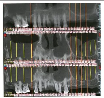
An X-ray orthopantomogram was performed before the surgical procedure (Figure 1).
Patients were evaluated for sinus pathology for a period of 6–48 months after bone transplantation and implant insertion using preoperative and post-operative panoramic radiological imaging and CBCT scan for the purposes of diagnosing their sinus pathology (Figure 2).
Furthermore, CT is employed in the diagnosis and follow-up of patients who are candidates for, or have been submitted to grafting techniques, which allows bone areas to be evaluated by means of measurements and different densities with great
precision.13,14
Imaging was employed to monitor the progress of all patients.
The above CBCT scan examinations have shown that 26 patients of Group A (Control Group) were not showing alterations of the anatomy and physi-ology of the maxillary sinus, such as thickening of the Schneider membrane or radiographic opacifi-cation which could suggest the presence of an inflammatory process.
In the other 30 patients of Group B (Test Group), on the basis of the radiographic findings, it was possible to hypothesize the presence of opacification of the maxillary sinus.
All patients were questioned for complaints and symptoms of sinusitis preoperatively, and any pos-itive findings were assessed by an otolaryngology consultation prior to the surgical procedure.
Surgery preparation
All the patients underwent a careful hematologic and cardiologic evaluation (blood routine, cardio-logical examination, ECG) in specialist wards to optimize and customize the prophylaxis and the preoperative pharmacologic therapy (details shown in surgical procedure).
The bone loss was assessed by means of radio-logical investigations and CBCT; the choice of implants was case-specific, according to the reha-bilitative necessity (Stone® -I.D.I.-EVOLUTION, Monza, Italy).
Before each surgical treatment, a full mouth
disinfection protocol18 consisting of scrupulous
oral hygiene was practiced by supragingival and subgingival scaling with adjuncts of antimicro-bial (chlorhexidine, CURASEPT 0,2%, Curadent, Italy). Moreover, before surgery, the authors decided to administer per os amoxicillin/clavu-lanic acid (NEODUPLAMOX, Procter & Gamble, Italy, of 2 g, 1 h before surgery and 1 g 6 h after surgery).
Preparation of PRF®
As reported from previous experience,19 the
proto-col is performed in accordance with Choukroun’s procedure, introduced with the European Directive no. 2004/23/CE of 31 March 2004.
The patients were subjected, before surgery, to complete hemogram analysis. Before starting the surgical procedure, 5 mL of the patient’s venous
Figure 1. X-ray showing the preoperative condition of a patient enrolled in our study.
Figure 2. Particular of maxillary CT in order to evaluate the residual bone and the sinus pathology.
blood was drawn from the antecubital vein in a sterile 10 mL glass test tube (free from anticoagu-lant) and centrifuged at 3000 rpm for 10 min (Centrifugette model 4206, ALC International).
The upper straw-colored layer was then removed and the middle fraction was collected, 2 mm below lower dividing line, which was the PRF.
This PRF was used in two ways: a part of it was included in a sterile cup and mixed with Bio-Oss®/ Sint-Oss®, until a homogeneous material is obtained; the other part was divided into two sterile compresses, one of which was modeled as a resist-ant fibrin membrane, transferable to the Schneiderian membrane, and the other was transferred to the material placed before closing the wound.
Surgical procedure
The authors opted for the surgical procedure of one-stage sinus lift (implant insertion occurred concurrently with sinus lift, and the healing and integration time was 6–9 months), except for eight selected cases in which the height of the residual bone was lower than 5 mm.
After local and regional anesthesia administra-tion (20 mg/mL mepivacaine chloridrate CARBOPLYINA-Dentsply, Italy,) containing adrenaline as a vasoconstrictor (1:100,000), all patients underwent a lateral window approach, in
accordance with Tatum’s technique.19
The crestal mucosa was incised and two release incisions were also made. The muco-periosteal flap was then turned over in order to expose the bone surface beneath.
The osteotomy to delimit the bone window was performed by Surgybone® Piezosurgery (Silfradent Co., Sofia, Italy) (Figure 3) and the Valsalva maneuver was performed to be sure of the
mem-brane integrity.10
The sinus membrane was carefully elevated from the sinus floor and medial sinus wall; radio-graphic depth gauge was used to confirm that the depth was correct.
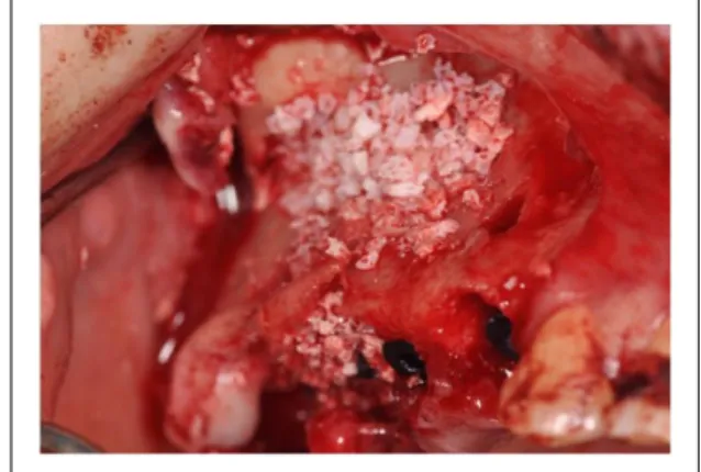
PRF was used in combination with autogenous bone, an organic bone material, and organic bone substitutes (Bio-Oss® and Sint-Oss®).
This mixture was inserted in the surgical alveo-lus by means of a syringe or an amalgam carrier (Kavo Kerr Group, Italy), and pushed upwards with the osteotome, in order to lift the Schneiderian membrane and keep clear the first part of the implant bed (Figure 4), and at the end of this stage, two membranes were placed.
They were obtained from the flattening of the remaining PRF between two sterile gauzes and then placed in order to close the access area to the sinus site.
At this phase, implants were inserted, and the length was in the range of 2–4 mm in excess, com-pared to the height of the available bone.
Lastly, the bone window was closed to avoid traumatisms, the flap was replaced and then the mucosal areas were sutured through a resorbable
material.19
During implant insertion, which had to be very slow, the filling material displaced the Schneiderian membrane and was placed around the implant, thus avoiding the collapse of the membrane on its surface.
At the end of surgery, an endoral periapical radi-ography with a positioner was performed to assess the implant position compared to the maxillary sinus and the implants did not make contact with the sinus membrane in any of the patients.
In the eight patients in which the height of the residual bone was lower than 5 mm, the fixture
Figure 3. Vestibular osteotomy performed by Piezosurgery (Surgybone® Silfradent Co., Sofia, Italy).
was inserted in a later stage (4 months after the grafting procedure), in order to allow the stabiliza-tion and integrastabiliza-tion of the grafted material, and then the restoration of an adequate bone quantity to ensure the predictability and stability of implant rehabilitation.
Patients were given postoperative instructions, and prescribed antibiotics (amoxicillin/clavu-lanic acid 1000 mg every 12 h, for 7 days; NEODUPLAMOX, Procter & Gamble, Italy), analgesics/anti-inflammatory drugs (intramuscular injection of ARTROSILENE, Dompè SpA, Italy; ketoprofen 160 mg, every 12 h for 5 days), and mouthwash with clorexidine ( CURASEPT 0.12%, Curadent, Italy, every 12 h for 14 days).
Patients in Group B also received instructions for aerosol therapy consisting of 2 mL of nebulized corticosteroid (hydrocortisone and diprofillin, CORT-INAL, Teofarma SRL, Italy) and 500 mg/4 mL of antibiotic (Thiamphenicol glycinate acetyl-cysteinate; FLUIMUCIL ANT.IN.TOP, Zambon, Italy), twice per day for 10 days before and 10 days after surgery.
Follow-up
Synthetic absorbable sterile surgical suture stitches (Vicryl® sizes 3.0–4.0) were removed after 7 days.
After implant placement, clinical parameters were recorded and evaluated at baseline, 3, 6, 9, and 12 months. The clinical parameters recorded
were the modified plaque index,20 modified
gingi-val index,21 probing depth around the implant at
four sites using the Tissue pressure sensitive (TPS)
probe,22 implant mobility according to clinical
implant mobility scale,23 and the absence or
pres-ence of any infection around the implant.
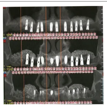
X-ray orthopantomogram and a second-level radi-ographic examinations (Denta-Scan) were per-formed at 15, 60, 90, 120, and 180 days and 6, 12, 24, 36, and 48 months after surgery (Figures 5 and 6).
At 1-year follow-up, patients had been instructed on how to brush, in order to improve oral hygiene by auxiliary cleaning methods.
Results
A total of 175 dental implants were inserted in 56 patients (32 men, 24 women) between 2009 and 2010.
All procedures were successfully concluded with-out significant procedural or postprocedural compli-cations or implant failure. Healing, in general, was
uneventful with minimal discomfort to all the patients. All the sites maintained excellent peri-implant soft and hard tissue conditions.
In 30 patients with a pre-existing radiographic opacification which could suggest the presence of an inflammatory process (54% of both groups), the sinus pathology regressed or remained unchanged in 18 patients (32% in Group B), while in two patients in Group B the sinus membrane pathology was limited to evaluation by periapical X-rays and in six patients (11% in Group B) it completely disappeared as shown in CBCT scan examinations.
All the patients receiving the procedure reported a subjective improvement of nasal breathing with increased perception of the air passageway.
Figure 5. X-ray showing the postoperative condition.
Figure 6. Particular of maxillary CT in order to evaluate the bone and sinus healing.
From a clinical point of view, the authors noticed peri-implant tissue healing, with a successful implant-prosthetic rehabilitation. All patients reported no pain to percussion, no sign of tissue suffering to the soft peri-implant tissues, and the presence of an optimal primary stability of the inserted implants, as confirmed by the fulfillment
of Albrektsson’s implant success criteria24
The radiological examination, performed after 6 months, revealed the presence of newly-formed bone tissue, well amalgamated with the residual bone, and also showed an average increase in the peri-implant bone density of 31% (Figures 5 and 6).
In the apical region, the authors also noticed close contact of implants with the newly-formed bone.
One year after loading there were no dropouts and no failure of the definitive prosthesis occurred.
Discussion
The basis of reduction of the volume of the maxil-lary sinus is multifactorial and includes congenital and acquired variants; among the latter are neo-plastic, traumatic, iatrogenic, inflammatory, and
systemic causes.4,25
Maxillary sinus grafting is a predictable and reliable procedure that has been routinely
per-formed for more than 30 years.5
The complication rate is low, but some cases may require additional surgery, and the outcome of
oral rehabilitation may be affected.6
The use of Piezosurgery involves considerable advantages for the oral surgeon: an osteotomy with micrometric cut for complete surgical accuracy and a high intraoperative sensibility; a site-specific selective cut to minimize the damage to the soft tissues, which is an important quality to preserve the sinus membrane while creating the trans- cortical access window in sinus lift; and an excel-lent intraoperative visibility thanks to a bloodless surgical field, ensured by the effect of cavitation.
Both the measurements previously performed on radiograms and the tactile sensation of contact with the cortical surface allowed the experienced clinician to appreciate the contiguity with the sinus floor.
In the present study, even in cases where the sur-gery was considered risky and not suitable for the presence of a clear pathological state of departure that could affect the final result and exposure to a higher risk of complications, there was full success in centering the objective of regenerative bone
therapy and the authors also performed the collat-eral objective of no negative evolution in the sinusitis.
For this purpose, the choice of the ‘filling’ mate-rial represents an essential aspect for a complete and real achievement of the above-mentioned objectives.
In fact, in recent years, various techniques have been proposed and several types of materials have been assessed (autologous and eterologous); these were critically reviewed by means of clinical
stud-ies and histological investigations.26
The continuous pursuit of the ideal filling mate-rial has often focused the attention on the adoption and marketing of several bone substitutes, such as autologous bone, demineralized and freeze-dried bone, hydroxyapatite, and different combinations of these materials, often with satisfactory results in terms of biocompatibility, induction of bone
for-mation, and of course, implant stability.26
As a supplement to procedures of tissue regen-eration, a platelet concentrate called PRF was tested for the first time in France by Choukroun et al. and introduced with the European Directive no.
2004/23/CE of 31 March 2004.27
PRF belongs to a new generation of platelet concentrates; unlike Platelet Rich Plasma (PRP), PRF is obtained without the addition of anticoagu-lants such as heparin, EDTA, bovine thrombin,
and so on.27
During the production of PRF, apart from plate-lets, other cellular elements are activated, such as leukocytes, which release cytokines after the artifi-cial hemostatic and inflammatory phenomenon
induced by centrifugation.28
There are, therefore, three pro-inflammatory cytokines (IL-1β, IL-6, and TNFα), an anti-inflam-matory cytokine (IL-4), and an angiogenesis
pro-moter (VEGF).27
Hence, PRF is able to regulate inflammation and
stimulate the immune process of chemotaxis.28
PRF is an autologous graft material that elimi-nates any risk of disease transmission; besides, its gelatinous consistency enhances clotting and graft
stability.19,26 However, it has an important defect:
as it is a biomaterial directly obtained from the patient’s blood, its quantity is modest.
PRF has the special feature to undergo polym-erization during centrifugation, naturally and slowly; active thrombin and fibrinogen concentra-tions are almost physiological, because the mate-rial does not require any addition of bovine
As a result, the fibrin assumes a three-dimensional conformation, and the combination of fibrin mono-mers leads to the formation of a trimolecular struc-ture, thus forming a soft and permeable mesh network
and allowing rapid colonization of the scar cells.27
This natural material actually seems to acceler-ate the physiological wound healing; besides, in association with bone grafts, it seems to accelerate
new bone formation.29
Most of the authors who used PRF in their den-tal practice affirm that there was no risk of
infec-tion, disease transmission, or side effects.26
In this regard, a careful revision of literature on the possible effects of the therapeutic use of growth fac-tors (GFs) (including PRF), with relation to carcino-genesis, to their influence on tissues with epithelial dysplasia or oral carcinoma, and to their relation with growth and tumor infiltration is absolutely necessary.
Besides, it is essential to underline that little is known about the ideal concentration of each GF or the optimal dosage for each actual therapeutic situa-tion, and it seems that concentrated growth factor (CGF) is similar to PRF and may be useful in the
future to increase the success rate of bone grafting.29
In the present study, all patients participated until the end of the study with no clinical dropout reported; all patients showed good compliance in oral hygiene maintenance and followed other instructions and no complications were recorded in any of the cases.
Furthermore, the use of PRF® and Piezosurgery reduced the healing time, favoring optimal bone regeneration and allowing sinus membrane integ-rity to be maintained during surgical procedures,
according to evidence-based dentistry.30
In conclusion, the authors conclude by stressing the need to continue this study on a wider cohort of patients, and to carry out large-scale analyses which could provide statistically significant crite-ria about the success of this procedure.
Declaration of conflicting interests
The author(s) declared no potential conflicts of interest with respect to the research, authorship, and/or publication of this article.
Funding
This research received no specific grant from any funding agency in the public, commercial, or not-for-profit sectors.
References
1. Esposito M, Grusovin MG, Maghaireh H et al. (2013) Interventions for replacing missing teeth: Different
times for loading dental implants. Cochrane Database of Systematic Reviews 3: CD003878.
2. Trisi P (2008) Why science? Why research? Implant Dentistry 17: 373–374.
3. Woo I and Le BT (2004) Maxillary sinus floor eleva-tion: Review of anatomy and two techniques. Implant Dentistry 13: 28–32.
4. Fokkens W, Lund V and Mullol J (2012) European position paper on rhinosinusitis and nasal polyps. Rhinology 50: 1–198.
5. Kfir E, Goldstein M, Abramovitz I et al. (2014) The effects of sinus membrane pathology on bone aug-mentation and procedural outcome using minimal invasive antral membrane balloon elevation. Journal of Oral Implantology 40: 285–293.
6. Tonetti MS and Hämmerle CH; European Workshop on Periodontology Group C (2008) Advances in bone augmentation to enable dental implant placement: Consensus Report of the Sixth European Workshop on Periodontology. Journal of Clinical Periodontology 35: 168–172.
7. Barone A, Santini S, Marconcini S et al. (2008) Osteotomy and membrane elevation during the max-illary sinus augmentation procedure. A comparative study: Piezoelectric device vs. conventional rotative instruments. Clinical Oral Implants Research 19: 511–515.
8. Wallace SS, Mazor Z, Froum SJ et al. (2007) Schneider- ian membrane perforation rate during sinus elevation using piezosurgery: Clinical results of 100 consecu-tive cases. International Journal of Periodontics and Restorative Dentistry 27: 413–419.
9. Misch CE and Dietsch F (1993) Bone grafting materi-als in implant dentistry. Implant Dentistry 2: 158–167. 10. Vercellotti T, De Paoli S and Nevins M (2001) The
piezoelectric bony window osteotomy and sinus mem-brane elevation: Introduction of a new technique for simplification of the sinus augmentation procedure. International Journal of Periodontics and Restorative Dentistry 21: 561–567.
11. Bauer SE and Romanos GE (2014) Morphological characteristics of osteotomies using different piezo-surgical devices. A scanning electron microscopic evaluation. Implant Dentistry 23: 334–342.
12. Maiorana C, Farronato D, Pieroni S et al. (2014) A four-year survival rate multicenter prospective clini-cal study on 377 implants: Correlations between implant insertion torque, diameter and bone quality. Journal of Oral Implantology Feb 11. [Epub ahead of print].
13. Loubele M, Guerrero ME, Jacobs R et al. (2007) A com-parison of jaw dimensional and quality assessments of bone characteristics with cone-beam CT, spiral tomog-raphy, and multi-slice spiral CT. International Journal of Oral and Maxillofacial Implants 22: 446–454.
14. Benavides E, Rios HF, Ganz SD et al. (2012) Use of cone beam computed tomography in implant dentistry: The International Congress of Oral Implantologists consensus report. Implant Dentistry 21: 78–86. 15. Passaretti F, Tia M, D’Esposito V et al. (2014)
Growth-promoting action and growth factor release by different platelet derivatives. Platelets 25: 252– 256.
16. Diss A, Dohan DM, Mouhyi J et al. (2008) Osteotome sinus floor elevation using Choukroun’s platelet-rich fibrin as grafting material: A 1-year prospective pilot study with microthreaded implants. Oral Surgery, Oral Medicine, Oral Pathology, Oral Radiology and Endodontology 105: 572–579.
17. Dohan DM, Choukroun J, Diss A. (2006) Platelet-rich fibrin (PRF): A second-generation platelet concen-trate. Part I: Technological concepts and evolution. Oral Surgery, Oral Medicine, Oral Pathology, Oral Radiology and Endodontology 101: e37–44.
18. Eberhard J, Jepsen S, Jervøe-Storm PM et al. (2008) Full-mouth disinfection for the treatment of adult chronic periodontitis. Cochrane Database of Systematic Reviews 1: CD004622.
19. Inchingolo F, Tatullo M, Marrelli M et al. (2010) Trial with Platelet-Rich Fibrin and Bio-Oss used as graft-ing materials in the treatment of the severe maxillar bone atrophy: Clinical and radiological evaluations. European Review for Medical and Pharmacological Sciences 14: 1075–1084.
20. Mombelli A, Van Oosten MA and Schurch E (1987) The microbiota associated with successful or failing osseointegrated titanium implants. Oral Microbiology and Immunology 2: 145–151.
21. Apse P, Schmitt A and Lewis DW (1991) The longitu-dinal effectiveness of osseointegrated dental implants. The Toronto study: Peri-implant mucosa response. International Journal of Periodontics and Restorative Dentistry 11: 95–111.
22. Rams TE and Slots J (1993) Comparison of two pres-sure sensitive periodontal probes and a manual
perio-dontal probe in shallow and deep pockets. International Journal of Periodontics and Restorative Dentistry 13: 521–529.
23. Misch CE (2008) Contemporary implant dentistry. 3rd edn. Amsterdam: Elsevier.
24. Albrektsson T, Zarb GA, Worthington P et al. (1986) The long-term efficacy of currently used dental implants: A review and proposed criteria of suc-cess. International Journal of Oral and Maxillofacial Implants 1: 1–25.
25. Lawson W, Patel ZM and Lin FY (2008) The devel-opment and pathologic processes that influence max-illary sinus pneumatization. Anatomical Record 291: 1554–1563.
26. Scarano A, Degidi M, Iezzi G et al. (2006) Maxillary sinus augmentation with different biomaterials: A comparative histologic and histomorphometric study in man. Implant Dentistry 15: 197–207.
27. Simonpieri A, Del Corso M, Vervelle A et al. (2012) Current knowledge and perspectives for the use of rich plasma (PRP) and platelet-rich fibrin (PRF) in oral and maxillofacial surgery part 2: Bone graft, implant and reconstructive sur-gery. Current Pharmaceutical Biotechnology 13: 1231–1256.
28. Dohan Ehrenfest DM, Bielecki T, Jimbo R et al. (2012) Do the fibrin architecture and leukocyte con-tent influence the growth factor release of platelet concentrates? An evidence-based answer compar-ing a pure platelet-rich plasma (P-PRP) gel and a leukocyte- and platelet-rich fibrin (L-PRF). Current Pharmaceutical Biotechnology 13: 1145–1152. 29. Kim TH, Kim SH, Sándor GK et al. (2014)
Comparison of rich plasma (PRP), platelet-rich fibrin (PRF), and concentrated growth factor (CGF) in rabbit-skull defect healing. Archives of Oral Biology 59: 550.
30. Ballini A, Capodiferro S, Toia M et al. (2007) Evidence-based dentistry: What’s new? International Journal of Medical Sciences 6: 174–178.