4085
Rearing in captivity affects spermatogenesis and sperm quality
in greater amberjack, Seriola dumerili (Risso, 1810)
1R. Zupa,* C. Fauvel,† C. C. Mylonas,‡ C. Pousis,* N. Santamaria,* Μ. Papadaki,‡ I. Fakriadis,‡ § V. Cicirelli,* S. Mangano,# L. Passantino,*
G. M. Lacalandra,* and Aldo Corriero*2
*Department of Emergency and Organ Transplantation, Section of Veterinary Clinics and Animal Production, University of Bari Aldo Moro, Valenzano 70010 (Bari), Italy; †UMR MARBEC, IRD-UM2-CNRS-IFREMER, Station Ifremer,
Palavas 34250, France; ‡Institute of Marine Biology, Biotechnology and Aquaculture, Hellenic Center for Marine Research, Heraklion 71003, Crete, Greece; §Department of Biology, University of Crete, Heraklion 71003, Crete, Greece; and #Institute for Marine Coastal Environment, National Research Council, Capo Granitola 91021 (TP), Italy ABSTRACT: The greater amberjack, Seriola
dumer-ili (Risso, 1810), is a promising candidate for the diversification of European aquaculture production, but inconsistent reproduction in captivity prevents commercial production. Recent studies showed that greater amberjack confined in sea cages exhibited scarce gonad development and early interruption of gametogenic activity during the reproductive sea-son. The aim of the present study was to improve our understanding of the observed impairment of spermatogenesis. Adult wild and captive-reared males were sampled during 3 different phases of the reproductive cycle: early gametogenesis (EARLY; late April to early May), advanced gametogenesis (ADVANCED; late May to early June), and spawn-ing (SPAWNING; late June to July). Spermatogonial stem cells and proliferating germ cells were identi-fied through the immunohistochemical localization of Pou5f1 and proliferating cell nuclear antigen, respec-tively. Apoptotic germ cells were identified throughout the terminal deoxynucleotidyl transferase-mediated 2’-deoxyuridine 5’-triphosphate nick end labeling
method. Sperm quality of captive-reared fish was evaluated using computer-assisted sperm analysis. Captive-reared males exhibited seminiferous lobules of a smaller diameter, a precocious and progressive decrease of spermatogonial mitosis, and a high level of apoptosis at the beginning of the reproductive season, concomitant with a many-fold higher 17β-estradiol plasma concentration. The motile spermatozoa per-centage of captive greater amberjack was lower than in other teleosts, and a drastic decrease of sperma-tozoa motility duration, velocity, and ATP content occurred along the reproductive season. An abnormal increase of sperm concentration as well as an increase of dead spermatozoa occurred during the SPAWNING phase, probably because of lack of sperm hydration and ejaculation and consequent sperm ageing. The present study demonstrates the extreme susceptibility of greater amberjack to rearing stress and underscores the need for improvement of the rearing and handling procedures to ameliorate gametogenesis dysfunctions in commercial aquaculture production.
Key words: germ cell apoptosis, germ cell proliferation, greater amberjack,
rearing in captivity, Seriola dumerili, sperm quality
© 2017 American Society of Animal Science. All rights reserved. J. Anim. Sci. 2017.95:4085–4100
doi:10.2527/jas2017.1708
1This project has received funding from the European Union’s
Seventh Framework Programme for research, technological de-velopment and demonstration (KBBE-2013-07 single stage, GA 603121, DIVERSIFY). The identification and description of stem spermatogonia was performed thanks to a grant from the Apulian Region to R.Z. (Fondo di Sviluppo e Coesione 2007-2013 – APQ Ricerca Regione Puglia “Programma regionale a sostegno della specializzazione intelligente e della sostenibilità sociale ed am-bientale – FutureInResearch). Thanks are due to Peppe, Giovanni
and Vincenzo Billeci, and all the crew of the purse seine fishing vessel Graziella for their hospitality on board and assistance dur-ing wild greater amberjack sampldur-ing. Special thanks are due to Mr Tasos Raftopoulos of Argosaronikos Fishfarms S.A. (Greece) for the hospitality in his farm and the maintenance and sampling of captive-reared greater amberjack broodstock.
2Corresponding author: [email protected]
Received May 9, 2017. Accepted July 5, 2017.
INTRODUCTION
The greater amberjack, Seriola dumerili (Risso, 1810), is a highly valuable teleost considered a
prom-ising aquaculture species. However, proper commer-cial aquaculture production of the species has not developed so far, mainly due to its unpredictable re-production in captivity (Micale et al., 1999; Garcia et al., 2001; Kožul et al., 2001a; Mylonas et al., 2004), which prevented the development of hatchery produc-tion of juveniles and the conversion of the capture-based farming activity into a true aquaculture industry. A renewed effort to develop a technology for aquaculture production of greater amberjack is cur-rently in progress within the European Union project DIVERSIFY (http://www.diversifyfish.eu/; accessed 17 March 2017). A comparative study on the reproduc-tive development in capreproduc-tive-reared greater amberjack (Zupa et al., 2017) demonstrated reduced testis devel-opment and an early cessation of spermatogenic ac-tivity; this gametogenesis impairment was associated with important changes in sex steroid plasma concen-trations. Gametogenesis dysfunctions in fish reared in captivity involve inadequate pituitary gonadotropin synthesis and/or release (Zohar and Mylonas, 2001; Mylonas et al., 2010; Berkovich et al., 2013), which has been attributed to captivity-induced stress, lack of suitable environmental conditions (Mylonas et al., 2010), and/or nutritional deficiencies (Izquierdo et al., 2001). Spermatogenesis dysfunctions may result in a qualitative and quantitative decrease of sperm output (Rurangwa et al., 2004; Cabrita et al., 2009; Bobe and Labbé, 2010) and, therefore, in unsuccessful spawning and production of fertilized eggs.
The aim of the present study was 1) to compare male germ cell proliferation and apoptosis in wild and captive-reared greater amberjack sampled in differ-ent phases of the reproductive cycle and 2) to assess sperm quality of greater amberjack specimens reared in sea cages, in an effort to improve our understanding on the spermatogenesis impairment recently described in this species (Zupa et al., 2017).
MATERIALS AND METHODS
Sample Collection
This study did not fall within the obligations con-tained in the Italian decree number 26 of March 4, 2014, regarding the permission to carry out research stud-ies on experimental animals (http://www.gazzettauf-ficiale.it/eli/id/2014/03/14/14G00036/sg; accessed 12 April 2017) and did not need approval from any ethics committee, because the fish came from a registered aquaculture facility and from commercial catches and
the research did not involve any experiments on live animals. Captive-reared fish originally came from the fishery at 0+ year of age and were then reared at a registered aquaculture facility for 3 yr, according to routine farming practices, before they were recruited for this study, sacrificed, and sampled. No specific per-mission was required for wild greater amberjack sam-pling because these fish were commercially caught from an authorized purse seine fishing vessel during routine fishing operations. Immediately after death, those fish whose size was beyond that of first maturity (Kožul et al., 2001b) were purchased and sampled on board. The greater amberjack is classified as “Least Concern” in the International Union for Conservation of Nature Red List of Threatened Species (Smith-Vaniz et al., 2015).
In the present study, the same male wild and captive-reared greater amberjack used in Zupa et al. (2017) were analyzed. A total of 14 wild and 12 captive-reared greater amberjack males were sampled in 3 different phases of the reproductive cycle that were determined according to the available literature (Mandich et al., 2004; Sley et al., 2014): early ga-metogenesis (EARLY; late April to early May), ad-vanced gametogenesis (ADVANCED; late May to
early June), and spawning (SPAWNING; late June to July). Wild fish were caught by a professional purse seine fishing vessel during 2 consecutive fishing sea-sons (2014–2015) around the Pelagic Islands (Sicily, Italy). Immediately after capture, fish were placed on ice and left to die before sampling. Captive-reared fish belonged to a broodstock captured in the area of Astakos (Ionian Sea) in 2011 and transferred into a sea cage of Argosaronikos Fish Farms SA (Salamina Island, Greece) in September 2013. Fish (n = 28 of an approximately 1:1 male:female ratio) were reared for 2 yr according to standard farming practices and fed to apparent satiation with a commercial extruded broodstock diet (Vitalis Cal; Skretting SA, Stavanger, Norway) every other day. For each sampling, a por-tion of the whole populapor-tion of the captive-reared fish (approximately 12 fish) were confined to one side of the cage and were herded into a rectangular polyvinyl chloride anesthesia “bag” (15 m3 volume) and then lightly anesthetized with 0.01 mL/L clove oil. Then, one by one, individual breeders were gently directed into a polyvinyl chloride stretcher, brought on board a service vessel, and deeply anesthetized with 0.03 mL/L clove oil for sex recognition by means of go-nad cannulation. Subsequently, 10- to 20-mL blood samples were taken from the caudal vasculature using a heparinized syringe and then transferred to 10-mL tubes containing 200 IU sodium heparin/mL of blood. Then, fish were euthanized by decapitation, placed in
crushed ice, and transferred to the farm facility for the subsequent sample collection. The procedure was re-peated until a total of 4 males (and 4 females that were not examined in the present study) were sampled.
Biometric data (fork length [nearest cm], body mass [nearest kg], and testis mass [nearest g]), gonad-osomatic index (GSI = 100 × testis mass/body mass), and reproductive state are reported in Table 1 (data already presented in Zupa et al., 2017). One-centime-ter-thick cross-sections were taken from one of the testis of each fish and fixed in 10% buffered formalin
for further histological (germ cell type description), immunohistochemical (stem and proliferating germ cells), and apoptosis analysis. For the assessment of sperm quality, sperm samples were taken from all 12 captive-reared greater amberjack. After unsuccess-ful attempts to collect sperm by applying pressure on the fish’s abdomen, samples of intratesticular semen were obtained by squeezing the dissected testes. Part of each sperm sample was left undiluted and part was diluted 1:3 (vol/vol) in modified Leibovitz medium according to Fauvel et al. (2012); all the samples were stored at 4°C until analysis, which took place within 30 min.
Histology, Immunohistochemistry, and Identification of Apoptotic Germ Cells
Testis slices were dehydrated in ethanol, clari-fied in xylene, and embedded in paraffin wax. Four-micrometer-thick sections were cut and stained with hematoxylin and eosin or processed for immunohis-tochemistry and for detection of apoptotic cells. The identification of spermatogonial stem cells was per-formed through the immunohistochemical identifica-tion of Pou5f1, a transcripidentifica-tion factor involved in the maintenance and self-renewal of undifferentiated and pluripotent cells, which is considered a reliable mo-lecular marker for spermatogonial stem cells in fish (Schulz et al., 2010; Lacerda et al., 2014). The iden-tification of proliferating germ cells was performed through the immunohistochemical localization of the proliferating cell nuclear antigen (PCNA), a poly-merase delta accessory protein that is synthesized in the late G1 and S phases of the cell cycle and is, there-fore, used as a nuclear marker of proliferation.
The immunohistochemical detection of Pou5f1 and PCNA was performed using the same protocol, with the exception of an antigen retrieval procedure that was ap-plied to only Pou5f1 immunostaining. This procedure was performed by boiling testis sections in citrate buffer (0.01 M, pH 6.0; 4 × 5-min cycles) in a microwave oven on high power (750 W). Endogenous peroxidase was inhibited by treating sections for 10 min with 3% H2O2 and then rinsing them with distilled water and PBS (0.01 M, pH 7.4, containing 0.15 M NaCl). Subsequently, sec-tions were incubated for 30 min in normal horse serum (Vector Laboratories, Inc., Burlingame, CA) to block nonspecific binding sites for immunoglobulins and then incubated overnight in a moist chamber at 4°C with rabbit polyclonal antibodies raised against synthetic peptide of Pou5f1 (Abnova Corp., Taipei, Taiwan) and monoclonal antibodies to PCNA (Santa Cruz Biotech-nology, Inc., Dallas, TX). Anti-Pou5f1 and anti-PCNA antibodies were diluted 1:500 and 1:100, respectively, Table 1. Biometric data, gonadosomatic index (GSI), and
maturity state of wild and captive-reared greater amber-jack males sampled during the reproductive season in the Mediterranean Sea1
Fish
state Sampling date FL,
2
cm BM,
3
kg TM,
4
g GSI, % Reproductive state Early gametogenesis
Wild May 1,
2015 111 14 300 2.14 All spermatogenic stages; luminal spermatozoa 112 20 450 2.25 112 15 300 2.00 113 19 400 2.10 117 19 550 2.89 Captive Apr. 24,
2015 92 12 65 0.54 All spermatogenic stages; few luminal spermatozoa 94 12 60 0.50 94 13 60 0.46 101 15 95 0.63 Advanced gametogenesis Wild May 31,
2014 99 14 1,150 8.21 All spermatogenic stages; plenty of luminal spermatozoa 102 13 650 5.00 115 19 2,200 11.57 124 22 1,900 8.63 Captive June 4, 2015 90 9 370 4.11 Ended spermatogenesis; plenty of luminal spermatozoa 97 14 295 2.10 98 13 600 4.61 All spermatogenic stages; plenty of luminal spermatozoa 103 15 690 4.60 Spawning Wild June 29,
2015 100 12 650 5.41 All spermatogenic stages; plenty of luminal spermatozoa 102 14 700 5.00 104 16 950 5.93 June 30, 2014 99 11 577 5.24
100 11 400 3.63 Partially spent; residual luminal spermatozoa Captive July 2, 2015 91 10 70 0.70 Ended spermatogenesis; small number of luminal spermatozoa 95 11 155 1.40 96 13 140 1.07 96 12 130 1.08
1Table modified from Zupa et al. (2017). 2FL = fork length.
3BM = body mass. 4TM = testis mass.
in PBS containing 0.1% BSA (Sigma-Aldrich S.r.l., Mi-lan, Italy). After rinsing for 10 min in PBS, immuno-histochemical visualization was obtained using the Vec-tastain Universal Elite Kit (Vector Laboratories, Inc.). This method uses the avidin–biotin–peroxidase complex
procedure. Peroxidase activity was visualized by incu-bating for 10 min with a Vector DAB (3,3’-diaminoben-zidine) Peroxidase Substrate Kit (Vector Laboratories, Inc.), which produces a brown precipitate. To confirm the specificity of the immunoreaction, a control-staining procedure was performed by replacing the primary anti-body with normal horse serum and PBS.
The localization of apoptotic germ cells was per-formed using the terminal deoxynucleotidyl transfer-ase-mediated 2’-deoxyuridine 5’-triphosphate nick end labeling (TUNEL) method with an in situ Cell Death Detection Kit, AP (Roche Diagnostics Deutschland GmbH, Mannheim, Germany) that was used in accor-dance with the manufacturer’s instructions. Prior to incubation with the reaction mixture, the sections, af-ter their rehydration through graded ethanol solutions, were incubated in a permeabilization solution of 0.1% Triton X-100 (Sigma-Aldrich S.r.l.) in 0.1% sodium citrate for 8 min at 37°C. Terminal deoxynucleotidyl transferase was diluted 1:10 in TUNEL Dilution Buf-fer (Roche Diagnostics Deutschland GmbH). A ready-to-use solution of nitro-blue tetrazolium chloride/5-bromo-4-chloro-3’-indolyphosphate p-toluidine salt (Roche Diagnostics Deutschland GmbH) served as a substrate for the signal conversion.
Seminiferous Lobule Diameter and Quantification of Germ Cell Proliferation and Apoptosis
At least 50 seminiferous lobules were randomly selected and measured from the sections used for germ cell proliferation and apoptosis analyses. The density of anti-PCNA–positive single A spermatogonia (num-ber of cells/mm2 germinal epithelium) and the density of anti-PCNA–positive spermatocysts (i.e., number of cysts containing type A and type B spermatogonia or primary spermatocytes/mm2 germinal epithelium), as well as the surface occupied by TUNEL-positive apop-totic cells (μm2/mm2 germinal epithelium), were mea-sured on 5 randomly selected fields of each testicular section. All these parameters were measured from mi-crophotographs taken with a digital camera (DFC 420; Leica Microsystems, Cambridge, UK) connected to a light microscope (DIAPLAN; Ernst Leitz GmbH, Wet-zlar, Germany), using image analysis software (Leica Application Suite, version 3.3.0; Leica Microsystems).
Sex Steroid Plasma Level Measurement
Plasma was separated from the blood by centrifuga-tion (2,408 × g for 5 min at room temperature) and then was kept at −80°C until assayed for sex steroid determi-nation. For the quantification of 17β-estradiol (E2), an ELISA kit was used (Cayman Chemical Company, Ann Arbor, MI). For the E2 extraction, 200 μL of plasma was extracted twice with 2 mL diethyl ether. Extraction was done by vigorous vortexing (Vibramax 110; Heidolph Instruments GmbH & Co.KG, Schwabach, Germany) for 3 min. After vortexing, samples were frozen for 10 min at −80°C and the supernatant organic phase was collected in new tubes and evaporated under a stream of nitrogen (Pierce Reacti-Vap III, Thermo Scientific, Rockford, IL. Samples were reconstituted in reaction buffer for running for the ELISA.
Analysis of Sperm Quality
The analyses of the captive-reared greater amber-jack sperm quality were performed at the sampling site (Argosaronikos Fish Farms SA), using a microscope (Eclipse 50i; Nikon Instruments Europe BV, Amster-dam, The Netherlands) equipped with a video camera (Sony ExWaveHAD, Model No. SSC-DC58AP, Tokyo, Japan) recording 25 frames per second. To estimate sper-matozoa (spz) concentration (spz/mL), sperm samples were diluted to 1:500 (vol/vol) in tap water and placed on a cell counting chamber (Thoma; 0.1 mm depth; Pre-cicolor, HBG, Giessen-Luetzellinden, Germany). Sper-matozoa were allowed to settle on the counting cham-ber for 10 min, and then pictures were taken with the microscope at 20x magnification, focused so as to get highly contrasted spermatozoa and a slightly apparent grid. The particles were then counted on a cropped part of each picture adjusted to a known number of squares using the free software ImageJ (National Institutes of Health, Bethesda, MD).
To assess motility, a 20-μL sperm sample from each fish was initially diluted (1:10, vol/vol) in modified Leibovitz medium and then activated by mixing with 1 mL seawater containing 2% BSA, for a final dilution of 1:500 (vol/vol); concomitantly, the 25-frames-per-second video record (.avi format) was launched so as to record sperm activity from its beginning. Immedi-ately after short mixing by a vigorous shake, 1 μL of activated sperm was placed in a prefocused, 10-µm-deep, dedicated cell (Leja Products B.V., Nieuw-Ven-nep, The Netherlands) on the microscope. The record-ing was stopped at the cessation of any progressive spermatozoa movement. The time between activation and first possible motility analysis was around 10 s. For computer-assisted sperm analysis (CASA), the vid-eos were subsequently transformed into sequences of
frames using the free software Virtualdub (http://www. virtualdub.org/; 20 April 2014); then, sequences of 2 s (e.g., 50 frames) were analyzed every 10 s using the plugin CASA developed by Wilson-Leedy and Inger-mann (2007) for ImageJ software. Due to variations of motility recording quality at the different times of sampling, the image treatment settings were adjusted to each sampling time and each sample, whereas the parameters of CASA associated with motility evalua-tion were common for all analyses. The only motility features that showed variations among the 3 different phases of the reproductive cycle and were relevant were the motility (% of motile spz) and the velocity on a smoothed trajectory called average path velocity (VAP; µm/s). Therefore, only these values are present-ed.
To determine spermatozoa ATP content, 1- and 10-μL aliquots of each sperm sample were prepared
according to Boryshpolets et al. (2009) and assessed using an ATPlite luminescence kit (PerkinElmer, Inc., Waltham, MA). The integrity of spermatozoa plasma membrane was tested using a LIVE/DEAD Sperm Vi-ability Kit (Molecular Probes, Eugene, OR) after dilu-tion to 1:100 (vol/vol) and prefixadilu-tion for 4 min in 4% glutaraldehyde as described by Beirão et al. (2009). This procedure stains live, dying, and dead
spermato-zoa in fluorescent green, green + red, and red, respec-tively.
Statistical Analysis
Differences in GSI, mean diameter of seminiferous lobules, density of anti-PCNA–positive single A sper-matogonia, density of anti-PCNA–positive spermato-cysts (spermato-cysts containing type A and type B spermatogo-nia + cysts containing primary spermatocytes), surface occupied by apoptotic germ cells, and sex steroid con-centrations were evaluated by a 2-tailed Student’s t test in the following groups: wild specimens sampled in consecutive phases of the reproductive cycle, captive-reared specimens sampled in consecutive phases of the reproductive cycle, and wild vs. captive-reared speci-mens sampled in the same phase of the reproductive cycle. Prior to the Student’s t test, the raw data of GSI and apoptotic surfaces were arcsine transformed, as appropriate with proportions (Sokal and Rohlf, 1981).
Differences in the sperm quality indexes among sampling phases were assessed either by ANOVA (after angular transformation in the case of % of motile sper-matozoa), or by nested design ANOVA (in the case of sperm velocity where individual spermatozoa perfor-mances were taken into account for each male at the dif-ferent sampling periods). Means were compared using Duncan’s new multiple range post hoc test. All the results
are presented as means ± SE, and the statistical probabil-ity significance was established at the P < 0.05 level.
RESULTS
Changes in Gonadosomatic Index and Diameter of Seminiferous Lobules
Both in wild and captive-reared fish, GSI and semi-niferous lobule diameter significantly increased (P < 0.05) from the EARLY to ADVANCED periods and decreased thereafter (Table 2). Wild fish had higher GSI compared with captive-reared fish at all 3 phases (P < 0.05), and seminiferous lobules were significantly larger (P < 0.05) in wild than in captive-reared greater amberjack during the EARLY and SPAWNING phases.
Histological and Immunohistochemical Analysis of the Testes
In sections stained with hematoxylin and eosin, 2 types of single A spermatogonia were identified: a smaller cell type (8.0 ± 0.1 µm diameter), having a spherical nucleus, with heterochromatin dots and 2 nucleoli, surrounded by a thin acidophilic cytoplasm (Fig. 1a), and a larger cell type (10.6 ± 0.2 µm di-ameter), showing a roundish/ovoidal nucleus with a prevalent euchromatic appearance and sparse hetero-chromatic patches, with a preeminent nucleolus and an acidophilic cytoplasm (Fig. 1a). The immunohisto-chemical staining with antibodies against the stemness marker Pou5f1 labeled single intralobular cells as well as sparse interlobular cells (Fig. 1b). Intralobular anti-Pou5f1–positive cells were more frequently observed beneath the tunica albuginea and, according to their size, they presumptively corresponded to the smaller type A single spermatogonia.
Spermatogonia contained in cysts also appeared as 2 different cell types: larger cells (8.8 ± 2.0 µm mean diameter) with one or more nucleoli and a mod-erately acidophilic cytoplasm (presumptively type A spermatogonia; Fig. 1a) and smaller cells (4.8 ± 0.8 µm mean diameter), showing a small heterochromatic nucleus and thin rim of a weakly acidophilic cyto-plasm (Fig. 1a), making part of larger cysts (presump-tively type B spermatogonia).
The appearance of primary (4.4 ± 0.9 µm) and secondary (3.3 ± 0.8 µm) spermatocytes differed ac-cording to the different phases of meiosis; metapha-sic figures were often observed within spermatocyte I and spermatocyte II cysts (Fig. 1a). Spermatids had a mean diameter of 2.6 ± 0.7 µm and were characterized by a compact and strongly basophilic nucleus (Fig. 1a). Flagellated spermatozoa showed an oval head
Table 2. Mean (SE) gonadosomatic index (GSI) and seminiferous lobule diameters of wild and captive-reared greater amberjack males sampled during the reproductive season in the Mediterranean Sea
Fish state Early gametogenesis Advanced gametogenesis Spawning
GSI, %
Wild 2.3 (0.2)a 8.3 (1.3)a,z 5.1 (0.4)a,z
Captive 0.5 (0.1)b 3.8 (0.6)b,z 1.1 (0.1)b,z
Seminiferous lobule diameter, µm
Wild 135.6 (1.3)a 182.6 (2.6)z 171.9 (1.8)a,z
Captive 109.7 (1.1)b 180.0 (2.2)z 152.5 (1.8)b,z
a,bSignificant differences between wild and captive-reared individuals at the same phase of the reproductive cycle (Student’s t test, P < 0.05). zStatistically significant differences versus the preceding phase within the same group (wild or captive reared; P < 0.05).
Figure 1. Micrographs of testis sections of greater amberjack sampled in different periods of the reproductive cycle. (a) Hematoxylin-eosin stained testis
section from a specimen sampled in late May showing the different germ cell types. Magnification bar = 25 µm. Single A spermatogonia are indicated by black arrows and arrowheads; the latter point to cells likely corresponding to the intralobular anti-Pou5f1–positive spermatogonia (undifferentiated stem spermatogonia). (b) Peripheral region of the testis and detail of a seminiferous lobule (inset) of an individual sampled in early May, immunostained with anti-Pou5f1 antibodies. Interlobular- (yellow curved arrows) and intralobular- (yellow arrowheads) positive cells are stained in brown. Magnification bar = 50 µm; in inset = 10 µm. The white dashed line encircles a type A spermatogonial cyst; the yellow dashed line encircles a type B spermatogonial cyst. sd = spermatid cyst; scI = primary sper-matocyte cyst; scII = secondary spersper-matocyte cyst; sz = spermatozoa.
within cysts or in the lumina of seminiferous lobules after cyst breakdown (Fig. 1a).
Germ Cell Proliferation and Apoptosis
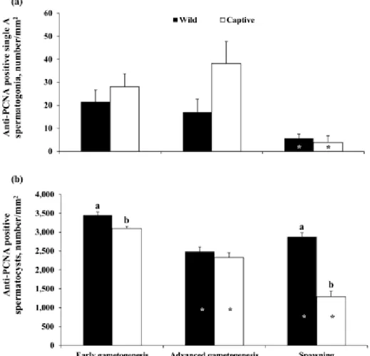
Anti-PCNA immunostaining was observed in the nuclei of single A spermatogonia, spermatogonia con-tained in cysts, and primary spermatocytes (Fig. 2a). A weak staining of the nuclei of secondary spermatocytes was also observed, but these cells were not included in the quantitative analysis. The relative quantification of anti-PCNA–positive single A spermatogonia and sper-matocysts throughout the sampling period is shown in Fig. 3. In wild greater amberjack, anti-PCNA–positive single A spermatogonia gradually decreased through-out the 3 examined phases, although a statistically significant change (P < 0.05) was detected only at the SPAWNING stage (Fig. 3a); anti-PCNA–positive spermatocysts decreased from the EARLY phase to the ADVANCED phase and then slightly increased in the SPAWNING phase (P < 0.05; Fig. 3b). In captive-reared greater amberjack, the density of anti-PCNA single A spermatogonia was stable throughout the EARLY phase and the ADVANCED phase and dramat-ically decreased in the SPAWNING phase (P < 0.05; Fig. 3a); a progressive decrease of anti-PCNA–posi-tive spermatocysts density was observed in capanti-PCNA–posi-tive- captive-reared specimens throughout the examined phases of the reproductive cycle (P < 0.05; Fig. 3b). During the EARLY and SPAWNING phases, significantly higher (P < 0.05) densities of anti-PCNA–positive spermato-cysts were observed in wild specimens compared with captive-reared specimens (Fig. 3b).
All the captive-reared and most of the wild greater amberjack showed TUNEL-positive germ cells. Ap-parently, the TUNEL reaction involved mainly single A spermatogonia, spermatogonia contained in cysts and primary spermatocytes (Fig. 2b). In wild males, the surface occupied by apoptotic germ cells signifi-cantly increased (P < 0.05) from the EARLY phase to the ADVANCED phase and remained stable thereaf-ter, whereas in captive-reared individuals, the surface occupied by apoptotic cells was already high at the EARLY stage and remained unchanged during the 3 sampling phases and was comparable to the highest levels of the wild specimens (Fig. 4). The surface oc-cupied by apoptotic cells was significantly higher (P < 0.05) in captive-reared specimens than in wild speci-mens during the EARLY phase.
17β-Estradiol Plasma Levels
The trend of E2 plasma levels of wild and captive-reared greater amberjack is shown in Fig. 5.
Many-fold higher (P < 0.05) E2 plasma levels were observed in captive-reared fish during the EARLY phase, but these levels significantly decreased in the following phases and were similar to those in wild fish.
Sperm Quality in Captive-Reared Greater Amberjack
The spermatozoa concentration of captive-reared greater amberjack was stable throughout the EARLY (2.3 ± 0.5 × 10¹⁰ spz/mL) and ADVANCED (3.6 ± 0.4 × 10¹⁰ spz/mL) phases and significantly increased dur-ing the SPAWNING period (4.6 ± 0.6 × 10¹⁰ spz/mL; ANOVA, P < 0.05). For all 3 sampling phases, the highest spermatozoa motility (%) was reached within the first 20 s after activation and was followed by a pro-gressive decrease until complete cessation of move-ment (Fig. 6a). However, sperm movemove-ment within the first 20 s presented variations linked to the sampling time, with the highest mean percentage of swimming spermatozoa recorded in the ADVANCED phase (59 ± 16.9% of motile spz) and the lowest mean value reg-istered in the SPAWNING phase (21 ± 9.7% of motile spz). The mean VAP of the spermatozoa varied during the 3 different phases, with the highest mean value 10 s after activation recorded in the ADVANCED phase (102.7 ± 7.0 µm/s) and the lowest mean VAP recorded during the SPAWNING phase (36.5 ± 3.3 µm/s); the highest maximum value of individual velocity was reached during the ADVANCED phase (164 µm/s; Fig. 6b). Finally, a progressive significant decrease (P < 0.05) of sperm motility duration was observed from the EARLY phase to the SPAWNING phase (Fig. 6c).
The ATP level of captive-reared greater amberjack sperm was generally very low and close to the detec-tion threshold for several samples (data not shown). A progressive, but not statistically significant,
de-crease of spermatozoa ATP concentration occurred from the EARLY phase (4.7 ± 1.7 nmol/109 spz) to the ADVANCED phase (1.9 ± 0.6 nmol/109 spz) and the SPAWNING phase (1.2 ± 0.4 nmol/109 spz). In terms of spermatozoa viability, there were significant variations among fish within each sampling time. Not-withstanding this individual variability, a significant increase (P < 0.05) of the proportion of dead and live spermatozoa was observed from the ADVANCED phase to the SPAWNING phase, whereas the propor-tion of dying spermatozoa did not significantly vary (Fig. 7).
DISCUSSION
The negative influence of captivity on reproduc-tive function has been widely demonstrated in all
vertebrate classes, including fishes (Zohar and My-lonas, 2001; Corriero et al., 2009, 2011; Mylonas et al., 2010; Schreck, 2010; Rosenfeld et al., 2012; Zupa et al., 2013). As part of a larger effort to improve our understanding of the gametogenesis dysfunctions re-cently reported in greater amberjack maintained in captivity (Zupa et al., 2017), the present study adds
further information on the effects of confinement in captivity on greater amberjack germ cell proliferation and apoptosis as well as on sperm quality. Recently, Zupa et al. (2017) reported scarce gonad development and precocious cessation of the spermatogenic activity as well as abnormal testosterone (T), 11-ketotestoster-one (11-KT), and 17,20β-dihydroxy-4-pregnen-3-11-ketotestoster-one Figure 2. Micrographs of greater amberjack testis sections sampled in different periods of the reproductive cycle. (a) Testis section of a wild
in-dividual sampled in early May, immunostained with antibodies against the proliferating cell nuclear antigen (PCNA), which stains brown the nuclei of proliferating cells. Magnification bar = 40 µm. Arrowheads indicate an anti-PCNA–positive single spermatogonium, double arrows indicate an anti-PCNA– positive spermatogonial cyst, and dashed arrows indicate a primary spermatocyte cyst. (b) Testis section of a captive-reared individual sampled during late April stained with the terminal deoxynucleotidyl transferase-mediated 2’-deoxyuridine 5’-triphosphate nick end labeling (TUNEL) method, with apoptotic
cells appearing as dark blue dots. Magnification bar = 150 µm. Arrows indicate a TUNEL-positive single spermatogonium and curved arrows indicate a TUNEL-positive spermatocysts.
Figure 3. Changes in mean (±SE) anti-proliferating cell nuclear antigen (PCNA)–positive germ cell density in wild and captive-reared greater
am-berjack males during the reproductive season. (a) Anti-PCNA–positive single A spermatogonia. (b) Anti-PCNA–positive spermatocysts. *White asterisks indicate statistically significant differences (Student’s t-test, P < 0.05) versus the preceding phase in wild fish. *Black asterisks indicate statistically signifi-cant differences (Student’s t test, P < 0.05) versus the preceding phase in captive fish. a,bDifferent letters represent significant differences between wild and
captive individuals within the same sampling phase (Student’s t test, P < 0.05).
Figure 4. Changes in mean (±SE) surface occupied by apoptotic germ cells in wild and captive-reared male greater amberjack sampled during the
reproductive season in the Mediterranean Sea. *A white asterisk indicates a statistically significant difference (Student’s t test, P < 0.05) versus the preced-ing phase in wild fish. a,bDifferent letters indicate a significant difference between wild and captive individuals sampled in the same phase (Student’s t-test,
(17,20β-P) plasma levels. On the other hand, vitello-genesis proceeded without any noticeable impairment (C. Pousis, unpublished data). The present study sug-gests that the earlier-reported dysfunctional spermato-genesis of captive-reared greater amberjack involves also smaller seminiferous lobules in the EARLY and SPAWNING phases, an altered pattern of germ cell proliferation, and an increased number of apoptotic germ cells as well as abnormally high E2 plasma con-centrations during the EARLY spermatogenesis phase. The smaller seminiferous lobules of captive-reared greater amberjack reported here were corre-lated with a lower GSI and indicate a reduced capacity of the testes to develop and reach full maturation. In turn, this reduced capacity to reach full gonad devel-opment was likely related to the lower sex steroid (T, 11-KT, and 17,20β-P) plasma concentrations reported
by Zupa et al. (2017). In terms of germ cell prolifera-tion, the basic histology and immunohistochemistry used in the present study made it possible to distin-guish and describe 3 different type A spermatogonia, of which 2 types were single cells and a third type was represented by cells being part of spermatocysts, type B spermatogonia, primary and secondary spermato-cytes, spermatids, and spermatozoa. Only 1 of the 2 type A single spermatogonia immunoreacted with anti-Pou5f1 antibodies, revealing stemness properties. This stem spermatogonium type, likely corresponding to the type A undifferentiated* spermatogonium of the classification used by Schulz et al. (2010), is respon-sible for germ cell self-renewal. The second type of A single spermatogonia found in greater amberjack is non-stem spermatogonia, whose activity is likely re-lated to differentiation and rapid proliferation toward meiosis. This cell type may correspond to the type A undifferentiated spermatogonia referred to by Schulz et al. (2010), although these authors did not exclude a
residual stem capacity for this cell type. In greater am-berjack testes, anti-PCNA–positive undifferentiated single type A spermatogonia (both positive and nega-tive cells to the stemness marker Pou5f1), differenti-ated spermatogonia (type A and B spermatogonia that are part of cysts), and primary spermatocytes were detected during all investigated reproductive phases.
In the present study, the density of proliferating single spermatogonia remained at the highest levels throughout the EARLY and ADVANCED phases and dramatically decreased during the SPAWNING phase, with differences between wild and captive-reared
in-dividuals. This trend of proliferating activity of sin-gle spermatogonia during the reproductive season is consistent with the decreasing trend of T and 11-KT plasma levels during the SPAWNING phase (Zupa et al., 2017). The absence of significant differences in the density of proliferating single spermatogonia between wild and captive-reared greater amberjack is appar-ently in contrast with the lower T and 11-KT plasma levels observed in fish kept in captivity (Zupa et al., 2017). However, this apparent incongruence may be explained by 1) an increased spermatogonial self-re-newal activity stimulated by abnormally high E2 lev-els during the EARLY phase and/or 2) a diminished capacity of spermatogonia to proceed toward meiosis (lower density of PCNA-positive spermatocytes; see below), resulting in a comparatively higher number of less-developed germ cells in captive-reared fish. The lower spermatogonial capacity of captive-reared fish to proceed toward meiosis might have resulted from the combined effects of higher E2 and lower T/11-KT plasma concentrations. In fact, although E2 in male fish stimulates spermatogonial self-renewal (Miura et al., 1999; Schulz and Miura, 2002; Schulz et al., 2010), supraphysiological concentrations of this hormone in-hibit spermatogenesis through negative feedback ef-Figure 5. Mean (±SE) plasma 17β-estradiol (E2) concentrations in wild and captive-reared greater amberjack males sampled during the
reproduc-tive season in the Mediterranean Sea. *The asterisk indicates a statistically significant difference (Student’s t-test, P < 0.05) versus the preceding phase.
fects on the brain and the pituitary, involving downreg-ulation of the testicular androgen production capacity (Schulz and Nóbrega, 2011).
In captive-reared individuals, the density of matocysts containing proliferating type A and B sper-matogonia plus primary spermatocytes was lower than
in wild fish in the EARLY and SPAWNING stages and showed a progressive decrease from the EARLY phase to the SPAWNING phase. This is in agreement with previous observations showing that captive-reared greater amberjack were already in a spent condition during the SPAWNING phase of the wild population Figure 6. (a) Sperm motility percentage, (b) average path velocity (VAP), and (c) motility duration in captive-reared greater amberjack during 3
phases of the reproductive season in the Mediterranean Sea. In (b), black lines illustrate the mean VAP of sperm population for each phase, whereas gray lines show the maximum value of individual sperm velocity recorded. *In (c), black asterisks indicate significant differences versus the preceding phase (ANOVA, P < 0.05). spz = spermatozoa
and their T, 11-KT and 17,20β-P plasma levels were abnormally low (Zupa et al., 2017). Besides promoting germ cell proliferation, spermiogenesis, and spermia-tion, androgens have been proposed to act as survival factors for germ cells, both in mammals (Young and Nelson, 2001) and in fish (Corriero et al., 2009; Zupa et al., 2013, 2014). Withdrawal of androgens induces apoptosis in the testis (Nandi et al., 1999; Woolveridge et al., 1999), and reintroduction of steroid hormones can reduce apoptotic cell death (Nandi et al., 1999). In the present study, apoptotic germ cells, spermatogonia and primary spermatocytes, were observed in all speci-mens analyzed. In wild greater amberjack, apoptotic germ cell density was highest in the ADVANCED and SPAWNING phases, corroborating the physiological role of apoptosis in the quantitative control of germ cell populations and in the prevention of aberrant germ cell development, as proposed for other large pelagic fish such as the Atlantic bluefin tuna (Thunnus thyn-nus; Corriero et al., 2009; Zupa et al., 2013, 2014) and the swordfish Xiphias gladius; Corriero et al., 2007). In captive-reared greater amberjack, a high density of germ cell apoptosis was observed at the beginning of the reproductive season (EARLY phase). The high inci-dence of apoptosis at the onset of spermatogenesis, far from playing a physiological role, was likely correlated to the low androgen and high E2 plasma levels reported in captive individuals and may have been co-respon-sible for the reduced sperm concentration (see below). Incidentally, the administration of high doses of E2 in
male gilthead seabream (Sparus aurata) induced apop-tosis of spermatogonia (Chaves-Pozo et al., 2007).
In a mammalian model (rat), gonadotrophin with-drawal following hypophysectomy and the consequent decline of sex steroid circulating levels were found to induce testicular atrophy, reduction of germ cell pro-liferation, and increase of apoptosis (Tapanainen et al., 1993). In wild-caught captive-reared Atlantic bluefin
tuna, an increase of 11-KT plasma levels produced by gonadotropin-releasing hormone agonist (GnRHa) ad-ministration stimulated spermatogonial proliferation and reduced the rate of apoptotic germ cells (Corriero et al., 2009). The observed low androgen levels found in captive-reared greater amberjack may have been caused by a reduced release of gonadotropins from the pituitary and/or an altered steroid metabolism. In fact, testes of greater amberjack reared in captivity were found to have a much reduced amount of arachidonic acid (Zupa et al., 2017), a molecule that stimulates testosterone produc-tion by elevating cyclic adenosine monophosphate lev-els in a dose-dependent manner (Mercure and Van Der Kraak, 1995, 1996). Moreover, in birds (Newman et al., 2008; Dickens et al., 2011) and mammals (Williams, 2012), the exposure to different types of stressing fac-tors can result in aromatase upregulation with the conse-quent increase of E2 and decrease of androgens.
One objective of this study was to assess if the above-described dysfunctions resulted in decreased sperm quality. Unfortunately, due to the difficulties of operating in the field, it was not possible to collect and analyze sperm from wild individuals, which would Figure 7. Proportion of live, dying, and dead spermatozoa in captive-reared greater amberjack during 3 phases of the reproductive season in the
Mediterranean Sea. *Black and white asterisks indicate significant differences versus the preceding phase within the same spermatozoa condition (ANOVA,
have represented a valued reference for the assessment of sperm taken from captive-reared specimens. Con-trary to previous sperm sampling attempts performed in Croatia (Kožul et al., 2001a), but consistent with at-tempts made in Greece (Mylonas et al., 2004), in the present study, it was not possible to take sperm by strip-ping captive-reared greater amberjack. This failure is believed to be due to the strong abdominal muscula-ture of this species (Mylonas et al., 2004) and could be exacerbated by the lack of significant testicular hy-dration. It has been observed in various captive-reared greater amberjack broodstocks that it is not possible to collect sperm by abdominal stripping even from males belonging to actively spawning broodstocks that pro-duce large numbers of fertilized eggs daily (Fakriadis et al., 2017). In the case of the present study, however, our failure to collect sperm by stripping was probably more related to a lack of significant testicular hydra-tion. Indeed, during the dissection of the testes, it was observed that the vasa deferentia were not full of sperm and that only a direct strong squeezing of the testes allowed sperm to be obtained, so that the follow-ing discussion actually refers to intratesticular sperm, which might lack complete maturation and hydration and whose count could potentially be affected by the presence of somatic cells, immature germ cells, and cellular debris that can be mistaken for spermatozoa. The sperm concentration of captive-reared greater
am-berjack measured in this study was in the upper range of marine fish species (Suquet et al., 1994; Cosson et al., 2008b), which is consistent with a lack of hydration that, if realized, would have resulted in a physiological reduction of this parameter toward the spawning sea-son. To our knowledge, the observed increase of sperm concentration during the SPAWNING phase has never been reported in any other fish species; moreover, the histological analysis reported by Zupa et al. (2017) showed that although these specimens had ceased their spermatogenic activity, they still retained a moderate number of luminal spermatozoa in the testes. Altogeth-er, these observations seem to support the hypothesis of the lack of proper sperm hydration in captive-reared greater amberjack, probably in response to low sex steroid levels. It is known that sperm hydration with seminal fluid and release via the sperm duct are un-der endocrine control, and a key role in this process and in the intensification of sperm motility is played by 17,20β-P (Schulz and Miura, 2002; Milla et al., 2008; Scott et al., 2010), whose plasma levels were reported to be abnormally low in captive-reared greater amber-jack during the ADVANCED and SPAWNING phases (Zupa et al., 2017).
The sperm of captive-reared greater amberjack analyzed in the present study showed a general
motil-ity pattern similar to those of other fishes, with high initial spermatozoa motility percentage and velocity at activation followed by a decrease of both parameters until all movement ceased (Cosson et al., 2008a,b). However, despite the fact that the velocity of the fast-er spfast-ermatozoa in captive-reared greatfast-er ambfast-erjack sperm was similar to that of other species, such as the European sea bass (Dicentrarchus labrax; Fauvel et al., 2012) and the Atlantic bluefin tuna (Zupa et al., 2013), the maximum sperm motility recorded (about 60% of motile spermatozoa during the ADVANCED phase) was lower compared with most other studied species (Cosson et al., 2008b), and the percentage of motile spermatozoa, motility duration, and veloc-ity drastically declined during the SPAWNING phase. Moreover, the sperm ATP content decreased in cap-tive-reared greater amberjack from the EARLY phase to the SPAWNING phase. The ATP content is widely used as a sperm quality marker (Cosson et al., 2008b; Fauvel et al., 2010), because it is a key limiting factor for maintaining motility (Christen et al., 1987; Cos-son, 2010; Ulloa-Rodríguez et al., 2017). Therefore, the decrease of energy content observed in captive-reared greater amberjack in the present study might explain, at least partially, the lower percentage of motile spermatozoa. Finally, the assessment of sperm membrane integrity from captive-reared fish demon-strated that the percentage of dead spermatozoa sig-nificantly increased from the ADVANCED phase to the SPAWNING phase, which is consistent with the lack of sperm hydration and, presumably, ejaculation and consequent sperm ageing.
As to the underlying cause of the abovementioned dysfunctions in sex hormone levels, germ cell prolif-eration, and apoptosis, we can only speculate at this stage. The fish were acclimated for 2 yr in the facili-ties and were maintained in a large-volume sea cage (640 m3 rectangular shape) at low stocking densities (<1 kg/m3). The site has excellent water circulation and low aquaculture production capacity (<500 metric tons/yr) and the fish showed a good feeding behavior and growth rate during the 2-yr acclimation period, increasing in weight from 5 to 7 kg to 9 to 15 kg. Wa-ter temperatures during the year were typical of the Mediterranean Sea, ranging between 14.3 and 30.1°C. Apart from being maintained in a captive environment, preventing any migration, and being fed a commercial extruded broodstock diet as opposed to forage fish, the breeders were exposed to very low stress conditions during the whole year. So it is conceivable that the offered food was not optimal for reproductive matu-ration and required some formulation adjustment, as suggested by Zupa et al. (2017), although it cannot be said that it was grossly inappropriate. In fact, another
broodstock of the same source and age was maintained under identical conditions in the same facility, and during June, it reached advanced stages of gameto-genesis to be able to be induced to spawn and produce fertilized eggs using a GnRHa therapy (Mylonas et al., 2017). This suggests that it is possible for captive-reared greater amberjack to undergo gametogenesis to an extent that viable gametes (eggs and sperm) can be spawned, producing viable progeny. Although similar evaluations of spermatogenesis were not undertaken on the males of this latter spawning stock, the fact that fertilized eggs were produced indicates that adequate amounts of viable sperm were produced by the males, which were also capable of engaging in normal breed-ing activities to fertilize the eggs produced by the fe-males, albeit after an exogenous GnRHa therapy.
One aspect of the rearing conditions of the fish in the present study that might have exacerbated the prog-ress of an already dysfunctional spermatogenesis was the management/handling stress to which the fish were exposed during the sampling process. Due to facil-ity limitations, all fish used for the 3 samplings were maintained in the same sea cage. As a result of the sampling procedure (see Materials and Methods), ex-cept for the fish sacrificed during the first sampling, all other fish were exposed to a certain amount of handling or management stress a few weeks prior to their being sampled for the study. We believe that this handling or management stress during the reproductive period was responsible for the deterioration of the spermatogene-sis, which was already impaired at the beginning of the reproductive cycle (high plasma E2 concentration and germ cell apoptosis during the EARLY phase).
In conclusion, the present study demonstrated that rearing in captivity affected spermatogenesis in greater amberjack from its EARLY phase, when a high level of germ cell apoptosis was observed. Han-dling or mild management probably had an additional negative effect on spermatogenesis, resulting in a con-stant reduction of the rate of spermatogonia entering meiosis, which resulted in a precocious cessation of the spermatogenic activity. As a consequence of this spermatogenesis impairment, greater amberjack con-fined in captivity showed low sperm quality, in terms of sperm density and motility and velocity as well as ATP content and membrane integrity. This study pro-vides further information on the occurrence of severe reproductive dysfunctions in captive-reared greater amberjack males reported by Zupa et al. (2017) and further supports the need for an improvement of rear-ing technology. In particular, handlrear-ing procedures minimizing stress could be effective in alleviating re-productive deficiencies.
LITERATURE CITED
Beirão, J., F. Soares, P. Herraez, M. T. Dinis, and E. Cabrita. 2009. Sperm quality evaluation in Solea senegalensis during the re-productive season at cellular level. Theriogenology 72:1251– 1261. doi:10.1016/j.theriogenology.2009.07.021
Berkovich, N., A. Corriero, N. Santamaria, C. C. Mylonas, R. Vas-sallo-Aguis, F. de la Gándara, I. Meiri-Ashkenazi, V. Zlatnikov, H. Gordin, C. R. Bridges, and H. Rosenfeld. 2013. Intra-pitu-itary relationship of follicle stimulating hormone and luteiniz-ing hormone durluteiniz-ing pubertal development in Atlantic bluefin tuna (Thunnus thynnus). Gen. Comp. Endocrinol. 194:10–23. doi:10.1016/j.ygcen.2013.08.005
Bobe, J., and C. Labbé. 2010. Egg and sperm quality in fish. Gen. Comp. Endocrinol. 165:535–548. doi:10.1016/j.yg-cen.2009.02.011
Boryshpolets, S., B. Dzyuba, V. Stejskal, and O. Linhart. 2009. Dynamics of ATP and movement in Eurasian perch (Perca
fluviatilis L.) sperm in conditions of decreasing
osmolal-ity. Theriogenology 72:851–859. doi:10.1016/j.theriogenol-ogy.2009.06.005
Cabrita, E., V. Robles, and P. Herráez. 2009. Sperm quality assess-ment. In: E. Cabrita, V. Robles, and P. Herráez, editors, Meth-ods in reproductive aquaculture. Marine and freshwater species. CRC Press Taylor & Francis Group, Boca Raton, FL. p. 93–148. Chaves-Pozo, E., S. Liarte, L. Vargas-Chacoff, A. García-López,
V. Mulero, J. Meseguer, J. M. Mancera, and A. García-Ayala. 2007. 17Beta-Estradiol triggers postspawning in spermatogeni-cally active gilthead seabream (Sparus aurata L.) males. Biol. Reprod. 76:142–148. doi:10.1095/biolreprod.106.056036 Christen, R., J. L. Gatti, and R. Billard. 1987. Trout sperm
mo-tility: The transient movement of trout sperm is related to changes in the concentration of ATP following the activation of the flagellar movement. Eur. J. Biochem. 166:667–671. doi:10.1111/j.1432-1033.1987.tb13565.x
Corriero, A., S. Desantis, C. R. Bridges, D. E. Kime, P. Megalo-fonou, N. Santamaria, F. Cirillo, G. Ventriglia, A. Di Summa, M. Deflorio, F. Campobasso, and G. De Metrio. 2007. Germ cell proliferation and apoptosis during different phases of swordfish (Xiphias gladius L.) spermatogenetic cycle. J. Fish Biol. 70:83– 99. doi:10.1111/j.1095-8649.2006.01257.x
Corriero, A., A. Medina, C. C. Mylonas, C. R. Bridges, N. San-tamaria, M. Deflorio, M. Losurdo, R. Zupa, H. Gordin, F. de la Gándara, A. Belmonte Ríos, C. Pousis, and G. De Metrio. 2009. Proliferation and apoptosis of male germ cells in captive Atlantic bluefin tuna (Thunnus thynnus L.) treated with gonado-tropin-releasing hormone agonist (GnRHa). Anim. Reprod. Sci. 116:346–357. doi:10.1016/j.anireprosci.2009.02.013
Corriero, A., R. Zupa, G. Bello, C. C. Mylonas, M. Deflorio, S. Genovese, G. Basilone, G. Buscaino, G. Buffa, C. Pousis, G. De Metrio, and N. Santamaria. 2011. Evidence that severe acute stress and starvation induce rapid atresia of vitellogenic folli-cles in Atlantic bluefin tuna, Thunnus thynnus (L.) (Osteichthy-es: Scombridae). J. Fish Dis. 34:853–860. doi:10.1111/j.1365-2761.2011.01303.x
Cosson, J. 2010. Frenetic activation of fish spermatozoa fla-gella entails short-term motility, portending their precocious decadence. J. Fish Biol. 76:240–279. doi:10.1111/j.1095-8649.2009.02504.x
Cosson, J., A.-L. Groison, M. Suquet, C. Fauvel, C. Dreanno, and R. Billard. 2008a. Marine fish spermatozoa: Racing ephemeral swimmers. Reproduction 136:277–294. doi:10.1530/REP-07-0522
Cosson, J., A.-L. Groison, M. Suquet, C. Fauvel, C. Dreanno, and R. Billard. 2008b. Studying sperm motility in marine fish: An overview on the state of the art. J. Appl. Ichthyology 24:460– 486. doi:10.1111/j.1439-0426.2008.01151.x
Dickens, M. J., C. A. Cornil, and J. Balthazart. 2011. Acute stress differentially affects aromatase activity in specific brain nuclei of adult male and female quail. Endocrinology 152:4242–4251. doi:10.1210/en.2011-1341
Fakriadis, I., F. Lisi, I. Sigelaki, M. Papadaki, A. Raftopoulos, and C. C. Mylonas. 2017. Spawning kinetics of greater amberjack
Seriola dumerili in response to multiple GnRHa injections or
implants. In: Aquaculture Europe 2017, Dubrovnik, Croatia, October 16–20, 2017. (Accepted oral presentation; in press.) Fauvel, C., S. Boryshpolets, J. Cosson, J. G. Wilson Leedy, C.
Lab-bé, P. Haffray, and M. Suquet. 2012. Improvement of chilled seabass sperm conservation using a cell culture medium. J. Appl. Ichthyology 28:961–966. doi:10.1111/jai.12071
Fauvel, C., M. Suquet, and J. Cosson. 2010. Evaluation of fish sperm quality. J. Appl. Ichthyology 26:636–643. doi:10.1111/ j.1439-0426.2010.01529.x
Garcia, A., M. V. Diaz, and B. Agulleiro. 2001. Induccion hor-monal de la puesta y desarrollo embrionario de la seriola Medi-terranea (Seriola dumerilii, Risso). (In Spanish.) Monogr. Inst. Canar. Cienc. Mar. 4:561–566.
Izquierdo, M. S., H. Fernandez-Palacios, and A. G. J. Tacon. 2001. Effect of broodstock nutrition on reproductive perfor-mance of fish. Aquaculture 197:25–42. doi:10.1016/S0044-8486(01)00581-6
Kožul, V., B. Skaramuca, B. Glamuzina, N. Glavić, and P. Tutman. 2001a. Comparative gonadogenesis and hormonal induction of spawning of cultured and wild Mediterranean amberjack
(Se-riola dumerili, Risso 1810). Sci. Mar. 65:215–220. doi:10.3989/
scimar.2001.65n3215
Kožul, V., B. Skaramuca, M. Kraljević, J. Dulčić, and B. Glam-uzina. 2001b. Age, growth and mortality of the Mediterranean amberjack Seriola dumerili (Risso 1810) from the south-eastern Adriatic Sea. J. Appl. Ichthyology 17:134–141. doi:10.1046/ j.1439-0426.2001.00301.x
Lacerda, S. M. D. S. N., G. M. J. Costa, and L. R. de Fran-ça. 2014. Biology and identity of fish spermatogonial stem cell. Gen. Comp. Endocrinol. 207:56–65. doi:10.1016/j.yg-cen.2014.06.018
Mandich, A., A. Massari, S. Bottero, P. Pizzicori, H. Goos, and G. Marino. 2004. Plasma sex steroid and vitellogenin profiles dur-ing gonad development in wild Mediterranean amberjack
(Se-riola dumerilii). Mar. Biol. 144:127–138.
doi:10.1007/s00227-003-1185-6
Mercure, F., and G. Van Der Kraak. 1995. Inhibition of gonado-tropin-stimulated ovarian steroid production by polyunsaturat-ed fatty acids in teleost fish. Lipids 30:547–554. doi:10.1007/ BF02537030
Mercure, F., and G. Van Der Kraak. 1996. Mechanisms of action of free arachidonic acid on ovarian steroid production in the goldfish. Gen. Comp. Endocrinol. 102:130–140. doi:10.1006/ gcen.1996.0054
Micale, V., G. Maricchiolo, and L. Genovese. 1999. The reproduc-tive biology of the amberjack, Seriola dumerilii (Risso, 1810). I. Oocyte development in captivity. Aquacult. Res. 30:349–355. doi:10.1046/j.1365-2109.1999.00336.x
Milla, S., X. Terrien, A. Sturm, F. Ibrahim, F. Giton, J. Fiet, P. Prunet, and F. Le Gac. 2008. Plasma 11-deoxycorticosterone (DOC) and mineralocorticoid receptor testicular expression during rainbow trout Oncorhynchus mykiss spermiation: Impli-cation with 17alpha, 20beta-dihydroxyprogesterone on the milt fluidity? Reprod. Biol. Endocrinol. 6:19. doi:10.1186/1477-7827-6-19
Miura, T., C. Miura, T. Ohta, M. R. Nader, T. Todo, and K. Yam-auchi. 1999. Estradiol-17 β stimulates the renewal of spermato-gonial stem cells in males. Biochem. Biophys. Res. Commun. 264:230–234. doi:10.1006/bbrc.1999.1494
Mylonas, C. C., I. Fakriadis, N. Papandroulakis, A. Raftopoulos, G. Iakovopoulos, M. Papadaki, and I. Sigelaki. 2017. Broodstock management and spawning induction of greater amberjack
Se-riola dumerili reared in tanks and sea cages in Greece. In:
Aqua-culture Europe 2017, Dubrovnik, Croatia, October 16–20, 2017. (Accepted oral presentation; in press.)
Mylonas, C. C., A. Fostier, and S. Zanuy. 2010. Broodstock management and hormonal manipulations of fish reproduc-tion. Gen. Comp. Endocrinol. 165:516–534. doi:10.1016/j.yg-cen.2009.03.007
Mylonas, C. C., N. Papandroulakis, A. Smboukis, M. Papadaki, and P. Divanach. 2004. Induction of spawning of cultured great-er ambgreat-erjack (Sgreat-eriola dumgreat-erili) using GnRHa implants. Aqua-culture 237:141–154. doi:10.1016/j.aquaAqua-culture.2004.04.015 Nandi, S., P. P. Banerjee, and B. R. Zirkin. 1999. Germ cell
apopto-sis in the testes of Sprague Dawley rats following testosterone withdrawal by ethane 1,2-dimethanesulfonate administration: Relationship to Fas? Biol. Reprod. 61:70–75. doi:10.1095/bi-olreprod61.1.70
Newman, A. E., D. S. Pradhan, and K. K. Soma. 2008. Dehydro-epiandrosterone and corticosterone are regulated by season and acute stress in a wild songbird: Jugular versus brachial plasma. Endocrinology 149:2537–2545. doi:10.1210/en.2007-1363 Rosenfeld, H., C. C. Mylonas, C. R. Bridges, G. Heinisch, A.
Cor-riero, R. Vassallo-Aguis, A. Medina, A. Belmonte, A. Garcia, F. de la Gándara, C. Fauvel, G. De Metrio, I. Meiri-Ashkenazi, H. Gordin, and Y. Zohar. 2012. GnRHa-mediated stimulation of the reproductive endocrine axis in captive Atlantic bluefin tuna, Thunnus thynnus. Gen. Comp. Endocrinol. 175:55–64. doi:10.1016/j.ygcen.2011.09.013
Rurangwa, E., D. E. Kime, F. Ollevier, and J. P. Nash. 2004. The measurement of sperm motility and factors affecting sperm quality in cultured fish. Aquaculture 234:1–28. doi:10.1016/j. aquaculture.2003.12.006
Schreck, C. B. 2010. Stress and fish reproduction: The roles of al-lostasis and hormesis. Gen. Comp. Endocrinol. 165:549–556. doi:10.1016/j.ygcen.2009.07.004
Schulz, R. W., L. R. de França, J.-J. Lareyre, F. Le Gac, H. Chiari-ni-Garcia, R. H. Nóbrega, and T. Miura. 2010. Spermatogenesis in fish. Gen. Comp. Endocrinol. 165:390–411. doi:10.1016/j. ygcen.2009.02.013
Schulz, R. W., and T. Miura. 2002. Spermatogenesis and its endocrine regulation. Fish Physiol. Biochem. 26:43–56. doi:10.1023/A:1023303427191
Schulz, R., and R. H. Nóbrega. 2011. The reproductive organs and processes: Regulation of spermatogenesis. In: A. P. Farrel, editor, Encyclopedia of fish physiology from genome to en-vironment. 1st ed. Academic Press, London, UK. p. 627–634. doi:10.1016/B978-0-12-374553-8.00269-0
Scott, A. P., J. P. Sumpter, and N. Stacey. 2010. The role of the maturation-inducing steroid in male fishes: A review. J. Fish Biol. 76:183–224. doi:10.1111/j.1095-8649.2009.02483.x
Sley, A., A. Hadj Taeib, O. Jarboui, M. Ghorbel, and A. Bouain. 2014. Reproductive biology of greater amberjack Seriola
du-merili (Risso, 1810) from the Eastern Mediterranean Sea
(Tuni-sia, Gulf of Gabes). Cah. Biol. Mar. 55:421–430.
Smith-Vaniz, W. F., F. Pina Amargos, J. Brown, M. Curtis, and J. T. Williams. 2015. Seriola dumerili. The IUCN Red List of Threat-ened Species. http://www.iucnredlist.org/details/198643/0. (Accessed 28 April 2017.)
Sokal, R. R., and F. J. Rohlf. 1981. Biometry: The principles and practice of statistics in biological research. Freeman WH and Company, New York, NY.
Suquet, M., R. Billard, J. Cosson, G. Dorange, L. Chauvaud, C. Mugnier, and C. Fauvel. 1994. Sperm features in turbot (Scophthalmus maximus): A comparison with other freshwa-ter and marine fish species. Aquat. Living Resour. 7:283–294. doi:10.1051/alr:1994031
Tapanainen, J. S., J. L. Tilly, K. K. Vihko, and A. J. Hsueh. 1993. Hormonal control of apoptotic cell death in the testis: Gonado-tropins and androgens as testicular cell survival factors. Mol. Endocrinol. 7:643–650.
Ulloa-Rodríguez, P., E. Figueroa, R. Díaz, M. Lee-Estevez, S. Short, and J. G. Farías. 2017. Mitochondria in teleost spermato-zoa. Mitochondrion 34:49–55. doi:10.1016/j.mito.2017.01.001 Williams, G. 2012. Aromatase up-regulation, insulin and raised
intracellular oestrogens in men, induce adiposity, metabolic syndrome and prostate disease, via aberrant ER-α and GPER signalling. Mol. Cell. Endocrinol. 351:269–278. doi:10.1016/j. mce.2011.12.017
Wilson-Leedy, J. G., and R. L. Ingermann. 2007. Development of a novel CASA system based on open source software for charac-terization of zebrafish sperm motility parameters. Theriogenol-ogy 67:661–672. doi:10.1016/j.theriogenolTheriogenol-ogy.2006.10.003
Woolveridge, I., M. de Boer-Brouwer, F. Taylor, K. J. Teerds, F. C. W. Wu, and I. D. Morris. 1999. Apoptosis in the rat sper-matogenic epithelium following androgen withdrawal: Chang-es in apoptosis-related genChang-es. Biol. Reprod. 60:461–470. doi:10.1095/biolreprod60.2.461
Young, K. A., and R. J. Nelson. 2001. Mediation of seasonal tes-ticular regression by apoptosis. Reproduction 122:677–685. doi:10.1530/rep.0.1220677
Zohar, Y., and C. C. Mylonas. 2001. Endocrine manipulations of spawning in cultured fish: From hormones to genes. Aquacul-ture 197:99–136. doi:10.1016/S0044-8486(01)00584-1 Zupa, R., C. Fauvel, C. C. Mylonas, N. Santamaria, L. Valentini,
C. Pousis, M. Papadaki, M. Suquet, F. de la Gándara, G. Bello, G. De Metrio, and A. Corriero. 2013. Comparative analysis of male germ cell proliferation in wild and captive Atlantic bluefin tuna Thunnus thynnus. J. Appl. Ichthyology 29:71–81. doi:10.1111/j.1439-0426.2012.02045.x
Zupa, R., C. Rodríguez, C. C. Mylonas, H. Rosenfeld, I. Fakriadis, M. Papadaki, J. A. Perez, C. Pousis, G. Basilone, and A. Cor-riero. 2017. Comparative study of reproductive development in wild and captive-reared greater amberjack Seriola dumerili (Risso,1810). PLoS One 12(1):e0169645. doi:10.1371/journal. pone.0169645
Zupa, R., N. Santamaria, C. C. Mylonas, M. Deflorio, F. de la Gándara, R. Vassallo-Agius, C. Pousis, L. Passantino, G. Cen-toducati, G. Bello, and A. Corriero. 2014. Male germ cell pro-liferation and apoptosis during the reproductive cycle of cap-tive-reared Atlantic bluefin tuna Thunnus thynnus (Linnaeus). Aquacult. Res. 45:1733–1736. doi:10.1111/are.12110