Piotto, S. P., L. Sessa, S. Concilio and P. Iannelli. "Yadamp: Yet Another Database of Antimicrobial Peptides." Int J Antimicrob Agents 39, no. 4 (2012): 346-51.
Concilio, S., S. Piotto, L. Sessa, P. Iannelli, A. Porta, E. C. Calabrese, M. R. Galdi and L. Incarnato. "Antimicrobial Polymer Films for Food
Packaging." AIP Conference Proceedings 1459, no. 1 (2012): 256-258. Piotto, S., S. Concilio, L. Sessa, P. Iannelli, A. Porta, E. C. Calabrese, M. R.
Galdi and L. Incarnato. "Novel Antimicrobial Polymer Films Active against Bacteria and Fungi." Polymer Composites 34, no. 9 (2013): 1489-1492.
Piotto, S., S. Concilio, L. Sessa, A. Porta, E. C. Calabrese, A. Zanfardino, M. Varcamonti and P. Iannelli. "Small Azobenzene Derivatives Active against Bacteria and Fungi." Eur J Med Chem 68, (2013): 178-84. Bruno, A., C. Borriello, T. Di Luccio, G. Nenna, L. Sessa, S. Concilio, S.
Haque and C. Minarini. "White Light-Emitting Nanocomposites Based on an Oxadiazole–Carbazole Copolymer (Poc) and Inp/Zns Quantum Dots." Journal of Nanoparticle Research 15, no. 11 (2013): 1-10. Sessa, L., S. Piotto, S. Concilio, T. Robinson, A. Kϋchler and P. Walde. "A
Novel Photoresponsive Peptide with Antimicrobial Activity: A Biophysical Study." (submitted).
Sessa, L. , S. Concilio, S. Piotto, A. Porta, E. C. Calabrese and P. Iannelli. "Novel Materials with Antibacterial and Antifungal Activity." In II International Conference on Antimicrobial Research (ICAR). Lisbon, Portugal, 21-23 November 2012.
Piotto, S., L. Sessa, S. Concilio and P. Iannelli. "Molecular Simulation of Membrane Interacting Novel Antimicrobial Compounds." In II International Conference on Antimicrobial Research (ICAR). Lisbon, Portugal, 21-23 November 2012.
Sessa, L., S. Concilio, S. Piotto and P. Iannelli. "Engineered Amps for Nano Drug Delivery Systems." In 1st Workshop on Bio Nano Materials (BIONAM2013) Department of Pharmacy, University of Salerno, 18 October 2013.
Concilio, S., L. Sessa, S. Piotto and P. Iannelli. "Smart Materials for Biomedical Applications " In 1st Workshop on Bio Nano Materials (BIONAM2013). Department of Pharmacy, University of Salerno, 18 October 2013.
Table of contents
Table of contents ... I
Abstract ... V
Part I.Design of new polymers inspired by AMPs
Chapter 1. Antimicrobial peptides: an overview ... 3
1.1. Background ... 3
1.2. What are AMPs? ... 3
1.3. Classification by secondary structure ... 4
1.4. Principal families of cationic antibacterial peptides and their activity spectrum ... 9
1.5. Antimicrobial activity: mechanism of action ... 11
1.6. Bacterial resistance strategy ... 16
1.7. Limits of the antimicrobial peptides as therapeutic agents ... 18
1.8. AMPs in clinical trial ... 19
Chapter 2. Collection and building of a new AMPs database ... 23
2.1. Introduction ... 23
2.2. Data collection ... 24
2.3. Database content ... 33
2.4. Web database design: an overview ... 34
Chapter 3. Development of a modified antimicrobial peptide ... 39
3.1. Design of modified antimicrobial peptides ... 39
3.2. Our approach ... 40
3.3. Chemical modification of natural amino acid with a photoactive ligand. 42 3.4. Selection of an AMP as template ... 46
Chapter 4. Investigation of the different membrane interaction ... 51
4.1. Aim of the work ... 51
4.2. Materials used ... 52
4.3. Effect of synthetic peptides on GUVs membrane permeability ... 62
4.4. Effect of synthetic peptides on LUVET membrane permeability ... 67
4.5. LUVETs characterization ... 86
Chapter 5. Conclusion e discussion ... 91
Part II.Development of new antimicrobial and antifungal compounds Chapter 6. Small azobenzene derivatives active against bacteria and fungi ... 97
6.1. Introduction ... 97
6.2. In silico screening ... 100
6.3. Azo compounds synthesis ... 104
6.4. Thermal and optical properties ... 115
Chapter 7. Structure modifications using A4 as lead compound ... 127
7.1. Introduction ... 127
7.2. Synthesis of A4 analogues with modified second ring ... 130
7.3. Synthesis of A4 analogues with modified first ring ... 133
7.4. Thermal and optical properties of A4 analogues compounds ... 135
7.5. Antimicrobial activity of A4 analogues compounds ... 137
Chapter 8. Conclusions and outlook ... 139
Part III.Development of new antimicrobial and antifungal polymer films Chapter 9. Novel antimicrobial polymer films active against bacteria and fungi ... 143
9.1. Introduction ... 143
9.2. Thin film preparation techniques ... 146
9.2.1. Mold-casting method ... 147
9.2.2. Solvent casting method ... 147
9.3. Material used ... 148
9.4. Experimental part: film preparation ... 153
9.5. Thermal characterization of the films ... 161
9.6. Summary of films characteristics ... 167
9.7. Microbiological Characterization ... 169
9.8. Conclusions and outlook ... 175
Appendix-a MatLab Scripts ... 179
Appendix-c Dynamic light scattering results ... 195
Appendix-d 1H NMR spectra of azo compounds ... 201
Appendix-e Thermogravimetric characterization of azo compounds .. 205
Appendix-f Thermal characterization of azo compounds ... 207
Appendix-g UV-Vis spectra of azo compounds ... 211
Appendix-h Thermal characterization of thin films ... 213
Abbreviations, Acronyms and Symbols ... 217
Abstract
This PhD project is focused on the development of new polymeric materials with antibacterial and antifungal activity.
The first part of the work was dedicated to the synthesis and the insertion of a modified amino acid into an antimicrobial peptide (AMP), with the aim to retain the action mechanism on bacterial membranes. Understanding the mechanism of action of AMPs is important for the rational design of new drugs. For this reason, I have collected structural properties, antimicrobial activity values and biological origin of antimicrobial peptides from published data. I have calculated the most relevant chemical physical properties like charge, hydrophobic moment, helicity, flexibility, isoelectric point, Boman and instability index and penetration capabilities. This data collection work permitted us to create YADAMP (www.yadamp.unisa.it), a web database with detailed informations on AMPs. YADAMP database contains the highest number of active sequences with proven antimicrobial activity. YADAMP peremitted me to do a work of data mining that end up with the choice of a peptide, a defensine, to be used as template for developing a new photoresponsive peptide. The peptide, hereafter indicated as ALY, is a short α-helix, membrane active AMP with a tyrosine in the sequence. I have developed the modified analogue replacing the tyrosine by a modified tyrosine with azobenzene group in the side chain. The modified amino acid, named Fmoc-azoTyr, was synthesized according to the classic scheme of diazocopulation reactions. I have chosen the azobenzene group because it permits a reversible trans to cis photochemical isomerization. The photo-induced switch will, potentially, permit to turn on or off the peptide antimicrobial activity. As described in the following part, we have already confirmed the antimicrobial and antifungal activity of azobenzene group.
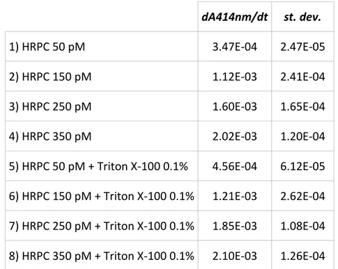
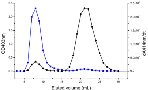
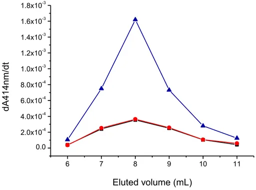
Therefore, the modified peptide might have a broader spectrum of activity due to the azobenzene presence, and it might act as a prodrug, generating in vivo the antimicrobial azo compound. I evaluated the difference in membrane permeability due to the introduction of the modified amino acid performing studies of membrane permeability during a period in the group of Prof. Peter Walde at the Swiss Federal Institute of Technology (ETH Zurich). Calcein leakage assays showed that the modified amino acid introduced into active sequence increased the membrane permeability of a suspension of giant unilamellar vesicles compared to the unmodified peptide at the same conditions. I have prepared large unilamellar vesicles formed by POPC/POPG (mixture 90:10 mol/mol), that contain HRPC enzyme, by mechanical extrusion and I performed measurements of enzyme leakage after interaction with the peptide. In this assay the membrane permeability was associated with an increase in enzyme activity. Preliminary results of enzyme leakage showed a greater membrane perturbation with time-dependence enzyme leakage from LUVs after interaction with modified peptide.
The second part of my research work was focused on the design and the synthesis of antimicrobial low molecular weight molecules with antimicrobial and antifungal activity. I used the phytoalexin resveratrol as template to design a new class of active compounds with azobenzene structure. I selected the best candidates by preliminary in silico test of ADMET properties and then I synthesized the azo compounds with lowest in silico toxicity values (A1, A2, A3, A4, A5, B10 and B11) according to the classic scheme of diazocopulation reaction. The antimicrobial activity and the thermal stability of each compound were evaluated. The majority of synthesized compounds exhibited high antibacterial activity against S. aureus and antifungal activity against C. albicans, but they were inactive against Gram-negative bacteria such as P. aeruginosa and S. Typhimurium. The different antibacterial activities of synthesized azo compounds suggest that these molecules interact with protein
receptors and that the interaction with membranes is of minor importance. To validate this hypothesis, I carried out structural modifications of azo compounds to enhance their biological activity. I have used the best antimicrobial azo compounds named A4 (4’-hydroxy-(4-hydroxy-3,5-dimethyl)-azobenzene) as lead compound to synthesize several analogues having modifications on the first and on the second azobenzene ring. This includes the moving of the phenolic hydroxyl group from para- to meta- position, the removal of the phenolic hydroxyl group, and the replacement of the phenolic hydroxyl group by the methoxy or the methyl group. In this way I obtained molecules with antibacterial and antifungal activity higher than lead compound.
The antimicrobial activities of these azo compounds and their thermal stability are very promising and indicate that these molecules may have interesting and therapeutically significant applications. A possible application is the insertion of azo compounds into polymeric matrices to produce composites with low production cost, good processability, and antimicrobial potential. Using solvent casting and mold casting methods, I realized active antimicrobial and antifungal films using polyolefins (such as PP and LLDPE) and biodegradable polymers (such as PLA, PVA and Mater-B), by introducing different percentages of antimicrobial azo dyes in polymer matrices. The obtained thin films retained the proprieties of the pure matrices without azo compounds such as thermal proprieties, flexibility and transparency; also I prepared transparent amorphous films, as confirmed by X-Ray analysis. The films exhibited antimicrobial activity and the capability to inhibit biofilms formation of S. aureus and C. albicans. Concentration of 0.01% (w/w) permitted the preparation of active, uncolored and transparent films. This is the first time that novel azobenzene based antimicrobial compounds have been added into polymer films. These preliminary tests confirmed that the new materials realized in this thesis are promising for future applications in the field where
an intrinsic antimicrobial ability of the material is required, like biomedical tools, antibacterial surfaces, and films for food packaging. Spectrophotometric investigation of the azo compound release from the polymer matrices is currently undergoing.
Part I.
Chapter 1. Antimicrobial peptides: an overview
1.1. Background
Since the discovery of the penicillin, the subsequent development of new antibiotics has considerably improved the public health. However, starting from the 80s, a slowdown was observed. First, very few antibiotics were discovered, but improvements within existing classes were still suggested. Second, a resistance to almost all antibiotics was observed in clinical use. However, the use and overuse of antibiotics has causing the emergence of multidrug-resistant microorganisms. Due to mutations and adaptation in bacteria, traditional antibiotics became more inefficient and the elimination of adapted strains has become increasingly difficult [1].
This is the reason why antimicrobial peptides (AMPs) are receiving much attention as a class of new antibiotics. Traditional antibiotics generally target a specific physiological process of bacteria, such as DNA replication, cell wall synthesis, etc. Instead, most AMPs target the bacterial cell membrane without specific receptors. Consequently their membrane interaction and broad activity spectra are becoming an ideal approach to overcome the resistance resulting from bacterial mutations [2].
1.2. What are AMPs?
The term AMP is generally used to define a large number of small proteins produced by multicellular organisms that are able to kill or to inhibit growth of various microorganisms [3].They are small peptides, highly variable, with 12– 50 amino acids, mostly in their common L configuration. Due to the presence of basic amino acids as Lys and Arg, they are positively charged, with net charge from + 2 to + 9. AMPs possess approximately 50% hydrophobic
residues, which allow an amphipathic conformation that permits them to interact with bacterial membranes [4]. These molecules are able to kill or inhibit a variety of organisms, including Gram-positive and Gram-negative bacteria, fungi, viruses, protozoa, parasites [5]. More than two thousand natural AMPs have been isolated and characterized from different sources and several thousands of synthetic variants have been developed.
1.3. Classification by secondary structure
AMPs can be divided into many subtypes following different criteria: origin, amino acid sequence, size, structure, biological action, mechanism of action and others; but it has been shown that secondary structure is the only meaning criterion to order them [6]. The secondary structure of AMPs include four major classes, α-helix, β-sheet stabilized by two or more disulfide bridges, extended helices with a predominance of one or more amino acids and loop structures with one disulfide bridge. As one of most widely distributed AMPs, α-helical antimicrobial peptides (αAMPs) have been thoroughly investigated.
1.3.1. α-helical antibacterial peptides
The α-helical peptides family is the largest, the most common in nature and the most studied class of cationic peptides. They have been found in plants, vertebrates and invertebrates. This subgroup contains linear peptides with antimicrobial activity, generally formed of less than forty amino acid residues other than cysteine [7]. They are highly positively charged and present notable amphipathic behavior. In aqueous solution these peptides are unstructured and fold into their α-helical configuration after binding the bacterial membrane. Here they could be absorbed onto bacterial surface or inserted into it. A direct correlation between α-helical conformation and antibacterial activity has been established [8].
Some known families are cecropins, andropins, melittins and ceratotoxins from insects; or magainins, bombinins, dermaseptins, esculentins and buforins II from amphibians.
Figure 1.1 shows two known example such as Magainin 2 from frogs and LL-37 from humans. The structures were taken from the RCSB Protein Data Bank (http://www.pdb.org/) and personally modified using YASARA software. The protein are colored by secondary structure elements, which means that α- helices are blue, β- strands are red, turns are green and random coil is cyan.
a) Name magainin 2 Chain GIGKFLHSAKKFGKAFVGEI MNS Source Xenopus laevis b) Name Cathelicidin LL-37 Chain LLGDFFRKSKEKIGKEFKRIV QRIKDFLRNLVPRTES Source Homo sapiens
Figure 1.1. 3D model structures representing to the structure of magainin 2, PDB ID: 2MAG (a); LL-37, PDB ID: 2K6O (b).
1.3.2. β-sheet antibacterial peptides
This subgroup is formed of β-sheet peptides that are conformationally forced and stabilized because of the presence from one to five disulfide bridges [7]. They adopt a sort of cyclic conformation. They already exist in their β-sheet conformation in aqueous solution and may be more stabilized after binding the bacterial membrane. The number of disulfide bridges has an impact on the overall structure as well as on the activity of the peptide. It has been shown that the cyclic structure is essential for antibacterial activity [9].
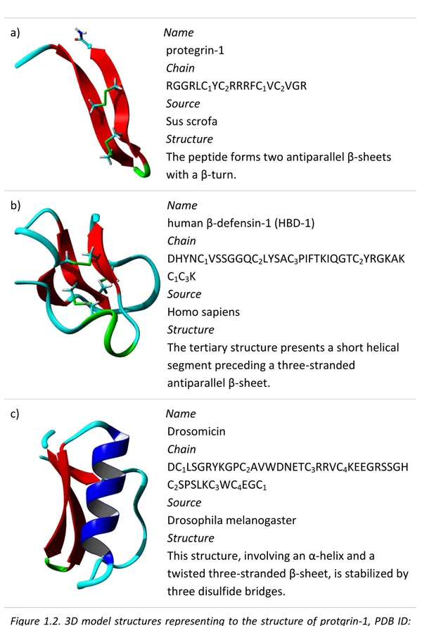
Some known examples with 2 disulfide bonds are protegrins from porcine leukocytes, or tachyplesins from horseshoe crabs, while examples with 3 disulfide bonds are defensins from several human tissues humans; finally peptides with more than 3 disulfide bonds are drosomycins in fruit flies. Figure 1.2 shows an example for each category, as reported for α-helical peptide. The structures are colored by secondary structure elements.
a) Name protegrin-1 Chain RGGRLC1YC2RRRFC1VC2VGR Source Sus scrofa Structure
The peptide forms two antiparallel β-sheets with a β-turn.
b) Name
human β-defensin-1 (HBD-1) Chain
DHYNC1VSSGGQC2LYSAC3PIFTKIQGTC2YRGKAK
C1C3K
Source
Homo sapiens Structure
The tertiary structure presents a short helical segment preceding a three-stranded
antiparallel β-sheet. c) Name
Drosomicin Chain
DC1LSGRYKGPC2AVWDNETC3RRVC4KEEGRSSGH
C2SPSLKC3WC4EGC1
Source
Drosophila melanogaster Structure
This structure, involving an α-helix and a twisted three-stranded β-sheet, is stabilized by three disulfide bridges.
Figure 1.2. 3D model structures representing to the structure of protgrin-1, PDB ID: 1PG1 (a); HBD-1, PDB ID: 1KJ5 (b) and Drosomicin, PDB 1MYN (c).
1.3.3. Linear extended antibacterial peptides
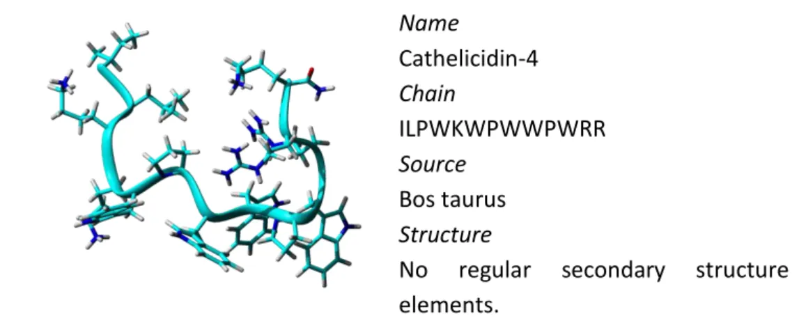
Linear extended peptides are very flexible in solution but they do not fold into regular secondary structure. These peptides are characterized by an overexpression of one or more amino acids, especially Arg, Trp or Pro residues. Some of them are rich in histidine residues, like histatin found in human saliva [10], others, such as abaecin and apidaecins from honeybees, are rich in proline residues or as prophenin from pigs, rich in proline and phenylalanine and indolicidin from cattle, rich in tryptophan and arginine residues (Figure 1.3). Name Cathelicidin-4 Chain ILPWKWPWWPWRR Source Bos taurus Structure
No regular secondary structure elements.
Figure 1.3. 3D model structures representing to the structure of Indolicidin derivative, PDB ID: 1QXQ
1.3.4. Loop antibacterial peptides
This is a quite small group of peptides which adopt a loop formation with one intramolecular disulfide bridge, often in the C-terminal part. Bactenecin, isolated from bovine neutrophils, is the first peptide in this group [11], other examples are peptides extracted from the frog skin, such as ranalexin or brevinin which contain a C-terminal loop and a long N-terminal tail, or thanatin from insect hemocytes (Figure 1.4).
Name Thanatin Chain
GSKKPVPIIYC1NRRTGKC1QRM
Source
Podisus maculiventris (Spined soldier bug) Structure
Thanatin adopts a well-defined anti-parallel beta-sheet structure from residue 8 to the C-terminus, including the disulfide bridge.
Figure 1.4. 3D model structures representing to the structure of Thanatin, PDB ID: 8TFV
1.4. Principal families of cationic antibacterial peptides and
their activity spectrum
A variety of different natural peptides have been identified and isolated from many organisms in addition, modified and mimetic peptides have been designed and synthesized.
Amongst the multi-drug resistant bacteria responsible for nosocomial infections are Staphyloccocus aureus, Pseudomonas aeruginosa and Escherichia coli. In this paragraph, the principal families of cationic AMPs active against these pathogens will be described.
1.4.1. Magainins
The name magainin derives from the Hebrew magain meaning shield. Magainins 1 and 2 were the first AMPs extracted thirty years ago from the skin of the African frog Xenopus laevis [12]; they can be found within amphibian skin as well as mammals. They are composed of 23 amino acids, exhibit a net charge of +4 and differ by two residues in position 10 and 22. They adopt an amphipathic α-helical structure and present a broad
antimicrobial activity spectrum against Gram-positive and Gram-negative bacteria with a MIC (Minimum Inhibitory Concentration) value between 50 and 150 μgmL-1. Different analogues have been synthesized and they showed enhanced activity. Their mechanism of action follows the toroidal pore model to permeabilize the bacterial membrane [13].
1.4.2. Cathelicidins
Cathelicidins are cationic peptides constituted by a highly conserved N-terminal domain of approximately 100 residues, named cathedin and with a structurally variable antimicrobial domain at the C-terminus. They have been isolated in neutrophilic granules and epithelial cells of many invertebrate and vertebrate species, including reptiles, fishes, birds, mammals and humans [14-16].
Among the most studied cathelicidins are SMAP-29 and LL-37. SMAP-29 has been isolated in sheep and is, nowadays, one of the most potent AMP known: its MIC against some bacteria being below 1 μgmL-1. It is composed of 29 residues, adopts an α-helical conformation and presents an amphipathic nature [17]. Its antibacterial activity is attributed to its N-terminal amphipathic α-helix. The only cathelicidin found in humans is LL-37, a cationic α-helical peptide formed by 37 amino acids with two leucine residues at the beginning. LL-37 disrupts the bacterial membrane though the toroidal pore model or through the carpet model [18].
1.4.3. Defensins
Defensins are cationic peptides rich in cysteine residues, adopting β-sheet structures. The first peptides were discovered in 1983 in rabbits [19] and only two years later in humans [20]. Since these discoveries many similar peptides were isolated from other organisms.
They have broad spectra of activity due to interact and disrupt the lipid membranes by multimerizing and forming pores, with subsequent lysis of the microbes.
Mammalian defensins contain three stabilizing disulfide bridges formed by six cysteine residues [21]. They are classified into three subclasses: α-defensins and β-defensins, adopting a triple-stranded β-sheet conformation, and θ-defensins, adopting a total circular conformation.
In contrast, insect defensins contain an α-helix domain bound to a β-sheet region via a disulfide bridge.
Plant defensins, commonly called as thionines, may contain up to eight cysteines, thus forming up to four disulfide bridges. They also contain both α-helix and β-sheet domains.
It is known that AMPs found in humans or in mammals are not limited to cathelicidins and defensins, but other anionic peptides into histatins and dermcidins families were also found in some tissue.
1.5. Antimicrobial activity: mechanism of action
1.5.1. Reaching of the site of action
Regardless of the precise mechanism, the interaction with the cell membrane is the key step for all AMPs [22]. For Gram-negative and Gram-positive bacteria, the bactericidal process of cationic AMPs is stepwise and generally rapid. Initially, driven by electrostatic forces, AMPs diffuse toward the cell membrane even from a relatively long distance. It is hypothesized that the negative charge on the outer bacterial envelope and the strong electrochemical gradient of the bacterial cytoplasmic membrane both contribute to the attraction of cationic AMPs [23].
Before looking at the mode of action, it is necessary to understand the membrane biology of bacteria, fungi and eukaryotic membranes, which are the primary targets for most AMPs.
Universally, all cell membranes can be seen as fluid mosaics of proteins and phospholipids, which are arranged as bilayers with hydrophobic and hydrophilic domains. However, a significant lipid compositional difference exists between the prokaryotic and eukaryotic membranes as well as among cell types.
Bacterial membranes (Figure 1.5) are made up of negatively charged phospholipids such as phosphatidylglycerol (PG), cardiolipin (CL), or phosphatidylserine (PS) [24], which are stabilized by divalent cations such as Mg+2 or/and Ca+2. Even though there is not much difference in lipid composition, Gram-negative bacteria differ from Gram-positive bacteria as the former have a smaller peptidoglycan layer and an outer membrane, in addition to a cytoplasmic membrane containing lipopolysaccharide (LPS), which acts as permeability barrier [25].
Interestingly, some peptides binding to Gram-negative bacterial membranes induce Mg2+ ion displacement between the LPS, thus destabilizing the membranes arrangement.
In contrast to bacteria, fungal membranes are rich in phosphomannans and other related constituents such as negatively charged phosphatidylinositol (PI), phosphatidylserine (PS) and diphosphatidylglycerol (DPG), which give a higher negative charge surface to the membranes [26].
Figure 1.5. Schematic structure of Gram-negative (on the left) and Gram-positive (on the right)[27]
On the other hand, mammalian membranes are rich in zwitterionic phospholipids with neutral net charge, including phosphatidylethanolamine (PE), phosphatidylcholine (PC), or sphingomyelin (SM). Moreover, cholesterol is present in significant amounts in mammalian membranes and can reduce the activity of AMPs by affecting the fluidity and dipole potential of phospholipids, in addition to stabilizing the lipid bilayers and delaying the binding of peptides to the membranes. Therefore, sterol in the mammalian membranes is thought to be involved in the specificity of action of the antimicrobial peptides [28], in addition membrane potential and asymmetric distribution of phospholipids in eukaryotic membranes contribute to prevention of AMPs binding [29].
1.5.2. Proposed models
After reaching the bacterial surface, cationic peptides competitively displace divalent cations due to different binding affinities. Divalent cations act as stabilizers for the LPS by binding the anionic phosphate groups. The loss of Mg2+ and Ca2+ results in the disturbance of the rigid outer membrane and promotes the uptake of more molecules. During this process, many linear AMPs fold into an amphipathic structure due to the hydrophobic environment of the cell membrane. The amphipathic nature of AMPs is essential for further
interaction with the hydrophobic components of the membrane. Consequently, the peptides contact the cytoplasmic membrane and disrupt the lipid bilayer in multiple ways.
During this folding process, it is proposed that AMPs adopt a parallel orientation to the membrane. Moreover, there is an accumulation of the peptides on the surface of the membrane, until a threshold in peptide interfacial concentration is reached. Parameters influencing the threshold concentration include the propensity of peptide self-assembly, peptide charge, amphipathicity and hydrophobicity, as well as membrane fluidity and composition [30, 31].
An overview of the widely accepted models proposed to clarify the membrane permeabilization is given in Figure 1.6: they include the carpet model, barrel-stave model and toroidal-pore model and all of them need the interaction of cationic AMPs with the negatively charged outer membrane surface in a parallel orientation.
Figure 1.6. Three expected models of interaction between cationic AMPs and cytoplasmic membrane [7].
a) In the carpet model the peptides initially associate with the membrane, align parallel to the surface of bilayer and cover the surface. This orientation destabilizes the packing of phospholipids and causes a change in membrane fluidity because of the moving of phospholipids by peptides. Thus, the stability of the local membrane is disturbed. When a threshold peptide concentration is reached, the membrane will break down due to unfavorable energetics. The general features of the carpet model include that peptides remain in contact with the lipid head groups with consequent membrane dissolving [32].
b) Barrel-stave model was first proposed by Ehrenstein and Lecar in 1977. According to this model, a variable number of individual peptide molecules are arranged to form a barrel-like pore or channel. In this mechanism, peptide hydrophobic surfaces interact with the acyl chains of lipid in the membrane, generating an aqueous pore consisting of at least four peptides. A crucial step in this model is that peptides have to recognize each other in the membrane bound state. It is highly energetically unfavorable for a single peptide to traverse the membrane, hence the peptides aggregate on the surface until the threshold concentration is reached, and then insert into the hydrophobic core of the membrane by undergoing a conformational phase transition, forcing polar–phospholipids head groups aside to induce localized membrane thinning. This event is followed, by additional recruitment of peptides around or into the channel, leading to an increase in pore size and stabilization, thus killing the microbe by leakage of intracellular components. Aggregation may be required simply because the amount of stabilization that one peptide provides is not sufficient for pore formation and stronger binding of peptides to pores could be due to packing reasons as lipid head groups are less tightly packed in pores [33].
c) Toroidal pores can be formed by a much greater variety of peptides. Prior to formation of both barrel-stave and toroidal pores, the peptide adsorbs
parallel to the membrane surface [34]. When a certain concentration on the surface is reached, the peptide either inserts perpendicularly into the membrane, or induces a positive curvature strain in the membrane, resulting in an opening, the so-called toroidal pore. The hydrophilic regions of the peptides keep the association with lipid head groups and the hydrophobic peptide regions associate with the hydrophobic core of the membrane lipids. Thus, the peptides and lipids together form well-defined pores, with the hydrophilic regions of the peptides and phospholipids head groups facing the center of the pore and producing an aqueous pore [23].
The effect of barrel-stave and toroidal mechanism is that peptides or other molecules could be translocated into the cytoplasm, resulting in enzyme inhibition or DNA and RNA binding.
Numerous AMPs are found to show antifungal activity and the mechanisms of action involve cell disruption by binding of the outer membrane. Moreover antifungal peptides interfere with cell wall synthesis [35]. AMPs also possess antiviral activity by interacting with virus proteins [36, 37] and different AMPs with antiparasitic activities have been discovered [38].
1.6. Bacterial resistance strategy
Presumably, bacteria have been exposed to AMPs for millions of years, but the development of resistance against AMPs has occurred to a much less degree.
Inability to develop resistance to AMPs is based on two points. Firstly, the bacterial membrane has to be modified and this is really hard for the microorganisms. Secondly, due to the presence of huge numbers of AMPs in the host, it is difficult for microbes to develop resistance against all peptides at the same time.
More recently, however, a few resistance mechanisms in bacteria against AMPs have been discovered and investigated. They include: surface modification, external trapping of AMPs, and active efflux of AMPs or proteolytic degradation.
The simplest way for a bacterium to increase its resistance towards AMPs is to reduce the negative net charge of its outer membrane. A small number of bacteria have developed some mechanisms to stop the first step of AMPs action in which they reduce the electrostatic interaction with the cationic peptides. These modifications concern the teichoic acid of peptidoglycan in Gram positive or the lipid A moiety of LPS in Gram negative bacteria. For example, some Staphylococcus aureus transport positively charged D-alanines residues from the cytoplasm to the anionic teichoic acids of the bacterial wall. These modified teichoic acids could have a role in the protection of some bacteria against antimicrobial peptides [39].
Other resistance mechanisms could involve the production of extracellular proteases, which are able to degrade cationic peptides. Interestingly, some bacteria such as Pseudomonas sp. and S. epidermidis, use other methods in addition to protease production, such as forming biofilms to prevent AMP insertion and pore formation [40]. AMP insertion and pore formation are prevented by this increased hydrophobicity of the membrane.
It has also been shown that ATP-Binding Cassette (ABC) transporters import AMPs into the bacteria, while efflux pumps export them. Overexpression of these efflux pumps can explain resistance to AMPs [41].
In conclusion, it is important to keep in mind that the primary target of an AMP is the bacterial membrane. To develop resistance against them, the bacteria should redesign their whole membrane a very long process. Considering that AMPs are part of the innate immune system, and that bacteria have had a huge time-span to adapt to their killing mechanisms, the resistance
phenomenon is extremely difficult. However, it is worth to notice that, among microbes, only bacteria have shown resistance mechanisms against AMPs.
1.7. Limits of the antimicrobial peptides as therapeutic
agents
AMPs have many advantages which make them potential substitute of conventional antibiotics. They have broad spectra of activity, are efficient towards multi-resistant bacteria and are not hindered by resistance. However AMPs possess different disadvantages that limit their development as therapeutic agents. Briefly, these limits are:
- Hemolytic activity for eukaryotic cells
Since the mechanism of action involves membrane interactions, the antimicrobial peptides could interact with eukaryotic cells and lyse them. It was observed that some natural AMPs with amidated C-terminal show higher hemolytic effect [42]. It is known that the process of amidation stabilizes the helix formations after membrane binding [43, 44]. To decrease the unwanted effect Ulrich et al. have prepared the deamidated analogues observing a decrease of hemolytic effect, maintaining the antimicrobial activity [45].
- Broad activity spectrum
As already stated, antimicrobial peptides possess broad spectrum of activity, but this could be a problem in term of destruction of the microflora during the therapy with consequent risk of diarrhea and other infections [46].
- Protease susceptibility
Proteolytic stability is essential for therapeutic use of AMPs because they are rapidly inactivated by proteases in the human body. This problem can be overcome using different strategies. Considering that AMPs taken into account in this thesis do interact with bacterial membranes, the most frequent modification includes the replacing of natural amino acids with D-amino acids
in the antimicrobial sequence, in this case the proteases cannot hydrolyze the unnatural residues and the modified peptide is consequently more active [47]. Designing polymeric analogues, such as amphiphilic arylamide polymers that mimic the AMPs structures, is another good strategy to increase the stability against proteases. In addition these peptide mimetics could be prepared in an easy and less expensive way [48]. Finally, in some studies has been observed that peptide cyclization improves the serum stability of AMPs [49].
- Salt sensitivity
AMPs are inactive in presence of high salt concentrations. The mechanism of inactivation is not so clear, but the general idea is that the interaction with the salt occurs after the interactions with cell membranes, when AMPs organize in the secondary structure [50]. Therefore, it is necessary to develop new AMPs with the secondary structure more stable.
- High cost of production
If we compare the cost of production of traditional antibiotics with the cost of the production of AMPs, the last one is really higher. Compared with the cost of traditional antibiotics, circa 1$/g, an AMP produced by fermentation can cost more than $400/g [2]. The high cost slowed the development of novel AMP drugs.
1.8. AMPs in clinical trial
Several companies are currently attempting to developed AMP-based drugs, but to date, none of them have reached the market. Nevertheless, many antimicrobial peptides are currently being tested in clinical trials. At the moment I am writing this thesis, a few peptides or mimetics are in (or have completed) clinical trials as antimicrobial or immunomodulatory agents. In Table 1.1 a list of AMPs into clinical trials is given, obtained by the web database of the U.S. National Institutes of Health [51].
Table 1.1. List of AMP into clinical trials and their current status
Peptide
name Company Structure
Phase of study Development for treatment of Omiganan Cadence Pharmaceuticals analogue of indolicidin Completed Phase III preventing catheter infections/ colonization in patients with Central Venous Catheters Completed Phase II topical skin antisepsis in healthy adult subjects hLF1–11 AM-Pharma aminoterminal amino acids 1– 11 of human lactoferrin Completed Phase II Candida infection PMX-30063 Polymedix oligomeric and small molecule mimetics of AMPs Completed Phase II acute bacterial skin and skin structure infections (ABSSSI) caused by S. aureus PAC 113 Pacgen Biopharmaceuti cals Corporation from histatin 3 and histatin 5 Completed Phase II oral candidiasis in HIV seropositive patients
DPK-060 Pergamum structurally derived from the endogenous human protein kininogen Phase II Atopic Dermatitis with an extension program in external otitis OP-145 Octoplus from cathelicidin antimicrobial peptide LL-37
Phase II chronic otitis media CLS001 Cutanea Life Sciences Cationic AMP pentahydro-chloride currently recruiting participants for Phase II Rosacea LTX-109 Lytix Biopharma AS synthetic antimicrobial peptido-mimetic currently recruiting participants for Phase II Impetigo HB1345 HelixBiomedix
Lipohexa-peptide Phase I skin infections
POL-7080 Polyphor Ltd protein epitope mimetics of the antimicrobial peptide protegrin I Phase I Pseudomonas specific antibiotic NZ2114 Novozymes defensin-like AMP plectasin
Preclinical pneumonia and septicaemia
The story of Pexiganan is interesting. It derives from magainin-2 and was developed by MacroChem Corporation in the 90s. It entered in Phase III trials
(ClinicalTrials.gov Identifier: NCT00563433) and was tested as topical cream vs. oral Ofloxacin in the Treatment of Infected Diabetic Ulcers. It was rejected by the Food and Drug Administration (FDA) in 1999 because its efficiency towards infected diabetic foot ulcers did not offer any improvement over the conventional treatment with ofloxacin, a fluoroquinolone antibiotic. Pexiganan is, however, now getting a new life as Locilex by Dipexium Pharmaceuticals of White Plains, New York. This company is negotiating with FDA to bring Pexiganan through clinical trials, as a way of treating bacterial infections associated with diabetic foot ulcers, particularly when the bacteria are resistant to standard antibiotics (ClinicalTrials.gov Identifier: NCT01594762).
Chapter 2. Collection and building of a new AMPs
database
2.1. Introduction
After 30 years of intensive research on AMPs, an accepted and universal model of action is still lacking. This is not due to lack of scientific research, but rather to the simple fact that a comprehensive model does not exist. AMPs utilize a wide variety of mechanisms, such as altering the membrane equilibrium, creating pores, disrupting the membrane, docking a protein receptor and so on. The first requirement to conduct a quantitative structure-activity relationship (QSAR) study of a series of peptides is to cluster those expected to act in the same way. A correct QSAR investigation is simply impossible if the same analysis set includes peptides acting with different mechanisms, such as membrane disruption or enzyme inhibition. In recent years, many novel AMPs have been discovered and characterized. Most of these data have been included in web databases. Unfortunately, several noteworthy features of AMPs, such as minimum inhibitory concentrations (MIC) or their spectrum of activity, are not always included in public databases. The need to have a more extended collection of AMPs, together with the need for physical-chemical parameters, motivated me and my research group to create a new web database called YADAMP: Yet another database of antimicrobial peptides [52]. It permits a quick and easy search of peptides on the base of their activity (expressed as MIC against several bacterial strains) and their structure. The philosophy behind YADAMP is to facilitate the access to important information on AMPs, to allow extensive QSAR analysis and the creation of the activity model against a particular bacterial target. With YADAMP, it is possible to create a uniform subset of
AMPs that is still large enough to allow meaningful statistical analysis. The database can be accessed via a web-based browser at:
http://www.yadamp.unisa.it.
2.2. Data collection
YADAMP collects data on AMPs scattered in scientific papers or web databases, providing structural data and information on antimicrobial activities. In YADAMP, a user can obtain information about peptide name, amino acid sequence, length, presence of disulfide bridges, date of discovery, activity and taxonomy. In addition, the most relevant chemical-physical properties were calculated such as charge, hydrophobic moment, helicity, flexibility, isoelectric point, Boman index, instability index and penetration capabilities.
2.2.1. Sequences
Sequences of active AMPs were mainly extracted from the scientific literature and were compared with data in public databases (UniProtKB/Swiss-Prot [53], APD [54], CAMP [55]. Each of the collected sequence was validated with literature available data. To facilitate future QSAR studies of antimicrobial peptides for peptide design, I also collected sequences of synthetic peptide derivatives. I collected relevant information on 2525 active sequences. Collecting and annotating large numbers of sequences is a perilous job as it can propagate errors in the original papers that might have been corrected later, or because it can introduce new errors from manual annotations. For this reason, YADAMP is frequently updated.
2.2.2. MIC values
More important, YADAMP is mainly focused on peptides activities. In microbiology, the MIC is the lowest concentration of an antimicrobial that will
inhibit visible growth of a microorganism. The MIC is necessary to perform a statistical analysis of peptides. YADAMP permits the selection of AMPs with the lowest MIC value. Experimental MIC values (expressed in μM) were manually extracted from careful reading the original papers. MIC values expressed in μg/mL were converted to μM to allow a quick comparison, using the formula:
(
)
( )
The most intensively studied organisms are Escherichia coli, Pseudomonas aeruginosa and Salmonella enterica serotype Typhimurium amongst Gram-negative organisms and Staphylococcus aureus and Micrococcus luteus amongst Gram-positives. These organisms are also the primary source of infections in humans. For these reasons, in YADAMP the fields corresponding to these MIC values are added, by default, to the Result page. There are also two areas reporting the activity against Bacillus subtilis and the fungus Candida albicans owing to their high frequency. Data against all other bacteria were inserted in fields: Other Gram−, Other Gram+ and Other for fungi and yeast. In addition, for each peptide, we added in YADAMP the link to the references from which information were extracted in order to check the data and the antibacterial assay conditions.
2.2.3. Biological classification
Biological classification is a method of scientific taxonomy used to group and categorize organisms into groups having attributes or traits in common. Taxonomy materials are important to understand and identify sequence patterns conserved across species. It is a hierarchical classification, in which each level is named rank. The data about taxonomic information was extracted
from NCBI Taxonomy database [56]. YADAMP permits a selection for five main ranks: phylum, class, order, family, and genus.
2.2.4. Calculated parameters
As already stated, YADAMP was created to be a resource for QSAR investigations on AMPs, thus, for an accurate QSAR analysis, it is essential to group peptides sharing some features, such as similar secondary structure, flexibility or charge. For this reason, YADAMP enriches the experimental data with some theoretical information.
Following descriptions about calculation performed, however in the Appendix-a it is possible to reAppendix-ad the MATLAB scripts Appendix-associAppendix-ated with eAppendix-ach pAppendix-arAppendix-ameter computed.
i. Charge
AMPs can act in very different pH conditions, depending on the tissue in which the bacteria are grown. I have calculated the charge of each peptide at three different pH values (pH 5, 7 and 9) by the formula:
∑
∑
where Ni is the number of the N-terminus and of the side chains of arginine,
lysine and histidine. The j-index refers to the C-terminus and the aspartic acid, glutamic acid, cysteine and tyrosine amino acids. pKai and pKaj values refer to
amino acids labeled with the index i and j.
This algorithm has some limitations, such as:
- the residues are assumed to be independent of each other; - N- and C-termini have fixed pKa values;
- only the 20 natural amino acids are considered;
I used pKa value taken from Lehninger Principles of Biochemistry [57]. A quick inspection at the database reveals that, mainly because of the wide variation in lysine abundance, the charge of certain peptides can largely vary at different pH. Peptides acting as antimicrobial compounds do not always experiment the neutral pH, so this parameter can be decisive for peptide simulations in specific tissues.
ii. Isoelectric point
The isoelectric point (pI) is the pH at which a protein has no net electrical charge. Below the pI proteins carry a net positive charge and above it they have a net negative charge. Theoretical pI values were calculated using a free online tool [58]. According to Bjellqvist et al. [59] it was assumed that the same pK value could be used for an amino acid residue in all polypeptides and in all positions in the peptide except for N- or C-terminally placed amino acids. For the pK values of the N-terminal amino groups the effect of the different substituents on the α-carbon were taken into account.
iii. Boman index
Most authors have agreed that a potential AMP should possess a positive net charge to facilitate binding to bacterial phospholipids as well as a certain degree of amphipathicity to allow molecule adaptation to a bacterial membrane. These criteria are not enough to predict the ability of a peptide to interact with cell membrane. Boman [60] introduced a parameter which shows a certain degree of discrimination between membrane-interacting and protein-interacting peptide. They established the tendencies of amino acids to leave water and move in a nonpolar condensed phase calculating the distribution coefficients for each side chain of the natural amino acids at pH 7. I calculated the Boman index for all sequences, as the sum of the free energies (kcal/mol)
of the respective amino acid side chains for transfer from cyclohexane to water divided by the total number of residues.
∑ ( )
The free energies values were taken from Radzeka and Wolfenden [61].
iv. Hydrophobicity
Hydrophobicity is another critical characteristic of amino acid residues that determines protein folding, protein subunit interaction, binding to receptors, and interactions of proteins and peptides with biological membranes. Calculation of hydrophobicity assigns a numerical hydrophobicity value to each type of amino acid, and then relates these hydrophobicities in a particular protein or fragment with some aspect of structure or function. The hydrophobicity of an amino acid residue is not a property that can be easily defined or simply measured. Nevertheless, several groups have attempted to derive numerical hydrophobicity scales using a variety of experimental and computational methods. The distribution of hydrophobic residues in amphipathic peptides is revealed by the hydrophobic moment, which depends on the spatial conformation of the peptide. The hydrophobic dipole moment can be estimated from a known peptide sequence, assumed that the polypeptide backbone follows some periodic arrangements such as an α-helix or a strand from a β-sheet. It is represented by the vectorial sum of all the hydrophobicity indices, divided by the number of residues. I estimated the hydrophobic moment in according to three different hydrophobic scales: CCS [62]; Kyte–Doolittle [63]; and Eisenberg [64], reporting in the web database the three different results for each peptide. For this calculation it was assumed that the hydrophobicity of each residue i can be represented by a vector of
length Hi, having a direction perpendicular to the axis of the helix or strand of
beta structure.
The value of estimated hydrophobic dipole moment (µH) is:
((∑ ( )) (∑ ( )) ) ⁄
where δ is the angle separating side chains along the backbone (e.g. δ = 100° for an α-helix).
Finally, I calculated the mean hydrophobicity as the total hydrophobicity (sum of all residue hydrophobicity indices) divided by the number of residues.
∑ (
)
v. Helicity
The secondary structure of a peptide is crucial for the investigation. If it is not experimentally available, peptide structure prediction is essential. In YADAMP, the prediction is based upon the DSC (Discrimination of protein Secondary structure Class) algorithm from King and Sternberg [65]. The method extracts the maximum information from the primary sequence and allows the prediction of the secondary structure from multiply aligned homologous sequences and linear statistics. The DSC Method is accessible as ‘Secondary Structure Prediction’ (SSP) option in Discovery Studio from Accelrys.
The DSC method gave the following results: 001 ALRLAIRKR DSC-SEC HHHHHHCCC PROB-H 779998421 PROB-E 000001000 PROB-C 331111689 NO. 1 2 3 4 5 6 7 8 9 RES a l r l a i r k r DSC-SEC H H H H H H C C C PROB-H 0.723 0.723 0.877 0.905 0.893 0.751 0.366 0.185 0.087 PROB-E 0.020 0.020 0.004 0.015 0.032 0.065 0.042 0.021 0.020 PROB-C 0.257 0.257 0.119 0.080 0.075 0.0184 0.592 0.794 0.893
The first section of the report shows 5 lines for each sequence: 1. the sequence
2. the predicted secondary structure type, DSC_SEC 3. the probability of being helix, PROB_H
4. the probability of being beta strand, PROB_E 5. the probability of being coil, PROB_C
Values of PROB_H, PROB_E, and PROB_C are between 0 and 9, with 9 being the highest probability.
The second section of the report shows similar information for each residue, but the probabilities are shown in real number values for each residue. I calculated the Helicity probability via a Perl script by summing of PROB-H number and dividing by the number of residues.
vi. Flexibility
The molecular flexibility of proteins is a crucial factor in determining their biological activity, including binding affinity, and for the theoretical understanding of peptide dynamics. The identification of regions in proteins
with the highest conformational flexibility and rigidity is essential for predicting the mechanism of protein folding. Consequently, there is considerable interest in predicting the flexibility or, conversely, the rigidity of peptides from their amino acid sequence. Obviously, prediction of the secondary structure of an AMP is a hard task due to the different conformations that a peptide shows in different chemical environments. Moving from the water bulk into the membrane, the structure of peptides varies considerably. I calculated the flexibility of α-AMPs according to a conformational flexibility scale for amino acids in peptides [66], which provides an absolute measure for the time scale of conformational changes in short unstructured peptides as a function of the amino acid type. This experimental scale derived from kinetic measurements of the collision frequency between the two ends of short random-coil polypeptides. These peptides were labeled with a fluorescent probe at the C-terminus and Trp as a fluorescence quencher at the N-terminus. The fluorescence lifetimes of fluorescent probe/Trp peptides provide the quenching rate constants (kq),
which measure the end-to-end collision frequency. The authors have shown different collision frequencies when the probe and the quencher were separated by different amino acids. This arrangement allowed them to correlate the collision frequency with the type of amino acid and build up a flexibility scale.
In YADAMP the flexibility was calculated by formula:
∑
For amino acids not found in the Huang work, I estimated missing values by comparison with reported kq constants.
vii. Instability index
A peptide cannot show activity if it is not sufficiently resistant to proteases. The instability index predicts the in vivo half-life of the peptide; this was added to YADAMP to investigate the correlation between activity and in vivo stability. To estimate the instability values I inspired in Guruprasad work [67]. They made a statistical analysis of 12 unstable and 32 stable proteins to reveal patterns in the occurrence of certain dipeptides. Some dipeptides appeared particularly frequent in stable proteins, whereas other dipeptides were common in unstable proteins. The contribution of each of the dipeptides towards instability was obtained by summing the instability weight values corresponding to the conditions satisfied by the dipeptide and termed as the dipeptide instability weight value (DIWV). I calculated the instability index (II), using the DIWV values for all 400 combinations reported in the Guruprasad paper by equation:
( ) ∑ ( )
where xiyi+1 is a dipeptide, L is the length of the sequence and 10 is a scaling
factor.
viii. CPP
This parameter is the acronym of Cell Penetrating Peptides and is an estimate of the tendency for a peptide to penetrate a cell membrane. The parameter can take values between 0 and 1, where 1 corresponds with the highest probability of a peptide to penetrate a membrane, and 0 indicates the impossibility to enter a membrane. To predict this ability I used a free online tool [68] in which I inserted the peptide sequences. The limit of this web server is that the prediction is limited to peptide with length between 5 and 30 amino acids.
2.3. Database content
YADAMP contains a large number of manually annotated sequences of AMPs. To date, it contains 2525 sequences of active peptides with length between 5 and 96 amino acids; amongst them, 469 sequences have one or more disulfide bridges. In cases in which the active structure of a peptide is not experimentally known, an estimation of helicity was made by an automated script. From helicity estimations, 1362 sequences have a helicity index >5, in a range between 0 and 9, in which 9 corresponds with the highest probability of α-helix secondary structure. The action mechanism of many peptides involves interaction with the membrane, therefore hydrophobic moment is a key parameter to characterize AMPs. In my collection, there are 619 sequences with mean hydrophobic moments >3.0 calculated using the CCS scale.
YADAMP has the largest collection of MIC values of AMPs. It contains 1015 sequences with an experimentally verified action against E. coli (the bacterium with the largest data set). Other bacteria well represented in YADAMP are S. aureus (957 sequences), P. aeruginosa (438 sequences), B. subtilis (253 sequences), S. Typhimurium (149 sequences) and M. luteus (129 sequences). Furthermore, there are 251 sequences active against other Gram-negative bacteria and 363 sequences active against other Gram-positive bacteria. Fungi are represented by C. albicans (486 sequences), but there are 128 sequences active against other fungi or yeast. A specific peptide may have activity against different bacteria, so it can be included twice or more. Finally YADAMP includes 911 sequences extracted from Amphibia, the most important source for active peptides, 260 sequences from Insecta, 277 sequences from Mammalia, 158 sequences from Magnoliopsida and 294 synthetic sequences.
2.4. Web database design: an overview
The web interface of YADAMP (Figure 2.1) is designed to offer a simple but practical use of the database.
Figure 2.1. Screenshot of YADAMP Homepage It includes seven sections:
- Home - About
In this section is possible to read the philosophy behind YADAMP and eventually news.
- Search Database
In this section is possible to query the database by length, sequence, name, taxon, or discovery period. Any query can be made combining as many fields as the user likes with AND and OR operators. We have provided the option to search structural parameters such as the presence of disulfide bridges, the value of charge at pH 7, the mean hydrophobic moment, the instability or
propensity to α-helix conformation. More importantly, it is possible to select AMPs with a proven activity against five organisms, chosen because of their importance and because they are a common target of AMPs. In this way, it is extremely easy to build sets of AMPs with characteristics suitable for further investigation.
- Theory
This section offers a description of the theoretical terms, included the references.
- Statistical Data
It provides an estimate of the database content and the date of the last update. - Literature
This section provides an automatic visualization of the 50 most recent papers through RSS (Rich Site Summary). It is due to the extraordinary interest in AMPs, as reflected by the high number of publications in this field.
- Contact
In this section is possible to send an e-mail for opinion and suggestions or to report eventual errors.
2.5. Using YADAMP
As an example of use, I simulated here the search of structural elements in peptides effective against E. coli. To develop a model of action against this bacterium, it is necessary to choose sequences sharing the same target (for example peptides acting on the cell membrane or against the cell wall), and it is critical to quantify the effectiveness of this action.
Figure 2.2. Screenshot of Search database page
As shown in Figure 2.2, it is possible to make a query to select peptides with a similar secondary structure (in this case with helicity >5), amphipathic (with hydrophobic moment > 2) and active against E. coli (MIC < 40 μM).
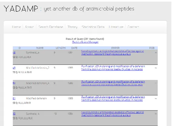
The query result page shows recovers of 391 sequences for further investigation (Figure 2.3). The peptides are listed by ID order and by clicking on this number detailed information for each selected peptide are accessible.
Figure 2.3. Query result page
The example of page about detailed information is given in Figure 2.4. To facilitate the immediate interpretation of the results, we separated the information by colors. In the blue section (Figure 2.4) I reported data about ID YADAMP, name and eventually number of disulfide bridges. In the green section I put the value of calculated parameters and the sequence of the peptide. In the orange section there are the activity values and in the second blue section there are taxonomic information, date of discovery and the link to papers in which I extracted the activity value. Finally, since some peptides possess an amphipatic helix structure, it is interesting to visualize the amphiphilic region in a sequence. Thus I used the helical-wheel Java applet [69] to obtain a projection of the amino acids side chains along the helix axis
(Figure 2.4, bottom). Each residue is numbered and colored according to its chemical nature.
Chapter 3. Development of a modified antimicrobial
peptide
3.1. Design of modified antimicrobial peptides
Due to the discussed limits of AMPs in the clinical therapy, some chemical modifications or sequence optimization are needed. In the last years many molecules that mimic AMPs function and structure have been synthesized, resulting in a number of distinctive AMPs classes, briefly reported in the following.
3.1.1. AMPs mimetics
This class is formed by synthetic molecules such as β-peptides, peptoids or arylamides. These compounds possess the same chemical-physical feature of AMPs, mimic the same mechanism of action but, lacking the peptide bonds, they are more stable in vivo [70, 71].
3.1.2. Hybrid AMPs
As suggested by the name, these molecules are the result of the combination of the active portions of two or three AMPs [72]. In this way it is possible to increase the antimicrobial activity towards a specific target.
3.1.3. AMPs congeners
A congener peptide is a modified AMP obtained by swapping out of a particular amino acid in the active sequence or by truncating of the N-terminus or the C-terminus ends. The goal is to individuate the residues responsible for the activity and to reach the smallest sequence length [73].
3.1.4. Cyclotides and stabilized AMPs
They are cyclic peptides of about thirty amino acids isolated from plants. They possess a characteristic head-to-tail ring structure stabilized from three disulfide bonds [74]. The high stability and microbiological activity as anti-HIV and insecticidal give them promising potential application as new stable peptide-based drugs [75].
3.1.5. AMP conjugates
AMPs can be coupled to an antibody or a ligand for a receptor on a definite bacterial pathogen, in this case they could be used at lower concentrations inducing lower side effects or could reach a specific target [76, 77].
3.1.6. Immobilized AMPs
Finally AMPs can be incorporated in different materials or adsorbed to surfaces where they still retain their ability to bind and kill bacteria. Immobilized peptides appear to interact and disrupt microbial membranes with high affinity in a similar fashion to native peptides in solution [78]. These classes of molecules are advertised to have selective or ‘targeted’ antimicrobial activities, improved retention or unique abilities that allow them to bind to specific surface. These groups of new peptides have smart medical and industrial application potentials, for example for treating antibiotic-resistant bacterial infections and septic shock, to preserve food or to sanitize surfaces both in vitro and in vivo.
3.2. Our approach
There is an urgent need for unconventional, molecular approaches to control antimicrobial function. Light is an attractive external trigger for the control of biological functions [79, 80] because it offers highly spatiotemporal resolution,
![Figure 1.6. Three expected models of interaction between cationic AMPs and cytoplasmic membrane [7]](https://thumb-eu.123doks.com/thumbv2/123dokorg/5731260.74604/30.892.146.713.637.945/figure-expected-models-interaction-cationic-amps-cytoplasmic-membrane.webp)
![Figure 4.13. Changes of the absorption spectrum of the reaction solution as a function of reaction time, with [ABTS 2- ] 0 = 0.25 mM, [H 2 O 2 ] 0 = 80 μM and [HRP] = 150](https://thumb-eu.123doks.com/thumbv2/123dokorg/5731260.74604/85.892.254.650.483.795/figure-changes-absorption-spectrum-reaction-solution-function-reaction.webp)