Università degli Studi di Pisa
Dottorato di Ricerca in Oncologia Sperimentale e
Molecolare
Settore scientifico disciplinare Bio/11-Anno 2003
Tesi di Dottorato:
"GFP transgene driven by Kit proto-oncogene
regulatory sequences is differentially expressed in
multiple stem and progenitor cells"
Relatore
Candidato
"We are books of blood: wherever we're opened we're red"
Clive Barker
"Diversi nell'aspetto siamo scritti in mille lingue, ma siamo libri di sangue"
RIASSUNTO
Nel corso degli ultimi anni, numerosi geni regolatori dello sviluppo embrionale e della differenziazione cellulare si sono rivelati importanti mediatori della tumorigenesi. Lo studio di questi geni ha inoltre evidenziato l’esistenza di meccanismi comuni nella regolazione di diversi sistemi differenziativi, tuttavia ancora poco si conosce della loro funzione ed interazione. Una migliore comprensione dei meccanismi d’azione e delle relazioni fra geni regolatori è quindi fondamentale per chiarire anche la loro possibile implicazione in processi patologici. Il mio progetto di tesi di Dottorato si è inserito nel contesto di ricerca del laboratorio diretto dalla Dr.ssa Maria Cristina Magli che si interessa da diversi anni della funzione di alcuni di questi geni nell’ematopoiesi ed in altri sistemi cellulari, in particolare a livello di cellule staminali e progenitori. Durante la mia tesi ho sviluppato una serie di studi sul ruolo di Kit, un protoncogene che codifica il recettore tirosin-chinasico per il fattore di crescita Stem Cell Factor (SCF). Il
pathway regolativo SCF/Kit riveste un ruolo chiave nella regolazione di diversi tipi
di cellule staminali come le ematopoietiche, germinali e cardiache. Inoltre Kit è espresso anche nell’intestino, a livello delle cellule interstiziali di Cajal, e recentemente è stato evidenziato per Kit un’attività oncogenica nello sviluppo dei tumori stromali gastro-intestinali (Gastrointestinal stromal tumors; GISTs).
Al fine di studiare la regolazione e la funzione di Kit ho utilizzato una linea transgenica murina, generata nel laboratorio diretto dal Prof. Sergio Ottolenghi, nella quale il gene repoter “enhanced green fluorescent protein (GFP)” è espresso sotto il controllo di regioni di regolazione di Kit.
Il primo obiettivo della mia tesi è stato verificare se il transgene Kit/GFP sia espresso nelle cellule staminali ematopoietiche (HSC) del midollo osseo (BM) e del
fegato fetale. Tramite analisi citofluorimetriche, saggi clonogenici e saggi di ripopolazione in vivo, ho dimostrato che il transgene è efficientemente espresso nelle HSC sia durante lo sviluppo embrionale che nella vita adulta. Inoltre, i risultati da me ottenuti forniscono la prima evidenza funzionale di una espressione differenziale di Kit nelle HSC rispetto ai progenitori ematopoietici, riproducendo quindi i pattern di espressione di altri importanti marcatori di cellule staminali. Questi risultati, in aggiunta ai dati precedentemente ottenuti riguardo alla espressione del transgene in altri tipi di cellule staminali e progenitori, dimostrano che il transgene contiene le regioni regolatorie necessarie per una corretta espressione che ricapitola l’espressione del gene Kit endogeno. Per queste ragioni, il nostro modello transgenico può essere considerato un prezioso sistema in vivo per “monitorare” cellule staminali e progenitori durante lo sviluppo e la rigenerazione tissutale.
In quest’ottica, il modello Kit/GFP è stato da me impiegato per analizzare il
trafficking fra midollo osseo e cuore in seguito ad infarto miocardico (MI). Infatti,
il secondo obiettivo della mia tesi e’ stato determinare se cellule del BM possano contribuire a ripopolare il pool di cellule staminali cardiache Kit+ in seguito a MI. A tal fine abbiamo trapiantato midollo Kit/GFP in animali riceventi wild type che, a distanza di quattro mesi, sono stati sottoposti a infarto miocardico. Dopo 2-3 settimane gli espianti cardiaci sono stati cresciuti in vitro; nelle colture cellulari derivate dai cuori infartuati abbiamo evidenziato la presenza di cellule GFP+ capaci di proliferare estesamente e di generare "cardiosfere", ovvero strutture contenenti cellule staminali cardiache, progenitori e cellule differenziate. Tutte le cardiosfere derivate da cuori infartuati sono risultate fluorescenti, cioè derivate dal midollo osseo trapiantato. Le stesse cardiosfere si sono inoltre dimostrate capaci di
differenziare in cellule cardiache in vitro e in vivo, se iniettate in topi riceventi
secondari wild type sottoposti a MI. Questi risultati indicano che, almeno in seguito a un danno a livello cardiaco, cellule derivate dal midollo osseo possono migrare nel tessuto cardiaco danneggiato e generare cellule con proprietà analoghe a quelle delle cellule staminali cardiache "endogene".
In conclusione, il nostro modello transgenico Kit/GFP costutuisce un potente strumento per lo studio delle dinamiche differenziative delle cellule staminali dei tessuti normali e patologici in cui il pathway Kit/SCF riveste una fondamentale funzione regolativa.
ABSTRACT
A significative number of genes that regulate embryo development and cell differentiation are also involved in oncogenesis. A considerable amount of data about these genes also strongly supports the idea that common regulatory mechanisms are involved in the control of different cell systems, although very little is known about their function and interaction. Elucidating the mechanisms of action and relationships among these regulatory genes is therefore a critical issue in order to understand their role in oncogenesis. My PhD thesis has been developed in Dr Magli’s laboratory within a project aimed at investigating the role of common regulatory mechanisms involved in hematopoiesis and other cell systems, particularly at the level of stem and progenitor cells. During my thesis I carried out studies focused on the Kit gene, encoding a tyrosine-kinase receptor for the Stem Cell Factor (SCF). The SCF/Kit signaling pathway is essential in the regulation of multiple stem cell types, such as hematopoietic, germinal, cardiac etc. Kit is also expressed in the gut, at the level of interstitial cells of Cajal, and it has been indicated as an oncogene in the development of gastrointestinal stromal tumors (GIST).
In order to study Kit regulation and function I used a transgenic mouse line, generated in the laboratory of Prof Sergio Ottolenghi, expressing the reporter gene “enhanced green fluorescent protein” (GFP) under the control of Kit regulatory elements. The first question addressed in my thesis is whether the Kit/GFP transgene is expressed in hematopoietic stem cells (HSCs) of bone marrow (BM) and Fetal Liver (FL). Using FACS analysis, in vitro clonogenic assays and in vivo bone marrow repopulation assays, I showed that indeed the transgene is
functionally efficient in HSCs both during embryonic development and in adult life. Furthermore, the results provide the first evidence suggesting that Kit might be differentially expressed in HSCs and in progenitor cells, thus reproducing the expression patterns of other important stem cell markers. Moreover, these findings, together with our previous data on the expression of the transgene in other types of stem and progenitor cells, demonstrate that the transgene contains the regulatory regions required for a correct expression that recapitulates the expression of the endogenous Kit. Thus, our transgenic model provides a powerful in vivo system to track stem and progenitor cells during development and tissue regeneration.
In this view, the second issue addressed in my thesis concerns the trafficking between bone marrow and heart after myocardial infarction (MI). In order to determine whether BM cells could contribute to repopulate the Kit+ cardiac stem cell pool after myocardial infarction we transplanted BM cells from transgenic
Kit/GFP mice into wild type recipients that, following hematological reconstitution,
were subjected to MI. After 2-3 weeks, heart explants were grown in vitro and cultures of the infarcted hearts contained GFP+ cells that were able to undergo extensive proliferation and generate typical "cardiospheres", that is structures including stem cells, progenitors and some differentiated cells. Indeed, all the cardiospheres developed from the damaged hearts were fluorescent, demonstrating their origin from the transplanted BM. Furthermore, they contained cells capable of differentiating into cardiac cells both in vitro and in vivo, upon injection into secondary wild type recipients subjected to myocardial infarction.
These results indicate that, at least following heart damage, bone marrow cells can home into the heart and generate cells with properties of resident cardiac stem cells.
In summary, our transgenic model provides a powerful in vivo system to monitor the expression of a gene critical for signaling in several stem/progenitor cells.
SUMMARY
1. INTRODUCTION page 1
1.1 Stem Cells "
1.1.2 Tumor stem cells page 6 1.1.3 Hematopoietic stem cells and hematopoiesis page 9 1.1.4 Ontogenesis of HSC page 16 1.1.5 Regulation of HSCs page 19 1.2 The Kit gene page 31 1.2.2 Gene and protein structure page 33 1.2.3 Kit expression and function page 34 1.2.4 Oncogenic potential of Kit page 36
2. AIM OF THE STUDY page 38
3. MATERIALS AND METHODS page 42
3.1 Mice "
3.2 Flowcytometric analysis and cell sorting "
3.3 In vitro colony assays page 43
3.4 PCR analysis page 44
3.5 Bone marrow transplantation " 3.6 Processing and isolation of cardiosphere forming cells page 45 3.7 Lentiviral vector production page 46 3.8 Immunohistochemistry and microscopy " 3.9 Myocardial infarction, cell injection and in vivo differentiation page 47
4. RESULTS page 49
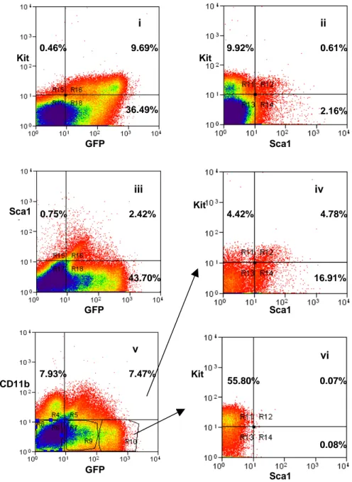
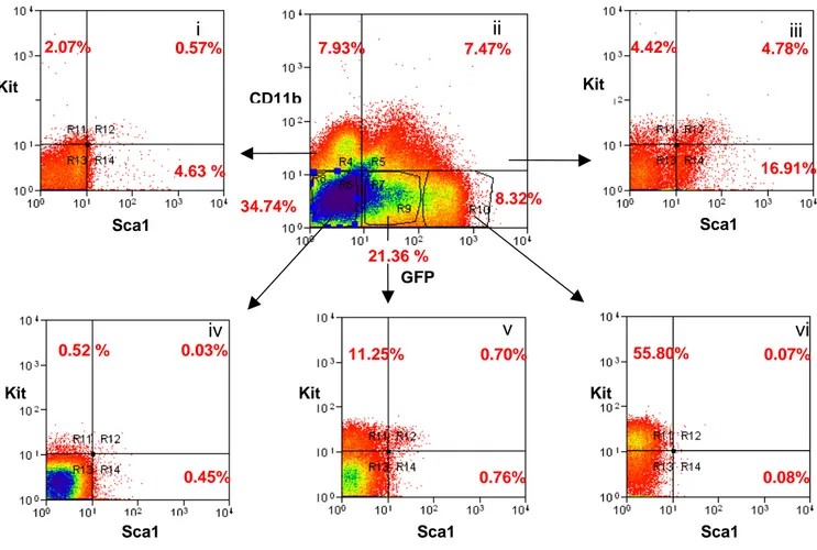
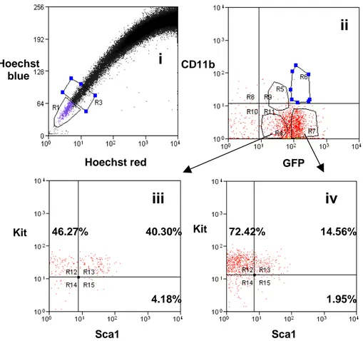
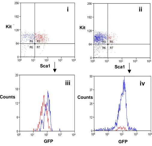
4.1.1 The transgenic Kit/GFP gene is expressed at intermediate
levels in cells with the immunophenotype of HSC page 49 4.1.2 HSC are contained in the Kit+/GFPint cell population page 58 4.2 Kit/GFP bone marrow derived cells can generate Kit/GFP+
cardiac stem cells in damaged heart page 62 4.2.1 Bone marrow-derived cells are able to generate
cardiospheres in vitro page 62 4.2.2 Cells from Kit/GFP bone marrow-derived CS express
cardiac markers page 66 4.2.3 BM-derived CSs are capable of cardiac regeneration
in vivo page 69
4.2.4 Analysis of CSs for hematopoietic potential page 71
5. DISCUSSION page 74
5.1 Transgenic Kit/GFP expression in hematopoietic Kit+ cells " 5.2 GFP expression levels in Kit+ cells discriminate between
HSCs and progenitors page 75 5.3 The Kit/GFP construct is expressed in multiple
stem cell types page 78
6. CONCLUSIONS page 82
1. INTRODUCTION
1.1 Stem Cells
In the last few years, great emphasis has been placed on the isolation, characterization and potential therapeutic uses of stem cells.
Stem cells are defined as cells capable of both self-renewal and commitment to differentiation into one or more mature cell types. However different types of stem cells can be distinguished on the basis of their developmental potential.
The real totipotent stem cells are the fertilized oocyte (the zygote) and its descendants of the first two divisions. These cells are indeed able to form the embryo and the trophoblasts of the placenta. After about 4 days, these totipotent cells begin to specialize, forming a hollow ball of cells, the blastocyst, and a cluster of cells called the inner cell mass (ICM) from which the embryo develops. The ICM cells are considered to be pluripotent, namely able to differentiate into almost all cells that arise from the three germ layers, but they are unable to give rise to the placenta and supporting tissues. ICM cells can be maintained in culture and the cell lines derived, known as embryonic stem (ES) cells, are also considered to be pluripotential. In the adult, most tissues have multipotential stem cells, defined as cells capable of producing a limited range of differentiated cell lineages appropriate to their location. Also adult stem cells are heterogeneous with respect to developmental potential. For example, small intestinal stem cells can produce all four indigenous lineages (Paneth, goblet, absorptive columnar, and enteroendocrine), while central nervous system (CNS) stem cells have tri-lineage potential, giving rise to neurons, oligodendrocytes, and astrocytes. At the bottom of the stem cell hierarchy are unipotential stem cells, capable of generating one specific cell type. Examples are epidermal stem cells in the basal layer that produce
only keratinized squames and certain adult hepatocytes that have long-term repopulating ability (Overturf et al., 1997). Tissue-specific stem cells appear to be present in most organs of the body, and share some common properties:
(1) Stem cells are a self-maintaining population.
This is achieved if, on average, each stem cell division gives rise to one replacing stem cell and one transit-amplifying cell. Equally well, stem cell numbers would remain constant if only symmetrical divisions occurred, provided that each time a stem cell gave rise to two daughter transit-amplifying cells, another stem cell gave rise to two daughter stem cells.
(2) Stem cells are a small percentage of the total cellularity.
In the mouse small intestine, there are perhaps 4–5 stem cells in a ring near the bottom of the crypt (Bjerknes and Cheng, 1999) out of a total crypt population of about 250 cells. Likewise, in skeletal muscle, satellite cells comprise about 5% of all nuclei. In the bone marrow, the multipotential hematopoietic stem cell (HSC) is even more rare, with a frequency of perhaps 1 in 104-105 bone marrow cells.
(3) Stem cells are undifferentiated.
In most tissues, stem cells do not have the specialized functions of the progeny that they originate.
(4) Stem cells are slowly cycling but highly clonogenic.
In theory, it would seem prudent to restrict stem cell division because DNA synthesis can be error-prone. Thus, in many tissues we see that stem cells divide less frequently than transit-amplifying cells. In the intestine, stem cells cycle less frequently than transit-amplifying cells, located more luminally (Wright, 2000) and in human epidermis, integrin-bright cells have a lower level of
proliferation as compared to other basal cells. In hair follicles, the hair shaft and its surrounding sheaths are produced by the hair matrix, which is itself replenished by the bulge stem cells. The bulge cells divide less frequently, but are more clonogenic than the transit-amplifying cells of the hair matrix (Oshima et al., 2001), thus showing an extensive proliferative ability.
In many tissues and organs, the identity of the stem cells has remained either elusive or at least equivocal. However, in the bone marrow the identification of cells with the properties of self-renewal and multi-lineage differentiation potential is well advanced. Indeed, such cells were functionally defined in the mouse back in 1961 by Till and McCulloch (Till and McCulloch, 1961) as cells that, upon transplantation, were able to form multilineage hematopoietic colonies in the spleen of lethally irradiated animals [colony forming units – spleen (CFU-S)].
In human bone marrow, the sialomucin CD34 is a hematopoietic cell surface antigen that has been extensively exploited for the selection of long-term repopulating cells with multi-lineage potential, although not all HSCs express this. Nowadays, murine HSCs are empirically recognized on the basis of their immunoprofile and known as KLS cells (selected using several markers; Kit+/Lin¯/Sca-1+). An alternative method for enriching HSCs exploits the fact that some cells have evolved a cellular protection mechanism against toxic metabolites and xenobiotics. This mechanism involves the activity of efflux pumps that belong to the ATP-binding cassette (ABC) superfamily of membrane transporters, and such cells are able to efflux a combination of Hoechst 33342 and Rhodamine 123, thus appearing at the bottom left corner of a dual parameter FACS analysis – hence called the side population (SP)(Bunting, 2002). SP cells have been found in many
other tissues, and the association between SP phenotype and stemness seems to be true in most of these tissues.
In the central nervous system, neural stem cells and probably their transit-amplifying descendants express both the intermediate filament nestin and a 39 kD RNA-binding protein known as Musashi1 (Kaneko et al., 2000; Keyoung et al., 2001). Musashi was first identified in Drosophila and thought to be responsible for the asymmetric divisions of sensory organ precursor cells (Sakakibara et al., 1996); it may also be a marker for intestinal crypt stem cells.
A tissue that has been traditionally considered completely post-mitotic is the heart. However, in the last few years a number of studies suggested the presence of a cardiac stem cell population capable of (re)generating the cardiac tissue throughout life, raising the possibility to speculate about new potentially therapeutic strategies for cardiac repair. Initial evidence, though in part controversial, has been presented that bone marrow cells injected into damaged myocardium or mobilized into the circulation may transdifferentiate into heart cell types (Orlic et al., 2001; Wojakowski et al., 2004; Wang et al., 2006; Kawada et al., 2004). Additional, exciting approaches for cardiac repair have been raised by the remarkable discovery that the heart contains a reservoir of stem and progenitor cells. These cells are positive for various stem/progenitor cell markers (Kit, Sca-1, Isl-1, and Side Population -SP- properties)(Hierlihy et al., 2002; Martin et al., 2004; Beltrami et al., 2003; Oh et al., 2003; Matsuura et al., 2004; Messina et al., 2004; Smith et al., 2007; Laugwitz et al., 2005), propagate in vitro and develop phenotypic features of heart cells after differentiation in vitro or in vivo. Cardiac stem and progenitor cell types characterized so far (Hierlihy et al., 2002; Martin et al., 2004; Beltrami et al., 2003; Oh et al., 2003; Matsuura et al., 2004; Messina et
al., 2004; Smith et al., 2007; Laugwitz et al., 2005) exhibit significant differences in their immunophenotypic, developmental and biological properties. Thus, the heart probably contains various types of stem and progenitor cells. This is in agreement with the emerging consensus that more than one stem cell may be present in a particular tissue (Cai et al., 2004), and with recent evidence that heart endothelial, cardiac and muscle cells may arise from a hierarchy of multipotent/bipotent stem progenitor cells (Moretti et al., 2006; Kattman et al., 2006; Wu et al., 2006).
The developmental origin(s) of stem and progenitor cells of the adult heart has been studied only to a very limited extent (Parmacek and Epstein, 2005).Mouse chimera experiments suggest that most (at least 95%) cardiomyocytes are derived from a relatively small embryonic founder population, ruling out any major contribution to the adult cardiomyocyte population from “immigrant” non-cardiac stem cells, at least under physiological conditions (Eberhard and Jockusch, 2005). On the other hand, several findings, including the presence of cardiac chimerism in patients receiving allogeneic bone marrow transplantation, suggest that immigrant bone marrow-derived cells could behave, under pathological conditions leading to heart damage, as precursors of cardiac stem cells (Quaini et al., 2002).
Recently (Mouquet et al., 2005), Mouquet et al showed that in mice transplanted with labelled GFP+ bone marrow GFP+ cells home to the heart after infarction, and
replenish the depleted pool of SP, Sca1+, CD31+ or CD31¯ cells, which are known to include a very small population of CSC (Pfister et al., 2005). However, they did not demonstrate the replenishment of the functional CSC pool by the transplanted cells.Thus, the important issue of extra-cardiac sources of CSCs, following cardiac damage, still needs to be addressed. If extra-cardiac cells can be induced, not only
to migrate to the heart, but also to reconstitute a pool of extensively replicating CSCs, exciting therapeutic possibilities may arise.
1.1.2 Tumor stem cells
During the last years, a growing body of evidence indicate that also neoplastic tissues can harbour stem cells: in particular, it is now commonly accepted that such "tumor stem cells" are responsible for drug-resistance and recidive after treatment in cancer patients. However, already back in 1971, McCulloch and collaborators noticed that only 1 in 100 to 1 in 10,000 murine myeloma cells had the ability to form colonies in vitro. Moreover, it had previously been demonstrated that only 1– 4% of transplanted murine lymphoma cells formed colonies in spleens of recipient animals (Ianus et al., 2003; Park et al., 1971; Bruce and van der Gaag, 1963; Ianus et al., 2003; Park et al., 1971). This low clonogenic potential was also observed in human leukemias (Sabbath et al., 1985; Griffin and Lowenberg, 1986). Furthermore, at this time, it was also found that solid organ cancer cells vary in their ability to proliferate in similar assays: for example, only 0,1%-0,02% of cells isolated from solid tumors (such as lung, ovarian and brain tumours) were capable of forming colonies in standard soft-agar assays (Hamburger and Salmon, 1977). These observations suggested the hypothesis that the whole cell population of a tumor could originate from a few tumor stem cells. Based on functional and immunophenotypic analysis of cell subpopulations with modern technologies, cancer has started to be increasingly considered as a stem-cell disorder, in which the continuous growth and propagation of the whole tumour depends on a small population of self-renewing cancer stem cells.
The HSC was the first adult somatic stem cell to be described. The existence of cancer stem cells was also first described in the hematopoietic system. The demonstration of a leukaemia-initiating cell, now commonly referred to as the leukaemia stem cell (LSC), in AML and other leukemias, along with the subsequent identification of cancer stem cells in breast and central-nervous-system (CNS) tumours, provides evidence for the broad applicability of the cancer-stem-cell model. John Dick and collaborators used the non-obese diabetic (NOD)/SCID mouse xenotransplantation stem-cell assay system, to demonstrate that in human AML there are LSC populations that can reproduce all the AML characteristics when transplanted in recipient mice (Hamburger and Salmon, 1977; Lapidot et al., 1994). This methodology has been fundamental to fractionate leukemia cells and test them for "stemness", thus demonstrating that only CD34+CD38– cells from AML are true LSC. In addition, a hyerachical organization of cells in AML has been postulated, similar to that existing in normal hematopoiesis, and this analogy seems to indicate that normal HSC can be the target for transformations leading to AML (Bonnet and Dick, 1997). Subsequently, several other groups contributed to define the LSC phenotype in AML: CD34+, CD38¯, IL-R+, CD71¯, HLA-DR¯, CD117¯ (Jordan et al., 2000; Blair et al., 1997; Blair et al., 1998; Blair and Sutherland, 2000). In addition, LSC have more recently been characterized in acute limphoyd leukemias (ALL) (Cox et al., 2004) and chronic myeloid leukemias (CML) (Jamieson et al., 2004).
A similar model has been hypothesized also for solid tumor, using the same criteria adopted for the definition of LSC. However, the identification of stem cells in non-hematopoietic tissues has been rather challenging, due to the lack of knowledge for specific cell markers. Nevertheless, tumor stem cells have been identified in breast,
brain and colon cancer (Al Hajj et al., 2003; Hemmati et al., 2003; Ignatova et al., 2002; Singh et al., 2003; Singh et al., 2004; O'Brien et al., 2007; Ricci-Vitiani et al., 2007). Breast cancer stem cells (BCSC) have been identified on the basis of the expression of membrane antigens and the ability to form tumors after injection into adipose mammary tissue of (NOD)/SCID mice. Less than 100 ESA+, Lin–, CD24–
/low, CD44+ cells are capable of generating tumors histologically similar to the
original when transplanted into (NOD)/SCID mice (Al Hajj et al., 2003; Hemmati et al., 2003; Ignatova et al., 2002; Singh et al., 2003; Singh et al., 2004). Moreover, cells re-derived from these tumors are still capable to generate tumors in secondary and tertiary recipients. These experiments demonstrated the long term self-renewal activity of BSCS.
In the nervous system, it is possible to isolate and propagate stem cells as "neurospheres", i.e. clonal cell aggregates derived from a single Neural Stem Cell (NSC) (Reynolds and Weiss, 1992). Recently, tumor NSCs have been isolated from pediatric tumors. They form neurospheres in culture, that have the ability to generate neurons and glia, and show a higher proliferative and self-replicative potential with respect to normal NSC. Tumor NSC have also the ability to form tumors in neonatal rat brain in vivo (Hemmati et al., 2003; Ignatova et al., 2002; Singh et al., 2003). These cells express CD133 (Singh et al., 2003), a NSC marker, and it has been recently demonstrated that only CD133+ subpopulations have the
ability to regenerate tumors in (NOD)/SCID mice (Singh et al., 2004).
CD133 seems to be an important marker also in colon cancer cells (Ricci-Vitiani et al., 2007; O'Brien et al., 2007). To assess the presence of cancer stem cells in colon cancer, the group of J. Dick used renal capsule transplantation in immunodeficient NOD/SCID mice to identify a human colon cancer-initiating cell (CC-IC).
Purification experiments established that all CC-ICs were CD133+; the CD133¯ cells, that comprised the majority of the tumor cells, were unable to initiate tumor growth. In limiting dilution analysis it was shown that there is one CC-IC in 5.7 x 10(4) unfractionated tumor cells, whereas there is one CC-IC in 262 CD133+ cells, representing >200-fold enrichment. CC-ICs within the CD133+ population were able to maintain themselves as well as differentiate and re-establish tumor heterogeneity upon serial transplantation (O'Brien et al., 2007).
Thus, it is realistic that in the next future the identification of new markers and new
in vitro and in vivo assays will lead to the identification of other tumor stem cell
populations in solid tumors.
1.1.3 Hematopoietic stem cells and hematopoiesis
The best-characterized stem cells are those responsible for blood cell production. All experimental strategies and conceptual paradigms applicable to stem cells in general were first defined in the hematopoietic system. The hallmark properties of the HSC were defined in 1963 by Till and Mc-Culloch (Siminovitch et al., 1963). They defined HSCs as cells that are individually capable of both self-renewal and multilineage differentiation, yielding greatly expanded numbers of lymphocytes, myeloid cells, and erythrocytes. In fact, around 2 × 10¹¹ erythrocytes
and 1010 white blood cells must be replaced each day to maintain adult human hematopoiesis. Terminally differentiated cells are by themselves incapable of further growth and must, therefore, be replaced through proliferation and development of HSCs (Goldman, 1982). The process, by which stem cells give rise to terminally differentiated cells, occurs through a hierarchy of progenitor cells,
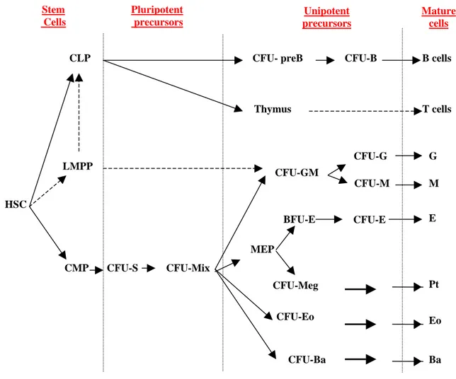
often overlapping in their hematopoietic capacity (Dexter et al., 1984; Golde, 1991). During commitment, cells can undergo extensive proliferation and sequential differentiation, accompanied by a decrease in self-renewal capacity. The primary function of this transit population is to increase the number of mature cells produced by each stem cell division (Figure 1).
The immediate progeny of HSCs in adult bone marrow are the common myeloid precursor (CMP) and the common lymphoid precursor (CLP) (Akashi et al., 2000;
Stem Cells
HSC
CLP
CMP
CFU- preB CFU-B
BFU-E CFU-E CFU-GM CFU-Eo CFU-Ba CFU-G CFU-M E G M Pt Eo Ba CFU-S CFU-Mix CFU-Meg B cells Thymus T cells Unipotent precursors Pluripotent precursors Mature cells
Figure 1: Schematic representaion of hematopoiesis LMPP
Hao et al., 2001; Manz et al., 2002). CLP is thought to give rise to all lymphoid lineage (B, T and NK), but little is known about its development (Spangrude and Cooper, 2000). CMP can be associated with 2 cells that can form in vivo (CFU-S) and in vitro (CFU-Mix) colonies containing multiple cell types. CFU-S (Colony Forming Unit-Spleen) can produce colonies on the spleen of irradiated mice injected with BM cells (Till and McCulloch, 1961). In particular, at 7-8 days after transplant most spleen colonies contain mature cells of a single lineage, predominantly erythroid, whereas by day 12, the majority of the colonies are composed of multiple cell types. Furthermore, late-appearing colonies do not represent further development of the early clones, since most of the day 7-8 spleen colonies disappear within the subsequent 72 hours. Therefore the early-appearing colonies derive from monopotent progenitors, whereas colonies present at day 11 or later originate from multipotential cells (Magli et al., 1982; Metcalf, 1999). The other pluripotent progenitor is the CFU-Mix (Colony Forming Unit-Mix) that generates mixed colonies in cultures containing an appropriate cocktail of cytokines. CMP gives rise to progenitors committed to the generation of erythrocytes and megakaryocytes (MEP) or granulocytes and macrophages (CFU-GM). These cells, capable of developing along more than one lineage, undergo commitment and become progressively restricted to a specific differentiative fate, such as Colony Forming Granulocyte (CFU-G) and Colony Forming Unit-Macrophage (CFU-M), the direct precursors of granulocytes and monocytes, respectively. Similarly, two stages of different maturation have been identified within the erythroid lineage: an early cell (Burst Forming Unit-Erythroid or BFU-E) and a late precursor (Colony Forming Unit-Erythroid or CFU-BFU-E) capable of forming large and small erythroid colonies, respectively.
Less than 0.01% of the hematopoietic cells in BM are pluripotent stem cells capable of long-term proliferation and self-renewal. In vitro clonogenic assays are a powerful means to quantitatively and qualitatively study a broad spectrum of progenitors (Metcalf, 1999), but they do not sustain HSC growth. Long-term cultures (LTC) in liquid media can detect primitive cells (LTC-initiating cells, LTC-ICs), however these cells represent a rather functionally heterogeneous population and the relationship between LTC-ICs and in vivo repopulating stem cells is not clear (Bhatia et al., 1997). The only valid test for HSCs is an in vivo assay that demonstrates the capacity for complete and sustained (>4 months) regeneration of the lymphohematopoietic system following transplantation into lethally irradiated histocompatible recipient. In particular, in competitive repopulation strategies the cells being evaluated are co-injected with genetically or phenotypically distinguishable reference cells. These reference cells serve the dual purpose of exerting a selective pressure to identify HSCs in the test population that possess superior proliferation potential, and enable stem cells to be identified among small grafts (<103 enriched HSCs) that may not ontheir own be sufficiently radioprotective as to allow their detection at later times (Harrison et al., 1978; Harrison et al., 1993). This approach demonstrated that a single donor stem cell can repopulate all the hematopoietic lineages for the entire lifespan of primary and secondary recipient animals. This method, named competitive repopulation assay, has given an estimated stem cell frequency of approximately 1 in 104 - 105 murine BM cells (Harrison et al., 1988; Harrison and Zhong, 1992; Okada et al., 1993; Kim et al., 2000).
There are a number of studies supporting the concept of cell cycle dormancy of stem cell: this allows the use in vivo or in culture of cytotoxic drugs such as
5-fluorouracil (5-FU) and hydoxyurea, that selectively kill dividing cells to eliminate many of the more mature cells that are typically rapidly dividing. Administration of 5-FU to mice typically results in a 5- to 10-fold increase in the frequency of HSCs in the BM cells obtained 2–4 days later and this strategy is commonly used to obtain enriched mouse HSC populations prior to other selection methods (Lemieux and Eaves, 1996).
Despite many exhaustive studies, researchers have yet to find a single molecular marker that is expressed exclusively by HSCs. However, there are several markers whose expression is gained (or lost) at different rates as primitive hematopoietic cells differentiate. By targeting various combinations of markers, it has thus become possible to subdivide this functionally heterogeneous mixture of cells into more homogeneous subpopulations. Primitive hematopoietic cells, including HSCs, do not express a variety of surface markers that are associated with the terminal maturation of specific blood cell types. Lack of expression of a number of these lineage (Lin) markers, which in combination include all of the mature hematopoietic cell types (RBCs, T-cells, B-cells, natural killer cells, monocytes, and granulocytes), can thus be used to distinguish immature cells from the more abundant differentiated cells in most populations of hematopoietic cells. Selection of Lin¯ cells typically gives a 20- to 500- fold enrichment of HSCs depending on the combination of Lin markers used (Morrison et al., 1995). The sialomucin CD34 was the first marker to be recognized on primitive hematopoietic cells: it is expressed in a subpopulation of cells containing both stem cells and early committed progenitors. Transplant studies in several species, including baboons and mice, have shown that long-term marrow repopulation can be provided by CD34 selected cells (Berenson et al., 1988). CD34 selection has been adopted as a
means to enrich for stem cells in clinical transplantation protocols. However, several recent studies suggest that there may be human and murine stem cells that do not express CD34. Xenograft repopulation assays using fetal sheep and immune-deficient mice have been crucial for the identification of human CD34¯stem cells capable of long-term repopulation and multilineage differentiation in vivo. Moreover, these cells were also able to repopulate secondary recipients, attesting to the extensive self-renewal potential of the engrafting cells (Nakauchi, 1998; Sato et al., 1999; Tajima et al., 2001). These results both in mouse and human suggest that CD34 may be a marker of activated and/or cycling stem cells, and that CD34+ stem cells can revert to quiescent CD34¯state (Sato et al., 1999). The same controversy holds true for another stem cell marker, CD38, whose expression might be influenced by stem cells activation state (Tajima et al., 2001). It is well established that the majority of murine BM HSCs express Stem Cell Antigen-1 (Sca-1), Kit (Stem Cell Factor Receptor) and Thy-1. Thus, isolation of adult BM cells on the basis of a single marker, for instance Sca-1 or Kit, can provide a significant one-step enrichment of HSCs, but to discriminate between HSCs and various subsets of lineage-committed progenitors and mature blood cells, strategies that exploit the use of specific combinations of markers are required. In the mouse, the Lin¯ Sca-1+ Thy-1low Kit+ population of bone marrow cells includes essentially all cells with the ability to repopulate irradiated mice (Uchida and Weissman, 1992). It has also been shown that single Lin¯Kit+ Sca-1+ mouse BM cells can reconstitute stable and long term, multi-lineage hematopoiesis after transplantation in 10–20% of transplanted mice (Szilvassy et al., 2003). This repopulation frequency of <100% suggests that either the Lin¯ Sca-1+ Thy-1low Kit+ cells are not all HSCs, or that some of the cells have transiently lost their engrafting potential, or that only a random portion of
transplanted cells will successfully home to appropriate niches in the BM and proliferate to form detectable clones of differentiated progeny. Even pure HSC populations are likely to show dynamic changes in cell-surface antigen expression as well as functional heterogeneity. As discussed previously, the regulated expression of the mouse CD34 antigen independent of HSC function is one notable example. Therefore, caution must be taken when attempting to extrapolate the phenotypic profiles of HSCs. Apart from cell-surface antigen expression, primitive hematopoietic cells from mouse, humans, and other species have been identified and isolated based on their ability to efflux certain fluorescent dyes, such as Rhodamine-123 (Rho) and Hoechst 33342 (Ho). The majority of the HSCs in adult human and murine tissues are Rho¯/lo (Spangrude and Scollay, 1990). Adult BM of many species also contain a rare population of Ho¯/lo cells that have been designated as SP (Goodell et al., 1996). SP cells in the BM of adult mice are primarily CD34¯ and are highly enriched in their HSC content (Goodell et al., 1996).
The same procedure has been adopted to successfully isolate SP populations from non-hematopoietic tissues as muscle, brain, liver, lung and gut (Gussoni et al., 1999; Asakura et al., 2002), thus suggesting that SP phenotype is not characteristic of hematopoietic cells (Jackson et al., 2001).
Recently it has been shown that, among SP cells, the so-called "Tip-SP" population can be regarded as a more purified HSC population. Single CD34¯Lin¯ Sca-1+ Kit+ cells have been proven to long term repopulate an irradiated recipient in 96% of the cases (Matsuzaki et al., 2004). However, a recent report showed that CD34¯ LSK cells are distributed almost equally between SP and non-SP population fractions, showing the same repopulative potential. Despite no information was provided
respect CD34+ LSK cells activity in SP and non-SP cells, the main indication seems to be that SP is a powerful tool for quick isolation and concentration of HSC, but SP should not be considered exclusively as a characteristic phenotype for HSC (Morita et al., 2006).
A new combination of Signaling Lymphocyte Activation Molecule (SLAM) family antigens has been recently utilized by Morrison and collaborators to separate hematopoietic mature, progenitor and stem cell populations within bone marrow and fetal liver cells (Kiel et al., 2005; Kim et al., 2006). Particularly, the CD150+CD244¯CD48¯ fraction seems to harbour almost all the hematopoietic stem cells, while multipotent and committed progenitors are, respectively, CD150¯CD244+CD48¯ and CD150¯CD244+CD48+. This simple antigen combination also allowed the direct characterization of the hematopoietic niches in bone marrow and spleen sections (Kiel et al., 2005). Furthermore, the study of SLAM receptors expression in non-hematopoietic tissues seems to indicate that CD150, CD244, and CD48 are not pan-stem cell markers, as they were not detectably expressed by stem cells in the fetal or adult nervous system (Kim et al., 2006).
1.1.4 Ontogenesis of HSC
The mammalian embryo contains at least two spatially separated sources of hematopoietic cells (Dzierzak, 2002). As it has been known for many years, subsequent to gastrulation and mesoderm formation, the first hematopoietic cells are generated in the yolk sac around embryonic day E7.5 (Palis and Yoder, 2001). These primitive erythrocytes produced in the yolk sac from putative hemangioblasts
(the common mesodermal precursor of endothelial and hematopoietic lineages) provide a temporary embryonic hematopoietic system. The first autonomous generation of more complex (lymphoid-myeloid-erythroid) hematopoietic progenitors and adult repopulating hematopoietic stem cells (HSCs) occurs later in the embryo proper in the para-aortic splanchnopleura (PAS) and aorta-gonad-mesonephros (AGM) region. The first HSCs capable of long term repopulation in adult irradiated recipients are found in the AGM at E10.5, and expand over the time until fetal liver is colonized by a wave of HSCs between day E11.5 and day E13. HSCs hugely expand in the fetal liver until birth, when hematopoiesis is almost exclusively sustained by the BM (Dzierzak et al., 1998; Ema and Nakauchi, 2000). Thus, the independent and multiple emergent hematopoietic events in the extra-and intraembryonic tissues suggest that these sites play different roles in the development of the rapidly expanding hematopoietic system in the mammalian embryo. Understanding the direct precursors of HSCs may provide insights into expansion strategies for these clinically relevant cells. Several recent studies have provided a detailed phenotypic description of HSCs in the mouse AGM, YS, and FL. Indeed, all HSCs in the AGM are Sca-1/GFP+ (de Bruijn et al., 2002), Kit+ CD34+ (Sanchez et al., 1996), Runx1+ (North et al., 2002), SCL+ (Sanchez et al., 2001), and Gata-2+ (Ling et al., 2004; Minegishi et al., 1999). Most or all HSCs are CD45+, CD31+, and VE-cadherin+ (North et al., 2002). The characterization of
functional HSCs and the localization of these cells within the embryonic tissues indicate the possible phenotype of the direct precursors of HSC. The vascular endothelial co-localization of Sca-1/GFP (de Bruijn et al., 2002) along with Runx1, CD34, CD31, VE-cadherin (North et al., 2002) suggests that the direct precursors of HSCs are endothelial cells or ‘hemogenic endothelium’. The vascular
endothelium of the human embryo also has blood-forming potential (Oberlin et al., 2002). However, these cells may not be fully functional endothelial cells and may just take on endothelial characteristics temporarily as they transit through the vessel wall. North et al. (North et al., 2002) showed that mesenchymal cells lying just beneath the ventral aortic endothelium could be the direct precursors of HSCs. These cells are Runx1+ (lacZ knock-in marker) and functional HSCs could be sorted based on a mesenchymal phenotype (CD45¯, CD31¯, and VE-cadherin¯). A more recent study suggests that HSCs are generated in Gata-3 and AA4.1 expressing hematopoietic foci underlying the ventral aorta (Bertrand et al., 2005). While transplantation data show that these cells from the subaortic patches have some adult repopulating activity in adult recipients (0.4–1.9% engraftment), they are clearly not as potent as the Runx1+ or Sca-1/GFP+ AGM HSCs that provide up to 100% engraftment of irradiated adult recipients. Thus, the precursor cells to the fully potent HSCs found in the aortic endothelium and hematopoietic clusters may be cells localized in the subaortic patches and mesenchyme underlying the aorta. These cells may acquire mature HSC function as they progress towards the aortic endothelium and emerge in the hematopoietic clusters. In the chick embryo, it was shown with DiI and retroviral marking approaches that cells in the hematopoietic clusters arise from the aortic endothelium (Jaffredo et al., 1998). While DiI tracing experiments in cultured whole mouse embryos have established a lineage relation between endothelial cells and primitive erythroid cells (Sugiyama et al., 2003), no DiI lineage tracing studies have established this relation for emerging hematopoietic clusters or functional HSCs. Another candidate precursor to AGM HSCs is the mesoangioblast (Minasi et al., 2002). Such cells were clonally isolated from the E10 mouse dorsal aorta and when introduced into chick embryos, donor
cells associated with the vasculature of the host, blood and hematopoietic tissues, along with chondrocytes, bone cells, and muscle cells were found. While the ability of aortic endothelial cells to give rise to hematopoietic clusters is well documented [reviewed in (Kubo and Alitalo, 2003; Nishikawa, 2001)], this finding, together with more recent temporal and spatial frequency mapping data (Mendes et al., 2005) demonstrates that the aorta contains cells with a wider differentiation potential. As such, the mesoangioblast may be a precursor to the hemangioblast or within the AGM to ‘hemogenic endothelium’.
1.1.5. Regulation of HSCs.
Over the years, several models have been advanced, proposing that hematopoietic lineage determination is driven extrinsically (through growth factors, stroma, or other external influences), intrinsically or both (Ogawa, 1993; Ogawa, 1999; Tenen et al., 1997). Within the hematopoietic microenvironment, the "niche", early precursors are maintained in specific compartmentalized niches where they interact with other cell types and with components of the extracellular matrix (Taichman and Emerson, 1998; Potocnik et al., 2000). The microenvironment has been reported to influence survival, proliferation, and differentiation (Torok-Storb et al., 1999). Despite some important progress, the genetic and cellular mechanisms that influence stem cells either to differentiate into developmentally restricted progenitor cells or to self-renew to replace cells that become committed to differentiation are still poorly understood.
The ability of adult stem cells to both self-renew and differentiate is critical for tissue homeostasis. The stem cell population would become depleted if cell differentiation overwhelmed renewal. Similarly, unchecked stem cell
self-renewal would expand the stem cell population excessively, risking tumorigenesis (Jones and Fuller, 2004). An important function of the stem cell niche, therefore, is to regulate the balance between cellular self-renewal and differentiation. One mechanism that ensures this balance is the control of asymmetric/symmetric stem cell division. Asymmetric division means that stem cells divide into 2 daughter cells; one daughter cell remains in the niche as a stem cell and the other leaves the niche to produce a large number of progeny. Symmetric division means that stem cells divide into 2 identical daughter cells, both remaining in the niche as stem cells. Switching between symmetric and asymmetric division can occur in multiple stem cells that occupy the same niche under different physiological conditions (Watt and Hogan, 2000; Fuchs et al., 2004). In Drosophila germ stem cells, the first stem cell population that has been demonstrated to be strictly regulated by a niche, cell division is asymmetric or symmetric depending on whether the orientation of the mitotic spindle is perpendicular or parallel to the interface between the stem cell and its niche. The mechanisms underlying the establishment of cell polarity and spindle orientation are complex and, in some cases, appear to involve overlapping subsets of factors including adenomatous polyposis coli (APC), centrosomin, and adherens junction–related cadherins and catenins (Fuchs et al., 2004; Wallenfang and Matunis, 2003; Yamashita et al., 2003). Whether stem cells normally undergo asymmetric division in mammals as they do in invertebrates is yet to be determined. The impact of the niche size on controlling the number of stem cells supports the model that stem cells, at least in the hematopoietic system, most likely execute asymmetric division under normal physiological conditions (Zhang et al., 2003; Calvi et al., 2003).
There are 2 classifications of HSCs, namely, long-term and short-term. Long-term HSCs (LT-HSCs) are capable of contributing to hematopoiesis for months or even a lifetime. Short-term HSCs (ST-HSCs) have a reconstitution ability that is limited to several weeks (Morrison and Weissman, 1994; Christensen and Weissman, 2001). LT-HSCs are maintained primarily as quiescent or slow-cycling cells, while ST-HSCs are actively cycling (Passegue et al., 2005). Data from conditional inactivation of bone morphogenic protein (BMP) receptor type IA (BMPRIA) mice reveal that an increase in the number of spindle-shaped N-cadherin+ CD45– osteoblastic (SNO) cells, a subset of osteoblastic lining cells, in the trabecular bone area correlates with an increase in the number of LT-HSCs, suggesting that SNO cells function as a key component of the niche to support HSCs (Zhang et al., 2003). Similarly, overexpression of parathyroid hormone (PTH) and PTH-related protein (PTHrP) receptor leads to increased osteoblast numbers, resulting in a parallel increase in the number of HSCs in the PTH/PTHrP receptor (PPR) transgenic mouse model (Calvi et al., 2003). Both studies point to the dependence of LT-HSCs on the osteoblastic niche. It appears that the ability of the osteoblastic niche to retain stem cells in a quiescent state is an important mechanism in maintaining sufficient stem cells (Arai et al., 2004).
Additional evidence supports the view that the osteoblastic niche plays a key role in sustaining and maintaining HSCs. Visnjic and colleagues have demonstrated that HSCs, and, consequently, the hematopoietic system, rely on osteoblasts for support (Visnjic et al., 2004). This result came from studies of transgenic mice that specifically express the herpesvirus thymidine kinase (TK) gene in developing osteoblasts under the control of the collagen αI promoter (Col2.3TK). The advantage of the Col2.3TK mice is the ability to inducibly ablate osteoblasts with
ganciclovir (GCV), which triggers TK function. Loss of osteoblasts in GCV-treated Col2.3TK mice led to substantial decrease in the number of lymphoid, erythroid, and myeloid progenitors (including osteoclast progenitors) in the BM, followed by a decrease in the absolute number of HSCs (although the percentage of HSCs appeared only slightly changed) and reduced BM cellularity. After withdrawal of GCV, osteoblasts reappeared in the bone, as did hematopoiesis in the BM. These observations further support the role of osteoblasts in the maintenance of HSCs and regulation of hematopoiesis, although the authors did not perform functional assays to test HSCs in this model. In another study osteoblasts were shown to facilitate engraftment of HSCs in an allogeneic environment. After purified osteoblasts were cotransplanted with marrow stem cells into allogeneic mouse strains, the transplanted recipient mice demonstrated excellent long-term survival, absence of disease, and complete engraftment by the donor cells. It appeared that the hematopoietic progenitor cells could engraft lethally irradiated mice, but these cells could not cross the MHC antigen barrier in the absence of cotransplanted osteoblasts (El Badri et al., 1998).
Hematopoiesis and vascularization occur concurrently during development. In fact, HSCs and endothelial cells are derived from the same progenitor cells (hemangioblasts) at the embryonic stage and are closely related to the ontogeny of hematopoiesis that occurs in the yolk sac, aorta-gonad-mesonephros, placenta, fetal liver, spleen, and adult BM (Hristov and Weber, 2004; Lacaud et al., 2001; Li, 2005). Indeed, HSCs expressing SLAM markers were detected on the osteoblastic surface of trabecular bone as well as adjacent to sinusoidal endothelial cells (Kiel et al., 2005). This is consistent with another study in which explanted endothelial cells were able to maintain HSCs in culture (Li et al., 2004). Together these observations
support the model that HSCs may use either osteoblasts or endothelial cells as their niche under different circumstances (Wagers, 2005). The discovery that sinusoid endothelial cells have the potential to act as part of an alternative HSC niche together with the known osteoblastic niche support a previously proposed model in which the osteoblastic niche provides a quiescent microenvironment and the vascular niche promotes proliferation and further differentiation (Kopp et al., 2005). This model is exemplified in studies of thrombopoietin mutant (TPO–/–) mice, which revealed that reconstitution of thrombopoiesis under stress involves recruitment of HSCs to the sinusoid endothelial surface. This action involves MMP-9, which mediates release of Stem Cell Factor (SCF), the soluble ligand for Kit. In this study HSCs left the osteoblastic niche and translocated to the vascular niche, where they differentiated into megakaryocyte progenitors for further megakaryocyte maturation as well as platelet release (Avecilla et al., 2004; Kopp et al., 2005). Other studies also support a more prominent role for osteoblasts in maintaining HSC quiescence and suggest that the sinusoidal endothelium promotes proliferation and differentiation of stem and progenitor cells by providing a more nutrient-rich microenvironment, with higher concentrations of oxygen and growth factors and in which mature blood cells are ultimately released into the peripheral circulation (Kopp et al., 2005; Abkowitz et al., 2003). A second plausible function for the vascular niche is to assist HSCs in transendothelial migration, important during both homing and mobilization (Lapidot et al., 2005; Cancelas and Williams, 2006). A third function of the vascular niche is assigned to cells located outside of the BM medullary space, such as in the spleen. These cells might serve to replace the BM niche when the BM niche is under stress, such as with marrow suppression (Kiel et al., 2005). Splenomegaly concomitant with extramedullary hematopoiesis
is a common phenomenon in both humans and genetically mutant mice that suffer from BM hematopoietic failure. Based on these observations it is reasonable to hypothesize that endothelial cells may also function as a “backup” niche to support HSC expansion and produce a large number of progeny for hematopoiesis when the BM is under stress.
It is well known that HSC circulation involves HSCs leaving the BM, entering the vascular system (mobilization), and returning to the BM (homing). However, the underlying physiological function of these events is not well understood (Wright et al., 2001). The BM vascular structure provides a barrier between the hematopoietic compartment and the peripheral circulation. Most primitive HSCs remain physiologically quiescent within the BM niche; however, a portion of HSCs leave this resting pool and initiate the process of mobilization (Heissig et al., 2002; Arai et al., 2004; Wagers, 2005). There are some controversial reports suggesting that mobilized HSCs may be more quiescent than those that remain in the BM. Evidence to support this notion comes mainly from a study in which the CD34+
marker was used to identify HSCs and progenitors (Steidl et al., 2002). Since the population of CD34+ cells included numerous progenitor cells, the comparison of the cell cycle profile of the CD34+ cells residing in BM and in circulating peripheral blood in this study might not reflect the most primitive HSCs. Indeed, Weissman and colleagues have definitively shown in mice that mobilized HSCs are more proliferative than HSCs in BM by comparing highly purified BM LT-HSCs and mobilized LT-HSCs (Passegue et al., 2005).
Multiple signaling and adhesion molecules are involved in stem cell–niche interactions. Well-studied signaling molecules involved in niche regulation include
SCF/Kit, Jagged/Notch, Ang1/Tie2, extracellular Ca2+ and Ca2+ receptors (CaR), BMP and Wnt signalling pathway.
SCF signaling through its receptor, Kit, has been well characterized in promoting both proliferation and survival of HSCs. Loss of SCF from supporting cells in steel mice, or loss of the receptor in HSCs in W/W mice, leads to hematopoietic failure, indicating essential roles for these molecules in niche function (Bernstein et al., 1991). These results are supported by more recent work revealing a direct role for SCF/Kit signaling in control of HSC activation and release from the niche (Heissig et al., 2002). It has been shown that, following 5-FU BM suppression, SDF1-mediated activation of Matrix metalloproteinase-9 (MMP-9) induces SCF release in the bone marrow, allowing recruitment of Kit+ stem/progenitors cells from the quiescent to proliferative niche, thus allowing differentiation and reconstitution of the stem/progenitor cell pool. Remarkably, in MMP-9¯/¯ mice, release of SCF and HSC motility are impaired, resulting in failure of hematopoietic recovery and increased mortality, while exogenous SCF restores hematopoiesis and survival after BM ablation (Heissig et al., 2002).
Notch is expressed in primitive HSCs, while the Notch ligand Jag1 is expressed by mouse osteoblasts and bone marrow stromal cells. Activation of the PPR can increase osteoblastic cell numbers concomitant with enhanced Jag1/Notch activity, resulting in expansion of the HSC population (Calvi et al., 2003). This observation is consistent with previous reports that activation of Notch signaling is able to maintain undifferentiated stem and progenitor cells and expand the HSC/HPC pools (Li et al., 1998; Varnum-Finney et al., 2000).
Ang-1 is expressed in osteoblasts, while Tie2, a tyrosine kinase receptor, is expressed in HSCs and endothelial cells. Ang-1 enhances the ability of HSCs to
become quiescent and can induce adhesion to osteoblastic cells through Tie2 (Arai et al., 2004).
Recently, CaR was found to facilitate retention of HSCs on the endosteal bone surface. Deficiency of CaR in knockout mice leads to release of HSCs into the bloodstream. Interestingly, although CaR deficiency permitted HSC homing to BM in these animals, the cells were unable to remain localized to the osteoblastic niche (Adams et al., 2006).
BMP and Wnt signaling pathways are also involved in stem cell regulation. BMP-4 is expressed in osteoblastic cells, but the type of BMP receptor expressed in HSCs is unknown (Li and Xie, 2005). Wnt signals are important for stem cell self-renewal (Reya et al., 2003; Staal and Clevers, 2005), but the identity of the niche (osteoblastic or vascular) that expresses any of numerous canonical and non-canonical Wnt molecules is unknown. The same is true for FGF and hedgehog, both of which affect HSC behavior in vitro (Bhardwaj et al., 2001; de Haan et al., 2003). However, whether and where these factors are present as niche signals remain unresolved.
The adhesion molecules important in niche function include N-cadherin/β-catenin, VCAM/integrin, and osteopontin/β1 integrin (OPN/β1 integrin) (Li and Xie, 2005; Nilsson et al., 2005; Stier et al., 2005). They may play a role either for the attachment of stem cells to the niche or for migration of stem cells. Two adherens junction molecules, N-cadherin and β-catenin, are asymmetrically localized at the interface between HSCs and the osteoblastic niche (Zhang et al., 2003). The essential roles of cadherins and β-catenin in supporting germ-line stem cells and determining the asymmetric division and orientation of germ-line stem cells in Drosophila have been reported (Yamashita et al., 2003; Song and Xie, 2002). The
specialized distribution of the adhesion complex composed of N-cadherin and β-catenin suggests a potential role for these molecules in facilitating anchoring of HSCs to the osteoblastic niche as well as their involvement in regulating asymmetric division of HSCs (Muguruma et al., 2006). The precise functions of these adhesion molecules in this regard are yet to be determined. The role of integrins in mediating HSC migration and homing has been shown by blocking integrin function with anti-integrin antibody (Papayannopoulou, 2004). Recently, OPN was found to contribute to HSC trans-marrow migration toward the endosteal region through its interaction with β1 integrin (Nilsson et al., 2005; Stier et al., 2005).
The vascular niche was only recently identified (Heissig et al., 2002; Kiel et al., 2005). As a result, the associated molecules involved in regulating HSCs within this niche remain largely unidentified. However, studies regarding recruitment of HPCs to the endothelial surface provide important insights into understanding HSC behavior as it relates to the vascular niche. Recruitment of HPCs to the vascular niche depends on FGF-4 and chemokines such as Stromal cell–Derived Factor 1 (SDF-1; also called CXCL12) (Avecilla et al., 2004; Kopp et al., 2005).
FGF-4 augments adhesion of megakaryocyte progenitors to the vascular niche, while SDF-1 is a potent chemotactic factor for the transendothelial migration of CD41+ megakaryocytes. FGF-4–stimulated megakaryocyte adhesion or
SDF-1-induced megakaryocyte migration can be blocked by chemical inhibitors of signal transduction pathways, such as PI3K, PKC, and p38 MAPK, suggesting that multiple downstream signaling cascades are involved. FGF-4 and SDF-1 enhance the expression of the adhesion molecule Very Late Antigen 4 (VLA-4) on megakaryocytes as well as Vascular Cell Adhesion Molecule-1 (VCAM-1) on
endothelial cells. VE-cadherin is essential for both proper assembly and mediation of adhesion of megakaryocyte progenitors to the vascular niche for survival and further maturation. Blocking VE-cadherin by antibodies results in impaired thrombopoiesis (Avecilla et al., 2004).
HSCs also express FGFR1, -2, and -3, and FGF has been shown to be able to stimulate HSC self-renewal and proliferation in vitro (de Haan et al., 2003). Therefore it is reliable that the FGF signals emanating from the vascular niche may play roles in recruiting HSCs/HPCs by forming a gradient from the osteoblastic niche (lower FGF expression) to the vascular niche (higher FGF expression) (Kopp et al., 2005). This proposed regulatory role of FGF is consistent with its known ability to induce cell migration during embryonic development (Thisse and Thisse, 2005).
The ability of HSCs to mobilize and then return, or home, to the niche relies on specific molecular recognition, cell-cell adhesion/disengagement, transendothelial migration, and finally anchoring to the BM niche (Muguruma et al., 2006; Lapidot et al., 2005; Cancelas and Williams, 2006). The chemokine SDF-1 and its receptor CXCR4 (Lapidot et al., 2005; Dar et al., 2005) as well as underlying signaling pathways, including the Rac family molecules, regulate HSC mobilization and homing but also play a role in cell survival and proliferation (Lapidot et al., 2005; Cancelas et al., 2005). Endothelial cells, osteoblasts, and other stromal cells constitutively express SDF-1, while HSCs express CXCR4 (Peled et al., 1999; Kortesidis et al., 2005). SDF-1 generated from endothelial cells induces HSCs to undergo transendothelial migration mediated by E- and P-selectins (Katayama et al., 2003). Activation of adhesion molecules such as VLA-4 and leukocyte function antigen-1 (LFA-1) is also required for this process and the subsequent migration in
BM toward the osteoblast surface (Kopp et al., 2005). Likewise, high levels of SDF-1 on the surface of osteoblasts attract HSCs to return home to the osteoblast niche. The Rho GTPases Rac1 and Rac2, in response to SDF-1 signals, are also involved in regulating HSC mobilization and homing (Yang et al., 2001).
G-CSF induces HSC and progenitor cell mobilization and is widely used clinically during stem cell–based transplantation procedures. Recent studies revealed that the mechanism involved is primarily a decrease of SDF-1 in osteoblasts, with an increase of SDF-1 in peripheral circulation after G-CSF treatment (Petit et al., 2002). G-CSF also induces proteolytic enzymes such as elastase, cathepsin G, MMP-2, and MMP-9 (required for cells to penetrate the endothelium), which inactivates SDF-1 through cleavage of its NH2-terminal signal sequence (McQuibban et al., 2001). In addition, G-CSF could regulate SDF-1 expression in BM at the transcriptional level (Semerad et al., 2005; Katayama et al., 2006). During stem cell homing HSCs expressing CXCR4 are attracted to the osteoblastic niche, which expresses high levels of SDF-1 (Avecilla et al., 2004; Kopp et al., 2005), and the adherens complex formed by N-cadherin and β-catenin may play a crucial role for anchoring HSCs to the osteoblastic niche (Muguruma et al., 2006; Zhang et al., 2003). Interestingly, a recent report showed that the sympathetic nervous system can actively control the attraction/release of stem cells into the niche, utilizing G-CSF-mediated mobilization (Katayama et al., 2006).
How hematopoietic precursors choose their differentiation pathways is still a controversial issue. According to one theory, commitment is directed by an inductive mechanism, determinated by the microenvironment’s nature and different signals that lead to hematopoietic cells (Trentin, 1971; Van Zant and Goldwasser, 1979). An alternative hypothesis considers commitment as a completely stochastic
event (Ogawa, 1993). Cytologic analysis of multilineage colonies of single cell origin showed a variety of lineage combinations, consistent with the concept that stem cell commitment is a stochastic process (Suda et al., 1983). Analysis of colonies derived from paired progenitors (two daughter cells derived from a single parent cell) showed dissimilar combinations of lineages in many instances (Suda et al., 1984). Not only were the types of lineages different but also the number of lineages expressed in each of the pairs of colonies differed. This observation indicated that the stochastic principle applies not only to the type but also to the number of lineages that are expressed during the differentiation process. It is intriguing to believe that gene expression is stochastic in early stages of commitment. Lineage decisions would be probabilistic depending on the random formation of transcription complex able to initiate transcription or to repress it. Some combinations of expressed or repressed genes would form an autocrine loop. These combinations would increase the probability of transcription of other genes such as growth factors receptors or integral membrane proteins, which, in turn, would modulate collectively the stem cell response to its niche or microenvironment, further reinforcing the commitment event. Then, the HSC genome would be in an open chromatin structure and available for binding of transcription factors, although at low probability (Hume, 2000; Bellantuono, 2004). Using the highly sensitive single cell RT-PCR approach or a quantitative real-time PCR method, it has been demonstrated that HSC co-expresses several lineage-restricted gene sets (Hu et al., 1997; Cheng et al., 1996). These data indicate the complexity of gene expression, from which distinct patterns must emerge during the lineage commitment process, by selective up-regulation of the relevant gene cohorts, accompanied by the inactivation of others. To take into account this new
concept of regulation, a model for the molecular nature of the uncommitted stem cell “state” has been postulated (Enver and Greaves, 1998). This model proposes that a HSC possesses a molecular “ground state” composed of low levels of transcripts normally associated with the function of various mature cell lineages (Bonnet, 2002). According to this model, a commitment process would involve selection and amplification of an appropriate subset of the available transcriptional program, and possible repression of the remainder of the transcriptional program. Interestingly, the features of the ground-state model are consistent with the clinical description of gene expression promiscuity in certain mixed leukemias (Bonnet, 2002). Therefore, several molecules have been shown to play roles in several aspects of hematopoietic development, but the elucidation of the molecular phenotype of the HSC has just begun. Indeed, key aspects of stem cell regulation are likely to be the emergent properties of interacting pathways and networks, the elucidation of which will require an extensive description of the genetic program available to the stem cell.
1.2 The Kit gene
The Kit proto-oncogene encodes for a transmembrane tyrosine-kinase receptor for Stem Cell Factor (SCF), and belongs to the same family of colony stimulating factor-1 receptor (CSF-1-R) and platelet derived growth factor receptor (PDGF α e β) (Yasuda et al., 1993).
v-kit, an oncogene identified in the HZ4-FeSV retrovirus (Hardy-Zuckerman 4 feline sarcoma virus), is the viral homolog of Kit (Besmer et al., 1986); this onocogene presents a deletion of the extracellular domain, of transmembrane
segment and of 50 aminoacids of the C-term. The result of this mutation is the continuous stimulation of proliferation, independently on SCF binding.
In the mouse, Kit is located into the White (W) locus (Besmer et al., 1986; Chabot et al., 1988; Geissler et al., 1988) on chromosome 5, and the locus was denominated from the white spots characteristics of mice having Kit mutations that abolish its expression in melanocytes.
Other mutations that produce similar alterations have been mapped on the Steel locus, on chromosome 10 (Zsebo et al., 1990), and subsequently it has been demonstrated that there is located the gene encoding for the Kit ligand, SCF (Flanagan and Leder, 1990; Nocka et al., 1990; Huang et al., 1990).
SCF is encoded as a secreted signal into the extracellular matrix, or can be retained as transmembrane signal involved in migration and cell adhesion (Flanagan et al., 1991): this trans-membrane SCF is composed of a signal peptide, an extracellular domain (189aa), a transmembrane segment (23aa) and a cytoplasmic domain (36aa) (Anderson et al., 1990); the soluble form of SCF is probably derived from the proteolytic cut of the transmembrane isoform and corresponds to its first 165 aminoacids (Nocka et al., 1990; Williams et al., 1990).
Upon SCF binding, two Kit protein dimerize and autophosphorylate the cytoplasmic domain: this recruits and phosphorylates other cytoplasmic mediators in a cascade leading to activation of specific transcription factors, resulting in a modulation of the expression of target genes (Ullrich and Schlessinger, 1990).
Kit and its ligand have a primary role in the regulation of definitive hematopoiesis: an homozygous inactivating mutation of Kit, for example, causes perinatal death in mice pups due to severe anemia (Nocka et al., 1990).
Kit and SCF have also a role in the development of mouse embryo: disruption of this pathway has enormous impact in migration, proliferation and survival of Primordial Germ Cells (PGC) from day E9.5, melanoblasts from day E11 and hematopoietic precursors from day E11-E12 (Buehr et al., 1993; Mintz and Russell, 1957; Cable et al., 1995).
1.2.2 Gene and protein structure
The mouse Kit gene spans over a 80 Kb region and consists of 21 exons, with a coding sequence of 3 Kb (Gokkel et al., 1992). Exons have dimensions ranging from 100 to 200 bp, with exclusion of the 21st that is 2.3 Kb; introns are more variable, from 100 bp to 15 Kb. The exon-intron boundaries show the classic consensus sequences for splicing donors and acceptor sites (GT….AG); only one alternative splicing site has been identified at the exon 9 level, where a distal splicing donor site exist, that determines the maturation of a 12 bp longer mRNA (Hayashi et al., 1991).
The nucleotide sequence of human and mouse Kit promoter does not contain CAAT or TATA box: in both cases a transcription-initiating site is present at -58 bp from the translation-initiating codon. In the mouse, a secondary transcription-initiating site is situated at -12 bp from translation-transcription-initiating codon, while in man it is situated at -62 bp.
The gene isolation allowed defining the protein structure. The Kit product is a 145KDa transmembrane glycoprotein organised in: a signal sequence (1-22aa), that drives the localization of the protein in the endoplasmic reticulum; an extracellular domain (exon 1-9), with 9 potential N-glycosilation sites and 12 cysteines