Brief Original Article
Application of Luminex Gastrointestinal Pathogen Panel to human stool
samples from Côte d’Ivoire
Veronica Di Cristanziano1, Monika Timmen-Wego1, Nadine Lübke1, Rolf Kaiser1, Herbert Pfister1, David
Di Cave2, Federica Berrilli2, Yolande Kaboré3, Rossella D´Alfonso3,4 1 Institute of Virology, University of Cologne, Cologne, Germany
2 Department of Experimental Medicine and Surgery, University of Rome Tor Vergata, Rome, Italy 3 Centre Don Orione pour handicapés physiques, Bonoua, Côte d’Ivoire
4 Department of Systems Medicine, University of Rome Tor Vergata, Rome, Italy
Abstract
Introduction: Gastrointestinal infections caused by viruses, bacteria, and parasites are endemic in most developing countries due to inadequate provision of safe water supplies, sanitation, and hygiene. To investigate the enteric pathogens infecting people living in Côte d’Ivoire, the Luminex Gastrointestinal Pathogen Panel (xTAG GPP) assay was used to analyze 34 human fecal samples. This study represents the first application of this technology to samples from a sub-Saharan African country.
Methodology: Thirty-four stool samples from asymptomatic and symptomatic patients, 1–15 years of age, were analyzed by xTAG GPP. The Luminex assay represents a qualitative bead-based multiplexed molecular diagnostic test able to identify concurrently 15 enteric pathogens, including bacteria, viruses, and parasites.
Results: Overall, 22 out of 34 (64.7%) fecal specimens were detected to be positive by xTAG GPP. Sixteen were from asymptomatic subjects, and 10 patients (45.4%) showed co-infections. G. duodenalis was detected in 15 patients, in both mono- and co-infections, representing the most frequent pathogen, followed by enterotoxigenic Escherichia coli (ETEC) LT/ST. Four norovirus isolates were also detected and assigned to genogroups I and II.
Conclusions: Considering the burden of enteric infections in developing countries, particularly among children, and the high rate of co-infections in asymptomatic subjects, this study shows the need for diagnostic tools such as xTAG GPP to improve diagnosis and treatment of these infections in endemic areas.
Key words:xTAG GPP; Luminex; enteric pathogen; gastroenteritis; Côte d’Ivoire. J Infect Dev Ctries 2015; 9(8):884-889. doi:10.3855/jidc.6460
(Received 21 December 2014 – Accepted 13 March 2015)
Copyright © 2015 Di Cristanziano et al. This is an open-access article distributed under the Creative Commons Attribution License, which permits unrestricted use, distribution, and reproduction in any medium, provided the original work is properly cited.
Introduction
Enteric infections are a major cause of morbidity and mortality in the developing world [1]. They are caused by a wide range of viruses, bacteria, and parasites, which are linked by the common fecal-oral transmission route and diarrheal disease. The highest burden of infections is concentrated in areas characterized by poor water, sanitation, and hygiene (WASH), and affects mostly children and young adults [2-4]. In sub-Saharan Africa, the mortality rate due to acute diarrhea varies from 1.9% in Gambia and 9% in Côte d'Ivoire, up to 37% in Nigeria [5-6]. Furthermore, previous data from endemic areas have shown that, in addition to symptomatic gastroenteritis, enteric pathogens can be detected in apparently asymptomatic individuals. However, the etiology and the impact of these silent infections on the health of
impoverished people are still uncertain. There are reports suggesting that constant exposure to fecal-oral contamination and sustained episodes of self-limited intestinal infections represent the main factors involved in the development of a subclinical condition known as environmental enteropathy (EE), which commonly affects people in developing countries. EE is characterized by small-intestinal inflammation, reduced absorptive capacity, and increased intestinal permeability. Consequently, asymptomatic enteric infections could represent not only a crucial source of new infections, but they also contribute to worsening the state of malnutrition and making people more susceptible to infectious diseases [2,7].
Overall, more than 30 pathogenic microorganisms are responsible for gastrointestinal infections, and differential diagnosis is a challenge in developed
countries. For this reason, several diagnostic tests must be combined to avoid overlooking important pathogens. So far, most of the research efforts have been focused on single enteric pathogen detection. Recently, the xTAG Gastrointestinal Pathogen Panel (xTAG GPP), a novel bead-based multiplexed molecular diagnostic test designed to simultaneously detect and identify the most common human enteric pathogens, was developed (Luminex Molecular Diagnostics, Toronto, Canada). The assay is able to identify concurrently 15 intestinal pathogens among viruses, bacteria, and parasites. In the present investigation, we used the xTAG GPP assay as a diagnostic tool on stool samples collected from patients living in the south of Côte d´Ivoire.
Methodology
Patients and sample collection
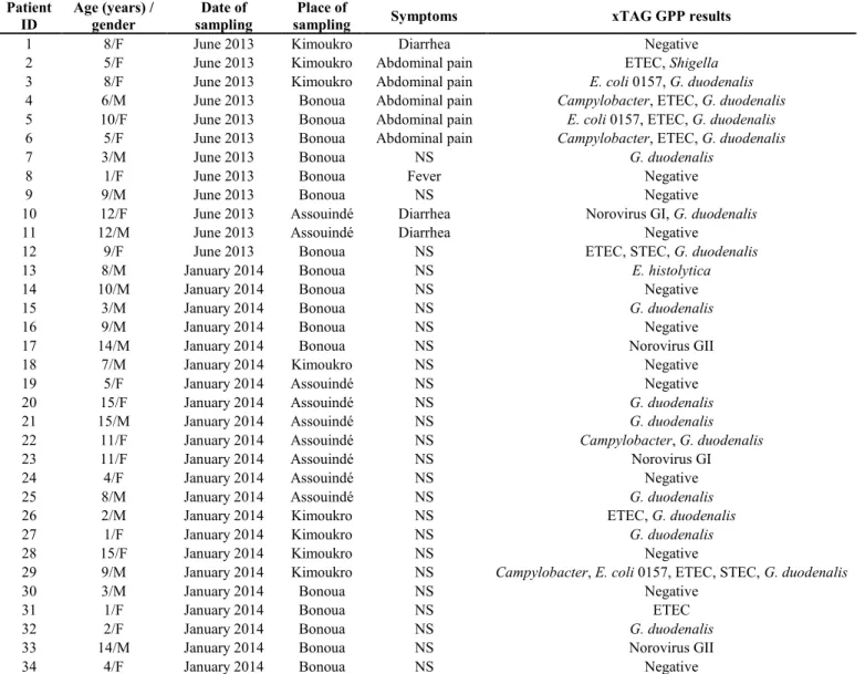
The study was based on 34 patients randomly and voluntary sampled in June 2013 and in January 2014 in three different localities (Bonoua, Kimoukro, and Assouinde) in the Sud-Comoé region of Côte d’Ivoire. The stool samples collected in 2013 were obtained from both asymptomatic and symptomatic subjects suffering from diarrhea, abdominal pains, and fever. The second group of samples, collected in 2014, was obtained from asymptomatic subjects (Table 1). All other relevant clinical information, including other diseases and therapy, were also noted. One fecal sample was collected in a sterile stool container from each patient and delivered to the clinical laboratory of the Centre don Orione in Bonoua within 12 hours. Table 1. Patients characteristics and enteric pathogens detected by Luminex xTAG GPP assay
Patient
ID Age (years) / gender sampling Date of sampling Place of Symptoms xTAG GPP results
1 8/F June 2013 Kimoukro Diarrhea Negative
2 5/F June 2013 Kimoukro Abdominal pain ETEC, Shigella
3 8/F June 2013 Kimoukro Abdominal pain E. coli 0157, G. duodenalis
4 6/M June 2013 Bonoua Abdominal pain Campylobacter, ETEC, G. duodenalis
5 10/F June 2013 Bonoua Abdominal pain E. coli 0157, ETEC, G. duodenalis
6 5/F June 2013 Bonoua Abdominal pain Campylobacter, ETEC, G. duodenalis
7 3/M June 2013 Bonoua NS G. duodenalis
8 1/F June 2013 Bonoua Fever Negative
9 9/M June 2013 Bonoua NS Negative
10 12/F June 2013 Assouindé Diarrhea Norovirus GI, G. duodenalis
11 12/M June 2013 Assouindé Diarrhea Negative
12 9/F June 2013 Bonoua NS ETEC, STEC, G. duodenalis
13 8/M January 2014 Bonoua NS E. histolytica
14 10/M January 2014 Bonoua NS Negative
15 3/M January 2014 Bonoua NS G. duodenalis
16 9/M January 2014 Bonoua NS Negative
17 14/M January 2014 Bonoua NS Norovirus GII
18 7/M January 2014 Kimoukro NS Negative
19 5/F January 2014 Assouindé NS Negative
20 15/F January 2014 Assouindé NS G. duodenalis
21 15/M January 2014 Assouindé NS G. duodenalis
22 11/F January 2014 Assouindé NS Campylobacter, G. duodenalis
23 11/F January 2014 Assouindé NS Norovirus GI
24 4/F January 2014 Assouindé NS Negative
25 8/M January 2014 Assouindé NS G. duodenalis
26 2/M January 2014 Kimoukro NS ETEC, G. duodenalis
27 1/F January 2014 Kimoukro NS G. duodenalis
28 15/F January 2014 Kimoukro NS Negative
29 9/M January 2014 Kimoukro NS Campylobacter, E. coli 0157, ETEC, STEC, G. duodenalis
30 3/M January 2014 Bonoua NS Negative
31 1/F January 2014 Bonoua NS ETEC
32 2/F January 2014 Bonoua NS G. duodenalis
33 14/M January 2014 Bonoua NS Norovirus GII
34 4/F January 2014 Bonoua NS Negative
After registration, the samples were immediately frozen at -20°C until transport in ice packs to the Institute of Virology of the University of Cologne, where they were kept at -80°C. The medical committee of Don Orione Centre provided approval and oversight of the study. Informed verbal consent was obtained from the parents or the guardians of children, and a local dialect interpreter was used to explain the study’s aims, procedures, and significance when necessary. All investigations and protocols followed the principles of the Helsinki Declaration.
Sample preparation and nucleic acid extraction
One hundred milligrams of each stool sample were suspended in 1 mL of phosphate-buffered saline (PBS). The total nucleic acids were extracted from 700 µL of the stool suspension using the automated platform VERSANT kPCR Molecular System and the VERSANT Sample Preparation 1.0 Reagents Kit (Siemens Healthcare Diagnostics, Erlangen, Germany) into an elution volume of 100 µL, according to the manufacturer’s instructions.
xTAG GPP assay
Fifteen human enteric pathogens were simultaneously tested by the xTAG GPP. This assay is a bead-based multiplexed molecular diagnostic test, which concurrently detects and identifies from a single stool specimen adenovirus subtypes 40/41, norovirus genogroups I and II (GI/GII), group A rotavirus,
Campylobacter spp., Clostridium difficile toxin A/B, Escherichia coli O157, enterotoxigenic Escherichia coli (ETEC) LT/ST, Salmonella spp.,
Shiga-like-toxin-producing E. coli (STEC) stx1/stx2, Shigella spp.,
Vibrio cholerae, Yersinia enterocolitica, Cryptosporidium hominis, C. parvum, Entamoeba histolytica, and Giardia duodenalis.
After extraction and purification of the total nucleic acids from each fecal specimen, a multiplex reverse transcription polymerase chain reaction (RT-PCR) reaction using 10 µL of eluate was performed using target-specific tagged primers and biotin-labeled primers. Each target results in PCR amplicons ranging from 58 to 293 bp. After sonication, 20 µL of the xTAG bead mix were aliquoted for each sample into a 96-well microtiter plate, and 5 µL of appropriate RT-PCR product were added. The xTAG 0.22 streptavidin, R-phycoerythrin conjugate (SAPE) was diluted with xTAG reporter buffer, and 75 µL of the reporter solution were added into each well. The RT-PCR and hybridization reactions were performed using Biometra T3 Thermocycler (Biometra GmbH, Göttingen, Germany). Following hybridization, the median fluorescence intensity (MFI) was generated for each xTAG bead population using the Luminex 100/200 instrument pre-heated to 45°C. The data were analyzed using the xTAG Data Analysis Software for the GPP (TDAS) to establish the presence or absence of bacterial, viral, or parasitic targets and the internal control in each sample.
Detection of norovirus by real-time PCR
In order to confirm the preservation of RNA during the storage and the shipping of stool samples, 5 µL of each eluate were analyzed by real-time PCR for the detection of norovirus GI/GII using the commercially available RealStar Norovirus RT-PCR Kit 2.0 (Altona Diagnostics, Hamburg, Germany) according to the manufacturer’s instructions.
Results
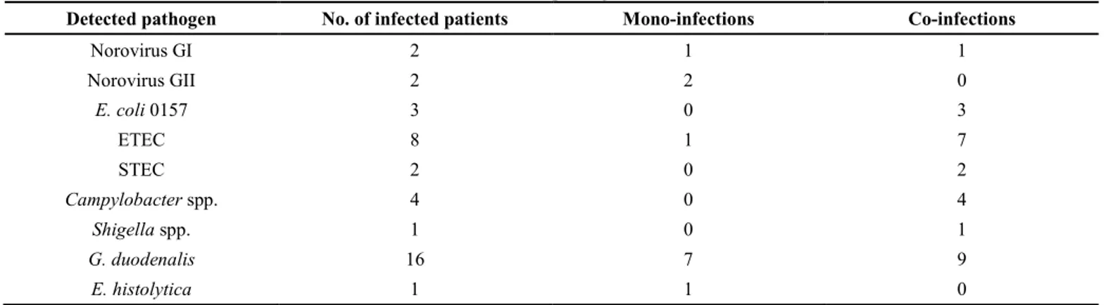
A total of 34 stool samples were examined from young patients (18 females and 16 males) between 1 and 15 years of age (average 7.6; median 8). Overall, Table 2. Number of mono- and co-infections for each detected pathogen
Detected pathogen No. of infected patients Mono-infections Co-infections
Norovirus GI 2 1 1 Norovirus GII 2 2 0 E. coli 0157 3 0 3 ETEC 8 1 7 STEC 2 0 2 Campylobacter spp. 4 0 4 Shigella spp. 1 0 1 G. duodenalis 16 7 9 E. histolytica 1 1 0
22 of 34 (64.7%) samples were detected positive by xTAG GPP (Table 1); 16 of 22 were from asymptomatic patients. Four norovirus isolates were detected and assigned to genogroups I and II, as confirmed also by real-time PCR. Among bacteria, E.
coli 0157 (3/22), ETEC (8/22), STEC (2/22), Campylobacter spp. (4/22), and Shigella spp. (1/22)
were identified. Concerning enteric protozoa, 15 samples were tested positive for G. duodenalis, whereas E. histolytica was detected in only one sample. No patient resulted positive for C. hominis or
C. parvum.
Of 22 positive children, 12 (54.6%) presented a mono-infection: 3 with norovirus GI/GII, 1 with ETEC, 7 with G. duodenalis, and 1 with E. histolytica. A total of 10 patients (45.4%) showed co-infections. In particular, 5/10 and 4/10 samples were detected positive for two and three different pathogens, respectively. A co-infection with four bacteria and one parasite species was revealed in a nine-year-old boy. All bacterial isolates were detected as co-infections, while only one patient was revealed to have a mono-infection by ETEC. No co-mono-infections with both bacteria and viruses were identified (Tables 1 and 2). Discussion
Gastroenteritis is recognized as one of the most important public health issues in developing countries, where low socio-economic level and favorable climatic conditions enable environmental contamination by pathogen microorganisms. In particular, the risk of contracting diarrheal disease is currently fivefold higher in sub-Saharan Africa than it is in industrialized countries. Diarrhea is often more severe or fatal in children suffering from malnutrition. In developing countries, the frequent combination of recurrent diarrhea and malnutrition can lead to a vicious cycle with dire consequences, especially for children under five years of age [10]. Moreover, laboratory diagnosis of infectious diseases still represents a challenge for most of the healthcare centers in low-income regions such as sub-Saharan Africa and, consequently, the appropriate therapeutic choice is frequently based only on clinical symptoms [11]. Most of the studies on enteric pathogens in sub-Saharan Africa over the last 20 years have focused on children under five years of age. Rotavirus,
Cryptosporidium spp., Shigella spp., enterotoxigenic E. coli (ETEC), Campylobacter jejuni, and Vibrio cholerae have been reported to be the main etiological
agents of pediatric diarrhea [12-14]. Fisher et al. (2010) systematically reviewed the etiology of
diarrhea in children older than five years of age, adolescents, and adults worldwide. They found that ETEC and V. cholerae were the most frequently isolated pathogens among hospitalized patients in low- and middle-income countries [15]. Recent data from Senegal showed that bacteria and parasites are equally frequent in all age groups, whereas viral infections are significantly more frequent in children under five years of age and during the dry season [16]. Lamberti
et al. (2014) underlined the impact of Shigella spp.
and ETEC on morbidity and mortality among older children, adolescents, and adults in South Asia and Africa [17].
The Luminex xTAG GPP is the first commercially available assay able to simultaneously detect viruses, bacteria, and parasites on total nucleic acid isolated from a single stool specimen [18]. The present study represents the first application of xTAG GPP to human stool samples collected in Côte d'Ivoire. Overall, the results obtained using the Luminex panel assay are in agreement with the data from studies focused on single pathogens or classes of microorganisms in sub-Saharan Africa [19-21]. Regarding viruses, only norovirus GI/GII was detected, and three of the four positive patients were asymptomatic. Huynen et al. (2013) reported a similar prevalence of norovirus in both symptomatic and asymptomatic patients in Burkina Faso [22]. Enteric infections among apparently asymptomatic individuals represent a serious issue in endemic areas [23-25]. This unrecognized source of infections and the low level of hygiene and sanitation constitute a hazardous combination, which contributes to the endemic transmission of EE [7]. Regarding bacteria, ETEC, STEC, E. coli 0157, Campylobacter spp., and Shigella spp. were identified, as previously reported in African countries [12,17,26]. Regarding parasites, whereas the high rate of G. duodenalis confirms our previous data from Tanzania and Côte d’Ivoire, no patients were detected positive for C. hominis and C. parvum [27-28]. This might be due to the low number of samples tested but also because the xTAG GPP is not able to identify C. meleagridis, a species frequently reported in humans in Africa [29]. Noteworthy was the molecular detection of E. histolytica, considering that the cysts are morphologically indistinguishable from those of the non-pathogenic E. dispar and E.
moshkovskii and the high prevalence of E. dispar in
the area of study [30]. Finally, the present study evidenced the issue of co-infections also in apparently healthy individuals.
Conclusions
In the context of the surveillance of diarrheal disease in sub-Saharan Africa, our results show evidence that the application of xTAG GPP might significantly improve the available knowledge about the etiology of symptomatic and asymptomatic enteric mono- and co-infections. Considering the impact of gastroenteritis in developing countries, new efforts from the international scientific community, including the application of innovative molecular technology, should be encouraged in order to improve diagnosis and treatment of intestinal infections in endemic areas. Acknowledgements
We are grateful to all the subjects for their participation and to the local interpreters for explaining the study aims. Logistical support and fecal sample collection were provided by the Hippocrates Project staff of Assomis Onlus.
References
1. The United Nations Children’s Fund/World Health Organization (2009) Diarrhoea: why children are still dying and what can be done. New York/Geneva: UNICEF/WHO. 68 p.
2. Ngure FM, Reid BM, Humphrey JH, Mbuya MN, Pelto G, Stoltzfus RJ (2014) Water, sanitation, and hygiene (WASH), environmental enteropathy, nutrition, and early child development: making the links. Ann N Y Acad Sci 1308: 118-128.
3. Berrilli F, Di Cave D, N'Guessan R, Kaboré Y, Giangaspero A, Sorge RP, D'Alfonso R (2014) Social determinants associated with Giardia duodenalis infection in southern Côte d'Ivoire. Eur J Clin Microbiol Infect Dis 33: 1799-1802. 4. Schmidlin T, Hürlimann E, Silué KD, Yapi RB, Houngbedji
C, Kouadio BA, Acka-Douabélé CA, Kouassi D, Ouattara M, Zouzou F, Bonfoh B, N'Goran EK, Utzinger J, Raso G (2013) Effects of hygiene and defecation behavior on helminths and intestinal protozoa infections in Taabo, Côte d'Ivoire. PLoS One 8: e65722.
5. Thapar N, Sanderson IR (2004) Diarrhoea in children: an interface between developing and developed countries. Lancet 363: 641-653.
6. Kotloff KL, Nataro JP, Blackwelder WC, Nasrin D, Farag TH, Panchalingam S, Wu Y, Sow SO, Sur D, Breiman RF, Faruque AS, Zaidi AK, Saha D, Alonso PL, Tamboura B, Sanogo D, Onwuchekwa U, Manna B, Ramamurthy T, Kanungo S, Ochieng JB, Omore R, Oundo JO, Hossain A, Das SK, Ahmed S, Qureshi S, Quadri F, Adegbola RA, Antonio M, Hossain MJ, Akinsola A, Mandomando I, Nhampossa T, Acácio S, Biswas K, O'Reilly CE, Mintz ED, Berkeley LY, Muhsen K, Sommerfelt H, Robins-Browne RM, Levine MM (2012) Burden and aetiology of diarrhoeal disease in infants and young children in developing countries (the Global Enteric Multicenter Study, GEMS): a prospective, case-control study. Lancet 382: 209-222.
7. Korpe PS, Petri WA Jr (2012) Environmental enteropathy: critical implications of a poorly understood condition. Trends Mol Med 18: 328-336.
8. Hickman D, Jones MK, Zhu S, Kirkpatrick E, Ostrov DA, Wang X, Ukhanova M, Sun Y, Mai V, Salemi M, Karst SM (2014) The effect of malnutrition on Norovirus infection. mBio 5: e01032-13.
9. Nhampossa T, Sigaúque B, Machevo S, Macete E, Alonso P, Bassat Q, Menéndez C, Fumadó V (2013) Severe malnutrition among children under the age of 5 years admitted to a rural district hospital in southern Mozambique. Public Health Nutr 16: 1565-1574.
10. Becker SL, Vogt J, Knopp S, Panning M, Warhurst DC, Polman K, Marti H, von Müller L, Yansouni CP, Jacobs J, Bottieau E, Sacko M, Rijal S, Meyanti F, Miles MA, Boelaert M, Lutumba P, van Lieshout L, N'Goran EK, Chappuis F, Utzinger J (2013) Persistent digestive disorders in the tropics: causative infectious pathogens and reference diagnostic tests. BMC Infect Dis 13: 37.
11. Nitiema LW, Nordgren J, Ouermi D, Dianou D, Traore AS, Svensson L, Simpore J (2011) Burden of rotavirus and other enteropathogens among children with diarrhea in Burkina Faso. Int J Infect Dis 15: 646-652.
12. Okeke IN, Lamikanra A, Steinrück H, Kaper JB (2000) Characterization of Escherichia coli strains from cases of childhood diarrhea in provincial southwestern Nigeria. J Clin Microbiol 38: 7-12.
13. Vargas M, Gascon J, Casals C, Schellenberg D, Urassa H, Kahigwa E, Ruiz J, Vila J (2004) Etiology of diarrhea in children less than five years of age in Ifakara, Tanzania. Am J Trop Med Hyg 70: 536-539.
14. Sire JM, Garin B, Chartier L, Fall NK, Tall A, Seck A, Weill FX, Breurec S, Vray M (2013) Community-acquired infectious diarrhoea in children under 5 years of age in Dakar, Senegal. Paediatr Int Child Health 33: 139-144.
15. Fischer Walker CL, Sack D, Black RE (2014) Etiology of diarrhea in older children, adolescents and adults: a systematic review. PLoS Negl Trop Dis 4: 768.
16. Sambe-Ba B, Espié E, Faye ME, Timbiné LG, Sembene M, Gassama-Sow A (2013) Community-acquired diarrhea among children and adults in urban settings in Senegal: clinical, epidemiological and microbiological aspects. BMC Infect Dis 13: 580.
17. Lamberti LM, Bourgeois AL, Fischer Walker CL, Black RE, Sack D (2014) Estimating diarrheal illness and deaths attributable to Shigellae and enterotoxigenic Escherichia coli among older children, adolescents, and adults in South Asia and Africa. PLoS Negl Trop Dis 8: 2705.
18. Wessels E, Rusman LG, van Bussel MJ, Claas EC (2014) Added value of multiplex Luminex Gastrointestinal Pathogen Panel (xTAG(®) GPP) testing in the diagnosis of infectious gastroenteritis. Clin Microbiol Infect 20: 182-187.
19. Trainor E, Lopman B, Iturriza-Gomara M, Dove W, Ngwira B, Nakagomi O, Nakagomi T, Parashar U, Cunliffe N (2013) Detection and molecular characterisation of noroviruses in hospitalised children in Malawi, 1997-2007. J Med Virol 85: 1299-1306.
20. Bonkoungou IJ, Haukka K, Österblad M, Hakanen AJ, Traoré AS, Barro N, Siitonen A (2013) Bacterial and viral etiology of childhood diarrhea in Ouagadougou, Burkina Faso. BMC Pediatr 13: 36.
21. Ouattara M, Silué KD, N'Guéssan AN, Yapi A, Barbara M, Raso G, Utzinger J, N'Goran E (2008) Prevalence and
polyparasitism of intestinal protozoa and spatial distribution of Entamoeba histolytica, E. dispar and Giardia intestinalis from pupils in the rural zone of Man in Côte d'Ivoire. Sante 18: 215-222.
22. Huynen P, Mauroy A, Martin C, Savadogo LG, Boreux R, Thiry E, Melin P, De Mol P (2013) Molecular epidemiology of norovirus infections in symptomatic and asymptomatic children from Bobo Dioulasso, Burkina Faso. J Clin Virol 58: 515-521.
23. de Moura C, Fregolente MC, Martini IJ, Domingos DF, da Silva EJ, Ferraz MM, Gatti MS, da Silva Leite D (2012) Prevalence of enteropathogens in normal feces from healthy children at an infant day care in Brazil. J Infect Dev Ctries 6: 176-180. doi:10.3855/jidc.1982.
24. Samie A, Guerrant RL, Barrett L, Bessong PO, Igumbor EO, Obi CL (2009) Prevalence of Intestinal Parasitic and Bacterial Pathogens in Diarrhoeal and Non-diarroeal Human Stools from Vhembe District, South Africa. J Health Popul Nutr 27: 739-745.
25. Qadri MH, Ai-Gamdi MA, Al-Harfi RA (1995) Asymptomatic Salmonella, Shigella and intestinal parasites among primary school children in the eastern province. J Family Community Med 2: 36-40.
26. Pazzaglia G, Bourgeois AL, Araby I, Mikhail I, Podgore JK, Mourad A, Riad S, Gaffar T, Ramadan AM (1993) Campylobacter-associated diarrhoea in Egyptian infants: epidemiology and clinical manifestations of disease and high frequency of concomitant infections. J Diarrhoeal Dis Res 11: 6-13.
27. Di Cristanziano V, Santoro M, Parisi F, Albonico M, Shaali MA, Di Cave D, Berrilli F (2014) Genetic characterization of Giardia duodenalis by sequence analysis in humans and animals in Pemba Island, Tanzania. Parasitol Int 63: 438-441. 28. Berrilli F, D'Alfonso R, Giangaspero A, Marangi M,
Brandonisio O, Kaboré Y, Glé C, Cianfanelli C, Lauro R, Di Cave D (2012) Giardia duodenalis genotypes and Cryptosporidium species in humans and domestic animals in Côte d'Ivoire: occurrence and evidence for environmental contamination. Trans R Soc Trop Med Hyg 106: 191-195. 29. Blanco MA, Iborra A, Vargas A, Nsie E, Mba L, Fuentes I
(2009) Molecular characterization of Cryptosporidium isolates from humans in Equatorial Guinea. Trans R Soc Trop Med Hyg 103: 1282-1284.
30. Ouattara M, N'guéssan NA, Yapi A, N'goran EK (2010) Prevalence and spatial distribution of Entamoeba histolytica/dispar and Giardia lamblia among schoolchildren in Agboville area (Côte d'Ivoire) PLoS Negl Trop Dis 4: e574.
Corresponding author Dr. Veronica Di Cristanziano
Institute of Virology, University of Cologne Fürst-Pückler-Straße 56
50935, Cologne, Germany Phone: +492214783927 Fax: +492214783902
Email: [email protected]