Department of Agriculture, Food and Environment,
University of Pisa, Italy
PhD thesis in Science of Crop Production
Trichoderma spp. in innovative substrates
for ornamentals plants
SSD AGR/12
Tutor Coordinator Prof. Giovanni Vannacci Prof. Alberto Pardossi Dr. Gianluca Burchi
Candidate Domenico Prisa
Examining Board: Prof. Alberto Pardossi, University of Pisa, Italy Prof. Aniello Scala, University of Florence, Italy
Dr. Mariarosaria Vergara, Scuola Normale Superiore of Pisa, Italy
ABSTRACT
Trichoderma spp. are free-living fungi commonly widespread in soil and root ecosystems. Recent discoveries show them as opportunistic, avirulent plant symbionts as well as parasites of other fungi. Some strains establish robust and long-lasting colonization of roots by entering into the first epidermal layers. Root colonization frequently results in enhancing of growth and development, crop productivity or induction of resistance to abiotic and biotic factors.
Peat, mainly imported from the northern and eastern European regions, is the basic constituent of organic substrates commonly utilized in the cultivation of ornamental plants in pots or in benches. During the past few years, the supply of the peat is hampered by environmental and economical constraints. Recently, the European Commission decided to exclude all substrates containing peat from the release of the Community Eco-Label Mark. In this optic the need to reduce peat in ornamental substrates devised great attention and resulted in pressing and increasing research activity to set up new and innovative substrates for ornamental market.
The aim of the present PhD thesis is to select beneficial fungi belonging to Trichoderma genus, to be add as soil inoculants, in order to develop an innovative, economical and suitable substrate alternative to peat for cultivation of seed plants (Limonium sinuatum and Cupressus sempervirens) and of acidophilus species (Camellia japonica) of ornamental interest. The activity involved the selection of Trichoderma spp. isolates for their ability to grow in the roots, as endophytes, or in the rhizosphere, to protect plants against plant pathogens or to act as plant growth promoters. The preliminary screening for endophytism resulted in 10 interesting isolates (out of 162) for Limonium sinuatum, 9 (out of 162) for Cupressus sempervirens and 8 (out of 202) for Camellia japonica. From following rounds of screening, three Trichoderma isolates, among which T. asperellum 2046 in
common for all the species, confirmed the best endophytic performance and improved growth.
The antagonistic activity of these selected strains, against fungal plant pathogens as Sclerotinia sclerotiorum, S. minor, Colletotrichum gleospoiroides and Rhizoctonia solani, has been evaluated in order to analyse if these isolates could be considered good beneficial fungi. In addition. T2046 was evaluated in biocontrol experiments on Limonium, against S. sclerotiorum and S. minor with mycoparasistim investigated as principal mechanisms.
Encouraging results herewith obtained, suggest that T. asperellum 2046 could be taken into account as bioactive ingredient of new biopesticide and/or biofertilizer to be used as inoculant for innovative substrates for ornamental plants.
Table of contents
ABSTRACT 2
TABLE OF CONTENTS 4
1. INTRODUCTION 6
1.1. Endophytism and ecological role of endophytic fungi 6 1.2. Biological control 9
1.3. Trichoderma spp. 11
1.4. Trichoderma: endophytism and plant growth promotion 13
2. AIM OF THE WORK 16 3. MATERIALS AND METHODS 17 3.1 Fungal isolates 17 3.2 Fungal inoculum 19 3.3 Screening of Trichoderma isolates as endophytes and growth promoters of
Limonium, Cupressus and Camellia 20
3.4 Evaluation of selected Trichoderma as inoculants and growth promoters for Cupressus sempervirens and Camellia japonica 25 3.5 Identification of Trichoderma spp. isolates 27 3.6 Antagonistic and mycoparasitic activity of selected Trichoderma isolates by
in vitro test. 28
3.7 Biocontrol of Sclerotinia sclerotiorum, Sclerotinia minor and Rhizoctonia
solani by T. asperellum 2046 on Limonium by in vivo test 30
4.1 Screening of Trichoderma isolates as endophytes and growth promoters of
Limonium, Cupressus and Camellia 32
4.2 Evaluation of selected Trichoderma as inoculants of innovative
substrates for Cupressus sempervirens and Camellia japonica 36 4.3 Identification of Trichoderma spp. isolates 40 4.4 Antagonistic and mycoparasitic activity of selected Trichoderma
isolates by in vitro tests 42 4.5 Effects of T. asperellum 2046 on Sclerotinia sclerotiorum, Sclerotinia
minor and Rhizoctonia solani by in vivo test 47
5. DISCUSSION 50
INDEX OF PICTURES 56
INDEX OF TABLES 59
1. INTRODUCTION
1.1. Endophytism and ecological role of endophytic fungi
The environment consists of a complex set of ecological niches and relationships between different organisms. Symbiotic associations are widespread in the biosphere and take on an important ecological role when involving autotrophic and heterotrophic organisms. These associations can be carried out by three kinds of symbiosis: harmonics, as the mutualism (i.e. mycorrhiza or bacterial root nodules) in which both organisms have a benefit; neutrals, as commensalism where any benefit for an organism doesn’t involve any damage to the other; inharmonious (antagonism), when an organism takes benefit from the other. In any case, this symbiosis is configured always a relationship of parasitism, where parasitic organism takes a benefit, sometimes without causing significant damage to the host (Graniti, 2002).
The association (interaction) microorganism/plant represents, especially for the first ones, an optimal system to satisfy its needs in terms of nutrition, water, protection to adverse environmental factors (water stress, radiation, extreme temperatures) and to competitors. Plants, therefore, represent a valuable habitat for many heterotrophic organisms and a source of nutrients. Endophytism is an important example of microorganism/plants interaction. The meaning of the term endophyte has undergone various transformations in the last decade and there is still considerable disagreement about what constitutes an endophyte.
The term endophyte was introduced by De Bary (1866) to describe microorganisms that colonize internal tissues of stems and leaves (cited by Wilson, 1995). De Bary’s definition has since modified many times. Two widely accepted definitions follow:
“Endophytes colonized symptomlessly the living, internal tissues of the host, even though the endophyte may, after incubation or latency period, cause disease” (Petrini, 1991) and “Endophytes are fungi or bacteria which, for all part of their life cycle….cause unapparent and asymptomatic infections entirely within plant tissue” (Wilson, 1995).
The results of a century of research on endophytic fungi suggest that these microorganisms establish relationships with plants (Siegel et al., 1987; Smith et al., 1996; Germinda et al., 1998, Rodrigues and Samuels, 1999; Gutierrezzmora and Martinez-Romero, 2001, Schena et al., 2003, Hoff et al., 2004; Raviraja, 2005, Rodriguez et al., 2009). The best-known endophytic fungi are mutualists as Neotyphodium species that infect cool-season grasses. They are of great economic importance and a superb model system to study the biology of interactions. Because Neotyphodium is an example of mutualism, there is widespread misconception (in the case of those who do not work with endophytes) or fantasy (in the case of those who do it) that most endophytes are mutualists as well. However, in most case a mutualistic relationship has not been demonstrates (Bayman, 2007).
Endophytic fungi have been examined in conifers (Petrini et al.,1993) including Pinus spp (Sieber et. al., 1999) Taxus spp. (Fisher and Petrini, 1987) and Juniperus spp. (Petrini and Muller, 1979; Petrini and Carroll, 1981) and they are presumed to be ubiquitous. Endophytic fungi have been described as playing a protective role against insect herbivory not only in grasses (Clay, 1990) but also in conifers (Carroll, 1991).
Fungal endophytes live internally, either intercellularly or intracellularly, and asymptomatically (i.e. without causing overt signs of tissue damage) within plant tissues. Endophytes usually occur in above-ground plant tissues, but also occasionally in roots, and are distinguished from mycorrhizae by lacking external hyphae or mantels (Saikkonen, 1998).
The endophytes can colonize the plant tissue in a systemic and localized manner (Stone et al., 2000). Furthermore, they can manifest a specifically preferred organ and tissue as a result of their adaptation to different physiological conditions in plants (Rodrigues and Samuels, 1999) and, therefore, only colonize the leaves or needles (Stone, 1986; Deckert et al. 2001), roots (Bacon and Hinton 1996), or adapt to grow in the cortex (Fisher and Petrini, 1990). The endophytic colonization of epigeal tissues (in particular leaves and buds) is different from that of the roots. Many studies showed that colonization of the shoots may be intracellular, and confined to individual cells or localized intercellular, while the endophytic colonization of the roots generally occurs extensively both intra and intercellular. The infection can occur through appressoria and haustoria, or thorugh the naturals opening such as stomata, lenticels and idatodi (Stone, 1987; Cabral et al., 1993; Stone et al., 1994).
Colonization can have a major impact on plants that is manifested by an increased tolerance to abiotic and biotic stresses, an increase of vigor, or with an alteration on physiology. Research in the last twenty years have demonstrated the extreme specialization of endophytic colonization in plants. It is evident that endophytes colonize all taxa of plants, from those less evolved like mosses and ferns to evoluted plants (gimnosperms and angiosperms) that grow in tropical, temperate and boreal forests; they also colonize poliannual and annual herbs that grow in extreme environments such as arctic, alpine and xeric (Zhang et al, 2006).
From a single plant is possible to isolate hundreds endophytic species, some with a specificity for host (Tan and Zou, 2001). This specificity may be influenced by environmental conditions such as changes seasonal climate, and the physiological conditions of the plant.
1.2. Biological control
Plant diseases need to be controlled to maintain the quality and abundance of food, feed and fibre produced by growers around the world. A number of different strategies may be used to manage and control plant pathogens.
The broad definition of biological control proposed by Cook and Baker (1983) is: “the reduction of the amount of inoculum or disease-production activity of a pathogen accomplished by or through one or more organisms than man”. This broad definition includes the use of less virulent pathogen, more resistant cultivars of the host, and microbial antagonists “that interfere with the survival or disease-production activity of the pathogen”.
Since biological control is a result of many different types of interactions among microorganisms, scientists have concentrated on characterization of mechanisms occurring in different experimental situations. In all cases, pathogens are antagonized by the presence and activities of other microorganisms that they encounter. Different modes of actions of biocontrol-active microorganisms in controlling fungal plant disease include mycoparasitism, antibiosis, competition for site and nutrient and induced resistance. The most effective biocontrol active microorganisms appear to antagonize plant pathogens employing several models of action (Cook, 1993).
Mycoparasitism: Mycoparasitism, the direct attack of one fungus to another one, is a very complex process that involves sequential events, including recognition, attack and subsequent penetration and killing of the host. The various mechanisms used by fungi to antagonize or parasitize their competitors include antibiotic production, secretion of lytic enzymes, hyphal interference and direct penetration of the host. Any particular fungus-fungus interaction may encompass more than one of these mechanisms either individually or simultaneously (Jeffries, 1997). Mycoparasitism involves morphological changes, such as coiling and formation of appressorium-like structures, which serve to penetrate the host and contain high
concentrations of osmotic solutes such as glycerol (McIntyre et al., 2004). Lysis of the host cell wall of the plant pathogenic fungi has been demonstrated to be an important step in the mycoparasitic attack (Kubicek et al., 2001; Howell, 2003). Antibiosis: In a general definition, antibiotics are microbial toxins that can, at low concentrations, poison or kill other microorganisms. It has been shown that some antibiotics produced by microorganisms are particularly effective against plant pathogens and the disease they cause (Homma et al., 1989; Howell and Stipanovic, 1980; Islam et al., 2005; Shanahan et al., 1992). In all cases, the antibiotics have been shown to be particularly effective at suppressing growth of the target pathogen in vitro and/or in situ conditions. Fungi have been demonstrated to produce a wide variety of toxic substances that have activity against a range of prokaryotic and eukaryotic organisms. The ability of a fungus to produce antibiotic may thus be very important in determining its ability to colonize or maintain its presence on a substrate (Faull, 1988).
Competition: Competition occurs when two (or more) organisms require the same resource and the use of this by one reduces the amount available to the other. The nutrient sources in the soil and rhizosphere are frequently not sufficient for microorganisms and starvation is the most common cause of death for microorganisms. For a successful colonization of phyllosphere and rhizosphere a microbe must effectively compete for the available nutrients. There is a general believe that competition between pathogens and non-pathogens for nutrient resources is an important issue in biocontrol. It is also believed that competition is more critical for soil borne pathogens, including Fusarium and Pythium species that infect through mycelial contact than foliar pathogens that germinate directly on plant surfaces and infect through appressoria and infection pegs (Elad and Baker, 1985; Keel et al., 1989; Loper and Buyer 1991). Competition for rare but essential micronutrients, such as iron, has also been shown to be important in biological
disease control. Competition is also possible for oxygen, space and, in the case of autotrophs, light.
Induction of resistance: Plants actively respond to a variety of environmental stimulating factors, including gravity, light, temperature, physical stress, water and nutrient availability and chemicals produced by soil and plant associated microorganisms. Such stimuli can either induce or condition plant host defences through biochemical changes that enhance resistance against subsequent infection by a variety of pathogens. Induction of host defences can be local and/or systemic in nature, depending on the type, source and amount of stimulating agents (Audenaert et al., 2002; De Meyer and Hofte, 1997; Kloepper et al, 1980; Leeman et al., 1995).
1.3. Trichoderma spp.
The genus Trichoderma consists of anamorphic fungi isolated primarily from soil and decomposing organic matter, with teleomorphs, when known, belonging to the ascomycete genus Hypocrea (order Hypocreales).
Fungal species belonging to this genus are worldwide in occurrence and easily isolated from soil, decaying wood and other plant organic matter. Trichoderma isolates are characterized by a rapid growth rate in culture and by the production of numerous spores (conidia) with varying shades of green. Their lifestyle is generally saprotrophic with minimal nutritional requirements; they are able to grow rapidly on many substrates, can produce metabolites with demonstrable antibiotic activity and may be mycoparasitic against a wide range of pathogens (Grondona et al., 1997). The abundance of Trichoderma spp. in various soils, coupled with a wide metabolic versatility, a dynamic colonization of plant rhizosphere and the ability to antagonize and repress a great number of plant pathogens are direct evidence of the role that these fungal species may play in biological control (Papavizas, 1985; Chet, 1987).
A number of isolates of Trichoderma have been found to be effective biocontrol agents of various soil-borne plant pathogenic fungi under greenhouse and field conditions. The knowledge of mechanisms of interaction of Trichoderma spp. with plant pathogenic fungi and the plant host is of importance to enhance the practical application of these beneficial microorganisms. They can work against fungal phytopathogens either directly through mechanisms such as mycoparasitism, competing for nutrients and space, modifying environmental conditions and antibiosis or indirectly promoting plant growth and plant defensive mechanisms. In the direct interactions between Trichoderma spp. and the plant pathogenic fungi, mycoparasitism is one of the mechanisms observed with the antagonist that coils around the hyphae of the pathogen, develops hook like structures known as appressoria coupled with production of lytic enzymes and then penetrates the pathogen hyphae (Chet 1987; Kubicek et al., 2001, Rocha-Ramirez et al. 2002; Howell 2003).
Trichoderma spp. have also been reported to produce a plethora of secondary metabolites showing antimicrobial activity (Vinale et al. 2008). The chemical composition of these compounds depends on the strains and they may be classified as volatile, water-soluble or water-insoluble compounds (Ghisalberti and Sivasithamparam, 1991).
The competition for space, infestation sites and nutrients has also been shown to be possible mechanisms involved in the biocontrol activities of Trichoderma spp. (Dennis and Webster 1971a, b; Chet 1987; Tronsmo and Hjeljord 1998).
The first demonstration of induced resistance was reported in 1997 (Bigirimana) who described the acquisition of resistance of bean plants towards Botrytis cinerea and Colletotrichum lindemuthianum after inoculation of the root with the strain T-39 of Trichoderma harzianum (Yedidia et al. 1999). Certain Trichoderma isolates invade the vascular tissue or epidermal cells of plant root, giving rise to accumulation of signal molecules, salicylic acid (SA) and jasmonic acid (JA).
These compounds induce the PR genes function coding pathogenesis-related proteins (PR protein), expressed by plant to defence pathogen infection (Hurtado, 2004; Wasternack et al., 2006). The PR proteins were classified into 14 families: among them the degrading enzymes chitinase and β-1,3-glucanases that are capable to lyses the fungal plant pathogen cell wall. Different reports revealed species diversity of Trichoderma spp. in tomato seed production fields and its effectiveness against Fusarium wilt (Saksirirat et al., 2005; Saepaisan, 2006).
1.4. Trichoderma: endophytism and plant growth promotion
In recent years, Trichoderma spp. have been widely used in agriculture as biocontrol agents and inoculants to provide plant growth promotion. They are involved in fundamental activities that ensure the stability and productivity of both agricultural and natural ecosystems.
Some Trichoderma strains, described as rhizosphere competent and selectively used for commercial development, can cause an asymptomatic infection of roots, where the fungus colonization is limited to the outer cortical regions. These fungi behave as endophytes, colonizing the root epidermis and outer cortical layers and release bioactive molecules. At the same time, the transcriptome and proteome of plants are substantially altered. This intimate interaction with the plant provides a number of benefits only recently recognized for their variety and importance, including increased resistance of the plant to various biotic stresses through induced or acquired systemic resistance and to abiotic stresses such as water deficit/excess, high salinity and extreme temperature; enhanced nitrogen use efficiency by improved mechanisms of nitrogen reduction and assimilation and reduced overexpression of stress genes or accumulation of toxic compounds during plant response to pathogen (Shoresh et al, 2010).
An additional benefit to consumer comes from an increased content of antioxidants in the fruit from plants treated by selected Trichoderma strains (Lorito et al., 2010).
Moreover, it was also observed that the fertility of soils treated with some Trichoderma strains could be significantly improved beyond disease control, which increased the attractiveness of these fungi for a general use in crop production. The effect could be particularly strong in terms of root growth promotion, even though it has been not unusual to detect an increase in stem length and thickness, leaf area, chlorophyll content and yield (size and/or number of flowers or fruits) (Harman et al., 2004).
The molecular mechanisms supporting this highly desirable beneficial effect of plant growth promotion are not fully clarified and include improvement of nutrient availability and uptake for the plant (Altomare et al., 1999, Lorito et al., 2010). As example, maize plants grown from seeds treated with T. harzianum T-22, grown using 40% less of nitrogen in the fertilizer, have obtained a maximum of efficiency equal to that of untreated plants but with a supply of nitrogen optimal (Harman, 2000). Further analysis show a general increase in the absorption of many elements such as Pb, Mn, Zn, Al and the ability to solubilize some nutrients in the soil, such as phosphates, ions Fe3+, Cu2+, Mn4+, many times not easily available from the plant. (Altomare et al., 1999).
Moreover, the involvement of growth phytormones from both plant and fungal origin could be involved in the phenomenon of plant growth promotion (Vinale et al. 2008).
In combination with the direct effects on plant pathogens and with the ability of promote plant growth, Trichoderma spp. have also been found to stimulate plant defence mechanisms. The presence of Trichoderma in plants involves an induction of resistance, often localized or systemic (Harman et al., 2004). This phenomenon, also observed in field, has been attributed to a fungus-root biochemical cross talk involving many bioactive metabolites produced by the biocontrol agents (Harman et al., 2004; Shoresh et al, 2010; Woo et al., 2006). The first demonstration of induced resistance was reported in 1997 (Bigirimana) who described the
acquisition of resistance of bean plants towards Botrytis cinerea and Colletotrichum lindemuthianum after inoculation of the root with the strain Trichoderma harzianum T-39 (Yedidia et al. 1999). Many Trichoderma strains colonize plant roots of dicots and monocots. During this process Trichoderma hyphae coil around the roots, form appressoria-like structures, and finally penetrate the root cortex. During the intercellular Trichoderma growth in the root epidermis and cortex the surrounding plant cells have been induced to deposit cell wall material and to produce phenolics compounds. This plant reaction limits the Trichoderma growth inside the root (Vinale et al., 2008). Effective Trichoderma srains are able to induce a stronger response in the plant compared to pathogen-triggered immunity by producing a variety of microbe-associated molecular patterns (MAMP) as hydrophobins, expansin-like proteins, secondary metabolites, and enzymes having direct antimicrobial activity such as peroxidase, chitinase and glucanase. In addition, there is an accumulation of antimicrobial compounds and phytoalexins (Lorito et al., 2010).
For all these reasons, the use of Trichoderma spp. strains as inoculants of substrates to be employed in nursery could confer an additional value both in order to control soilborne pathogens, to induce resistance or to promote growth of plants.
2. Aim of the Work
Due to the recent European Commission decision to exclude all substrates containing peat from the release of the Community Eco-Label Mark, the aim of the present PhD’s thesis was to develop an innovative, economical and suitable substrate alternative to peat for production of seed plants (Limonium sinuatum and Cupressus sempervirens) and for growing acidophilic plants (Camellia japonica). This activity was performed within the project “SUBARTIFLOR: Messa a punto di substrati artificiali innovativi per il florovivaismo” funded by the Italian Ministry of Agricultural, Food and Forestry Policies (MIPAAF).
The activity involved the use of a fungal collection maintained by the Mycology Lab of the Department of Agriculture, Food and Environment (University of Pisa), consisting of about thousand Trichoderma spp. isolates, in order to select beneficial isolates to be used as inoculants of selected substrates. Such fungal isolates should be able to grow in the rhizosphere or as endophytes in the roots and/or to be able to act as antagonists or to induce resistance to pathogens and/or to have effect as plant growth promoters.
The optimization of the recipe of new plant growth substrates for the reduction of the peat and the selection of beneficial fungi, an added value to the new substrates focussed on:
- the definition of the fungal collection on which operate the selection; - the definition of the screening procedure;
3. Materials and Methods
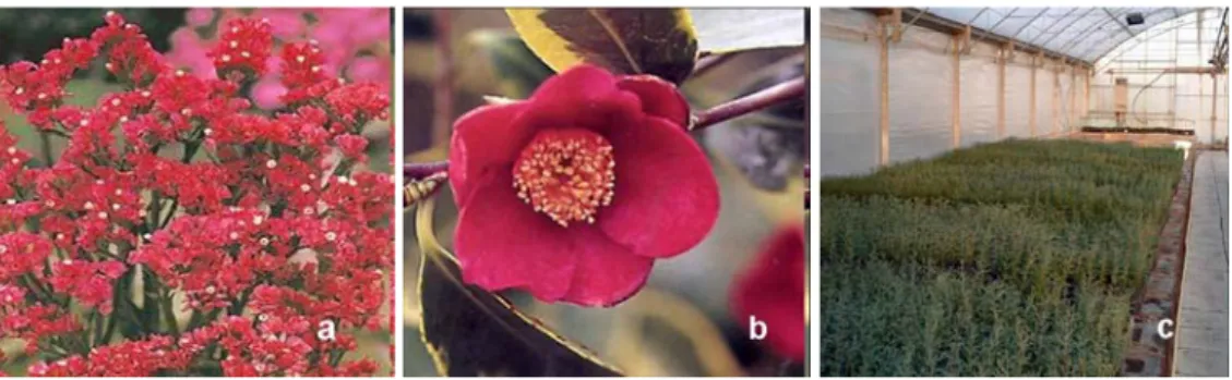
3.1 Fungal isolatesA screening of 162 Trichoderma spp. isolates for Limonium sinuatum (herbaceous plant, reproduced by seed, with a short “cycle”) and Cupressus sempervirens (reproduced by seed, with a medium “cycle”) and 202 for Camellia japonica cv Margherita and cv Woronzoff (an acidiphilous plant, reproduced by cuttings, with a long “cycle”), all included within a larger fungal collection maintained by the Department of Agriculture, Environment and Food (University of Pisa) was performed, in order to select endophytes and growth promoters of the three chosen species (Fig. 1)
Fig. 1. Flowers of Limonium sinuatum (a), Flower of Camellia japonica (b), greenhouse cultivation of Cupressus sempervirens (c).
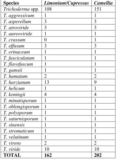
The fungal isolates, belonging to more than 20 different species of Trichoderma Fig. 2) are shown in Tab. 1.
Tab. 1. Trichoderma spp. isolates used for the screening on Limonium,
Cupressus and Camellia.
Species Limonium/Cupressus Camellia
Trichoderma spp. 108 151 T. aggressivum 1 1 T. asperellum 3 3 T. atroviride 3 3 T. aureoviride 1 1 T. crassum 0 1 T. effusum 3 3 T. erinaceum 1 1 T. fasciculatum 1 1 T. flavofuscum 1 1 T. gamsii 1 1 T. hamatum 2 2 T. harzianum 13 9 T. helicum 1 1 T. koningii 4 4 T. minutisporum 1 1 T. oblongisporum 1 1 T. polysporum 1 1 T. saturnisporum 1 1 T. sinensis 1 1 T. stromaticum 1 1 T. velutinum 1 1 T. virens 2 2 T. viride 10 10 TOTAL 162 202
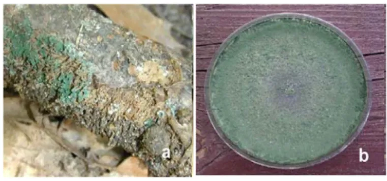
Fig. 2. Morphological aspect of Trichoderma spp. in the environment (a) and on agar plate (b).
These fungi were isolated from different matrices such us agricultural soil, natural parks soil, desert sand, peat, compost, plant parts, seeds, decaying organic matter, animal pellets, tree bark or unusual substrates such as Chernobyl Nuclear Power Plant sarcophagus, ant nest or mummy skin. The isolates are of different geographic origins such as Europe (largest part), North Africa, North and South America, Middle and Far East, Australia and New Zealand, mostly from temperate regions.
3.2 Fungal inoculum
In order to reduce the percentage of peat in new substrates for ornamental plants and set up a fermentation procedure to prepare Trichoderma inoculum, a preliminary survey was performed to assess the effect of the addition of an organic residue of processed barley, the Biomax, at different concentrations in the peat-based substrate usually used, on the germination of Limonium and Cupressus. Among all different tested combinations, substrate containing 90% of peat added by 10% of Biomax showed the highest percentage and a lower time of germination for both species and was used for all the fungal screening procedures.
Biomax resulted also a suitable growth substrate for the fermentation of Trichoderma spp. to prepare the fungal inoculum for peat/Biomax substrate.
Fungal inoculum was prepared in glass jars containing 40 g of Biomax, 5 mL of water and 1 mL of conidial suspension (approximately 106 spore mL-1). Inoculation of the fungus occurred 10 days before its addition to peat and jars were incubated at 24°C, photoperiod 12h/12h light/dark. After incubation, fungal inoculum was added to the peat at the final concentration of 10% inoculated Biomax, 90% peat (Fig. 3).
Fig. 3. Fungal inoculum (a), fermentation of inoculated Biomax (b) and mix of peat (90%) and Biomax (10%).
3.3 Screening of Trichoderma isolates as endophytes and growth promoters of Limonium, Cupressus and Camellia
- Limonium sinuatum and Cupressus sempervirens
The screening procedure provided for 4 rounds of tests for Limonium sinuatum and 3 rounds for Cupressus sempervirens in order to select the best Trichoderma isolates.
In the first round, 162 Trichoderma isolates were tested. Limonium and Cupressus were sown in 160 holes plateau, with 2 replicates for each thesis, 24 seeds (holes) for replicate. Peat inoculated with Biomax (10%) previously fermented with each
Trichoderma isolate as described before, was used for inoculated thesis; 100% peat and peat added with 10% not fermented. Biomax were used as uninoculated controls. Plateaus were kept in a growth chamber at 15°C until at least a single plateau reached a pre-established percentage of germination (30% for Limonium and 20% for Cupressus), then all the plateaus were moved to the greenhouse. Percentages of germination were periodically registered until the number of germinated seeds didn’t change with time. Percentages of germination and time of germination was submitted to statistical analysis assuming P<0.05 as significant level (ANOVA). All thesis with values equal or higher than control (90% peat + not fermented Biomax 10%) were selected (Fig. 4).
Fig. 4. Plateaus in a growth chamber (a) and Cupressus seeds germination (b).
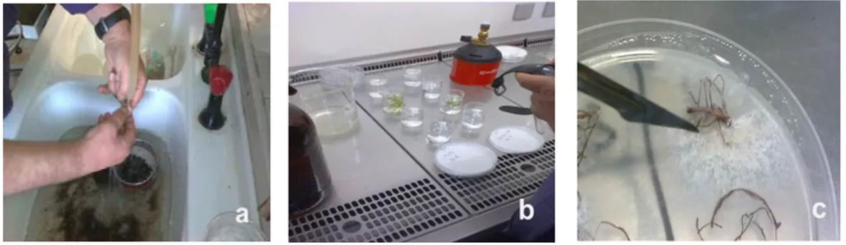
Six plants from each replicates of every selected thesis were collected (after 1 month for Limonium and 3 months for Cupressus); roots were excised, washed, superficially sterilized in an aqueous solution of NaClO (1% active Chlorine) and Ethanol (50%) for 5 minutes and rinsed three times in distilled sterilized water. Three small radical portions for each seedling were plated on P190, a Trichoderma semi-selective agar medium (PDA added with Plantvax 190 ppm, Streptomycin 50 ppm, Bacitracin 7500 u.i. l-1 and Hymexazol 0.3 g l-1). Plates were incubated at 24°C, 12h/12h darkness/light and percentages of endophytism were assessed on the basis of radical portions colonized by each Trichoderma spp. isolate. Colonies grown up from sterilized roots were isolated on PDA and submitted to
monoconidial cultures. Resulting colonies were submitted to a phenotypic analysis to compare their morphology with those of the inoculated strains (Fig. 5).
Fig. 5. Wash of roots (a), sterilization (b) and development of Trichoderma spp. Isolates from plated radical portions (c).
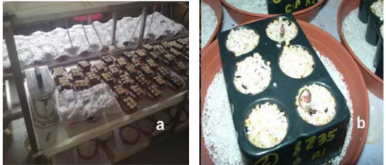
Trichoderma isolates showing 100% of endophytism were submitted to a second round of screening that was performed by a farmer, with an automatic seeder, in order to exactly simulate the usual commercial procedure of Limonium and Cupressus cultivation in nursery. 240 seed x 4 replicates were used for each thesis (inoculated thesis and uninoculated controls); endophytism and different growth parameters (leaves number, leaf area and dry weight for Limonium and height for Cupressus) were recorded, in order to confirm the endophytic fitness and the biostimulation effects of the selected strains (Fig. 6).
Fig. 6. Plateaus of Cupressus (a) and automatic seeder (b).
According to results deriving from this experiment, the best Trichoderma isolates, for each plant species, were chosen for a third round of screening, following the same experimental procedure as previously described. From this third round two isolates for Limonium and two isolates for Cupressus, confirming the 100% of endophytism and showing promising activity in terms of growth stimulation, were finally selected. A fourth test using the two selected isolates was performed for Limonium.
- Camellia japonica
The Camellia screening procedure provided for only 2 rounds of experiments, due to the long production cycle of this ornamental species. In the first round, 202 Trichoderma isolates were evaluated for their growth stimulating effects. The screening was performed on two different commercial cultivars, Margherita and Woronzoff. Ten days before transplanting in pots, 2 ml of a conidial suspension (approximately 106 spore mL-1) of each Trichoderma was inoculated into the rolls
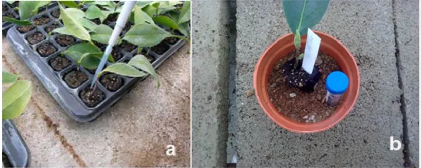
of turf of each plant. In addition, every Trichoderma isolates was fermented on Biomax for ten days, according to the protocol described for Limonium and Cupressus. At the transplant, fermented Biomax was added to peat at the final concentration of 10%. One cutting x isolate x cultivar was transplanted. Uninoculated cuttings transplanted in 100% peat and in 90% peat + 10% Biomax were used as uninoculated controls. During the cultivation cycle, the height of the stem and the number of leaves were measured as plant growth parameters. After 1 year, fungal isolates inoculated in plants showing height and number of leaves higher than in controls for both cultivars were chosen for a second round, to confirm the biostimulation effect and to evaluate the eventual endophytic activity (Fig. 7).
Fig. 7. Inoculation of conidial suspension into the roll of turf (a) and transplanting of Camellia into peat + inoculated Biomax (b).
The second round of screening was performed only on cv Margherita using 20 cuttings for 3 replicates for each thesis according to the same protocol as described for the first screening. The height of the stem and the number of leaves were measured at the beginning and at the end of the trial (one year) and data were submitted to statistical analysis (ANOVA) assuming P<0.05 as significant level. In order to evaluate the endophytic ability of the isolates, roots were excised, washed, superficially sterilized in an aqueous solution of NaClO (1% active Chlorine) and Ethanol (50%) for 5 minutes and rinsed three times in distilled sterilized water. and 50 small radical portions from each root were plated on P190. Plates were incubated at 24°C, 12h/12h darkness/light and percentages of endophytism were assessed on the basis of radical portions colonized by each Trichoderma isolate. Phenotypic analysis on monoconidial cultures of Trichoderma isolates developed from Camellia roots was performed as described for Limonium and Cupressus (Fig. 8).
Fig. 8. Camellia cuttings (a) and Camellia plants after 1 year (b) in nursery.
3.4 Evaluation of selected Trichoderma as inoculants and growth promoters for Cupressus sempervirens and Camellia japonica
At the end of all the concurrent screenings, T. asperellum 2046 resulted to be the most interesting isolate in terms of endophytism and plant growth promotion for Limonium sinuatum and Cupressus sempervirens and only plant growth promotion for Camellia japonica. In addition, T. viride 8238 showed interesting results in Cupressus. In order to follow the effects of T. asperellum 2046 and T. viride 8238 on Cupressus plants growth and of T. asperellum 2046 on Camellia plants growth, a further experiment was performed.
The experiment was performed by using peat added with Biomax, uninoculated or inoculated with Trichoderma isolates.
Fungal inoculum for Cupressus, was prepared in bags containing 800 g of Biomax, 100 mL of water and 20 mL of conidial suspension (approximately 106 spore mL
-1). Inoculation of the fungus occurred 10 days before addition to peat and bags
were incubated at 24°C, photoperiod 12h/12h light/dark. After incubation, fungal inoculum was added to the peat at the final concentration of 10% fermented Biomax, 90% peat.
After three months from sowing, plants of Cupressus were transplanted in pots (7x7x10 cm) with peat + Biomax inoculated or uninoculated, 15 plants for replicate, 4 replicates for thesis. Plant height was recorded once per month.
In Camellia, ten days before rooted cuttings transplanting in pots, 2 ml of a conidial suspension (approximately 106 spore mL-1) of each Trichoderma was inoculated into the rolls of turf of each cutting. In addition, every Trichoderma isolates was fermented on Biomax for ten days, according to the protocol described for Cupressus. At the transplant, fermented Biomax was added to peat at the final concentration of 10%.
Five months old Camellia cuttings (cv Margherita and cv Sea Foam) were transplanted in 8 pots (12cm diameter) with peat + Biomax inoculated or uninoculated, with 3 replicates for the first cv and 6 replicates for the second cv for each thesis. Plant height and the number of leaves per plant was recorded once per month.
At the fourth month, stomatal conductance (Gs), net photosyntesis (Pn), transpiration (Tr), internal CO2 concentrations (Ci) and Water Use Efficiency
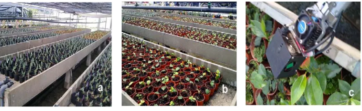
(WUE) were evaluated by a CIRAS equipment (Fig. 9) in both species of Camellia. All data have been subjected to analysis of variance (Anova) to evaluate the beneficial action of Trichoderma isolates on Cupressus and Camellia plants.
Fig. 9. Greenhouse experiments: Cupressus (a) and Camellia (b), equipment (CIRAS) for net photosynthesis measurement (c).
3.5 Identification of Trichoderma spp. isolates
Molecular identification of Trichoderma isolates considered interesting after the first part of screening procedures was based on DNA sequencing of the ribosomal ITS region. Mycelia were harvested after 2–4 days growth on PDA at 24°C and genomic DNA was isolated using the DNeasy Plant DNA extraction kit (Qiagen Inc., Valencia, CA, USA) following the manufacturer’s protocol.
Amplification of nuclear rDNA, containing the ITS1 and 2 and the 5.8S rRNA gene was done using primers ITS1 (5’- TCCGTAGGTGAACCTGCGG-3’) and ITS4 (5’- TCCTCCGCTTATTGATATGC-3’) (White et al., 1990), in a final volume of 50 µl by mixing 2 µl of DNA with 0.5 µM of each of the primers and 25 µl of 2x PCR Master Mix (Promega, Madison, WI, USA). Amplifications were conducted with an initial denaturation of 1 min at 94° C followed by 30 cycles of 30 sec denaturation at 94° C, 1 min primer annealing at 54°C, 1 min extension at 72° C and a final extension of 4 min at 72°C. Template DNA for sequencing was prepared directly from PCR products with the QIAquick PCR purification kit (Qiagen Inc.,Valencia, CA, USA). Both strands were sequenced for each isolate using both the forward and reverse primers.
Sequence identities were determined using both the different tools available online from the International Subcommission on Trichoderma and Hypocrea (ISTH, www.isth.info): TrichOKEY v.2.0 based on an oligonucleotide barcode within the ITS1 and ITS2 sequences (Druzhinina et al. 2005; Kopchinskiy et al. 2005). In some cases, BLAST analyses were also performed from the National Centre for Biotechnology Information (NCBI) available online.
3.6 Antagonistic and mycoparasitic activity of selected Trichoderma isolates by in vitro test.
T. asperellum 2046 and T. harzianum 8227, resulting to be the most interesting as endophyte of Limonium, were submitted to antagonistic tests against Rhizoctonia solani, Botrytis cinerea and Colletotrichum gleosporioides. Antibiosis and mycoparasitism were evaluated on PDA (Potato Dextrose Agar, 39 gl-1, Difco) and WA (Water Agar, agar 20 g l-1, Difco), respectively.
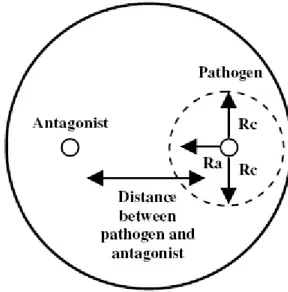
PDA disks of 6 mm diameter, cut from the edge of actively growing colony of each antagonist and pathogen, were placed at the opposite sides (at 4.5 cm each other) on PDA plates and on a sterile cellophane membrane laid on WA (Fig. 10).
Fig. 10. Confrontation plates for antagonistic tests.
Plates were incubated at 24 ± 2°C with 12 h/12 h darkness/light cycles. Radii of each pathogen approaching (Ra) and not approaching (Rc) the colony of antagonists and the distance between the two fungi were measured on PDA three times a day until the two colonies came in contact. Values were used to create
growth curves (Sigmaplot 10) and radial growth data were submitted to analysis of variance of regression in order to compare the slope and the elevation of curves in presence/absence of the antagonist, assuming P < 0.05 as a significant level.
Mycoparasitism was evaluated borth on PDA and WA plates. On PDA, after 14 days, overgrowth and sporulation of the antagonists on pathogens’ colonies were assessed. On WA plates, interaction zones and overlapping regions for each antagonist/pathogen combination were analysed by microscopic investigations and coilings and short loops around the host hyphae were recorded.
On the basis of its behaviour in in vivo biocontrol test (described in the following paragraph), T. asperellum T2046 was also used in a further in vitro test, aimed to evaluate mycoparasitic activity against sclerotia of Sclerotinia sclerotiorum and S. minor in a 24 wells microplate. Microplates containing PDA were inoculated with T2046 and incubated for one week at 24°C, 12h/12H light/darkness. Four sclerotia of each pathogen were sown in each well, a row of six wells was considerate as a replicates (24 sclerotia for replicate, 4 replicates). After 7 days (S. minor) and 14 days (S. sclerotiorum) of incubation in presence of the antagonist, firmness of sclerotia was evaluated by pressure. Hard sclerotia were surface sterilized in an acqueous solution of NaClO (1% active chlorine), in 50% ethanol for 5 minutes, washed in distilled sterilized water for three times, blotter-dried and plated on PDa in order to evaluate the ability of T2046 to internally colonize the resting stuctures (Fig. 11).
Fig. 11. Sclerotia of S. sclerotiorum (a), sclerotia of S. minor (b) and microplate test (c).
3.7 Biocontrol of Sclerotinia sclerotiorum, Sclerotinia minor and
Rhizoctonia solani by T. asperellum 2046 on Limonium by in vivo test
The ability of T. asperellum 2046 to reduce the attacks of Sclerotinia sclerotiorum, Sclerotinia minor and Rhizoctonia solani to Limonium plants was evaluated.
Biomax was inoculated with a conidial suspension of T2046 spp. as previously described and incubated for one week at 24°C, photoperiod 12h/12h light/dark. In parallel, 450g of peat were inoculated with 0.2g of sclerotia. Fermented Biomax was added to the peat at the final concentration of 10% and after two days Limonium seeds were sown. Four thesis were evaluated: i) Peat + Biomax (PB) as uninoculated control; ii) Peat + Biomax inoculated with T2046 (PB2046); iii) Peat inoculated with Sclerotinia spp. + Biomax inoculated with T2046 (PBS2046) and iv) Peat + Biomax, inoculated with Sclerotinia spp. (PBS). 6 replicates for each thesis, 16 seeds for replicate were arranged (Fig. 12).
Rhizoctonia solani was inoculated on millet seed and incubated for fourteen days at 24°C, while Biomax was inoculated with a conidial suspension of T2046 spp. as previously described. After ten days six millet seeds inoculated with R.solani were added to the peat with fermented Biomax. Four thesis were evaluated: i) Peat + Biomax (PB), as uninoculated control; ii) Peat + Biomax inoculated with T2046 (PB2046); iii) Peat inoculated with R. solani + Biomax inoculated with T2046
(PBR2046) and iv) Peat + Biomax, inoculated with R. solani (PBR). 6 replicates for each thesis, 16 seeds for replicate were arranged.
Percentages of emergence were periodically registered until the number of seedlings ceased in increasing (after 1 month) and submitted to statistical analysis (ANOVA).
4. RESULTS
4.1 Screening of Trichoderma isolates as endophytes and growth promoters of Limonium, Cupressus and Camellia
- Limonium sinuatum and Cupressus sempervirens
At the end of the preliminary screening aimed to find Trichoderma spp. isolates able to colonize, as endophytes, root apparatus and possibly stimulate plant growth, 10 out of 162 strains for Limonium and 9 out of 162 strains for Cupressus were chosen and submitted to successive round of experiments. These isolates were selected because of their ability to colonize 100% of internal root tissues of inoculated plants. In the second round of screening performed on Limonium, 5 isolates (T3148, T5961, T8227, T8233 e T8245) shown percentages of endophytism up to 95%.with T. asperellum T2046 and T. harzianum T8227 as the most interesting. These last two isolates were chosen for a further analysis and they resulted able to confirm 100% of root colonization after a third and a fourth experiment, also showing interesting plant growth promotion ability (Tab. 2). T2046 caused a statistically significant improvement in leaves number and leaf area whereas T8227 resulted in significant improvement of leaf area (Fig. 13). At this growth stage no significant differences in dry weight could be detected. Tab. 2. Effects of T. asperellum T2046 and T. viride T8227 on Limonium after 1 month of growth. Thesis Leaves number Leaf area (cm2) Dry weight (g) Control 7.9b* 6.2c 4.1a T2046 9.6a 7.9a 3.7a T8227 7.5b 7.3b 3.6a
*At different letters, within the same column, correspond values statistically different (ANOVA, P<0.05).
Fig. 13. Effects of selected endophytic Trichoderma isolates on Limonium sinuatum plants. T.
asperellum T2046 (a), T. harzianum T8227 (b) and uninoculated control (c).
On Cupressus, among the nine selected Trichoderma isolates, six resulted to be very interesting during the second round of selection (T8139, T8144, T8234, T8235, T8238 and T2046) with T. asperellum T2046 and T. viride T8238 as the most interesting for further analysis. These two Trichoderma isolates showed 100% of endophytism in a third test. In the same experiment they resulted also able to increase plant growth after 3 months (Tab. 3). In details, T2046 was able to significantly improve the height of Cupressus plants whereas a small improvement, but not statistically significant, was registered for T8238 (Fig. 14).
Tab. 3. Effect of T. asperellum T2046 and T. viride T8238 on Cupressus after 3 months of growth.
Thesis Height (cm)
Control 2.2b*
T2046 3.1a
T8238 2.7ab
*At different letters, within the same column, correspond values statistically different (ANOVA, P<0.05).
Fig. 14. Effects of T. asperellum T2046 on Cupressus sempervirens (A). In (B) uninoculated control. Plants growth after 5 months in trays, just after transplanting in 7x7x10 pots.
- Camellia japonica
On Camellia, after the first round of selection, lasted for one year, 8 isolates (T4762, T7630, T7660, T7664, T7666, T7677, T7785 and T8111) were chosen on the basis of their ability to improve height and number of leaves in both cultivars and employed for a second round of screening. In this test also T. asperellum T2046 was included, on the basis of the positive effects registered both on Limonium and Cupressus.
After one year of cultivation, T2046 resulted to be the most interesting isolate among those used for the second screening, causing a statistically significant improvement of plant growth, as shown in Tab. (Fig. 15). An increase, but not statistically significant, was registered also for leaves number.
Tab. 4. Effect of Trichoderma on Camellia plant growth after 1 year.
Thesis Height (cm) Leaves (number) Control 17.7b 10a T2046 28.9a 16a
*At different letters, within the same column, correspond values statistically different (ANOVA, P<0.05).
Fig. 15. Effect of T. asperellum T2046 (A and D) on stem height (a) and root development (b) of
Camellia. Uninoculated control plants (B and C).
Concerning endophytic activities, any Trichoderma isolates, including T2046, developed on the semiselective medium P190, showing no ability to persist into the radical tissues of Camellia after one year.
A B C D
a b
A B C D
4.2 Evaluation of selected Trichoderma as inoculants of innovative
substrates for Cupressus sempervirens and Camellia japonica
-
Cupressus sempervirensData registered during the first 5 months of growth, showed that T2046 had a positive effect on stems growth of Cupressus (Fig. 16a).
From the analysis of variance of regression on growth values, T2046 was able to significantly improve the height of plants compared to control (Pslope 0.006). In
(Fig. 16b) the effects of T. asperellum T2046 are shown.
Different situation occurred for plants grown in soil inoculated with T8238. The growth rate was not significantly higher than control as show by analysis of variance of regression (Pslope=0.928 and Pelevation =0.151).
Fig. 16. Growth rate of plants grown in soil inoculated with T2046 and T8238, compared with control (a). Effect of T2046 (b) on Cupressus (A, treated; B, control).
- Camellia japonica
The presence of T2046 in soil resulted in beneficial effects on growth of both cultivars of Camellia, as shown in (Fig. 17). T. asperellum T2046 was able to increase growth of cv Margherita and cv Sea Foam (Fig. 18 and Fig.19).
Fig. 17 Effect of T2046 on growth of Camellia cv Margherita (A-B) and cv. Sea Foam (C-D). Untreated controls (A-C).
Fig. 18. Growth rate, expressed as stem height of Camellia cv Margherita (a) and cv Sea Foam (b).
Physiological parameters evaluated by CIRAS confirmed the beneficial effect of T2046 on both Camellia cultivars. When inoculated in soil used for cv Margherita, the antagonist was able to increase net photosynthesis (Pn) and the Water Use Efficiency (W.U.E.) as showed in Tab. 5.
Tab. 5. Effect of T2046 on physiological parameters (cv Margherita)
Thesis Pn CI Gs Tr W.U.E. Control 3.57 b 255.00a 85.73a 1.68a 2.18 b
T2046 4.52 a 257.60 a 92.60 a 1.69a 2.90 a
*At different letters, within the same column, correspond values statistically different (ANOVA, P<0.05). Pn = net photosynthesis (µmol CO2 m-2s-1); Gs = stomatal conductance
(mmol CO2 m-2s-1); Tr = transpiration (mmol H2O m-2s-1); CI = internal co2 concentrations
(ppm); W.U.E = water use efficiency (Pn/Tr, (Pn/Tr, µmolCO2/mmol H2O).
On cv Sea Foam all analyzed parameters but transpiration, showed value significantly higher in plants grown in presence of T2046, as shown in Tab. 6.
Tab. 6. Effect of T2046 on physiological parameters (cv Sea Foam)
Thesis Pn CI Gs Tr W.U.E. Control 2.39 b 240.53b 48.77b 1.07a 2.22 b
T2046 3.44 a 262.87 a 56.70 a 1.15a 3.05 a
*At different letters, within the same column, correspond values statistically different (ANOVA, P<0.05). Pn = net photosynthesis (µmol CO2 m-2s-1); Gs = stomatal conductance
(mmol m-2s-1); Tr = transpiration (mmol H
2O m-2s-1); CI= internal co2 concentrations
4.3 Identification of Trichoderma spp. isolates
With the aim of checking the taxonomic position of the Trichoderma spp. isolates preliminary selected on the basis of the endophytic activity on Limonium, Cupressus and Camellia, DNA sequencing of the ribosomal ITS region was performed. Amplified fragments of almost 600 bp (Fig. 20), obtained by PCR, were purified and submitted to sequencing and resulting sequences were compared with those deposited in databases (Genebank and Trichokey). In Tab. 7 results from molecular identification are reported.
Fig. 20. Amplified ITS fragments of Trichoderma spp. obtained by PCR.
- 1000 bp - 750 bp - 500 bp - 1000 bp - 750 bp - 500 bp
Tab. 7. Molecular identification, based on ITS sequences, of Trichoderma isolates preliminary selected for endophytic abilities on Limonium, Camellia and Cupressus.
Isolate Morphological Identification
Molecular Identification
T8233 T. atroviride H. atroviridis/ T. atroviride
T8227 T. harzianum H. lixii/ T. harzianum
T3148 T. harzianum H. lixii/ T. harzianum
T8245 NI Clade XII
T5961 T. harzianum H. lixii/ T. harzianum
T8139 T. atroviride H. atroviridis/ T. atroviride
T8235 T. atroviride H. atroviridis/ T. atroviride
T8234 T. harzianum H. lixii/ T. harzianum
T8144 T. viride H. rufa/ T. viride
T8238 T. viride H. rufa/ T. viride
T8111 T. viride H. rufa/ T. viride
T7785 NI T. asperellum
T7677 T. viride H. rufa/ T. viride
T7666 T. viride H. rufa/ T. viride
T7664 T. viride H. rufa/ T. viride
T7660 NI T. asperellum
T7630 T. viride H. rufa/ T. viride
T4762 T. crassum H. crassa/ T. crassum
4.4 Antagonistic and mycoparasitic activity of selected Trichoderma isolates by in vitro tests
Antagonistic tests were performed in order to investigate the ability of T2046 and T8227 isolates to in vitro inhibit growth of R. solani, B. cinerea and C. gleospoiroides, potential pathogens of Limonium. Growth rates of each pathogen/antagonist combination were submitted to regression analysis in order to compare slope and elevation of pathogen’s growth curves in presence/absence of the antagonist. All growth slopes resulted to be highly significant (R2>0.96, P<0.0001).
Comparison of slopes shown that T2046 was not able to statistically reduce growth rate of any pathogens (Pslope >0.069), whereas significant P elevation resulted from
all combinations, suggesting a delay in starting the exponential growth phase by the pathogens in presence of T2046 (Fig. 21).
When growth curves of the pathogens grown in presence of T8227 were submitted to regression analysis, a similar behaviour emerged. The isolate was not able to inhibit any pathogen, as shown by P slope values (Pslope >0.118). A highly
significant P elevation resulted only for Colletotrichum (P=0.000), underlining a delay in starting the exponential growth phase for this pathogen (Fig. 22).
The lack of significance in slope differences suggests the exclusion of the involvement of antibiotic diffusible compounds as potential biocontrol mechanism by T. asperellum T2046 and T. harzianum T8227 towards the three pathogens.
Fig. 21. Growth curves of R. solani (a), B. cinerea (b) and C. gleospoiroides (c) in presence/absence of T. asperellum T2046. Time (h) 0 10 20 30 40 50 60 70 Ra dial gr owth (mm) 0 5 10 15 20 25 30 R. solani R. solani vs T2046 Time (h) 0 10 20 30 40 50 60 70 Ra di a l gr o w th (m m) 0 5 10 15 20 25 30 B. cinerea B. cinerea vs T2046 Time (h) 0 10 20 30 40 50 60 70 Ra di a l gr o w th (m m) 0 5 10 15 20 25 30 C. gleospoiroides C. gleosporioides vs T2046 Pslope=0.322 Pelev=0.046 Pslope=0.069 Pelev=0.000 Pslope=0.195 Pelev=0.004
a
b
c
Time (h) 0 10 20 30 40 50 60 70 Ra dial gr owth (mm) 0 5 10 15 20 25 30 R. solani R. solani vs T2046 Time (h) 0 10 20 30 40 50 60 70 Ra di a l gr o w th (m m) 0 5 10 15 20 25 30 B. cinerea B. cinerea vs T2046 Time (h) 0 10 20 30 40 50 60 70 Ra di a l gr o w th (m m) 0 5 10 15 20 25 30 C. gleospoiroides C. gleosporioides vs T2046 Pslope=0.322 Pelev=0.046 Pslope=0.069 Pelev=0.000 Pslope=0.195 Pelev=0.004a
b
c
Fig. 22. Growth curves of R. solani (a), B. cinerea (b) and C. gleospoiroides (c) in presence/absence of T. harzianum T8227. Time (h) 0 10 20 30 40 50 60 70 Radi al growt h (mm) 0 5 10 15 20 25 30 R. solani R. solani vs T8227 Time (h) 0 10 20 30 40 50 60 70 Radi al growt h (mm) 0 5 10 15 20 25 30 B. cinerea B. cinerea vs T8227 Time (h) 0 10 20 30 40 50 60 70 Radi al growt h (mm) 0 5 10 15 20 25 30 C. gleosporioides C. gleosporioides vs T8227 Pslope=0.118 Pelev=0.000 Pslope=0.124 Pelev=0.112 Pslope=0.554 Pelev=0.162
a
b
c
Time (h) 0 10 20 30 40 50 60 70 Radi al growt h (mm) 0 5 10 15 20 25 30 R. solani R. solani vs T8227 Time (h) 0 10 20 30 40 50 60 70 Radi al growt h (mm) 0 5 10 15 20 25 30 B. cinerea B. cinerea vs T8227 Time (h) 0 10 20 30 40 50 60 70 Radi al growt h (mm) 0 5 10 15 20 25 30 C. gleosporioides C. gleosporioides vs T8227 Pslope=0.118 Pelev=0.000 Pslope=0.124 Pelev=0.112 Pslope=0.554 Pelev=0.162a
b
c
Mycoparasitic ability of T2046 and T8227 was expressed as the ability to produce coilings around pathogens hyphae. Both Trichoderma isolates succeeded in coiling R. solani hyphae, whereas no coilings were detected against the other two pathogens (Fig. 23).
Fig. 23. Coilings of T. asperellum T8227 around R. solani hyphae.
Overgrowth and sporulation, on PDA, of the antagonists on pathogens’ colonies were evaluated as further signs of mycoparasitism: T2046 was able to grow and sporulate over R. solani, B. cinerea and C. gleospoiroides colonies whereas T8227 was able to overgrow and sporulate profusely on R. solani and B. cinerea and scarcely on C. gloeosporioides colony (Fig.24).
Fig. 24. Overgrowth and sporulation of T. asperellum T2046 and T. harzianum T8227 on R.
solani, B. cinerea and C. gloeosporioides.
When evaluated for its mycoparasitic activity on sclerotia of S. sclerotiorum and S. minor, T2046 has was able to degrade 100% of resting structures of both the pathogen (Fig. 25), providing evidence that the mycoparassitism of these quiescent structures could be one of the mechanisms for the control of disease incidence (as shown in the next paragraph) on Limonium.
T8227
R. solani B. cinerea C. gleospoiroides
Fig. 25. Microplates test on S. minor sclerotia (a) and valutation of sclerotia after 1 week of incubation (b).
4.5 Effects of T. asperellum 2046 on Sclerotinia sclerotiorum,
Sclerotinia minor and Rhizoctonia solani by in vivo test
Results obtained by in vivo biocontrol test on Limonium, showed that T. asperellum T2046 had a significant effect on Sclerotinia sclerotiorum and Sclerotinia minor (Tab. 8). When in presence of Sclerotinia sclerotiorum in soil T2046 was able to increase the percentage of emergence of Limonium of almost 60% (Fig. 26). When alone S. sclerotiorum caused the death of all seedlings. The same trend occurred in presence of Sclerotinia minor where T2046 was able to increase the percentage of emerged seedlings of more than 30%. In presence of this pathogen alone, only 20% of emergence occurred. The effect of T2046 on Limonium was also confirmed in soil inoculated only with the beneficial fungus that allowed to obtain significant higher emergence of plants.
T. asperellum T2046 had a significant effect also on Rhizoctonia solani (Tab. 9). When in presence of R. solani in soil T2046 was able to increase the percentage of emergence of Limonium of almost 40% (Fig. 27). When alone R. solani showed
b a
only 21% of emergence of Limonium. Also in this test, the effect of T2046 on Limonium was confirmed, in soil inoculated only with the beneficial fungus.
Tab. 8. Biocontrol of S. sclerotiorum and S. minor by T2046 Thesis % emergence S. sclerotiorum S. minor control PB2046 PBS2046 PBS 63.47a 68.67a 61.57a 0.00b 62.52a 62.63 a 54.33 a 19.24 b
*At different letters, within the same column, correspond values statistically different (ANOVA, P<0.05). PB2046 = soil inoculated with T2046; PBS2046 = soil inoculated with T2046 and Sclerotinia spp.; PBS = soil inoculated with
Sclerotinia spp.
Fig. 26. Effect of T2046 on S. sclerotiorum on Limonium. Uninoculated soil used as control (a), soil inoculated with T2026 (b), soil inoculated with T2046 and Sclerotinia minor (c) and soil inoculated with S. minor (d).
Tab. 9. Biocontrol of R.solani by T2046 Thesis % emergence R.solani control PB2046 PBR2046 PBR 72.64a 73.47a 38.92b 21.32c
*At different letters, within the same column, correspond values statistically different (ANOVA, P<0.05). PB2046 = soil inoculated with T2046; PBR2046 = soil inoculated with T2046 and R.solani; PBR = soil inoculated with R.solani
Fig. 27. Effect of T2046 on R. solani on Limonium. Uninoculated soil used as control (a), soil inoculated with T2026 (b), soil inoculated with T2046 and R. solani (c) and soil inoculated with
R. solani (d).
a b c d
5. DISCUSSION
Peat is actually the most basic component of substrates used in nursery, expecially for ornamental plants. For some crops peat is used as it is while, in most cases, it is employed in mixtures with other components. Peats are identified as those materials containing more or less decomposed plant residues, with an ash content below 10%. It is worldwide collected from natural deposits called peat lands. The deeper layers of such deposits are ten thousand years old and can be assigned to the late glacial or post-glacial period, while commonly used peat lands are almost one thousand years old. Due to its characteristics (homogeneity, high water absorption capacity, good aeration, structural stability, a limited nutrient content, pH around 3) sphagnum peat represents the starting material more frequently utilised for the production of substrates (D’Angelo et al., 1992).
In recent years, the price has increased as a consequence of the higher costs of energy for extraction and transport from the producing Countries in Northern Europe or Canada (Rea, 2005). In addition, there is an increasing demand for “Peat free” substrates as a result of the environmentalist campaign against the exploitation of peat lands because of the natural and archaeological value of these areas and the fact that peat is not a renewable resource (Armstrong, 2004).
It should be also noted that, in 2001, the European Commission excluded all substrates containing peat from the release of the Community Eco-Label Mark. In many Countries such as Holland (Armstrong, 2004) and also in our country (Project PROBIORN of Arsia, 2004-06; Project FLORPRO of M.i.P.a.a.f., 2007-10; Project SUBARTIFLOR of M.i.P.a.a.f., 2009-12) projects to find materials alternatives to peat, that combine low cost with optimal physical, chemical and biological properties, have been proposed.
In this optic, marketing investigation underlined the increasing demand of alternative substrates that could solve all these questions, mostly the reduction of peat amount, making new substrates available for ornamental field.
The research activity of the present PhD thesis has been focussed within the wider project “SUBARTIFLOR: Messa a punto di substrati artificiali innovativi per il florovivaismo” project funded by the Italian Ministry of Agricultural, Food and Forestry Policies (MIPAAF). The main aim was to develop an innovative, economical and suitable substrate alternative to peat for cultivation of seed plants (Limonium sinuatum and Cupressus sempervirens) and of acidophilus species (Camellia japonica) of ornamental interest.
The first step for reaching the prefixed scope was to select beneficial fungi belonging to Trichoderma genus to be add as soil inoculants, in order to give an added value to the new substrates but, at the same time, maintaining an ecofriendly approach. Our screening started from a large collection of Trichoderma spp. isolates, belonging to at least 20 different species and isolated from a wide variety of environment spread over different geographic origins, such as agricultural soil, natural parks soil, desert sand, peat, compost, plant parts, seeds, decaying organic matter, animal pellets, tree bark or unusual substrates as Chernobyl sarcophagus, ant nest or mummy skin, from Europe (largest part), North Africa, North and South America, Middle and Far East, Australia and New Zealand, mostly from temperate regions. The choice to start from this large collection derived also by the knowledge that there is a great diversity of useful characters in this fungal genus, and efficient biological control agents or endophytic plant symbionts are usually selected among many, sometimes, hundred or thousands, less active wild strains, as recognized by studies on rhizosphere competence. In fact, most of the investigations have been conducted with elite strains extensively tested for their efficacy in the lab and field. In addition, even selected strains often fully express
their beneficial effects (e.g., disease control, abiotic stress resistance, etc.) only on plants under stress conditions (Lorito et al., 2010).
In the present work Trichoderma spp. isolates were tested on three completely different host plants, two seed species as Limonium sinuatum and Cupressus sempervirens and an acidophilic plant, Camellia japonica, cultivated starting from cuttings. Despite of this wide variety of Trichoderma isolates and physiological diversity of host plants, at the end of the concurrent selection, started from 162 isolates for Limonium and Cupressus and 202 for Camellia, only one, T. asperellum T2046 was able to positively affect growth of all the tested plants. Trichoderma spp. are known for mutualistic relationships with plants (Harman et al., 2004) and many biocontrol strains can interact intimately with roots, colonizing the outer epidermis layers and acting as opportunistic, avirulent plant symbionts (Shoresh et al, 2010;). In the few cases that have been examined thoroughly, Trichoderma
isolates colonize root surfaces sometimes with morphological features reminiscent of those seen during mycoparasitism and hyphae invade the root epidermis
(Harman et al., 2004). Some Trichoderma strains can colonize only local sites on roots, but rhizosphere-competent strains colonize entire root surfaces for several weeks or months (Harman et. al, 2004).
An association between high Trichoderma populations and plant growth promotion was indicated for Trichoderma harzianum treated petunias and chrysanthemums, plants of ornamental interest (Chang et. al., 1986). However, the study did not specifically examine the rhizosphere and also did not consider the temporal dynamics of T. harzianum. Other studies related qualitative observations of
Trichoderma root colonisation and penetration to plant growth promotion (Chacòn et al., 2007) and biocontrol activity (Miranda et al., 2006). Similarly, a significant
correlation between Trichoderma population levels in the rhizosphere and its ability to antagonise Sclerotium rolfsii and improve plant growth was determined for axenically grown tomato plants (Tsahouridou and& Thanassoulopoulos, 2002).