University of Sassari
Department of Biomedical Sciences
INTERNATIONAL PHD SCHOOL IN BIOMOLECULAR AND
BIOTECHNOLOGICAL SCIENCES
XXVIII Cycle
MOLECULAR AND ANTIGENIC CHARACTERIZATION
OF Streptococcus suis serotype 2 ISOLATES IN
CENTRAL VIETNAM
Director:
Prof. LEONARDO A. SECHI
Tutor: Prof. ALBERTO ALBERTI
Co-tutor: VAN AN LE MD. PhD.
PhD thesis of
Dr. HOANG BACH NGUYEN
ABBREVIATION
ITS : 16S–23S ribosomal (r) DNA intergenic spacer region 6-PGD: : 6-phosphogluconate-dehydrogenase
ABC : Ammonium bicarbonate ACN : Acetonitrile
AmyA : Amylopullulanase BBB : blood-brain barrier BHI : Brain Heart Infusion
BMEC : Brain microvascular endothelial cell BMEC : brain microvascular endothelial cells CNS : central nervous system
CPEC : Choroid plexus epithelial cell CSF : Cerebrospinal fluid
GAPDH : Glyceraldehyde-3-phosphate dehydrogenase LPXTG : Leu-Pro-any-Thr-Gly
LTA : Lipoteichoic acid
NET : Neutrophil-extracellular trap PCR : Polymerase chain reaction PSF : peptide-spectrum match SS2 : Streptococcus suis serotype 2 TFA : Trifluoroacetic acid
TABLE OF CONTENTS
ACKNOWLEDGEMENT ABBREVIATION TABLE OF CONTENTS I. ABSTRACT ... 1 II. INTRODUCTION ... 22.1. Streptococcus suis: taxonomy and main biological features ... 2
2.2. Streptococcus suis serotype 2 and meningitis ... 4
2.3. Invasion of Streptococcus suis ... 7
2.3.1. Colonization: adherence and invasion of epithelial surfaces... 7
2.3.2. S. suis survival in blood and dissemination ... 9
2.3.3. Inflammatory activation and septicemia ... 11
2.3.4. Central nervous system invasion and meningitis ... 12
2.4. Epidemiology ... 15
2.5. Diagnosis and treatment of Streptococcus suis meningitis ... 16
III. RESEARCH OBJECTIVES ... 19
VI. MATERIALS AND METHODOLOGIES ... 20
4.1. Ethical approval ... 20
4.2. Media and buffers ... 20
4.3. Sampling ... 21
4.4. Bacterial strain, culture and isolation ... 22
4.5. DNA extraction ... 23
4.6. Capsular serotyping confirmation ... 23
4.7. 16S-23S rRNA intergenic spacer fragment amplication ... 24
4.8. Gene profile analysis ... 25
4.9. Whole protein extracts ... 26
4.10. SDS-PAGE ... 26
4.11. Western immunoblotting ... 26
4.12. Pooled WPE againts pooled patient sera by western immunoblotting ... 27
4.13. 2-D PAGE ... 28
4.14. Band and spot excision and Coomassie-stained destaining ... 29
4.15. In-gel tryptic digestion and extraction of peptides digestion products ... 29
4.16. LC-MS/MS ANALYSIS ... 30
V. RESULTS ... 32
5.1. Species identification ... 32
5.2. Capsular serotype identification ... 33
5.3. 16S-23S Intergenic spacer phylogenetic analysis ... 33
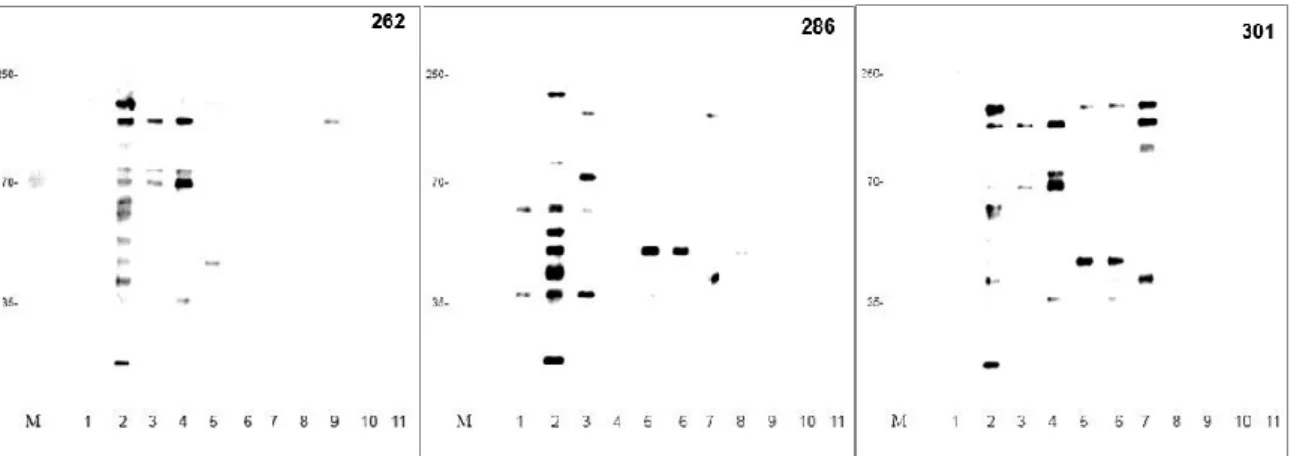
5.4. SDS-PAGE, western immunoblotting and immunogenic protein identification ... 36
5.5. 2-D electrophoresis, western immunoblotting and immunogenic protein identification ... 40
VI. DISCUSSION ... 44
VII. APPENDIX ... 51
Nguyen Hoang Bach - Molecular and antigenic characterization of Streptococcus suis serotype 2 isolates in central Vietnam - Doctorate Thesis of International PhD School In Biomolecular and Biotechnological Sciences, University of Sassari
1
I. ABSTRACT
Streptococcus suis (S. suis) is a Gram-positive bacterium with clinical relevance in pigs and is emerging in human. Based on differences in antigenic properties of the polysaccharide capsule, 35 S. suis serotypes have been distinguished to date, of which serotype 2 is most commonly associated with disease in human and pigs, worldwide. Recently, S. suis emerged as a zoonotic agent in human in contact with infected pigs or with their products(Wertheim et al.
2009)(Wertheim et al. 2009)(Wertheim et al. 2009)(Wertheim et al. 2009). In 2006
a major S. suis outbreak resulted in more than 200 human cases with a fatality rate of nearly 20% in China. More recently, several studies from Thailand, Hong Kong, Taiwan, Singapore and Viet Nam demonstrated that S. suis can cause adult endocarditis, septicemia, arthritis and especially meningitis with high fatality rate and severe neurological sequelae. Multilocus enzyme electrophoresis, restriction endonuclease analysis with HaeIII, ribotyping, repetitive extragenic palindromic (REP) or enterobacterial repetitive intergenic consensus (ERIC), arbitrarily primed PCR, and pulsed-field gel electrophoresis (PFGE) have been used to determine S. suis strains epidemiological relationships. Characterization of the 16S-23S ribosomal (r) DNA intergenic spacer region (ITS) has also been used to compare bacterial strains and to identify species within the genus Streptococcus, including S. suis.
In this study, a specific and sensitive PCR assay for fast detection of S. suis serotype 2 is crucial to meningitis treatment based on the S. suis serotype 2 cps2J
gene was developed and evaluated for diagnosis in meningitis patients hospitalized
in Thua Thien Hue, Viet Nam. The intergenic spacer region was amplified by traditional PCR from strains obtained in this study and sequenced. ITS - based phylogenetic analysis is also presented and discussed. We also identified and characterized the main immunodominant antigens of Streptococcus suis serotype 2 based on the immunoproteomics approach.
Nguyen Hoang Bach - Molecular and antigenic characterization of Streptococcus suis serotype 2 isolates in central Vietnam - Doctorate Thesis of International PhD School In Biomolecular and Biotechnological Sciences, University of Sassari
2
II. INTRODUCTION
2.1. Streptococcus suis: taxonomy and main biological features
In the early 1950s, Jensen et al. (1951) and Field et al. (1954) reported streptococcal septicemia as cause of meningitis and arthritis in pigs and piglets in the United Kingdom and in the Netherlands. Later, de Moor el al. (1963) described similar hemolytic streptococci that had been isolated from septicemic pigs and could be differentiated biochemically and serologically from the known streptococcal species. These streptococci were placed into two Lancefield groups. Strains isolated in newborn pigs were designated “group S”, whereas strains from older pigs were designated “group R” (Staats et al. 1997).
There were reports indicating that some strains of this species contained streptococcal group D antigens. However, later reports indicate that group R and group D antigens are similar but not identical, and observed reaction with group D antisera was due to cross-reaction. S. suis is a very heterogeneous species. So far, 35 capsular serotypes (types 1 to 34 and type 1/2) have been identified based on the difference of antigenic carbohydrate types of their capsule (Staats et al. 1997). The discovery of additional serotypes of this species led to a change in nomenclature used to indicate capsular types. Group R, the most common strain identified, is type 2, and group S is type 1. The important change to clinical microbiologists is that the only serotype identified from human has been type 2 (group R). There are difficulties when using the original method for serological grouping of S. suis with Lancefield procedure. Therefore, it is recommended that a capsular reaction should be used for identification of the various serotypes. This reaction is similar to the quelling reaction used to type S. pneumoniae. A modified Lancefield extraction used to identify type 2 by using group-precipitating antiserum. (Gottschalk et al. 1991, Staats et al. 1997, Facklam 2002).
Nguyen Hoang Bach - Molecular and antigenic characterization of Streptococcus suis serotype 2 isolates in central Vietnam - Doctorate Thesis of International PhD School In Biomolecular and Biotechnological Sciences, University of Sassari
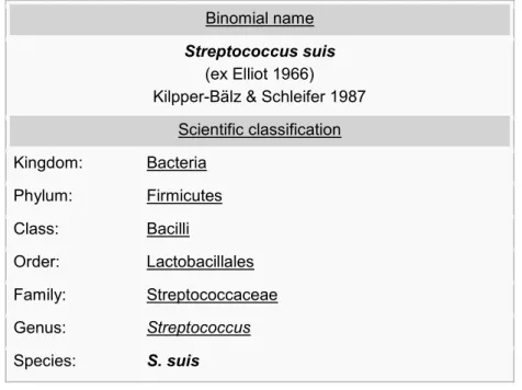
3 Table 1. Binomial nomenclature of Streptococcus suis (NCBI taxonomy ID: 1307)
Binomial name
Streptococcus suis
(ex Elliot 1966) Kilpper-Bälz & Schleifer 1987
Scientific classification Kingdom: Bacteria Phylum: Firmicutes Class: Bacilli Order: Lactobacillales Family: Streptococcaceae Genus: Streptococcus Species: S. suis
Streptococcus suis is a Gram-positive facultative anaerobe that belongs to Lancefield groups R and S. It is α- or β-hemolytic on sheep or horse blood agar, respectively. The organism is coccoid, ovoid, and occurs singly, in pairs or short chains (Fig. 1).
Figure 1. Gram-positive cocci (Streptococcus suis), single or in pairs, visible in Gram stain (original magnification, ×100; left). Scanning electron micrograph (SEM) graphic of
Nguyen Hoang Bach - Molecular and antigenic characterization of Streptococcus suis serotype 2 isolates in central Vietnam - Doctorate Thesis of International PhD School In Biomolecular and Biotechnological Sciences, University of Sassari
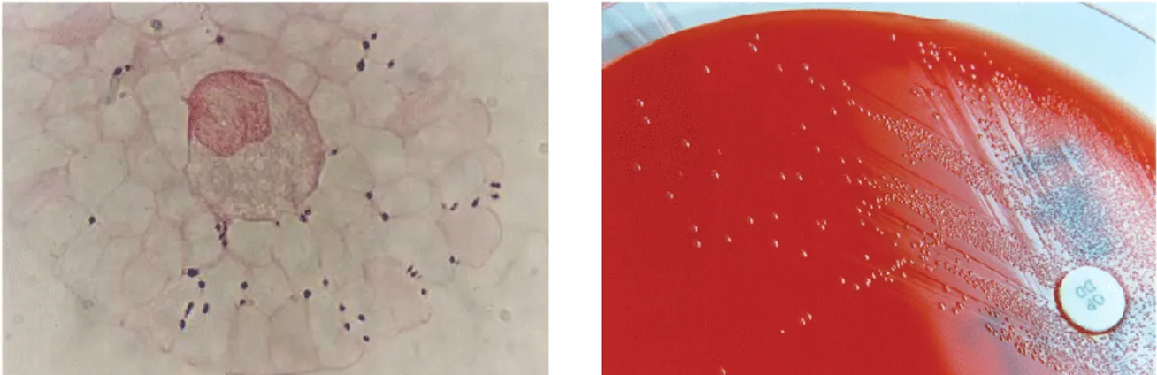
4 Streptococcus suis is one of the most important pathogens impacting the swine industry worldwide as it is responsible for important economic losses. It causes a wide variety of signs in pigs, including meningitis, septicemia and endocarditis. Among the 35 serotypes originally described based on capsular polysaccharide (CPS) antigens, serotype 2 is not only prevalent in swine diseases, but is also considered to be an emerging zoonotic agent causing meningitis and streptococcal toxic shock-like syndrome (STSLS) in human (Gottschalk et al. 2010). Nowadays, S. suis has gained more attention since recent recognition of its high prevalence in human meningitis cases in south-east and east Asia, and reports of outbreaks that resulted in high mortality rates (Wangkaew et al. 2006, Yang et al. 2006, Wangsomboonsiri et al. 2008, Wertheim et al. 2009, Tsai et al. 2012).
Figure 2. Gram-positive cocci (Streptococcus suis), single or in pairs, visible in direct
Gram stain of a CSF sample (original magnification, ×100; left). Growth of S. suis colonies on blood agar with optochin disk (right). (Wertheim et al. 2009)
2.2. Streptococcus suis serotype 2 and meningitis
The most serious form of meningitis is acute bacterial meningitis. Even when treated, bacterial meningitis can be fatal in severe cases. Indeed if bacterial meningitis progresses rapidly, in 24 hours or less, death may occur in more than half of those who develop it, even with proper medical treatment (CDC 2015).
Nguyen Hoang Bach - Molecular and antigenic characterization of Streptococcus suis serotype 2 isolates in central Vietnam - Doctorate Thesis of International PhD School In Biomolecular and Biotechnological Sciences, University of Sassari
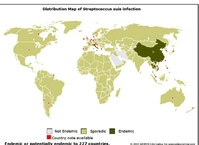
5 S. suis is a swine pathogen and a zoonotic agent afflicting people in close contact with infected pigs or pork meat. Although sporadic cases of human infections have been reported worldwide during the last 45 years, deadly S. suis outbreaks emerged in Asia (Fig 1.3).
Figure 3. Distribution map of S. suis infection (GIDEON Informatics et al. 2015)
Wangkaew et al. (2006) reported that forty-one patients (32 men and 9 women, mean age 51 years) with S. suis infection were identified. Three patients had a history of exposure to pig or pork and one patient had a history of raw beef consumption. Clinical manifestations included infective endocarditis, meningitis, sepsis, spondylodiscitis, and endophthalmitis in 16, 13, 10, 1, and 1 patient, respectively. The overall mortality rate was 19.5%. On univariabe analysis,
Nguyen Hoang Bach - Molecular and antigenic characterization of Streptococcus suis serotype 2 isolates in central Vietnam - Doctorate Thesis of International PhD School In Biomolecular and Biotechnological Sciences, University of Sassari
6
patients, who had low serum albumin, high serum total bilirubin, low platelet, and rapid onset of illness were significantly correlated with high mortality rate. All bacterial isolates were sensitive to penicillin (mean MIC90Z0.027 mg/ml) (Wangkaew et al. 2006).
An outbreak of S. suis serotype 2 occurred in villagers after direct exposure to deceased or sick pigs in Sichuan, China. Prohibition of slaughtering in backyards brought the outbreak to a halt. From June 10th to August 21st, 2005, 68
laboratorial confirmed cases of human S. suis infections were reported. All were villagers who gave an evidence of direct exposure to deceased or sick pigs in their backyards where slaughtering was performed. Twenty-six (38%) presented with toxic shock syndrome of which 15 (58%) died. Other presentations were septicemia or meningitis. A virulent strain of this bacterium is speculated to be in circulation, and is responsible for the unusual presentation of toxic shock syndrome with high case fatality (Yang et al. 2006).
Nguyen et al. (2012) reported that 450 patients were prospectively studied with suspected bacterial meningitis. In patients infected with S. suis, bacterial DNA load at hospital admission and during treatment was analyzed in cerebrospinal fluid (CFS) specimens using quantitative real-time polymerase chain reaction (real-time PCR). S. suis was the most common pathogen and was detected in 33.6% of the patients. Fifty of these 151 patients reported exposure to pigs or pork. Mortality was low but mild to severe hearing loss occurred in 66.4% patients of S. suis infection. S. suis serotype 2 is the most frequent cause of bacterial meningitis in adults in southern Vietnam and is associated with substantial morbidity attributable to hearing loss (Mai et al. 2008).
Nguyen Hoang Bach - Molecular and antigenic characterization of Streptococcus suis serotype 2 isolates in central Vietnam - Doctorate Thesis of International PhD School In Biomolecular and Biotechnological Sciences, University of Sassari
7
Table 2. Common causes of bacterial meningitis by age group (CDC 2015) Age Group Causes
Newborns Group B Streptococcus, Escherichia coli, Listeria
monocytogenes
Infants and Children Streptococcus pneumoniae, Neisseria meningitidis, Haemophilus influenzae type b
Adolescents and Young Adults
Neisseria meningitidis, Streptococcus pneumoniae, Streptococcus suis
Older Adults Streptococcus pneumoniae, Neisseria meningitidis, Listeria monocytogenes
2.3. Invasion of Streptococcus suis
Pigs may get infected by S. suis via both vertical and horizontal transmission. Bacteria can persist in the tonsils of colonized animals and may never develop disease (carrier animals). Conversely, some carrier piglets will eventually develop bacteremia, septicemia and/or meningitis due to dissemination of S. suis from tonsils and/or other mucosal surfaces, usually when maternal antibodies decline. To cause disease, bacteria must breach epithelial barriers, reach and survive in the bloodstream, invade different organs and cause exaggerated inflammation (Madsen et al. 2002, Cloutier et al. 2003).
Humans can get infected through skin lesions or through the oral route, although carriage of S. suis by human without clinical signs (usually slaughterhouse workers) has also been described (Gottschalk et al. 2010). Infection of human may also begin with colonization followed by invasion, bacteremia and septicemia with or without meningitis (Fittipaldi et al. 2012).
2.3.1. Colonization: adherence and invasion of epithelial surfaces
To date, the early mechanisms used by S. suis to colonize the host are poorly known. The bacteria may survive in swine tonsils for long periods of time. The tonsillar lymphoid tissue is overlaid by mucosal epithelium. After adhesion and invasion of epithelial cells in tonsils, the bacteria may escape the host immune systems by hiding in epithelial invaginations within the lymphoid tissue. Clinically
Nguyen Hoang Bach - Molecular and antigenic characterization of Streptococcus suis serotype 2 isolates in central Vietnam - Doctorate Thesis of International PhD School In Biomolecular and Biotechnological Sciences, University of Sassari
8 healthy pigs can carry S. suis in their nasal cavities, tonsils and upper respiratory tract, contributing to the dissemination of this pathogen. Although, S. suis is usually found in very low quantities in tonsils of clinically healthy pigs in herd, it might cross the first natural line of the host defense and initiate disease. This thing can be explained by that the S. suis breaches the mucosal epithelium in the upper respiratory tract of pigs (Torremorell et al. 1998, Cloutier et al. 2003, Fittipaldi et al. 2012).
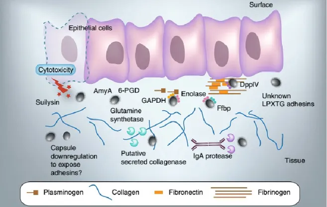
Human may be infected by S. suis via the oral route. Upon infection, bacteria interact with epithelial cells either at the epidermal surface or in the intestine (Wertheim et al. 2009, Gottschalk et al. 2010). The bacterial adhesion and invasion of epithelial cells are usually associated with the first steps of colonization by mucosal pathogens. There are a few mechanistic studies are available regarding the interactions between S. suis and epithelial cells. Virulent S. suis strains can adhere to epithelial cells from the respiratory tract of human (Figure 4) (Norton et al. 1999, Lalonde et al. 2000, Benga et al. 2004).
Figure 4. Interactions of Streptococcus suis with epithelial cells and extracellular matrix proteins (Fittipaldi et al. 2012).
Nguyen Hoang Bach - Molecular and antigenic characterization of Streptococcus suis serotype 2 isolates in central Vietnam - Doctorate Thesis of International PhD School In Biomolecular and Biotechnological Sciences, University of Sassari
9 Several factors involved in adhesion are bacterial surface proteins such as enolase, GAPDH, 6-PGD, AmyA, as well as a glutamine synthetase. Unidentified LPXTG-containing adhesins (including pili) have been suggested to participate in adhesion to epithelial cells. The actual contribution of these factors has not yet been demonstrated.
Invasion of epithelial cells other than choroid plexus epithelial cells by encapsulated S. suis serotype 2 is still controversial. In the case of suilysin-positive strains, expression of this hemolysin may be instrumental in breaching the epithelium. IgA protease-producing bacteria may also take advantage of released Fab fragments after IgA proteolysis to enhance their surface hydrophobicity and thus adhesion to host cells. S. suis binds extracellular matrix proteins such as fibronectin, plasminogen and collagen. Enolase, a fibronectin fibrinogen binding protein, and a dipeptidyl peptidase IV all bind human fibronectin and fibrinogen. Enolase also mediates binding to plasminogen. Also necessary for bacterial-extracellular matrix proteins interactions are LPXTG-containing proteins, since deletion of SrtA impairs S. suis binding to these host factors. Collagen degradation by means of the secretion of a putative collagenase has been proposed. Capsule down regulation upon the interactions could facilitate the display of adhesions (Fittipaldi et al. 2012).
2.3.2. S. suis survival in blood and dissemination
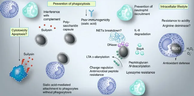
Cell invasion or cell disruption of S. suis may lead to the first step of systemic disease development. It has been proposed that S. suis may gain entry to the systemic circulation primarily through the palatine tonsils, after adhesion and invasion of epithelial cells and interaction with cells of the myeloid lineage. When S. suis reaches deeper tissues and/or the bloodstream, it is subject to the action of phagocytic cells of the innate immune system. Because there are no specific antibodies, S. suis could be resist phagocytosis and persist in blood at high concentrations, with inflammatory consequences. Bacterial survival largely depends on the production of CPS. There were many reports of the role of CPS in survival
Nguyen Hoang Bach - Molecular and antigenic characterization of Streptococcus suis serotype 2 isolates in central Vietnam - Doctorate Thesis of International PhD School In Biomolecular and Biotechnological Sciences, University of Sassari
10 capability of S. suis, which could protect S. suis from neutrophil and monocyte/macrophage-mediated phagocytosis and killing (Fig. 5) (Gottschalk 2011).
Figure 5. Suggested mechanism of avoidance innate immune response of the host (Fittipaldi et al. 2012).
Without phagocytosis, it is found with the phagocytes a mechanism of immune avoidance. Capsular sialic acid is considered as the factor that mediates this attachment. N-deacetylation of the peptidoglycan also reduces killing by neutrophils, probably by providing the bacterium with enhanced resistance against the action of lysozyme. The enhancement of resistance to host antimicrobial peptides and to resistance to neutrophil killing. S. suis through the contribution from D-alanylation of the LTA would bring out a cell wall-anchored DNase, which probably join NETs breakdown. The bacterium also secretes the serine protease SspA, capable of degrading IL-8, a major chemoattractant of neutrophils. It is believed that Secreted suilysin is toxic to phagocytes and also have the influence on complement activity. In order to resist the intracellular environment, SOD and the
Nguyen Hoang Bach - Molecular and antigenic characterization of Streptococcus suis serotype 2 isolates in central Vietnam - Doctorate Thesis of International PhD School In Biomolecular and Biotechnological Sciences, University of Sassari
11
arginine deiminase system can be applied by S. suis in the process of internalization (Fittipaldi et al. 2012).
2.3.3. Inflammatory activation and septicemia
Despite the fact that the activation of the immune system during microbial infection states protective, a consequence of excessive or poorly regulated immune response to the offending organism can be found as a septic shock (Tsiotou et al. 2005). Because human outbreaks of toxic shock-like syndrome and by septic shock cases caused by S. suis (in short incubation period, speedy disease development and a great mortal rate) have been found in Europe and Asia, it is suggested that proinflammatory mediators should be released in the time of S. suis systemic infections (Gottschalk et al. 2010). Therefore, a remarkable biological relevance can be connected to the ability of S. suis to induce cytokine production. The production of various proinflammatory cytokines by porcine, murine and human cells(Segura et al. 2002, Segura et al. 2002, Segura et al. 2004) is proved to be induced by S. suis serotype 2 virulent strains (Segura and Gottschalk 2002, Segura, Vadeboncoeur et al. 2002,Segura, Gottschalk et al. 2004) . It is said that the sudden early death of animals is caused by the high levels of systemic cytokines TNF-a, IL-6 and -12, IFN-g and the chemoattractants CCL2/MCP-1, CXCL1/KC, and CCL5/RANTES observed in vivo within 24 h postinfection.
The regulatory cytokine IL-10 was upregulated following the onset of most proinflammatory cytokines, indicating a negative feedback mechanism to control the extent of the inflammatory response. S. suis-infected mice treated either with neutralizing antibodies against IL-10 or with recombinant IL-10 was revealed from the observations of changes in septic shock. These observations in mice can be seen as the development of the infection from swine pathogen and zoonotic agent to pigs and human and the exactly infected individuals can provide accurate inflammatory cytokines and chemokines level (Vanier et al. 2009, Ye et al. 2009).
Cell receptor recognition probably comes from the Lipoproteins existence in the cell wall (Wichgers Schreur et al. 2010). A supposed prolipoprotein
Nguyen Hoang Bach - Molecular and antigenic characterization of Streptococcus suis serotype 2 isolates in central Vietnam - Doctorate Thesis of International PhD School In Biomolecular and Biotechnological Sciences, University of Sassari
12 diacylglyceryl transferase present in S. suis cell wall is essential for the innate immune activation (Segura et al. 2006, Graveline et al. 2007, Lecours et al. 2011, Wichgers Schreur et al. 2011). Suilysin was shown to activate phagocytes and to induce the release of proinflammatory cytokines (Lun et al. 2003, Segura et al. 2006). In addition, suilysin might release hemoglobin from red blood cells, contributing to raise the levels of proinflammatory mediators by acting in synergy with S. suis cell wall components (Dominguez-Punaro et al. 2007). A surface-associated subtilisin-like protease (SspA) has recently been shown to induce the secretion of different proinflammatory cytokines and chemokines by macrophages. Interestingly, a low concentration of SspA was associated with secretion of high amounts of CCL5, whereas the use of the same protein at high concentrations resulted in low amounts of CCL5 being detected, likely due to a proteolytic degradation of that chemokine by the same SspA (Figure 2) (Bonifait et al. 2011). Similar results had been observed by Vanier et al., who suggested that S. suis can induce an exacerbated release of inflammatory mediators resulting in massive recruitment of leukocytes and subsequent release of inflammatory mediators; however, S. suis may modulate this response, and improve its survival, by actively degrading the chemokines and thus delaying recruitment of neutrophils to the site of inflammation (Vanier et al. 2009).
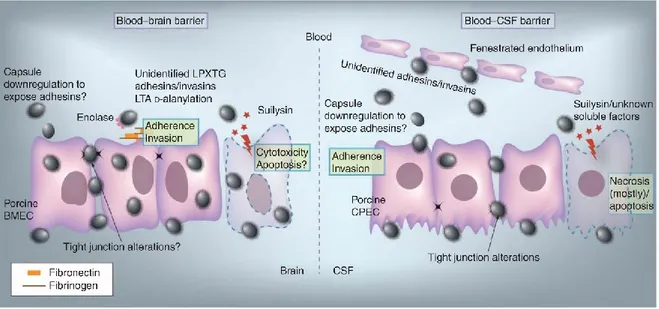
2.3.4. Central nervous system invasion and meningitis
As other blood borne pathogens, S. suis must cross the blood-brain barrier (BBB) and/or the blood-cerebrospinal fluid (CSF)-barrier in order to cause central nervous system (CNS) infections. The BBB is an anatomically and functionally unique barrier that separates the brain from the intravascular compartment and maintains the homeostasis of the CNS environment (Rubin et al. 1999).
The main cellular type of the BBB is brain microvascular endothelial cells (BMEC). Adhesion to, but not invasion of human BMEC has been demonstrated for S. suis (Charland et al. 2000). The join of the CPS is seen dissimilarly despite the fact that bacterial factors related to adhesion are not elucidated (Bonifait et al.
Nguyen Hoang Bach - Molecular and antigenic characterization of Streptococcus suis serotype 2 isolates in central Vietnam - Doctorate Thesis of International PhD School In Biomolecular and Biotechnological Sciences, University of Sassari
13 2010). In contrast, it has been demonstrated that pathogen is able to both attach and to invade immortalized porcine BMEC (Fig. 6), it is proved in antibiotic protection essays and electron microscopy. S. suis survived up to 7 h within porcine BMEC which can be considered as remarkable discovery because the main factor for the growth of meningitis is the capacity of pathogens to get through the BBB in the role of live bacteria (Vanier et al. 2004, Kim 2006). Benga et al. (2005) also showed internalization of S. suis when using the same cell line. However, this study did not consider the number of internalized bacteria to be significant and reported inability of S. suis to invade porcine BMEC (Benga et al. 2005). It might have different summaries because no general criteria have been found for the designation that if bacterial strains invasive or not on the base of the number of internalized bacteria. In fact, some researchers have arbitrarily defined a threshold to define the event as bacterial invasion (Benga et al. 2005). Vanier et al. (2007) also showed bacterial invasion of primary porcine BMEC (Vanier et al. 2007). As suggested for epithelial cells, the CPS of S. suis partially interferes with the adhesion/ invasion abilities of the pathogen, perhaps because it hinders the display of putative adhesins (Fig. 6) (Vanier et al. 2004, Benga et al. 2005). Further characterization of the invasion process suggested the involvement of proteinaceous adhesins/ invasins and cell wall components such as the LTA (Vanier et al. 2007). Mutants impaired in LTA d-alanylation adhered and invaded porcine BMEC to a significantly lesser extent than the wildtype strain (Fittipaldi et al. 2008). A S. suis SrtA mutant strain had reduced capacity to adhere and invade these cells, suggesting that LPXTG cell wall anchor proteins may also serve as adhesins/invasins (Vanier et al. 2008). Serum components may also participate in the interactions between S. suis and porcine BMECs (Vanier et al. 2004, Benga et al. 2005). Among them, only fibronectin was shown to play an important role (Vanier et al. 2007); in addition, antibodies against enolase (an important fibronectin-binding protein in S. suis) significantly decreased adhesion and invasion of porcine BMEC (Esgleas et al. 2008). Suilysin positive strains may also disrupt the BBB through cytotoxic effects, since at high bacterial doses suilysin-positive strains were toxic for porcine BMEC (Fig. 6). However,
Nguyen Hoang Bach - Molecular and antigenic characterization of Streptococcus suis serotype 2 isolates in central Vietnam - Doctorate Thesis of International PhD School In Biomolecular and Biotechnological Sciences, University of Sassari
14 suilysin was not indispensable for invasion, as a suilysin-negative mutant successfully invaded these cells (Vanier et al. 2004).
The blood-CSF barrier CPECs might be another CNS entry portal for S. suis (Fig.6). The blood-CSF barrier still show its significant part in bacterial translocation as well as in leukocyte transmigration despite of its smaller surface area than the BBB. It has recently proved that there is the appearance of in vitro invasion and translocation of S. suis across the blood-CSF barrier (inverted trans well model) (Tenenbaum et al. 2009). This invasion was suggested to involve three potential steps: invasion of porcine CPEC from the basolateral side; transport within membrane-bound endocytic vacuoles to the apical side; and exocytosis onto the apical membrane of the blood-CSF barrier. S. suis adhered and invaded cells better when applied to basolateral membranes (Tenenbaum et al. 2009).
Figure 6. Invasion of the central nervous system.
Proteinaceous adhesins/invasins, and cell wall components such as the lipoteichoic acid (including lipoteichoic acid D-alanylation) have shown its connection to the invasion to and invasion of porcine BMEC. It is also necessary to consider the interaction with host extracellular matrix proteins (such as
Nguyen Hoang Bach - Molecular and antigenic characterization of Streptococcus suis serotype 2 isolates in central Vietnam - Doctorate Thesis of International PhD School In Biomolecular and Biotechnological Sciences, University of Sassari
15 fibronectin/fibrinogen). As suggested for epithelial cells, since the bacterial capsule partially interferes with the adhesion/invasion abilities of the pathogen; a possible down regulation of capsular polysaccharide expression has been suggested. LPXTG cell wall anchor proteins and enolase (through adhesion to fibronectin) may play a role as adhesins/invasins. Suilysin-positive strains may also disrupt the BBB through cytotoxic effects. Invasion and translocation of Streptococcus suis across the blood-CSF barrier has been shown. S. suis adhered better to porcine CPEC when applied to basolateral membranes, suggesting that direct access to the extracellular matrix was required. The capsular polysaccharide clearly compromised bacterial CPEC invasion, as demonstrated by the use of unencapsulated mutants, and indicating that bacterial cell wall components and/or surface proteins are needed. However, the nature of these adhesins/invasins remains largely unknown. S. suis is also able to affect the blood-CSF barrier function and integrity further facilitating trafficking of bacteria and leukocytes. It has been shown that S. suis induce CPEC necrosis, although apoptosis might also play a role in the process of cell death. Although other soluble factors might also be involved, suilysin plays an important role as a toxin affecting the blood-CSF barrier function (Fittipaldi et al. 2012).
2.4. Epidemiology
By the end of 2012, a total of 1,584 cases had been reported in the literature (including 189 probable cases identified in 3 outbreaks), mainly from Thailand (36%), Vietnam (30%), and China (22%). More than half (53%) were in the Western Pacific region; 36% were in the South East Asia region, 10.5% in the European region, and 0.5% in the Americas. The highest cumulative prevalence rate was in Thailand (8.21 cases/million population), followed by Vietnam (5.40) and the Netherlands (2.52) (country population data for 2008-2012 by World Bank). The pooled mean age of the patients was 51.4 years, and 76.6% were men. All case-patients were adults, except 1 female infant reported in Thailand (Vilaichone et al. 2002). The pooled proportion of case-patients with occupational exposure was 38.1%; this proportion was higher for industrialized countries than for other
Nguyen Hoang Bach - Molecular and antigenic characterization of Streptococcus suis serotype 2 isolates in central Vietnam - Doctorate Thesis of International PhD School In Biomolecular and Biotechnological Sciences, University of Sassari
16 countries (83.8% for the United Kingdom, Netherlands, and Japan together) (Vu Thi Lan et al. 2014). Recent contact with pigs or pork was reported for 15.5% of single cases but for 33.9% in the meta-analysis. History of eating meals containing pork was reported mainly in Asia (Thailand and Vietnam); the pooled estimate was 37.3% (95% CI 20.2%-58.3%). For Thailand only, the proportion was 55.8% (95% CI 33.7%-75.9%). In other countries, only 1 patient in France was reported eating artisanal dry sausage, and 1 patient in the United States ate raw pork while traveling in the Philippines before the infection. Skin injury was shown for one fourth of patients, and alcohol use was evident in approximately one third of case-patients. However, a case-control study in Vietnam did not identify alcohol use as an independent risk factor after adjustment for other risk factors and confounders. The most commonly reported preexisting condition was diabetes. Other conditions included underlying heart disease, hypertension, cirrhosis, and cancer. Smoking was mentioned in 5.2% of patients in the single-case dataset. (Vu Thi Lan et al. 2014)
2.5. Diagnosis and treatment of Streptococcus suis meningitis
S. suis diagnosis is hampered by many bacteria caused purulent meningitis, which normally have similar clinical signs that make necessary the use of laboratory tools for rapid and specific detection. To diagnosis S. suis meningitis, CSF examination is the “gold standard” and mandatory. CSF culture is the standard procedure for diagnosis, and it is obligatory to obtain the in vitro susceptibility of the pathogenic bacteria and antibiotic treatment (van de Beek et al. 2006, Brouwer et al. 2010). Characteristic CSF findings for bacterial meningitis consist of polymorphonuclear pleocytosis, hypoglycorrhachia, and raised CSF protein level. CSF culture remains the gold standard for the diagnosis of bacterial meningitis. But sometimes, the yield of CSF culture is usually negative for patients who have received antibiotic pretreatment before lumbar puncture (van de Beek et al. 2006). In a study of McClelland et al. (2007),there were 103 patients with clinically defined meningococcal meningitis, only 13% had positive CSF cultures (McClelland et al. 2007).
Nguyen Hoang Bach - Molecular and antigenic characterization of Streptococcus suis serotype 2 isolates in central Vietnam - Doctorate Thesis of International PhD School In Biomolecular and Biotechnological Sciences, University of Sassari
17 The biochemical property allows S. suis classification on the basis of the metabolism of some molecules, such as esculine, trehalose, glycogen, lactose, saccharose…. Physiological and biochemical tests carried out on multiple serotype of S. suis with API 20 STREP, API 50CH and RAPID ID32STREP galleries (bioMérieux SA, Marcy l'Etoile, France), demonstrated a profile similar to that of entire S. suis serotypes. S. suis strains are postive in esculine, trehalose, glycogen, lactose, sacharose, starch, leucine aminopeptidase, alanine-phenyl-alanine-proline arylamidase tests, while most are noticeably negative in Voges-Proskauer, hipurate, ribose, arabinose and sorbitol tests. S. suis strains are in high percentage positive in arginine dihydrolase, β-glucoronidase, α-galactosidase, β-galactosidase, methyl-β-dglucopyranoside, glycyl-tryptophan arylamidase and inulin tests. The major disadvantage of these methods is that they are time consuming (Chatellier et al. 1998, Stanojković А. 2014).
Latex agglutination is a diagnostic test that has been utilized for the etiological diagnosis of bacterial meningitis, providing results in less than 15 min. A
ready-to-use chessboard system for Streptococcal group and serotyping of S. suis has been developed in form of latex particles coated with group specific streptococcal antiserum (A, B, C, D, F, G or L) or multiple serotype of S. suis antiserum raised in rabbits (0.0975 % sodium azide as preservation). The limitation of this method is intended for serotyping of pure cultures of capsulated Streptococcus sp.
PCR assays have been evaluated for their effectiveness in detecting the presence of bacterial DNA in CSF from patients with suspected and proven bacterial meningitis. Many studies has focused in detecting the presence of 16S rRNA gene, glutamate dehydrogenase gene (gdh), polysaccharide capsular gene (cps) (Harris et al. 2003, Okwumabua et al. 2003, Marois et al. 2004, Bronska et al. 2006, Nga et al. 2011, Wang et al. 2012, Kerdsin et al. 2014). Bronska et al. (2006) has used crgA and nested 16S rRNA primers were used to detect N. meningitidis, and siaD primers were used to identify serogroups B and C of N. meningitidis. There was 21 patients with meningococcal meningitis diagnosed either by culture or
Nguyen Hoang Bach - Molecular and antigenic characterization of Streptococcus suis serotype 2 isolates in central Vietnam - Doctorate Thesis of International PhD School In Biomolecular and Biotechnological Sciences, University of Sassari
18 by PCR, the data of this study showed positive CSF cultures for 9% of patients receiving antibiotic pretreatment and 50% for those who did not (Bronska et al. 2006). Marois et al. (2004) has developed a multiplex PCR assay for the detection of S. suis serotype 2 and ½ in specimen based on a the amplification of the gene coding for 16S rRNA and cps2J gene coding for the capsule of S. suis. The detection threshold of the test was 28 S. suis CFU/ml. The specificity and the sensitivity of the multiplex PCR test and the presence of an internal control allowed the analysis of biological samples without a culture step. (Marois et al. 2004). Nga et al. 2011 developed an internally controlled real-time PCR for detection of S. suis serotype 2 in CSF samples targeted at the cps2J gene (Nga et al. 2011)
Antibiotics commonly used for the treatment of S. suis meningitis are penicillin G or ceftriaxone (Wertheim et al. 2009). In a large cohort of patients with S. suis meningitis, all strains were susceptible to penicillin, ceftriaxone, and vancomycin, but resistance to tetracycline (83%), erythromycin (20%), and chloramphenicol (3%) occurred (Mai et al. 2008). Resistance to cephalosporin has also been described and was related to genetic variation in the bacteria (Holden et al. 2009).
Nguyen Hoang Bach - Molecular and antigenic characterization of Streptococcus suis serotype 2 isolates in central Vietnam - Doctorate Thesis of International PhD School In Biomolecular and Biotechnological Sciences, University of Sassari
19
III. RESEARCH OBJECTIVES
Main objective of this work is the development and evaluation of a specific and sensitive PCR assay for rapid and specific detection of Streptococcus suis serotype 2 based on the cps2J gene of S. suis for diagnosis hospitalized patients with meningitis.
The second major objective is analysis of genetic relationship based on sequencing and phylogenic analysis of a fragment of rRNA genes, including the 16S–23S rDNA ISR of S. suis serotype 2 isolated from human cases in central Vietnam.
The third major objective is identification and characterization of immunoreactive proteins of S. suis serotype 2 recognized by natural infection patient sera by immunoproteomics assay.
Nguyen Hoang Bach - Molecular and antigenic characterization of Streptococcus suis serotype 2 isolates in central Vietnam - Doctorate Thesis of International PhD School In Biomolecular and Biotechnological Sciences, University of Sassari
20
VI. MATERIALS AND METHODOLOGIES
4.1. Ethical approval
The participants were explained about the research, its benefits and risk and asked to sign consent to participate.
Study protocols were approved by Hue University of Medicine and Pharmacy Institutional Review Board.
4.2. Media and buffers
Nutrition Agar
Peptic digest of animal tissue 5 g/L Sodium chloride 5 g/L Beef extract 1.5 g/L Yeast extract 1.5 g/L Agar 15 g/L Water up to 1 L Final pH ( at 25°C) 7.4±0.2
Blood Agar Base
Beef heart peptone 10 g/L Tryptose 10 g/L Sodium chloride 5 g/L Agar 15 g/L Water up to 1L Final pH ( at 25°C) 7.3±0.2
5% anti-coagulated Rabbit's blood Chocolate agar
Heating after the blood has been added to the Blood Agar Base
Brain-Heart Infusion broth
Calf brain, infusion 200 g/L Beef heart, infusion 250 g/L
SDS running buffer 25 mM Tris 0.192 M Glycine 0.1%(w/v) SDS H2O MiliQ to volume Transfer buffer 25 mM Tris-HCl (pH 7.6) 192 mM Glycine 20% (v/v) Methanol H2O MiliQ to volume PBS-T 1X PBS buffer 0.05% (v/v) Tween-20 H2O MiliQ to volume Blocking solution 1X PBS-T 5% (w/v) Skim milk H2O MiliQ to volume Rehydration buffer 6 M Urea 2 M Thiourea
Nguyen Hoang Bach - Molecular and antigenic characterization of Streptococcus suis serotype 2 isolates in central Vietnam - Doctorate Thesis of International PhD School In Biomolecular and Biotechnological Sciences, University of Sassari
21 Proteose peptone 10 g/L Disodium phosphate 2.5 g/L Sodium chloride 5 g/L Dextrose 2 g/L Final pH ( at 25°C) 7.4±0.2 Water up to 1 L Lysic buffer H2O MiliQ to volume 4% CHAPS 1 M DTT* % SERVALYT™ Carrier Ampholytes pH 3-10 Deionized water (Milli Q) SDS Equilibration buffer 50 mM Tris-HCl pH 8.8 6 M Urea 30% (v/v) Glycerol 2% (w/v) SDS H2O MiliQ to volume Laemmli 1X Buffer , pH 6.8 63 mM Tris HCl 10% Glycerol 5% (v/v) β- mercaptoethanol 2% SDS 0.0025% bromophenol blue H2O MiliQ to volume
Trypsin working solution
10µl of 100ng/µL trypsin in 0.01% TFA
90 µl of 50 mM ABC
4.3. Sampling
Cerebrospinal fluid
Forty cerebrospinal fluid (CSF) samples were collected between July 2013 - July 2014 by clinicians before antimicrobial therapy from hospitalized patient diagnosis of acute bacterial meningitis with clinical features:
General poor feeling Sudden high fever
Severe, persistent headache Neck stiffness
Nguyen Hoang Bach - Molecular and antigenic characterization of Streptococcus suis serotype 2 isolates in central Vietnam - Doctorate Thesis of International PhD School In Biomolecular and Biotechnological Sciences, University of Sassari
22 Nausea or vomiting
Discomfort in bright lights
Drowsiness or difficulty awakening Joint pain
Confusion or other mental changes
Clinical specimens were transported to microbiological laboratory within 2 hours after collection for microbiological analysis. CSF samples were processed for bacterial isolation immediately after arrival in laboratory. Also, aliquots of CSF samples were used for DNA extraction.
Patient serum
Collection of 3ml of venous blood of hospitalized patients after one week, two weeks and three weeks of the onset of infection was done in a serum vacuum blood collection tube (BD, New Jersey, USA). When the whole blood samples were transported to laboratory, the patient serum was separated as following steps: Allowing the blood to clot by leaving it undisturbed at room temperature in 30 minutes. Removing the clot by centrifuging at 2,000 × g for 10 minutes at 4oC.
Immediately transferring patient serum into the clean tubes. The patient serum will be was apportioned into 0.5 ml aliquots, stored at -80°C until use
4.4. Bacterial strain, culture and isolation
The CSF is purulent (very cloudy) was examined immediately without centrifugation. In all other cases, the CSF was centrifuged in a sterile tube at 10.000 × g for 10 minutes. Removed the supernatant using a sterile Pasteur pipette fitted with a rubber bulb, and transferred it to another tube for chemical and/or serological tests. Used the sediment for further microbiological tests.
Sediment were cultured on blood agar base (BA) and chocolate agar (CA)
(HIMEDIA, Mumbai, India) at 37oC, 5% CO
2 for 24 hour. Samples were also
cultured in nutrition agar and brain-heart infusion broth (HIMEDIA, Mumbai, India) at 37oC for 24 hours. Identification of bacterial isolates was performed by
Nguyen Hoang Bach - Molecular and antigenic characterization of Streptococcus suis serotype 2 isolates in central Vietnam - Doctorate Thesis of International PhD School In Biomolecular and Biotechnological Sciences, University of Sassari
23
Briefly, S. suis was identified on basis of colony morphology, Gram stain and microscopic examination, and biochemical tests such as negative catalase reaction, optochin resistance, esculine hydrolysis, negativity for Voges-Proskauer test with API 20 Strep ((BioMérieux SA, Marcy l'Etoile, France) (Tarradas et al. 1994).
4.5. DNA extraction
The iVApDNA Extraction Kit (Viet A Technology Corporation, Ho Chi Minh City, Vietnam) was used for DNA extraction. Briefly, 100 μL of bacterial suspension or 200 μL CSF were treated as recommended by manufacturer. After the extraction procedure, DNA pellet was resuspended in a final volume of 50 μL in MiliQ water. Concentration and purity of total DNA were evaluated by using NanoDrop 2000 spectrophotometer (Thermo Scientific, Massachusetts, USA). DNA samples were stored at - 80oC until use.
4.6. Capsular serotyping confirmation
Real-time PCR assay was performed by using forward primer (5’-GGT TAC TTG CTA CTT TTG ATG GAA ATT-3’), reverse primer (5’-CGC ACC TCT TTT ATC TCT TCC AA-3’) and the TaqMan probe (5’ FAM-TCA AGA ATC TGA GCT GCA AAA GTG TCA AAT TGA-TAMRA 3’), which specifically target 85-bp of the cps2J gene, a gene involved in the biosynthesis of serotype 2 specific S. suis polysaccharide capsule (Smith et al. 1999, Smith et al. 2000, Nga et al. 2011). 10 ng DNA extractions from pure culture bacteria or 100 ng extractions from CSF samples, 0.4 µM for each primer, 0.1 µM of probe, 0.2 mM for each NTPs and 0.5
units of Platinum®Taq DNA Polymerase (Invitrogen, Thermo Fisher Scientific,
Massachusetts, USA) were combined in a 25 µL total volume reaction. PCR amplification was profiled as follows: initial denaturation at 95oC for 10 minutes,
followed by 40 cycles of 95oC for 30 s, 55oC for 30 s, 72oC for 30 s in Mx3000P
Nguyen Hoang Bach - Molecular and antigenic characterization of Streptococcus suis serotype 2 isolates in central Vietnam - Doctorate Thesis of International PhD School In Biomolecular and Biotechnological Sciences, University of Sassari
24
Negative controls (no-template control) and other pathogenic bacteria (S. pneumoniae, N. meningitides, H. influenzae, S. suis serotype 1 were coupled to samples in each experiment for confirming specificity of primers and probe. A DNA sample extracted from the reference strain of S. suis serotype 2, provided by the Department of Microbiology, Hue Central Hospital, was used as positive control.
PCR was considered positive if the negative controls were all negative and a FAM signal with a cycle threshold value (Ct) of ≤38 was obtained.
4.7. 16S-23S rRNA intergenic spacer fragment amplication
Pure culture DNA samples obtained from 18 S. suis serotype 2 isolates
confirmed by real-time PCR were used for amplifying ITS fragments by traditional PCR. PCR analysis was accomplished using a newly designed forward primer (5’-GCT GCA ACT CGC CTA CAT GA-3’) located at position 1259 of 16S rDNA and
reverse primer (5’-ACT TAC AGC TCC CCA AGG CA-3’) located at position 93
of 23S rDNA of Streptococcus suis 98HAH33 strain complete genome (Accession no. CP000408.1) by Geneious v8.1 software, which specifically amplify approximately a 729-bp amplicon including full length ITS fragment (Kearse et al. 2012).
100 ng genomic DNA, 0.4 µM for each primers, 0.2 mM for each dNTPs,
0.5 units of Platinum®Taq DNA Polymerase (Invitrogen, Thermo Fisher Scientific
Inc.) were combined in a 50 µL total volume reaction. PCR amplification was 40 cycles 30 sec 30 sec 30 sec 10 min 95oC 95oC 60oC 72oC
Nguyen Hoang Bach - Molecular and antigenic characterization of Streptococcus suis serotype 2 isolates in central Vietnam - Doctorate Thesis of International PhD School In Biomolecular and Biotechnological Sciences, University of Sassari
25
performed as follows: initial denaturation at 95oC for 10 min, followed by 36 cycles
of 95oC for 30 s, 55oC for 30 s, 72oC for 30 s; then a final extension at 72oC for 5
minutes in Veriti® Thermal Cycler (Applied Biosystems, CA, USA):
PCR products were separated by electrophoresis on 1% agarose gel with 1X
GelRedTM (Biotium Inc.) and digitalized with Essential V4 Gel Documentation
(Uvitech Limited, Cambridge, UK).
4.8. Gene profile analysis
PCR products were purified using QIAquick PCR Purification Kit (QIAGEN, Hilden, Germany), following manufacturer instructions. Ten ng of purified ITS fragments and 0.32 µM of primer were used for direct sequencing. To sequence both strands, two specific PCR primers were run for each ITS sample. Chromatograms were analyzed with Geneious software v8.1 and compared with
ITS sequence data strains available in the Genbank
(http://www.ncbi.nlm.nih.gov/genbank/) by using the Blastn plugin of Geneious software (Kearse et al. 2012). All sequences were aligned using ClustalX (Larkin et al. 2007). The ITS sequences of S.suis serotype 2 from different countries (China, Taiwan, Germany, Denmark, France and Canada) were obtained from the GenBank for comparisons and construction of phylogenetic trees. Phylogenetic and molecular evolutionary analyses were performed using MEGA6 program. Phylogenies were reconstructed by using Maximum Likelihood method with bootstrap values calculated over 100 replicate runs (Tamura et al. 2013).
36 cycles 30 sec 30 sec 30 sec 10 min 95oC 95oC 55oC 72oC 72oC 10 min
Nguyen Hoang Bach - Molecular and antigenic characterization of Streptococcus suis serotype 2 isolates in central Vietnam - Doctorate Thesis of International PhD School In Biomolecular and Biotechnological Sciences, University of Sassari
26
4.9. Whole protein extracts
S. suis serotype 2 pellets were harvested in 10 ml BHI broth after 24 hours incubation by centrifuging at 8000 ×g/25oC in 10 minutes. Discarded completely
the medium and washed 3 times with MiliQ water. Resuspended the pellets in 2% SDS (plus Tris-HCl pH 8.8) and vortex well. Incubated on a thermo-mixer in 10 minutes at 99°C with agitation. Chilled the sample on ice and disruption and homogenization by using bead mill (TissueLyser LT, Qiagen, Hilden Germany ) in 5 minutes, then sonicated in Digital Ultrasonic Unit T490 (ELMA, Singen, Germany). The remaining bacterial particles and non-lysed cells were removed and collected the supernatant by centrifugation at 8.000 × g/25oC in 10 minutes. The
whole protein extracts were mixed with 1X Laemmli buffer then heated at 95oC in 5
minutes for SDS-PAGE running or subjected to precipitation step for the 2-D electrophoresis.
For the 2-D PAGE procedures, whole protein extracts were precipitated with 2-D Clean-up Kit (GE Healthcare, Palo Alto, USA) following the manufacture instruction.
4.10. SDS-PAGE
5-15 µg of WPE extracted from SS2 were mixed in 1X Laemmli buffer. The samples were loaded in Mini-PROTEAN® TGX™ Precast Gels (Bio-Rad, CA, USA) and performed on a Protean Tetra Cell (Bio-Rad, CA, USA) at 50V in 15 minutes (when the blue line reaches to the separating gel), 200V in 35 minutes (the blue line reaches to bottom of gel), following the manufacturer instructions. After run, gels were alternatively stained with SimplyBlue™ SafeStain (Life Technologies) then digitalized with the ImageScanner™ III (GE) or subjected to Western immunoblotting or band excisions step.
4.11. Western immunoblotting
SDS-PAGE resolved proteins with single reference 1 mm comb were transferred onto nitrocellulose membrane (Hybond-C Extra, Amersham, GE) with a
Nguyen Hoang Bach - Molecular and antigenic characterization of Streptococcus suis serotype 2 isolates in central Vietnam - Doctorate Thesis of International PhD School In Biomolecular and Biotechnological Sciences, University of Sassari
27
Mini-Trans-Blot Cell (Bio-Rad, CA, USA) at 250 mA (100V) for one hour at 4°C.
Checking the quality of protein transfer before Western blotting, membranes were incubated 50 ml of Ponseau solution (Sigma) in 5 minutes. Destaining Ponseau color with washing buffer. Then membranes were blocked with PBS-T containing 5% (w/v) skim milk. For screening the patient serum, membranes were incubated
for one hour with a patient serum 1:500 - 1:1500 dilutions in PBS-T containing 2%
(w/v) skim milk in a Mini-PROTEAN II Multiscreen Apparatus (Bio-Rad, CA,
USA). Membranes were washed five times for 5 minutes each with PBS-T and
incubated with the Goat Anti-Human IgG-HRP-conjugated (Southern Biotech, Alabama, USA) as secondary antibodies diluted 1:50,000 in PBS-T containing 5% (w/v) skim milk in one hour.
After five washes, membranes were developed with Luminata™ Forte Western HRP substrate (Merck Millipore Corp., Darmstadt, Germany) and images were acquired with a ChemiDoc™ XRS+ System and were analyzed by Image Lab Software (Bio-Rad, CA, USA).
4.12. Pooled WPE againts pooled patient sera by western immunoblotting
100 µg of a pool of whole protein extracts of 3 S. suis strains were used to perform the western immunoblotting followed the previous describes. SDS-PAGE
resolved proteins were transferred onto Trans-Blot® Turbo™ Mini Nitrocellulose
Transfer Packs with Trans-Blot® Turbo™ Transfer System (2.5A, 25V, 7 minutes) (Bio-rad, CA, USA). Membranes were blocked with PBS-T containing 5% (w/v) skim milk and then were incubated for one hour with a pool of 2 patient sera with 1:1500 dilutions in PBS-T containing 2% (w/v) skim milk. Membranes were
incubated with the Anti-Human IgG (whole molecule)-Peroxidase antibody produced in goat (Sigma-Aldrich, Missouri, MO, USA) as secondary antibodies diluted 1:500,000 in PBS-T containing 5% (w/v) skim milk in one hour.
The membranes were developed with Chemiluminescent Peroxidase Substrate-1(Sigma-Aldrich, Missouri, MO, USA) and images were acquired with the ImageScanner™ III (GE Healthcare, Palo Alto, CA, USA).
Nguyen Hoang Bach - Molecular and antigenic characterization of Streptococcus suis serotype 2 isolates in central Vietnam - Doctorate Thesis of International PhD School In Biomolecular and Biotechnological Sciences, University of Sassari
28
4.13. 2-D PAGE
Prior to 2-D PAGE, 300 µg of whole protein extracts were resuspended in rehydration buffer with 1% SERVALYT™ Carrier Ampholytes pH 3-10 (SERVA Electrophoresis GmbH, Gemany). Passive rehydation the samples into 11 cm SERVA IPG BlueStrips pH 3-10 NL (Serva Electrophoresis GmbH, CA, USA) in 6 hours. In order to improve the reproducibility and quality of 2-D gels by preventing streaking and to eliminate extra spots caused by non-specific oxidation of protein, 150 µl of DeStreak Rehydration Solution (GE Healthcare, Palo Alto, CA, USA) were supplied to the paper wick to cover the anode end of strips (Friedman et al. 2009). The strips were focused on Ettan IPGphor 3 system (GE Heathcare, Palo Alto, CA, USA) for 38,000 Vh with the following program:
No step Volt Gradient Time
1 250V Linear 3h 2 500V Linear 2h 3 1000V Linear 2h 4 2500V Linear 1h 5 5000V Linear 1h 6 8000V Linaer 1h 7 8000-20000V/h Step ‘n’ hold 8 250V Linear 12h
After focusing, strips were equilibrated in equilibrating buffer supplemented with 2% (w/v) dithiothreitol (DTT) for 15 min with agitation, and then with 2.5% (w/v) iodoacetamide (IAA) for 15 minutes with agitation. Washed the strips with 1X Running buffer. The second dimension (SDS-PAGE) was conducted on Any kD™ Criterion™ TGX™ Gel, 11 cm IPG/prep+1 well, on Criterion™ Cell
(Bio-Nguyen Hoang Bach - Molecular and antigenic characterization of Streptococcus suis serotype 2 isolates in central Vietnam - Doctorate Thesis of International PhD School In Biomolecular and Biotechnological Sciences, University of Sassari
29 rad, CA, USA) following manufacturer’s instructions. The running condition: 50V in 15 minutes, 100V in 15 minutes and 200V until the blue line reach the bottom of the gel. After the run, gels were alternatively stained with SimplyBlue™ SafeStain (Life Technologies, Grand Island, NY, USA) then digitalized with the ImageScanner™ III (GE) or subjected to spot picking.
4.14. Band and spot excision and Coomassie-stained destaining
The specific immunoreactive protein bands and spots obtained upon SDS-PAGE, 2-D PAGE separation were matched through overlapping images of the blot and gel images. Desired protein band and spots were manually excised from gels:
- Put the gels on the clean glass.
- Excised spots by a 1 mm diameter micropipette tip (protein bands were excised by the clean scalpel).
- Transferred gel pieces into a clean tube.
- Shrink gel pieces by adding appropriately volume of acetonitrile (ACN) to cover completely the gel pieces and incubating for 30 minutes at RT. - Added 50mM ammonium bicarbonate (ABC) and incubated at RT for 10
minutes, then shank with acetonitrile for 10 minutes.
- Repeat sequentially until the gel pieces are completely destained.
4.15. In-gel tryptic digestion and
extraction of peptides digestion
products
Gel pieces were covered by 50 to 100 ng of trypsin depending on their intensity. Put the gel pieces in a 2-8oC fridge for 60 minutes. Removed the excess
of trypsin solution and added 50 mM ABC solution to completely cover the gel pieces. Gel pieces were then subjected to an O/N tryptic digestion at 37°C in 50 mM ABC, pH 8.0.
Chilled the tubes containing the gel pieces to room temperature. Spinned down and transfer the supernatant to a new tube. Added acetonitrile to completely cover the gel pieces and incubated for 10 minutes. Collected the supernatant by
Nguyen Hoang Bach - Molecular and antigenic characterization of Streptococcus suis serotype 2 isolates in central Vietnam - Doctorate Thesis of International PhD School In Biomolecular and Biotechnological Sciences, University of Sassari
30 centrifuging and repeated extracting the peptides from the ABC and ACN. Peptides were then acidified with TFA 20%, dried in SpeedVac® (Eppendorf, Hamburg, Germany), resuspended in 0.2% formic acid until mass spectrometry analysis or stored at -20°C.
4.16. LC-MS/MS ANALYSIS
Peptide mixtures, obtained by spots and bands, were analyzed by LC-MS/MS on a XCT Ultra 6340 ion trap equipped with a 1200 HPLC system and a chip cube (Agilent Technologies, Palo Alto, CA), as describe previously by Biosa et al. (2011)(Biosa et al. 2011). After loading, peptides were concentrated and desalted at 4 μL/min on a 40 nL enrichment column, with 0.2% formic acid and then fractionated on a C18 reverse-phase (75 μm×43 mm, Agilent Technologies Chip) at a flow rate of 300 nL/min, with a linear gradient of eluent B (0.2% formic acid in 95% acetonitrile) in A (0.2% formic acid in 2% acetonitrile) from 3% to 60% in 20 min. ESI parameters were as follows: Capillary voltage 1730 V; dry gas (N2), 5.00 L/min; dry temperature, 325 °C; trap drive, 100; skimmer 30 V; lens 1, −5.00 V; octopole RF amplitude, 200 Vpp; capillary exit, 90 V. The ion trap mass spectrometer was operated in positive ion mode.
Trap ICC smart target was 300,000 units and maximal accumulation time was 100 ms. MS/MS was operated at a fragmentation amplitude of 1.3 V, and
threshold ABS was 6000 units. Scan speed was set in “standard-enhanced” mode at
8100 (m/z)sec−1 for MS and “ultra scan” mode at 26,000 (m/z)sec−1 for MS/MS
scans. Peptide analysis was performed scanning from m/z 250 to m/z 2200 in AutoMS (n) precursor selection mode of the three most intense ions (fragmentation mass range from 100 to 2200 m/z). Dynamic exclusion was used to acquire a more complete survey of the peptides by automatic recognition and temporary exclusion (0.15 min) of ions from which definitive mass spectral data had previously acquired.
Nguyen Hoang Bach - Molecular and antigenic characterization of Streptococcus suis serotype 2 isolates in central Vietnam - Doctorate Thesis of International PhD School In Biomolecular and Biotechnological Sciences, University of Sassari
31
4.17. Mass spectrophotometry data analyses
Data Analysis software (6300 Series Ion Trap LCMS), provided by the manufacturer, was used to analyze raw MS and MS/MS spectra and to generate peak lists The peak list for each samples was analysed by Proteome Discoverer (version 1.4; Thermo Scientific, Bremen, Germany) using an in-house Mascot server (version 2.3; Matrix Science) for protein identification, according to the following criteria: database UniProtKB/Swiss-Prot (release 2015 05), enzyme: trypsin with two missed cleavages, taxonomy: Streptococcus, precursor mass tolerance 300 ppm, fragment mass tolerance 0.6 Da, methionine oxidation as dynamic and cysteine carbamidomethylation as static modification. The percolator algorithm was used for protein identifications in band and target-decoy peptide-spectrum match (PSM) validator for protein identifications in spots.
Nguyen Hoang Bach - Molecular and antigenic characterization of Streptococcus suis serotype 2 isolates in central Vietnam - Doctorate Thesis of International PhD School In Biomolecular and Biotechnological Sciences, University of Sassari
32
V. RESULTS
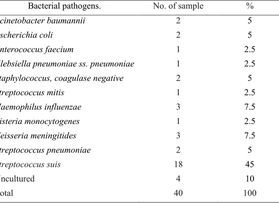
5.1. Species identification
Forty CSF samples were processed for bacterial isolation. A total of 36 bacterial isolates were rescued and identified by culture-dependent methods. Four (10%) samples tested negative when seeded in cultures. Among bacterial isolates, 18 (45%) were identified as S. suis by API®20 STREP with 85-90 % confidence
while 2 (5%) were identified as S. pneumoniae. H. influenzae and N. meningitidis
were detected in 6 (15 %) samples and L. monocytogenes was detected in 1 (2.5%).
Nine (22.5%) isolates were identified as A. baumannii, E. coli, E. faecium, K. pneumoniae ss. pneumonia, Staphylococcus coagulase negative, and S. mitis. Co-infections with two bacterial species were not detected. (Table 1).
Table 1. Bacterial pathogens identified by culture-dependent methods from CSF samples
Bacterial pathogens. No. of sample %
Acinetobacter baumannii 2 5
Escherichia coli 2 5
Enterococcus faecium 1 2.5
Klebsiella pneumoniae ss. pneumoniae 1 2.5
Staphylococcus, coagulase negative 2 5
Streptococcus mitis 1 2.5 Haemophilus influenzae 3 7.5 Listeria monocytogenes 1 2.5 Neisseria meningitides 3 7.5 Streptococcus pneumoniae 2 5 Streptococcus suis 18 45 Uncultured 4 10 Total 40 100
Nguyen Hoang Bach - Molecular and antigenic characterization of Streptococcus suis serotype 2 isolates in central Vietnam - Doctorate Thesis of International PhD School In Biomolecular and Biotechnological Sciences, University of Sassari
33
5.2. Capsular serotype identification
The real-time PCR assay for capsular serotype 2, all of 18 S. suis strains (45%) was positive (30-33 Ct value range on 3 replicates). Additionally, DNA extracted from CFS sample that was negative by isolation test was positive by real-time PCR with a Ct value ranging from 33 to 37. All negative controls (non-template controls, other bacterial pathogens such as S. pyogenes, S. pneumonia, S. aureus, Enterococcus, E. coli) were negative (Table 2).
Table 2. Specificity of S. suis serotype 2 real-time PCR on culture-confirmed CSF samples and uncultured-confirmed samples.
Bacterial pathogens. No. of sample Real-time PCR positive Real-time PCR negative Acinetobacter baumannii 2 0 2 Escherichia coli 2 0 2 Enterococcus faecium 1 0 1
Klebsiella pneumoniae ss. pneumoniae 1 0 1
Staphylococcus, coagulase negative 2 0 2
Streptococcus mitis 1 0 1 Haemophilus influenza 3 0 3 Listeria monocytogenes 1 0 1 Neisseria meningitides 3 0 3 Streptococcus pneumoniae 2 0 2 Streptococcus suis 18 18 0 Un-growth 4 1 3 Total 40 19 21
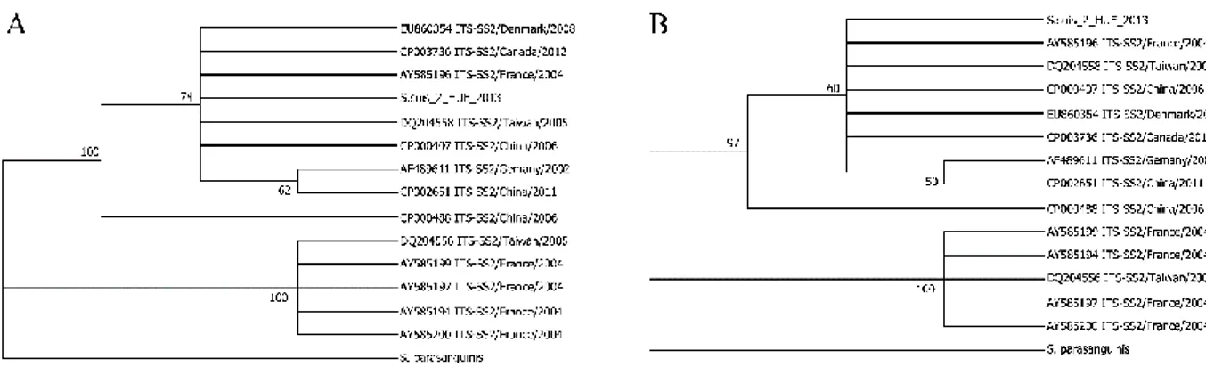
5.3. 16S-23S Intergenic spacer phylogenetic analysis
Amplification of the 16S-23S intergenic spacer was confirmed by gel
electrophoresis of the amplified fragments. The expected size of about 700bp was obtained (lanes 3-8) with all S. suis serotype 2 tested (Figure 1).