Received: October 28, 2015; Revised: December 5, 2016; Accepted: December 8, 2016
© The Author 2016. Published by Oxford University Press. All rights reserved. For Permissions, please email: [email protected]. doi:10.1093/carcin/bgw203
Advance Access publication December 26, 2016 Original Manuscript
218
original manuscript
Prevalence of HPV infection in racial–ethnic subgroups
of head and neck cancer patients
Camille Ragin
1,2,3,*
, Jeffrey C.Liu
4, Gieira Jones
2, Olubunmi Shoyele
5,
Bukola Sowunmi
1, Rachel Kennett
1, Denise Gibbs
1,3, Elizabeth Blackman
1,2,3,
Michael Esan
1, Margaret S.Brandwein
6, Karthik Devarajan
7, Francesco Bussu
8,
Rebecca Chernock
9, Chih-Yen Chien
10, Marc A.Cohen
11, Samir El-Mofty
9,
Mikio Suzuki
12, Gypsyamber D’Souza
13, Pauline Funchain
14, Charis Eng
14,
Susanne M.Gollin
15, Angela Hong
16, Yuh-S Jung
17, Maximilian Krüger
18,
James Lewis Jr
19, Patrizia Morbini
20, Santo Landolfo
21, Massimo Rittà
21,
Jos Straetmans
22, Krisztina Szarka
23, Ruth Tachezy
24, Francis P.Worden
25,
Deborah Nelson
2, Samuel Gathere
3,26and Emanuela Taioli
3,271Cancer Prevention and Control Program, Fox Chase Cancer Center – Temple Health, Philadelphia, PA 19111, USA,
2Department of Epidemiology and Biostatistics, Temple University, College of Public Health, Philadelphia, PA 19111,
USA, 3African Caribbean Cancer Consortium, Philadelphia, PA 19111, USA, 4Department of Otolaryngology, Fox Chase
Cancer Center, Temple University, Philadelphia, PA 19111, USA, 5Department of Pathology and Laboratory Medicine,
Western Connecticut Health Network, Danbury Hospital, Danbury, CT 06810, USA, 6Department of Pathology and
Anatomical Sciences, SUNY at the University at Buffalo, Buffalo, NY 14214-3005, USA, 7Department of Biostatistics,
Fox Chase Cancer Center – Temple Health, Philadelphia, PA 19111, USA, 8Institute of Otolaryngology, Università
Cattolica del Sacro Cuore, Policlinico Agostino Gemelli, Rome, Italy, 9Department of Pathology and Immunology,
Washington University School of Medicine, St Louis, MO 63110, USA, 10Department of Otolaryngology, Kaohsiung
Chang Gung Memorial Hospital and Chang Gung University College of Medicine, Kaohsiung, Taiwan, 11Department
of Surgery, Head and Neck Service, Memorial Sloan Kettering Cancer Center, New York, NY 10065, USA, 12Department
of Otorhinolaryngology, Head and Neck Surgery, Graduate School of Medicine, University of the Ryukyus, Okinawa,
Japan, 13Department of Epidemiology, Johns Hopkins Bloomberg School of Public Health, Baltimore, MD 21205, USA,
14Genomic Medicine Institute, Cleveland Clinic Lerner Research Institute, Cleveland, OH 44195, USA, 15Department
of Human Genetics, Graduate School of Public Health, University of Pittsburgh, Pittsburgh, PA 15261, USA, 16Central
Clinical School, The University of Sydney, Sydney, NSW, Australia, 17Department of Otolaryngology, Research Institute
and Hospital, National Cancer Center, Gyeonggi-do, Korea, 18Department of Oral and Maxillofacial Surgery - Plastic
Surgery, University Medical Center of the Johannes Gutenberg-University, Mainz, Germany, 19Department of Pathology,
Microbiology, and Immunology, Vanderbilt University, Nashville, TN 37232, USA, 20Department of Molecular Medicine,
Unit of Pathology, University of Pavia, and à IRCCS Policlinico S. Matteo Foundation, Pavia, Italy, 21Department of
Sciences of Public Health and Pediatrics, University of Turin, Turin, Italy, 22Department of Otorhinolaryngology-Head
and Neck Surgery, GROW Institute, Maastricht University Medical Centre, Maastricht, The Netherlands, 23Department
of Medical Microbiology, Faculty of Medicine, University of Debrecen, Debrecen, Hungary, 24Department of Immunology,
Institute of Hematology and Blood Transfusion National Reference Laboratory for Papillomaviruses, Prague, Czech
Republic, 25Department of Internal Medicine, Division of Hematology-Oncology, University of Michigan, Ann Arbor,
MI 48109, USA, 26Non Communicable Diseases Research Programme, Kenya Medical Research Institute, Nairobi,
Kenya and 27Departments of Population Health Science and Policy, of Thoracic Surgery, and Institute For Translational
Epidemiology, Icahn School of Medicine at Mount Sinai, New York, NY 10029, USA
doi:10.1093/carcin/bgw203
Advance Access publication December 26, 2016 Original Manuscript
Abstract
The landscape of human papillomavirus (HPV) infection in racial/ethnic subgroups of head and neck cancer (HNC) patients has not been evaluated carefully. In this study, a meta-analysis examined the prevalence of HPV in HNC patients of African ancestry. Additionally, a pooled analysis of subject-level data was also performed to investigate HPV prevalence and patterns of p16 (CDNK2A) expression amongst different racial groups. Eighteen publications (N = 798 Black HNC patients) were examined in the meta-analysis, and the pooled analysis included 29 datasets comprised of 3129 HNC patients of diverse racial/ethnic background. The meta-analysis revealed that the prevalence of HPV16 was higher among Blacks with oropharyngeal cancer than Blacks with non-oropharyngeal cancer. However, there was great heterogeneity observed among studies (Q test P < 0.0001). In the pooled analysis, after adjusting for each study, year of diagnosis, age, gender and smoking status, the prevalence of HPV16,18 in oropharyngeal cancer patients was highest in Whites (61.1%), followed by 58.0% in Blacks and 25.2% in Asians (P < 0.0001). There was no statistically significant difference in HPV16,18 prevalence in non-oropharyngeal cancer by race (P = 0.682). With regard to the pattern of HPV16,18 status and p16 expression, White patients had the highest proportion of HPV16,18+/p16+ oropharyngeal cancer (52.3%), while Asians and Blacks had significantly lower proportions (23.0 and 22.6%, respectively) [P < 0.0001]. Our findings suggest that the pattern of HPV16,18 status and p16 expression in oropharyngeal cancer appears to differ by race and this may contribute to survival disparities.
Introduction
Head and neck cancer (HNC) is the sixth most common can-cer in the world, accounting for approximately 4% of all cancan-cer cases (1). In 2012, there were an estimated 599 637 new cases of cancer of the oral cavity, larynx and oropharynx, and 324 794 deaths attributed to the disease worldwide (1). Although tobacco and alcohol use are the primary risk factors for developing HNC, human papillomavirus (HPV) is also an established risk factor for cancers arising in the oropharynx (2,3). Recently, HPV has also been reported to be associated with a subset of oral cav-ity cancers (4,5), but an etiological role has not been clearly demonstrated.
A recent review and meta-analysis from our group of HNC survival in relation to HPV demonstrated a survival advantage for all HPV-positive patients (6), but the survival advantage was only significant for patients with cancer of the oropharynx. Compared to patients with HPV-negative oropharyngeal cancer, the risk of death and risk of recurrence for patients with HPV-positive oropharyngeal cancer was reduced by ~28% and ~49%, respectively. In the USA, a clear disparity in HNC survival has been reported between Black and White patients, particularly for oropharyngeal cancers. Poor survival rates for Black Americans compared to White Americans have been observed (7), and some studies have suggested that this disparity may be explained at least partially by a difference in prevalence of HPV infection (8–10). Comparisons of HPV prevalence in cancer of the oral cav-ity and larynx between various racial/ethnic populations have been reported in a recent meta-analysis (11). However, a sum-mary of HPV prevalence for Black patients was only reported for oral cavity cancer in this study (11). Furthermore, an assess-ment of attributed survival differences for oropharyngeal cancer between racial/ethnic populations was not conducted.
The goal of this study was to develop a more complete per-spective of the landscape of HPV infection in ethnic subgroups
of HNC patients by examining the published literature. We con-ducted a meta-analysis examining the prevalence of HPV in the Black population. We also performed a pooled analysis of cases reporting HNC and HPV status using subject-level data from the published literature to investigate HPV segregation and preva-lence amongst different ethnic groups.
Materials and methods
This study was approved by the Fox Chase Cancer Center Institutional Review Committee.
Literature review and data collection
A PubMed search was conducted (from inception to December 2014) using the search terms, [‘human papillomavirus’ (All Fields) OR ‘HPV’ (All Fields)] AND [‘squamous cell carcinoma’ (All Fields) OR ‘cancer’ (All Fields)] AND [‘oropharyngeal’ (All Fields) OR ‘oropharynx’ (All Fields) OR ‘head and neck’ (All Fields) OR ‘tonsil’ (All Fields)]. All abstracts and full text of articles from the PubMed search were reviewed independently by two reviewers. When there was a discrepancy between reviewers, a third reviewer evalu-ated the article(s) to resolve the discrepancy. All studies that tested for the presence of HPV in HNC tissues from patients diagnosed with squamous cell carcinoma of the head and neck (oral cavity, oropharynx, larynx and hypopharynx) were eligible for inclusion in this analysis. The bibliogra-phies of several review articles were also examined in order to identify additional publications that might have been missed by our PubMed search (11–15). This review identified 291 original articles that qualified conditionally for the analysis. Studies that used serology methods to detect HPV antibodies were excluded from the analysis, as this method does not identify which tissue is infected by HPV. Studies that primarily evaluated HPV in lip cancers were excluded from this analysis, with the exception of studies where it was impossible to distinguish lip cancer data from the other head and neck subsites. In addition, case reports and stud-ies that included only HPV-positive HNC tumors/patients were excluded. Additional exclusion criteria includes studies of HNC patients who were co-infected with other diseases, such as HIV; studies in which the cancer tissues were sampled via cytobrushing and not biopsy or surgery; studies that classified HNC as HPV-related or HPV non-related tumors based on tumor site without directly testing that tissue for HPV; studies in which fewer than 80% of the eligible cases were tested for HPV; and studies that selected patient samples non-randomly, but applied pre-defined criteria for patient inclusion (e.g., patients with undifferentiated carcinoma only, metastasis only, positive lymph nodes only, advanced stage only, patients who underwent a specific treatment regimen, studies where smoking and drinking patient tissues were matched with nonsmoker and nondrinker patient tissues, etc.). For overlapping studies, the publication with the largest population and/or more complete information was included in this analysis. After accounting for these inclusion and exclusion criteria,
Abbreviations
FF fresh frozen
FT fresh tissue
FFPE formalin-fixed paraffin-embedded HPV human papillomavirus
HNC head and neck cancer
HR hazard ratios
140 articles with data for all racial/ethnic populations were eligible for inclusion in this study. Of these, only 18 articles presented data that could be abstracted and were included in the meta-analysis of Black cancer patients. All 140 articles were eligible for inclusion in the pooled analysis. A flow diagram of study selection is illustrated in Figure 1.
Meta-analysis of Black HNC patients
From each of the 18 articles that included data from Black HNC patients (818 cases), information on the number of patients, HPV prevalence, HPV genotype, tumor subsite, mean age, year of cancer diagnosis, geo-graphic location of the study, tissue source, HPV test methodology and HPV-infected cancer site were extracted and tabulated. All data were abstracted independently by two reviewers and cross-referenced to con-firm that there were no data entry errors. Three studies that included data for fewer than 10 Black patients (16–18) were therefore excluded from the meta-analysis, leaving 15 studies including 798 cases.
Pooled analysis
All investigators from the 140 studies were invited to submit their subject-level data for this pooled analysis; data from 22 studies were obtained. The remaining study investigators either did not respond or did not wish to participate. Common data elements included in the pooled analysis were HPV test method, HPV status, HPV genotype, DNA source, geographic location of the study, age at diagnosis, gender, race/ethnicity, p16 status, tobacco and alcohol use, clinical variables (such as tumor site, histology and stage) and survival variables (such as vital status and follow-up time). Seven additional articles reported demographic, clinical, HPV results, tobacco, alcohol and survival data in the publications, which enabled us to create pseudo-datasets for inclusion in the pooled analysis. All patients included in this analysis were diagnosed with cancers of the oral
cavity, oropharynx or larynx. Patients with hypopharyngeal cancers were grouped with the patients with cancers of the larynx. Patients with metas-tases or unknown primaries were excluded from this analysis. In total, there were 29 datasets including a total of 3129 HNC cases.
Statistical analysis
The Meta-proportion of any HPV and HPV16 only was calculated for all HNC subsites combined as well as separately for oropharynx and non-oro-pharynx data. All statistical analyses were performed using Intercooled STATA SE (version 10) software (StataCorp. LP, College Station, TX). Meta-analyses of the proportion of HPV-positive HNC were performed using the metaprop command in STATA. HPV proportions were calculated for each individual study and the reported confidence intervals were based on Clopper–Pearson exact binomial procedures (19). Pooled proportions of the multiple studies were estimated using a random effects model. The Meta-prevalence estimates were calculated by multiplying the Meta-proportion and confidence interval values by 100. The Q-statistics were used to test for heterogeneity between the studies included in the meta-analyses. The I2 metric was also calculated to quantify variation between studies (20).
Large between-study variation was observed when the I2 values were ≥50%
while moderate between-study heterogeneity was denoted by I2 values
between 25 and 50%. Evidence of publication bias or small study effects (P < 0.05) was assessed using the Egger’s test (21).
For the pooled analysis, unequal variance in age was observed between categories of race. Therefore, a square root transformation of age at diagnosis was performed. Adjusted HPV prevalence and 95% confi-dence intervals for each racial/ethnic group was calculated from logistic regression estimates for HPV-positive status, adjusting for study, year of diagnosis, square root of age, sex, history of alcohol drinking and smok-ing history. The adjusted prevalence refers to the average HPV prevalence
while averaging the values of the covariates in the regression model. The logistic coefficients and standard errors are provided in Supplementary Materials, available at Carcinogenesis Online. A Likelihood Ratio chi-square test was performed to evaluate differences between the adjusted preva-lence according to race and an analysis of variance (ANOVA) was used to compare the mean square root of age at diagnosis between racial groups (P values for pairwise comparisons were Bonferroni adjusted). P values < 0.05 were considered statistically significant. Mean age at diagnosis for each stratum was back transformed and reported. Follow-up time for overall survival refers to the interval between date of diagnosis and the date of last contact (if the patient was alive) or date of death. Hazard ratios (HR) were calculated and adjusted for each study and other confounders for risk of death or risk of disease progression (i.e., disease persistence, recurrence and/or metastasis). HR < 1.0 represents an overall survival ben-efit and HR > 1.0 represents poor overall survival.
Results
Description of studies: meta-analysis
Table 1 summarizes all published studies from which data were available to estimate HPV (any HPV or HPV16) prevalence in Black populations. Study size ranged from 13 to 161 patients. The majority (13/15, 87%) of studies included polymerase chain reac-tion (PCR)-based methods to test for the presence of HPV DNA. For all site strata (all head and neck, oropharynx and non-oro-pharynx), large heterogeneity was observed between the studies (Q test P-value range from 0.000 to 0.048; I2 values range from 62.1 to 94.6%). Nevertheless, as expected, the prevalence of any HPV or HPV16 was higher among oropharyngeal cancer patients (any HPV: 31.5%, 95% CI = 17.7–47.1; HPV16: 45.7%, 95% CI = 25.5–66.6) in comparison to non-oropharyngeal cancer patients (any HPV: 14.5%, 95% CI = 1.4–36.0; HPV16: 1.1%, 95% CI = 0.0–6.0). There was no evidence of publication bias or small study effect. The reasons for underlying heterogeneity were explored by strati-fying the dataset according to geographic region (Sub-Saharan Africa versus USA) as well as HPV test methods (ISH versus PCR/ RT-PCR). Large heterogeneity remained when stratified by HPV test method (data not shown). When stratified by geographic region (see Supplementary Table 1, available at Carcinogenesis Online), large heterogeneity was still observed except when data were limited to HPV16 infections only. For all head and neck subsites combined, the meta-prevalence of HPV16 in patients from Sub-Saharan Africa (N = 4 studies) was 1.0% (95% CI = 0.0–3.9), Q test P-value was 0.129, I2 was 47.0%. Large hetero-geneity was still observed between the remaining eight studies that included patients from the USA (Q test P <0.0001, I2 = 89%). Further stratification of the Sub-Saharan Africa studies accord-ing to head and neck subsite resulted in a meta-prevalence of HPV16 in non-oropharyngeal cancers at 0.1% (95% CI = 0.0–1.8, Q test P value = 0.768, I2 = 0.0%). The only study in the USA that reported HPV16 data for non-oropharyngeal cancer showed a higher prevalence (13.6%, 95% CI = 1.9–31.7) than that of patients in Saharan Africa. There were no studies in Sub-Saharan Africa that reported data for HPV16 in oropharyngeal cancer patients and the large heterogeneity remained for the USA studies that reported HPV16 data in Black oropharyngeal cancer patients.
Description of studies: pooled analysis
There were a total of 3129 patients included in this analysis (Table 2). Variations among the 29 studies were noted with regard to study size, the geographic region where the study was conducted, tumor site and the tissue source. Studies var-ied in size from 15 to 489 patients and were conducted mostly in Europe (48%, 14/29 studies), followed by the USA (31%, 9/29),
Asia (17%, 5/29) and a single study in Australia. Most of the studies (65%, 19/29) involved patients diagnosed with can-cers at both oropharyngeal and non-oropharyngeal sites (oral cavity, larynx, hypopharynx and non-oropharyngeal sites not otherwise specified). The remaining studies included patients diagnosed with oropharyngeal cancers only. Formalin-fixed paraffin-embedded (FFPE) tissues were examined in 66% of studies to test for the presence of HPV, rather than Fresh Frozen (FF) or Fresh Tissue (FT). All except for four studies used PCR methodology to detect HPV DNA, using either consensus or type-specific primers, and of these, five also evaluated HPV status using DNA in situ hybridization combined with PCR. Two studies detected HPV RNA using only RT-PCR and the other two detected both HPV RNA and DNA using RT-PCR and PCR. CDKN2A (p16) expression was evaluated in 16 studies using immunohistochemistry. With regard to race/ethnicity, the pooled dataset was diverse with patients representing African, African American, Asian and White populations. There was one study that included Aboriginal Australian patients. These patients were combined with the African and African American patients and classified as Black. There were 82 patients clas-sified as other race (for 63 patients race was unknown and 19 patients included Pacific Islander, Middle Eastern, Indian, Hispanic or other not otherwise specified). These patients were grouped and classified as other race. Follow-up time was avail-able for 19 studies and ranged from 0.03 to 244.5 months with a mean follow-up of 41.7 months and a median follow-up of 30.6 months.
Prevalence of HPV16 and HPV18 according to race and head and neck subsite
The prevalence of HPV16 and/or HPV18 (HPV16,18) stratified by race was calculated for all HNCs, oropharyngeal cancers only and non-oropharyngeal cancers only after adjusting for study, year of diagnosis, age, gender, alcohol drinking, and smoking status (Table 3, and Supplementary Table 2, available at Carcinogenesis Online, which summarizes the logistic coefficients and stand-ard errors). As expected, the overall mean age for HPV-positive patients diagnosed with oropharyngeal cancers was lower than the mean age of HPV-positive patients diagnosed with non-oropharyngeal cancers irrespective of whether the patient car-ried HPV16 or HPV18 in their tumor. The mean age at diagnosis was 56.3 years for HPV16,18+ oropharyngeal cancer patients, and 60.1 years for HPV16,18+ non-oropharyngeal HNC patients (P < 0.0001). There was no statistically significant difference in the mean age at diagnosis of HPV16,18+ oropharyngeal cancer patients according to race. However, for non-oropharyngeal HNC patients, a Bonferroni post hoc test shows that Asians were statistically significantly older compared to Whites (HPV16,18: Asians, 64.1 years versus Whites, 54.9 years, P = 0.038).
As expected, the prevalence of HPV16,18 was higher in oro-pharyngeal cancer tissues compared to non-orooro-pharyngeal cancer tissues (HPV16,18: 48.7% versus 18.2%). HPV16 was the predominant genotype carried in all patient tissues, 46.6% of oropharyngeal cancer patients and 13.4% of non-oropharyngeal HNC patients were positive for this genotype. In contrast, only approximately 1–2% of patients carried HPV18, irrespective of whether the cancer was diagnosed in the oropharynx or at a non-oropharyngeal head and neck site.
For oropharyngeal cancers, there was a statistically signifi-cant difference in the prevalence of HPV16,18 according to race. White patients had the highest prevalence of HPV16,18+ cancers followed by Blacks then Asians, however, only the prevalence in Asian patients was statistically significantly lower (61.1 versus
58.0% and 25.2%, respectively; P <0.0001). A similar pattern was observed for the prevalence of HPV16 infections. However, for HPV18, Black patients had the highest prevalence (14.8%) com-pared to Asians (1.6%) and Whites (1.1%) and this difference was statistically significant (P = 0.0025). For the non-oropharyngeal cancer patients, there was no statistically significant difference in HPV16 and/or 18 prevalence according to race.
Expression of p16 and HPV16,18 DNA according to race in oropharynx cancer patients
Twelve studies (1397 patients) presented with both HPV16,18 and p16 data. Among oropharyngeal cancer patients, the pat-tern of combined HPV16,18 and p16 status differed according to race, and this difference was statistically significant (Figure 2A, P < 0.0001). White patients had the highest proportion of cancers that were HPV16,18+/p16+ (52.3%). In contrast, Asian and Black
patients had lower proportions of tumors with HPV16,18+/p16+ cancers (23.0 and 22.6%, respectively). In addition, Black patients had a higher proportion of cancers that were HPV16,18+, but p16− compared to Asian and White patients (31.1 versus 10.5% and 4.7%, respectively). The proportion of patients with HPV16,18−/p16− disease also differed significantly by race. Asian patients had the highest proportion of HPV16,18−/p16− cancers, in contrast to Black and White patients (66.8 versus 37.7% and 29.6%, respectively).
When the oropharyngeal cancer patients were stratified according to smoking history, the pattern of combined HPV16,18 and p16 status according to race co-segregated with the frac-tion of patients that were ever smokers (Figure 2B). Among never smokers (Figure 2C), as expected, patients with HPV16,18+/ p16+ cancers comprised the predominant fraction among Asian, Black and White patients. However, White patients still Table 1. Meta-analysis of HPV prevalence in populations of African descent
Study HPV test method HPV types detected N
Any HPV N%, 95% CI
HPV 16 N%, 95% CI All head and neck
Van Rensburg et al. (22) ISH 66 0.0% (0.0–5.4) 0.0% (0.0–5.4)
Gillison et al. (23) PCR 16, 18, 33, 31 48 20.8% (10.5–35.0)
Boy et al. (24) PCR 16,18 21 9.5% (1.2–30.4) 0.0% (0.0–16.1)
Agrawal et al. (25) ISH 16 13 0.0% (0.0–24.7) 0.0% (0.0–24.7)
Lewis et al. (26) ISH, PCR 16, 33 26 11.5% (2.4–30.1)
Jalouli et al. (27) PCR X 20 65.0% (40.8–84.6) Jiron et al. (28) PCR 6,33,11,16,18,31,52,35,45,51,56 161 24.8% (18.4–32.3) 20.5% (14.5–27.6) Stephen et al. (29) qRT-PCR 16 31 16.1% (5.4–33.7) 16.1% (5.4–33.7) Babiker et al. (30) PCR 16, 18, 33, 31 100 8.0% (3.5–15.2) 5.0% (1.6–11.3) Isayeva et al. (31)a qRT-PCR 16, 18 30 60.0% (40.6–77.3) 43.3% (25.5–62.6) Ndiaye et al. (32) PCR 16, 35, 45 110 3.6% (1.0–9.0) 0.9% (0.0–5.0) Salazar et al. (33) PCR, RT-PCR 16 57 15.8% (7.5–27.9) 15.8% (7.5–27.9) Worsham et al. (10) q-PCR NR 49 30.6% (18.2–45.4) 30.6% (18.2–45.4) Isayeva et al. (34)a qRT-PCR 16, 18 22 22.7% (7.8–45.4) 13.6% (2.9–34.9) Liu et al. (35)a PCR 16 44 72.7% (57.2–85.0) Total 798 17% (8.8–27.0) 13.7% (1.5–26.4) P value, Q test 0.000 0.000 I2 test 89.8% 93.8%
P value, Egger’s test 0.419 0.643
Oropharynx
Lewis et al. (26) ISH, PCR 16, 33 26 11.5% (2.4–30.1)
Jiron et al. (28) PCR 6,33,11,16,18,31,52,35,45,51,56 36 25.0% (12.1–42.2) Isayeva et al. (31)a qRT-PCR 16, 18 30 60.0% (40.6–77.3) 43.3% (25.5–62.6) Salazar et al. (33) PCR, RT-PCR 16 23 34.8 (16.4–57.3) 34.8% (16.4–52.3) Worsham et al. (10) q-PCR NR 49 30.6% (18.2–45.4) 30.6% (18.2–45.4) Liu et al. (35)a PCR 16 44 72.7% (57.2–85.0) Total 146 31.5% (17.7–47.1) 45.7% (25.5–66.6) P value, Q test 0.003 0.000 I2 test 75.5% 84.1%
P value, Egger’s test 0.997 0.807
Non-oropharynx
Van Rensburg et al. (22) ISH 66 0.0% (0.0–5.4) 0.0% (0.0–5.4)
Boy et al. (24) PCR 16, 18 21 9.5% (1.2–30.4) 0.0% (0.0–16.1) Jalouli et al. (27) PCR X 20 65.0% (40.8–84.6) Jiron et al. (28) PCR 6,33,11,16,18,31,52,35,45,51,56 125 24.8% (17.5–33.3) Ndiaye et al. (32) PCR 16,35,45 105 3.8% (1.0–9.5) 1.0% (0.0–5.2) Isayeva et al. (34)a qRT-PCR 16,18 22 13.6% (2.9–34.9) Total 337 14.5% (1.4–36.0) 1.1% (0.0–6.0) P value, Q test 0.000 0.048 I2 test 94.6% 62.1%
P value, Egger’s test 0.685 0.424
NR, not reported; X, HPV genotype unknown.
had the highest proportion, and Asian patients had the low-est (White: 80.1%, Black: 62.5% Asian: 39.6%, P < 0.0001). Even among never smokers, Asians continued to have the largest pro-portion of patients with HPV16,18−/p16− cancers (37.4%), which was almost equal to the proportion of HPV16,18+/p16+ cancers (39.6%) observed in this subgroup.
Predictors of overall survival for oropharyngeal cancer patients according to race
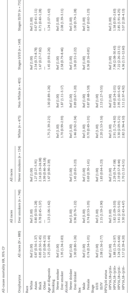
Independent predictors of overall survival for oropharyngeal can-cer patients were age at diagnosis, smoking history, late stage (III/ IV) at diagnosis, and combined HPV16,18 and p16 status (Table 4). Patients with HPV16,18−/p16+ cancers had an increased risk of death compared to patients with HPV16,18+/p16+ oropharyngeal cancers. There was also an even greater increased risk of death for patients with p16− cancers irrespective of HPV status. When stratified according to smoking history, among never smokers, HPV16,18−/p16− patients were the only group with a statistically significantly increased risk of death compared to HPV16,18+/p16+ patients (Hazard Ratio[HR]: 2.70, 95% Confidence Interval [CI] 1.12–6.51). Patients with HPV16,18+/p16− or HPV16,18−/p16+ oro-pharyngeal cancers also had an increased risk of death compared to patients with HPV16,18+/p16+ oropharyngeal cancers, but the hazard ratios were not statistically significant. When stratified according to race, non-White patients differed in comparison to White patients regarding risk of death based on HPV16,18/p16 status. Table 4 shows that p16 status rather than HPV DNA sta-tus appeared to be a predictor of overall survival for non-White
patients, but not for White patients. For non-Whites, the risk of death was statistically significantly increased for patients with p16-negative oropharyngeal cancers, irrespective of HPV16,18 status (HPV16,18+/p16−: HR = 2.95, 95% CI = 1.60–5.42, HPV16,18−/ p16−: HR = 3.11, 95% CI = 1.97–4.92 versus HPV16,18−/p16+: HR = 0.69, 95% CI = 0.24–2.01). In contrast, the risk of death for White patients with p16+ cancers was dependent upon HPV16,18 status. White patients with HPV16,18-/p16+ oropharyngeal can-cers had an increased risk of death (HR = 2.91, 95% CI = 1.72–4.92) in comparison to White patients with HPV16,18+/p16+ oro-pharyngeal cancers.
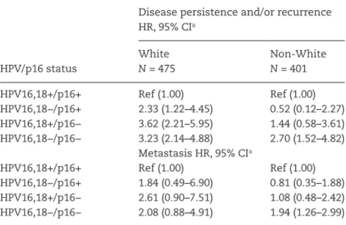
The risk of disease persistence, recurrence or metastasis based on HPV16,18/p16 status differed between White and non-White oropharyngeal cancer patients and is presented in Table 5. White patients that did not have HPV16,18+/p16+ disease had an increased risk of disease persistence and/or recurrence in comparison to patients diagnosed with HPV16,18+/p16+ disease. In contrast non-white patients with HPV16,18−/p16− were the only subgroup with a greater risk of disease persistence and/or recurrence in comparison to HPV16,18+/p16+ disease (HR = 2.70, 95% CI = 1.52–4.82). The risk of metastasis was only associated with non-White patients carrying HPV16,18−/p16− oropharyngeal cancers.
Discussion
This study expands on our prior reported meta-analysis of HPV and HNC (6). In that study, we showed that the presence of HPV infection, specifically in the oropharynx had a significant effect Table 2. Description of studies included in the pooled analysis
Author (Ref) Study size Tissue source HPV testing method p16 expression Geographic region Race/ ethnicity Tumor site FU, months (median)
Cruz et al. (36)a 35 FF PCR — Europe W NO —
Tsuhako et al. (37)a 88 FFPE PCR — Asia AS NO, O —
Koskinen et al. (38)a 61 FF PCR — Europe W NO, O —
De Petrini et al. (39) 70 FF PCR — Europe W NO, O 30.4
Ragin et al. (16) 125 FFPE PCR IHC USA W NO, O 48.4
Armas et al. (40,41) 280 FFPE PCR IHC Asia AS NO, O 18.6
Cohen et al. (42)a 35 FFPE PCR — USA UNK O —
Worden et al. (43) 70 FFPE PCR — USA AA, W NO, O 13.5
Szarka et al. (44) 33 FF PCR — Europe W NO, O 25.4
Szarka et al. (45) 55 FF PCR — Europe W NO, O 77.4
Straetmans et al. (46) 81 FFPE PCR/ISH IHC Europe W O —
Tachezy et al. (47) 135 FFPE PCR — Europe W NO, O 47.7
D’Souza et al. (48) 246 FFPE PCR/ISH — USA AA, AS, W NO, O 31.0
Eng et al. (49) 15 FFPE PCR IHC USA W NO, O 69.8
Chernock et al. (8) 266 FFPE PCR/ISH IHC USA AA, AS, W O —
Kabeya et al. (50)a 31 FF PCR IHC Asia AS NO —
Hoffman et al. (51)a 78 FF PCR IHC Europe W NO, O —
Park et al. (52) 89 FFPE PCR IHC Asia AS O 20.9
Heusinkveld et al. (53)a 41 FFPE PCR — Europe W NO, O —
Bussu et al. (54,55) 136 FT RT-PCR/HC2 IHC Europe A, W NO, O 12.5
Isayeva et al. (31,34) 315 FFPE RT-PCR IHC USA AA, AS, W NO, O 27.5
Deng et al. (56,57) 131 FF PCR — Asia AS NO, O 25.1
Morbini et al. (58) 52 FFPE PCR/ISH IHC Europe W NO, O 50.5
Hong et al. (59) 489 FFPE PCR IHC Australia W, AB, AS O 49.0
Kruger et al. (12) 88 FF PCR — Europe W NO —
Liu (35) 44 FFPE PCR IHC USA AA O 18.9
Morbini et al. (60) 41 FFPE PCR/ISH IHC Europe W O 21.2
Total 3129 30.6
A, African; AA, African American; AB, Aboriginal Australian; AS, Asian; FU, follow-up; HC2, hybrid capture 2; ISH, in situ hybridization; IHC, immunohistochemistry; NO, non-oropharynx; O, Oropharynx; PCR, polymerase chain reaction; RT-PCR, real-time PCR (mRNA); UNK, unknown race; UNKP, unknown primary; W, White.
Ta
ble 3.
Adjusted pr
ev
alence of HPV16 and HPV18 accor
ding to r
ace str
atified b
y head and nec
k subsite N HPV16+ mean age a (y ears ± SD) HPV16 pr ev alence b % (95% CI) N HPV18+ mean age a (y ears ± SD) HPV18 pr ev alence b N HPV16,18+ mean age a (y ears ± SD) HPV16,18 prev alence b All HNC Number of studies = 28 Number of studies = 24 Number of studies = 28 Asian 634 58.9 ± 0.64 28.4% (23.8–33.4) 631 61.9 ± 0.90 1.6% (0.7–3.9) 632 59.0 ± 0.68 26.0% (21.6–30.9) Blac k 158 56.6 ± 0.55 43.7% (34.2–53.8) 85 56.7 ± 0.20 9.8% (4.0–21.9) 131 56.7 ± 0.51 56.2% (45.1–66.7) White 2.123 56.5 ± 0.47 36.9% (34.0–40.0) 1778 54.5 ± 0.50 1.6% (0.9–2.8) 1915 56.6 ± 0.48 44.0% (40.8–47.3) Other c 58 55.3 ± 0.52 34.3% (16.2–58.5) 54 — 7.7% (1.0–39.6) 58 55.3 ± 0.51 44.9% (23.1–68.8) P = 0.0123 ¥ P = 0.0077 ¥ P < 0.0001 ¥ Total 2973 56.8 ± 0.50 35.0% (32.8–37.2) 2548 58.2 ± 0.70 1.9% (1.2–2.8) 2736 56.9 ± 0.51 39.3% (37.1–41.7) Or ophar ynx Number of studies = 25 Number of studies = 21 Number of studies = 25 Asian 433 56.8 ± 0.63 25.9% (21.1–31.4) 431 55.8 ± 1.02 1.6% (0.5–4.7) 432 56.7 ± 0.63 25.2% (20.5–30.7) Blac k 120 56.7 ± 0.58 51.1% (39.0–63.0) 65 57.0 ± 0.12 14.8% (5.6–33.7) 110 56.9 ± 0.54 58.0% (45.0–70.0) White 1317 56.2 ± 0.46 57.3% (53.1–61.4) 1100 56.5 ± 0.30 1.1% (0.4–2.5) 1229 56.2 ± 0.46 61.1% (56.8–65.3) Other c 44 55.9 ± 0.49 74.1% (35.4–93.7) 42 — — 44 55.9 ± 0.48 74.1% (35.5–93.7) P < 0.0001 ¥ P = 0.0025 ¥ P < 0.0001 ¥ Total 1914 56.3 ± 0.49 46.6% (43.7–49.4) 1638 56.4 ± 0.46 1.6% (1.0–2.7) 1815 56.3 ± 0.48 48.7% (45.9–51.6) Non-or ophar ynx Number of studies = 21 Number of studies = 19 Number of studies = 21 Asian 201 65.0 ± 0.50 27.1% (16.4–41.4) 200 64.6 ± 0.80 — 200 64.1 ± 0.64 20.9% (11.9–34.0) Blac k 38 54.6 ± 0.22 13.3% (5.0–30.8) 20 55.5 ± 0.90 3.2% (0.4–22.6) d 21 54.9 ± 0.31 30.2% (12.9–55.7) White 806 59.2 ± 0.52 11.3% (8.7–14.6) 678 51.9 ± 0.76 1.5% (0.6–3.8) d 686 58.7 ± 0.59 17.2% (13.5–21.5) Other c 14 — 6.7% (0.9–37.0) 12 — 11.6% (1.3–56.3) d 14 — 18.4% (4.2–53.3) P = 0.0553 ¥ P = 0.1434 ¥ P = 0.6344 ¥ Total 1059 60.5 ± 0.55 13.4% (11.0–16.3) 910 59.9 ± 0.88 1.4% (0.6–3.2) d 921 60.1 ± 0.63 18.2% (15.2–21.8) aAg e at dia gnosis w as bac k tr ansformed after ANO V A using squar e r oot tr ansformation.
bAdjusted for eac
h stud y, y ear of dia gnosis, squar e r oot a ge , g ender
, alcohol and smoking status.
cOther includes other r
ace/ethnic gr
oups and unkno
wn r ace . dSmoking status pr edicted HPV18 perfectl y and w as e xcluded as a co variate . ¥Chi-squar e P v
alue for the differ
ences betw
een the four r
ace/ethnic gr
oup cate
on disease-free survival and overall survival. Since the time of that publication, HPV-positive squamous cell carcinoma of the oropharynx has been well described and reported as a distinct clinical entity. Oropharyngeal cancer patients are often non-smokers, male, younger and White compared to traditional substance abuse-related (tobacco and alcohol) HNC. A dra-matic increase in oropharyngeal cancer prevalence has been identified over the last decade (2,61,62). The number of cases of oropharyngeal cancer exceeded the number of cervical cancer cases in 2010 in the United States, and the number of HPV+ oropharyngeal cancer is expected to exceed the incidence of cervical cancer by 2020 (2). In addition, the more favorable out-come of HPV+ oropharyngeal cancer is well-documented and has been confirmed in multiple studies (63,64). These tumors appear to be HPV-related, and a hallmark of favorable tumors is p16 positivity.
For unclear reasons, the prevalence and favorable outcome of HPV+ oropharyngeal cancer is seen mostly in Whites. Variations in the prevalence of HPV have been noted previously in studies of Black patients with oropharyngeal cancer, where some report lower prevalence and others report a prevalence that is higher and/or comparable to White oropharyngeal cancer patients (9,10,35). In the first part of this study, the meta-analysis of pub-lished HPV prevalence and HNC in Black patients echoes these findings. Consistent with what is expected when comparing HPV prevalence in oropharyngeal and non-oropharyngeal cancer sub-sites, we show that for Black patients, cancers in the oropharynx have a higher prevalence of HPV16 (45.7%), than non-oropharyn-geal sites (14.5%). There was large heterogeneity between the studies included in our meta-analysis. It is possible that differ-ences in the HPV detection methods used in different studies may have influenced HPV positivity rates. For example, DNA ISH assays lack sensitivity and in general, PCR may lack specificity for transcriptionally active virus. Nevertheless, we observed that the meta-prevalence of HPV16 among Black patients is similar to the prevalence reported in our pooled analysis (i.e., higher in the oropharynx and lower in non-oropharyngeal sites).
We performed a pooled analysis of published HPV and HNC data in racial/ethnic subgroups in order to obtain a broader per-spective. HPV status was obtained predominantly by PCR on FFPE tissues. Evaluation of HPV16, HPV18 and HPV16,18 preva-lence by subsite and race yielded multiple findings. First, it is clear that HPV, specifically HPV16 or HPV18 within the orophar-ynx is most common in Whites (61%). There is a similar yet lower rate of HPV16,18+ disease in Blacks (58%) and a significant difference in the rate of HPV16,18+ disease in Asians (25%). This highlights the major HPV prevalence difference between Whites and Asians. This finding is curious, since the prevalence of HPV in Black patients has been reported to be statistically signifi-cantly lower than what has been reported for White patients in the literature (61,65). However, our pooled analysis reflects data from multiple institutions which is more reliable than a single study. The observed differences in HPV prevalence between Asians and Whites is also interesting and is not consistent with the previously reported meta-analysis (11). This inconsistency might be explained by differences in the type of Asian popula-tions included in our study. This pooled analysis only included Asians from Taiwan (China) and Japan while the previously pub-lished meta-analysis included Asian populations from China and Korea. Significantly higher HPV prevalence was observed in Korean patients compared to Chinese patients and could explain the higher prevalence of HPV+ oropharyngeal cancer in Asians in that review (11).
An unexpected finding was the higher prevalence of HPV18 amongst Blacks. While HPV18 is rarely reported at either oro-pharyngeal (1.1%) or non-orooro-pharyngeal cancer sites (1.5%) in Whites, HPV18 is nearly 15 times more frequently detected in Black oropharyngeal cancer patients. This major difference was unexpected. It is unclear if this is due to a higher rate of HPV18 infection in HNC in Blacks or a lower rate of HPV16+ oropharyn-geal cancer in Blacks, thereby unmasking HPV18.
To better characterize oropharyngeal cancers, we evaluated by both HPV and p16 status. Canonical HPV oropharyngeal can-cer is characterized by a HPV+/p16+ signature and p16 status Figure 2. Proportions of combined HPV16,18 and p16 status among all (A), ever smoker (B) and never smoker (C) oropharynx cancer patients stratified by race.
Ta ble 4. Pr edictors of o ver all survi
val (all-cause mortality) for or
ophar
yng
eal cancer patients accor
ding to smoking status and r
ace All-cause mortality HR, 95% CI a Or ophar ynx All r aces ( N = 880) All r aces All r aces White ( n = 475) Non-White ( n = 401) Sta ges 0/I/II ( n = 149) Sta ges III/IV ( n = 731) Ev er smokers ( n = 746) Ne ver smokers ( n = 134) Race White Ref (1.00) Ref (1.00) Ref (1.00) Ref (1.00) Ref (1.00) Asian 0.87 (0.56–1.37) 0.80 (0.50–1.28) 1.27 (0.27–5.91) 2.54 (0.73–8.78) 0.67 (0.41–1.11) Blac k 0.85 (0.50–1.45) 0.78 (0.45–1.37) 2.14 (0.19–24.08) 1.47 (0.27–7.82) 0.71 (0.40–1.28) Other 0.57 (0.08–4.15) — 3.99 (0.46–34.18) — — Ag e at dia gnosis 1.25 (1.09–1.44) 1.23 (1.06–1.42) 1.47 (0.90–2.39) 1.75 (1.39–2.21) 1.00 (0.89–1.26) 1.45 (0.93–2.26) 1.24 (1.07–1.43) Smoking Ne ver smoker Ref (1.00) Ref (1.00) Ref (1.00) Ref (1.00) Ref (1.00) Ev er smoker 1.95 (1.34–2.83) 1.70 (0.99–2.93) 1.87 (1.11–3.17) 2.58 (0.79–8.46) 2.08 (1.39–3.11) Alcohol Ne ver drinker Ref (1.00) Ref (1.00) Ref (1.00) Ref (1.00) Ref (1.00) Ref (1.00) Ref (1.00) Ev er drinker 1.00 (0.80–1.26) 0.96 (0.76–1.22) 1.45 (0.65–3.23) 1.46 (0.92–2.34) 0.89 (0.67–1.20) 1.06 (0.51–2.22) 1.00 (0.79–1.28) Sex Male Ref (1.00) Ref (1.00) Ref (1.00) Ref (1.00) Ref (1.00) Ref (1.00) Ref (1.00) Female 0.74 (0.54–1.01) 0.74 (0.53–1.05) 0.69 (0.30–1.61) 0.70 (0.49–1.01) 0.87 (0.48–1.59) 0.36 (0.16–0.81) 0.87 (0.62–1.23) Sta ge 0/I/II Ref (1.00) Ref (1.00) Ref (1.00) Ref (1.00) Ref (1.00) III/IV 2.08 (1.56–2.77) 2.15 (1.59–2.90) 1.83 (0.64–5.23) 2.20 (1.53–3.16) 2.13 (1.27–3.55) HPV/p16 status HPV16,18+/p16+ Ref (1.00) Ref (1.00) Ref (1.00) Ref (1.00) Ref (1.00) Ref (1.00) Ref (1.00) HPV16,18−/p16+ 1.88 (1.19–2.97) 1.82 (1.09–3.03) 2.29 (0.74–7.08) 2.91 (1.72–4.92) 0.69 (0.24–2.01) 7.96 (2.08–30.43) 1.58 (0.95–2.63) HPV16,18+/p16− 3.24 (2.25–4.66) 3.41 (2.32–5.01) 2.19 (0.57–8.44) 3.30 (2.09–5.21) 2.95 (1.60–5.42) 7.22 (2.26–23.11) 2.85 (1.91–4.25) HPV16,18−/p16− 3.17 (2.39–4.20) 3.30 (2.43–4.47) 2.70 (1.12–6.51) 2.82 (1.94–4.10) 3.11 (1.97–4.92) 4.36 (1.51–12.60) 3.07 (2.28–4.13) aCo
variates included: squar
e r oot a ge , y ear of dia gnosis, r ace , se x, smoking, alcohol, sta ge at dia
gnosis combined HPV16,18 and p16 status and stud
has been reported previously as the best prognostic marker for this disease (63,66). Oropharyngeal cancer that develops in White nonsmokers is mostly likely to be HPV-associated. Our study confirmed this finding; nearly 80% of White nonsmok-ers were HPV+/p16+ (Figure 2C). As p16 loss is associated with smoking (67), amongst ever smokers, a much higher incidence of p16− disease was reported in all races. Although approxi-mately 45% of ever smokers continue to be HPV+/p16+, only half that frequency of HPV+/p16+ is reported in non-Whites. Amongst Blacks and especially Asians, HPV−/p16+ disease com-prises the majority of oropharyngeal disease, in distinction to Whites, where HPV+/p16+ disease is the predominant disease.
While it is not surprising that patients with HPV−/p16+ oro-pharyngeal cancer have a higher risk of death compared to patients with HPV+/p16+ oropharyngeal cancer, it was inter-esting to note that among non-Whites, the risk of death for patients with HPV−/p16+ oropharyngeal cancer was not dif-ferent from patients diagnosed with HPV+/p16+ oropharyn-geal cancer (HPV16,18−/p16+ HR: 0.69, 0.24–2.01). Unlike Whites (HPV16,18−/p16+ HR: 2.91, 1.72–4.92), the survival benefit among non-Whites appears to be attributed to p16 status rather than HPV. In Whites, the survival benefit appears to be attributed to HPV status rather than p16 status. However, it is possible that HPV16,18−/p16+ oropharyngeal cancers in non-Whites may be attributed to other high-risk HPV types. Further investigation of the possible role of high-risk HPV types other than HPV16,18 in non-White oropharyngeal cancer patients is needed. Overall, our findings suggest that the difference in HPV/p16 patterns according to race may impact survival differently. Given the multifactorial cause of racial survival disparities, such as poor socioeconomic status and poor access to care, the effect of HPV/ p16 patterns on racial disparities in survival is not easily identi-fied and further investigations are needed.
A limitation of this study is the use of publications as the source of patient data. Unlike database data, like SEER or The National Cancer Database, published data represent a sam-pling of the true population. A major assumption of our pooled analysis is that the landscape of the published literature is rep-resentative of the population as a whole. Given the dramatic differences noted in survival here between Whites and non-Whites, we feel it is highly unlikely that an error in sampling of the literature can explain these differences. A high fraction of cells with expression of p16 in both the nucleus and cytoplasm is the only good correlation with prognosis and with high-risk
HPV mRNA. For each of the studies included in the pooled analy-sis, we did not have detailed information on the cutoffs used to define p16 status (i.e., fraction of p16 expression in nuclei versus cytoplasm). This is also a limitation of our study, as this detail may have provided more accurate correlations of p16 expression and outcome according to race.
The reasons for this difference in patterns of HPV/p16 in oropharyngeal cancer are unclear. While smoking status has predicted p16 status (67), even amongst never smokers in this study, the prevalence of HPV+/p16+ disease is lower in non-Whites. Possible explanations include genetic and envi-ronmental causes. The development of HPV+ oropharyngeal cancer has been associated with differences in sexual behav-ior patterns and marijuana use (68). Differential sexual and behavior patterns amongst Whites versus non-Whites have not been studied well. While the number of oral sex partners has been identified in the risk of developing HPV+ oropharyn-geal cancer (68). The percentage difference in ever oral sex partners in individuals 45–60 years old between Whites and Blacks appears modest (about 15% difference in prevalence) from a few major studies (69,70), but this remains an area of active research. Other potential explanations are genetic dif-ferences between races and difdif-ferences in the host response to HPV infection, which merit further investigation. Intratypic variation of HPV16 is associated with geographical distribu-tion and may contribute to differences in outcome (71–76). For example, African and Asian-American intratypic variants of HPV16 show higher transforming potential in tumors of the anogenital tract. Therefore, in HNC, differential infection by HPV variants between races may also be an important area for investigation.
At this time, we do not have sufficient understanding to offer a clear recommendation as to how to reduce geal HPV infection or the risk of developing HPV+ oropharyn-geal cancer. This appears to be a problem of environment and biology, without a reversible modifiable factor to reduce risk. We hope that greater adoption of HPV vaccination will alter the incidence curve within about 20 years. Our study has examined HPV and HNC, with a focus on oropharyngeal cancer. This study demonstrates that while HPV-related oro-pharyngeal cancer (HPV+/p16+) represents the majority cause among White patients, Blacks and Asians have lower rates. Because HPV-related oropharyngeal cancer has a more favora-ble outcome regardless of race, the differential HPV prevalence amongst Blacks and Asians is expected to cause a significant outcome disparity in oropharyngeal cancer treatment. Further studies specifically examining racial differences in HPV+ oro-pharyngeal cancer are needed to corroborate these findings. However, this comprehensive pooled analysis of the published literature strongly supports a prevalence disparity in HPV+ oropharyngeal cancer that would predict an outcome/survival disparity.
Supplementary material
Supplementary data are available at Carcinogenesis online.
Funding
Supported by the American Cancer Society (RSG-14-033-01-CPPB to C.R.) and in part by National Cancer Institute (P30 CA006927) and Commonwealth of Pennsylvania. This work was also sup-ported in part by The Lagrange Project – CRT Foundation/ISI Foundation, Turin, Italy to M.R.
Conflict of Interest Statement: None declared. Table 5. Risk of disease progression for oropharyngeal cancer
pa-tients according to HPV/p16 status and race
Disease persistence and/or recurrence HR, 95% CIa HPV/p16 status White N = 475 Non-White N = 401 HPV16,18+/p16+ Ref (1.00) Ref (1.00) HPV16,18−/p16+ 2.33 (1.22–4.45) 0.52 (0.12–2.27) HPV16,18+/p16− 3.62 (2.21–5.95) 1.44 (0.58–3.61) HPV16,18−/p16− 3.23 (2.14–4.88) 2.70 (1.52–4.82) Metastasis HR, 95% CIa HPV16,18+/p16+ Ref (1.00) Ref (1.00) HPV16,18−/p16+ 1.84 (0.49–6.90) 0.81 (0.35–1.88) HPV16,18+/p16− 2.61 (0.90–7.51) 1.08 (0.48–2.42) HPV16,18−/p16− 2.08 (0.88–4.91) 1.94 (1.26–2.99)
aAdjusted for year of diagnosis, square root age, sex, race, stage, smoking,
References
1. Ferlay, J. et al. (2013) GLOBOCAN 2012 v1.0, Cancer Incidence and Mor-tality Worldwide: IARC CancerBase No. 11. Lyon, France: International Agency for Research on Cancer.
2. Chaturvedi, A.K. et al. (2011) Human papillomavirus and rising oro-pharyngeal cancer incidence in the United States. J. Clin. Oncol., 29, 4294–4301.
3. Gillison, M.L. et al. (2012) Prevalence of oral HPV infection in the United States, 2009–2010. JAMA, 307, 693–703.
4. Hobbs, C.G. et al. (2006) Human papillomavirus and head and neck cancer: a systematic review and meta-analysis. Clin. Otolaryngol., 31, 259–266.
5. Syrjanen, S. et al. (2011) Human papillomaviruses in oral carcinoma and oral potentially malignant disorders: a systematic review. Oral Dis., 17 (suppl. 1), 58–72.
6. Ragin, C.C. et al. (2007) Survival of squamous cell carcinoma of the head and neck in relation to human papillomavirus infection: review and meta-analysis. Int. J. Cancer, 121, 1813–1820.
7. Zandberg, D.P. et al. (2014) Oropharyngeal cancer is a driver of racial outcome disparities in squamous cell carcinoma of the head and neck: 10-year experience at the University of Maryland Greenebaum Cancer Center. Head Neck., 4, 564–572.
8. Chernock, R.D. et al. (2011) Human papillomavirus-related squamous cell carcinoma of the oropharynx: a comparative study in whites and African Americans. Arch. Otolaryngol. Head Neck Surg., 137, 163–169.
9. Settle, K. et al. (2009) Racial survival disparity in head and neck can-cer results from low prevalence of human papillomavirus infection in black oropharyngeal cancer patients. Cancer Prev. Res. (Phila Pa), 2, 776–781.
10. Worsham, M.J. et al. (2013) Improved survival with HPV among African Americans with oropharyngeal cancer. Clin. Cancer Res., 19, 2486–2492. 11. Ndiaye, C. et al. (2014) HPV DNA, E6/E7 mRNA, and p16INK4a detection
in head and neck cancers: a systematic review and meta-analysis. Lan-cet Oncol., 15, 1319–1331.
12. Kruger, M. et al. (2014) The prevalence of human papilloma virus (HPV) infections in oral squamous cell carcinomas: a retrospective analysis of 88 patients and literature overview. J. Craniomaxillofac.Surg., 42, 1506–1514.
13. Mehanna, H. et al. (2013) Prevalence of human papillomavirus in oro-pharyngeal and nonorooro-pharyngeal head and neck cancer-systematic review and meta-analysis of trends by time and region. Head Neck, 35, 747–755.
14. Dayyani, F. et al. (2010) Meta-analysis of the impact of human papil-lomavirus (HPV) on cancer risk and overall survival in head and neck squamous cell carcinomas (HNSCC). Head Neck Oncol., 2, 15. 15. Stein, A.P. et al. (2014) Prevalence of human papillomavirus in
oro-pharyngeal squamous cell carcinoma in the United States across time. Chem. Res. Toxicol., 27, 462–469.
16. Ragin, C.C. et al. (2006) 11q13 amplification status and human papil-lomavirus in relation to p16 expression defines two distinct etiologies of head and neck tumours. Br. J. Cancer, 95, 1432–1438.
17. Ukpo, O.C. et al. (2009) Human papillomavirus-associated oropharyn-geal squamous cell carcinomas: primary tumor burden and survival in surgical patients. Ann. Otol. Rhinol. Laryngol., 118, 368–373.
18. Wang, X.I. et al. (2012) Changing trends in human papillomavirus-asso-ciated head and neck squamous cell carcinoma. Ann. Diagn. Pathol., 16, 7–12.
19. Newcombe, R.G. (1998) Two-sided confidence intervals for the single proportion: comparison of seven methods. Stat. Med., 17, 857–872. 20. Higgins, J.P.T. et al. (2003) Measuring inconsistency in meta-analyses.
BMJ, 327, 557–560.
21. Egger, M. et al. (1997) Bias in meta-analysis detected by a simple, graph-ical test. BMJ, 315, 629–634.
22. Van Rensburg, E.J. et al. (1995) Detection of human papillomavirus DNA with in situ hybridisation in oral squamous carcinoma in a rural black population. S. Afr. Med. J., 85, 894–896.
23. Gillison, M.L. et al. (2000) Evidence for a causal association between human papillomavirus and a subset of head and neck cancers. J. Natl. Cancer Inst., 92, 709–720.
24. Boy, S. et al. (2006) HPV detection in primary intra-oral squamous cell carcinomas--commensal, aetiological agent or contamination? J. Oral Pathol. Med., 35, 86–90.
25. Agrawal, Y. et al. (2008) Oral human papillomavirus infection before and after treatment for human papillomavirus 16-positive and human papillomavirus 16-negative head and neck squamous cell carcinoma. Clin. Cancer Res., 14, 7143–7150.
26. Lewis, J.S., Jr. et al. (2010) p16 positive oropharyngeal squamous cell carcinoma:an entity with a favorable prognosis regardless of tumor HPV status. Am. J .Surg. Pathol., 34, 1088–1096.
27. Jalouli, J. et al. (2012) Human papilloma virus, herpes simplex virus and epstein barr virus in oral squamous cell carcinoma from eight different countries. Anticancer Res., 32, 571–580.
28. Jiron, J. et al. (2014) Racial disparities in human papillomavirus (HPV) associated head and neck cancer. Am. J. Otolaryngol., 35, 147–153. 29. Stephen, J.K. et al. (2012) Human papillomavirus outcomes in an
access-to-care laryngeal cancer cohort. Otolaryngol. Head Neck Surg., 146, 730–738.
30. Babiker, A.Y. et al. (2013) Screening for high risk human papilloma virus (HR-HPV) subtypes, among Sudanese patients with oral lesions. Int. J. Clin. Exp. Med., 6, 275–281.
31. Isayeva, T. et al. (2014) African Americans with oropharyngeal carci-noma have significantly poorer outcomes despite similar rates of human papillomavirus-mediated carcinogenesis. Hum. Pathol., 45, 310–319.
32. Ndiaye, C. et al. (2013) The role of human papillomavirus in head and neck cancer in Senegal. Infect. Agent Cancer, 8, 14.
33. Salazar, C.R. et al. (2014) Human papillomavirus-associated head and neck squamous cell carcinoma survival: a comparison by tumor site and initial treatment. Head Neck Pathol., 8, 77–87.
34. Isayeva, T. et al. (2015) The protective effect of p16(INK4a) in oral cavity carcinomas: p16(Ink4A) dampens tumor invasion-integrated analysis of expression and kinomics pathways. Mod. Pathol., 28, 631–653. 35. Liu, J.C. et al. (2015) High prevalence of discordant human
papilloma-virus and p16 oropharyngeal squamous cell carcinomas in an African American cohort. Head Neck. 38 (suppl. 1), E867–E872.
36. Cruz, I.B. et al. (1996) Age-dependence of human papillomavirus DNA presence in oral squamous cell carcinomas. Eur. J. Cancer B Oral Oncol., 32B, 55–62.
37. Tsuhako, K. et al. (2000) Comparative study of oral squamous cell carci-noma in Okinawa, Southern Japan and Sapporo in Hokkaido, Northern Japan; with special reference to human papillomavirus and Epstein-Barr virus infection. J. Oral Pathol. Med., 29, 70–79.
38. Koskinen, W.J. et al. (2003) Prevalence and physical status of human papillomavirus in squamous cell carcinomas of the head and neck. Int. J. Cancer, 107, 401–406.
39. De Petrini, M. et al. (2006) Head and neck squamous cell carcinoma: role of the human papillomavirus in tumour progression. New Micro-biol., 29, 25–33.
40. Al-Swiahb, J.N. et al. (2010) Prognostic impact of p16, p53, epidermal growth factor receptor, and human papillomavirus in oropharyngeal cancer in a betel nut-chewing area. Arch. Otolaryngol. Head Neck Surg., 136, 502–508.
41. Armas, G.L. et al. (2008) The impact of virus in N3 node dissection for head and neck cancer. Eur. Arch. Otorhinolaryngol., 265, 1379–1384. 42. Cohen, M.A. et al. (2008) Increased viral load correlates with improved
survival in HPV-16-associated tonsil carcinoma patients. Acta Otolar-yngol., 128, 583–589.
43. Worden, F.P. et al. (2008) Chemoselection as a strategy for organ preservation in advanced oropharynx cancer: response and survival positively associated with HPV16 copy number. J. Clin. Oncol., 26, 3138–3146.
44. Major, T. et al. (2005) The characteristics of human papillomavirus DNA in head and neck cancers and papillomas. J. Clin. Pathol., 58, 51–55.
45. Feher, E. et al. (2009) Investigation of the occurrence of torque tenovi-rus in malignant and potentially malignant disorders associated with human papillomavirus. J. Med. Virol., 81, 1975–1981.
46. Straetmans, J.M. et al. (2009) Human papillomavirus reduces the prog-nostic value of nodal involvement in tonsillar squamous cell carcino-mas. Laryngoscope, 119, 1951–1957.
47. Tachezy, R. et al. (2009) Demographic and risk factors in patients with head and neck tumors. J. Med. Virol., 81, 878–887.
48. D’Souza, G. et al. (2010) Moderate predictive value of demographic and behavioral characteristics for a diagnosis of HPV16-positive and HPV16-negative head and neck cancer. Oral Oncol., 46, 100–104. 49. Bennett, K.L. et al. (2010) HPV status-independent association of alcohol
and tobacco exposure or prior radiation therapy with promoter methyla-tion of FUSSEL18, EBF3, IRX1, and SEPT9, but not SLC5A8, in head and neck squamous cell carcinomas. Genes Chromosomes Cancer, 49, 319–326. 50. Kabeya, M. et al. (2012) Prevalence of human papillomavirus in mobile
tongue cancer with particular reference to young patients. Cancer Sci., 103, 161–168.
51. Hoffmann, M. et al. (2012) HPV DNA, E6*I-mRNA expression and p16INK4A immunohistochemistry in head and neck cancer - how valid is p16INK4A as surrogate marker? Cancer Lett., 323, 88–96.
52. Park, W.S. et al. (2012) Human papillomavirus in oropharyngeal squa-mous cell carcinomas in Korea: use of G1 cycle markers as new prog-nosticators. Head Neck, 34, 1408–1417.
53. Heusinkveld, M. et al. (2012) Systemic and local human papillomavirus 16-specific T-cell immunity in patients with head and neck cancer. Int. J. Cancer, 131, E74–E85.
54. Bussu, F. et al. (2013) HPV infection in squamous cell carcinomas arising from different mucosal sites of the head and neck region. Is p16 immuno-histochemistry a reliable surrogate marker? Br. J. Cancer, 108, 1157–1162. 55. Bussu, F. et al. (2014) Human papillomavirus (HPV) infection in
squa-mous cell carcinomas arising from the oropharynx: detection of HPV DNA and p16 immunohistochemistry as diagnostic and prognostic indicators--a pilot study. Int. J. Radiat. Oncol. Biol. Phys., 89, 1115–1120. 56. Deng, Z. et al. (2011) Prevalence and clinical features of human pap-illomavirus in head and neck squamous cell carcinoma in Okinawa, southern Japan. Eur. Arch. Otorhinolaryngol., 268, 1625–1631.
57. Deng, Z. et al. (2013) Viral load, physical status, and E6/E7 mRNA expression of human papillomavirus in head and neck squamous cell carcinoma. Head Neck, 35, 800–808.
58. Morbini, P. et al. (2013) Oral HPV infection and persistence in patients with head and neck cancer. Oral Surg. Oral Med. Oral Pathol. Oral Radiol., 116, 474–484.
59. Hong, A. et al. (2013) HPV status of oropharyngeal cancer by combina-tion HPV DNA/p16 testing: biological relevance of discordant results. Ann. Surg.Oncol., 20 (suppl. 3), S450–S458.
60. Morbini, P. et al. (2015) Identification of transcriptionally active HPV infection in formalin-fixed, paraffin-embedded biopsies of oropharyn-geal carcinoma. Hum. Pathol., 46, 681–689.
61. Fakhry, C. et al. (2015) Oropharyngeal cancer survivorship in Denmark, 1977–2012. Oral Oncol., 51, 982–984.
62. de Souza, D.L. et al. (2012) Trends in the incidence of oral cavity and oropharyngeal cancers in Spain. Head Neck, 34, 649–654.
63. Ang, K.K. et al. (2010) Human papillomavirus and survival of patients with oropharyngeal cancer. N. Engl. J. Med., 363, 24–35.
64. Fakhry, C. et al. (2008) Improved survival of patients with human pap-illomavirus-positive head and neck squamous cell carcinoma in a pro-spective clinical trial. J. Natl. Cancer Inst., 100, 261–269.
65. Zandberg, D.P. et al. (2015) Emergence of HPV16-positive oropharyngeal cancer in Black patients over time: University of Maryland 1992–2007. Cancer Prev. Res. (Phila), 8, 12–19.
66. Salazar, C.R. et al. (2014) Combined P16 and human papillomavirus testing predicts head and neck cancer survival. Int. J. Cancer, 135, 2404–2412.
67. Junor, E. et al. (2012) Benefit of chemotherapy as part of treatment for HPV DNA-positive but p16-negative squamous cell carcinoma of the oropharynx. Br. J. Cancer, 106, 358–365.
68. Gillison, M.L. et al. (2008) Distinct risk factor profiles for human papillo-mavirus type 16-positive and human papillopapillo-mavirus type 16-negative head and neck cancers. J. Natl. Cancer Inst., 100, 407–420.
69. D’Souza, G. et al. (2014) Differences in oral sexual behaviors by gender, age, and race explain observed differences in prevalence of oral human papillomavirus infection. PLoS One, 9, e86023.
70. Rettig, E.M. et al. (2016) Race is associated with sexual behaviors and modifies the effect of age on human papillomavirus serostatus among perimenopausal women. Sex Transm. Dis., 43, 231–237.
71. Xi, L.F. et al. (1997) Genomic variation of human papillomavirus type 16 and risk for high grade cervical intraepithelial neoplasia. J. Natl. Cancer Inst., 89, 796–802.
72. Villa, L.L. et al. (2000) Molecular variants of human papillomavirus types 16 and 18 preferentially associated with cervical neoplasia. J. Gen. Virol., 81, 2959–2968.
73. Berumen, J. et al. (2001) Asian-American variants of human papillo-mavirus 16 and risk for cervical cancer: a case-control study. J. Natl. Cancer Inst., 93, 1325–1330.
74. Hildesheim, A. et al. (2001) Human papillomavirus type 16 variants and risk of cervical cancer. J. Natl. Cancer Inst., 93, 315–318.
75. Xi, L.F. et al. (2002) Acquisition and natural history of human papillo-mavirus type 16 variant infection among a cohort of female university students. Cancer Epidemiol. Biomarkers Prev., 11, 343–351.
76. Tornesello, M.L. et al. (2008) Human papillomavirus genotypes and HPV16 variants in penile carcinoma. Int. J. Cancer, 122, 132–137.