0394-6320 (2007) Copyright © by BIOLIFE, s.a.s. This publication and/or article is for individual use only and may not be further reproduced without written permission from the copyright holder. Unauthorized reproduction may result in financial and other penalties
43
ROLE OF THE FRICTION FREE DISTALIZE APPLIANCE (2FDA)
PATIN THE MOLAR
DISTALIZATION: PHOTOELASTIC ANALYSIS AND ALKALINE-PHOSPHATASE (ALP)
ACTIVITY ON FIRST MOLAR AND BICUSPID
M. D’ATTILIO, S. TECCO, T TRAINI, G. SPOTO and F. FESTA
Department of Stomatology and Oral Sciences, Medical and Dental Schools, University of Chieti-Pescara, Chieti, Italy
Maxillary molar distalization is an increasingly popular option for the resolution of Class II malocclusions. This
study describes the effects of one particular molar distalizing appliance, the Friction Free Distalize Appliance (2FDA),
in a sample of 20 consecutively treated and growing patients to verify the osteoblastic activity in the compression and
traction sites of both the molars and the bicuspids when used as the anchorage teeth. The 2FDA appliances were
constructed utilizing a Nickel Titanium open coil spring of 200 gr force in order to distalize the maxillary first molar.
The reaction force was controlled utilizing the principle of low/free friction. The results show that the resin around
the root of the bicuspid did not discolour at all, which indicates an absence of a force load. On the other hand, on
the molar, the resin around the root of the molar became discoloured due to the fact that an orthodontic force was
involved with the tooth. To better understand whether the quantity of force that reached the tooth was able to produce
osteoblastic recruitment in the sites of tension of the molar and the bicuspid, we quantified an enzyme, the alkaline
phosphatase (ALP), present. This measurement allowed us to verify a regular increase of the ALP on the site of molar
traction. We also elaborated a mathematical model to evaluate the quantity of force of reaction that produces the
device on the bicuspid. Such force results as being 8.34 grams which equals half the pressure of the capillaries of the
parodontal ligament (18 grams). The 2FDA appliance compares favourably with other intra-oral distalization devices
for the resolution of patients with Class II malocclusions, and is the only distalizing appliance that does not determine
osteoclastic/osteoblastic recruitment on the “anchorage tooth”.
Mailing address: Michele D’Attilio DDS, MSD,
University “G. d’Annunzio”, Via dei Vestini, 31
66100 Chieti, Italy
Fax: ++39 0871 3554159 Tel: ++39 0871 3554159 e-mail: [email protected]
Key words: Class II, molar distalization, friction free, anchorage, non-compliance distalize appliance, alkaline-phosphatase, photoelastic resin
Class II malocclusion cases constitute a heterogeneous
group of patients that represents a significant portion of all
the patients who attend for orthodontic treatment. Class
II malocclusion is not a single clinical anomaly. It can
occur in various forms. In non-extraction Class II adult
patients, maxillary molar distalization may be used to
correct the molar relationship and to alleviate crowding
in the maxillary arch. A variety of techniques for molar
distalization have been suggested, including those that
require patient compliance (1-10) and those that reduce
the need for patient co-operation. In a survey by Sinclair
(11), all responding orthodontists reported using molar
distalization, and nearly all indicated that patient
co-operation was the most significant problem encountered
in distalizing maxillary molars.
Patients’ adherence is said to be decreasing and
co-operation with prescribed intraoral and extraoral devices
(12-16). Consequently, many non-compliance fixed
appliances have been developed to apply a distal force to
the maxillary molars, with the purpose of diminishing the
need for patient co-operation. (17-23) However, despite
the effectiveness of many of these appliances in moving
posterior teeth distally, they all produce a certain amount
of anterior anchorage loss—mesial movement of anchoring
teeth, proinclination of maxillary incisors and produce some
distal tipping of the maxillary molars. (24-27). International
literature (28-36)
reports a loss of anchorage between 15
and 65%; a molar tipping between -2.2° and 16.0°; and a
bicuspid tipping between -4.3° and 2.4°.
To try to minimize the adverse effects of the molar
distalization with a non-compliance device, we recently
developed a new distalize appliance named Friction Free
Distalize Appliance (2FDA
Pat). It has been proven that this
new device can distalize maxillary molars with very few
or, in several cases, without the disadvantages of the other
methods.
The use of the Friction Free Distalize Appliance
(2FDA), is clinically efficient, and in fact the
measurements of the dental casts of 20 patients showed
that there was a highly significant distal movement of the
maxillary first and second molars on the right side of 3.33
mm ± 1.46 mm (p ≤ 0.01). The rate of maxillary molar
distalization was 0.66 mm ± 0.29 mm per month on the
test side.
The measurement of the mean of the mesialization
(anchorage lost) of the first right bicuspid was 0.37 mm
± 0.005 mm (p= 0.219 n.s.) (Fig. 1)
The medium amount of space created was 6.4 mm,
the real distalization of the first molar was 5.4, therefore
the percentage of the distal movement obtained was
85.34%. From this mathematical proportion it is possible
to calculate the percentage of the lost anchorage by
subtracting the percentage of the molar distalization from
100. In this way the mesialization of the first premolar was
14.66%, which converted into millimeters was 0.37.
For this reason, the purpose of the present study is to
evaluate the nature of bone metabolism in the distalization
(the first molar zone) and anchorage site (the first bicuspid
zone). We evaluated longitudinally the levels of alkaline
phosphatase (ALP) activity in the gingival crevicular
fluid (GCF) of the teeth included in the 2FDA appliance.
It is known that bone turnover during orthodontic tooth
movement is characterized by a continual and balanced
process characterized by bone deposition at sites of tension
and bone resorption on the pressure sites (38-39). It is also
known that bone-forming cells have been shown to have
ALP activity, (40) and changes in this activity have also
been related to the time of treatment and the type of stress
exerted on the periodontium (tension or compression) by
the tooth movement.(41-42).
MATERIALS AND METHODS
Appliance Design. The 2FDA, which can be placed both in the upper and in the lower arch, is made up of: 1) a molar band (with a gingival tube, the slot size of which is .018’’ x .025’’ or .022’’ x .028’’) bonded to the upper molar to be distalized; 2) a coil spring, 5 mm long, and a Gurin screw or similar applied over each tube. (We use 3M Unitek nickel titanium 200g coil springs ); 3) a stainless steel wire (size: .016’’ x .022’’) extending from the first molar band to the first bicuspid band ending in a bayonet bend; 4) this sectional wire is solidarized to the first bicuspid by a double tube bracket. This bracket is made by two tubes that are placed one upon the other. The gingival tube has a rectangular shape with .018’’ x .025’’ size; the tube under the first one is round and contains a 3M Unitek Niti closed coil spring with 250 gr. of force, this coil is soldered onto the sectional wire. The 2FDA appliance is activated by sliding the Gurin screw close to the first molar once a month.
Study population. The subjects for this retrospective analysis of the 2FDA molar distalizing appliance consisted of twenty consecutively treated Cass II orthodontic patients (11 females; nine males) obtained from the private practice of two clinicians. The mean age of the patients at the time of the initial records was 12.6 (± 2.1).
The criteria for the subject selection included:
a) Need for non-extraction treatment (i.e. mild to moderate crowding); b) Molar distalization achieved only with the 2FDA in the first phase of treatment; c) availability of good quality radiographs and dental models (before treatment and after distalization); d) healthy systemic condition; e) no use of anti-inflammatory drugs in the month preceding the beginning of the study; f) probing depth values not exceeding 4 mm in the whole dentition; g) no loss of periodontal attachment exceeding
2 mm in any interproximal site; h) no radiographic evidence of periodontal bone loss after a full-mouth radiographic periapical examination; i) a mouth plaque score (FMPS) and a full-mouth bleeding score (FMBS) < 20%.
FMPS and FMBS were recorded as the percent of tooth surfaces with the presence of supragingival plaque or bleeding within 15 seconds after probing with a 20 g controlled-force probe (Vivacare TPS Probe, Vivadent, Schaan, Lichtenstein).
During the 2 months preceding the baseline examination, all subjects received repeated oral hygiene instructions on the correct use of a toothbrush, dental floss, and an interdental brush. Moreover, 2 weeks before the baseline examination, all patients underwent a session of supra- and subgingival ultrasonic scaling. Finally, the study subjects were not allowed to take any anti- inflammatory drugs during the study in order to avoid unreliable results. (43-45). Informed consent was obtained from the patients or the parents of patients under 18 years of age prior to the commencement of the study, and the protocol was reviewed and approved by the Ethical Committee of the G. d’Annunzio University Medical Faculty.
In this study, a maxillary first molar undergoing distal movement from each patient was used as the test molar (TM), with the controlateral (CM) first molar used as control. In addition, the first bicuspid (that was included in the 2FDA appliance and was considered as the anchorage element) was also tested (TB), with its controlateral (CB) as control.
The test teeth TM and TB were used to bond the 2FDA appliance. Orthodontic bands (3M-Unitek, Monrovia, CA) were used to build the 2FDA appliance. To distally move the TM, a nickel-titanium calibrated open coil spring, (3M-Unitek, Monrovia, CA) exerting a constant force over its range of activation of 200 g was included in the 2FDA appliance. Moreover, the teeth from the maxillary incisors to the maxillary cuspid were left without any orthodontic appliance. The CM and CB teeth were also included in the fixed appliance with another 2FDA appliance, (this appliance was absolutely passive). The two orthodontic 2FDA appliances were placed in a single clinical session. No orthodontic appliance was placed on the mandibular arch.
Evaluation of osteoblastic activity on mesial and distal sites of the first molar and first bicuspid Clinical monitoring and GCF collection. At the mesial and distal aspects of the TM, CM, AM, TB, CB and AB teeth, GCF was collected for the ALP activity assay. In each sampling site, the presence or absence of dental plaque (PL), the probing depth (PD), and the presence or absence of bleeding on probing (BoP) were assessed as clinical monitoring. GCF was collected immediately before the appliance placement and activation, as described above, and at 1 hour and at 15, 30 and 45 days after placement. Clinical examination consisted of assessing the PL visually and assessing BoP within 15 seconds after probing with a 20-g controlled force probe and the PD in 6 sites per tooth (mesio-buccal, mid-buccal, buccal, mesio-lingual, mid-lingual, and disto-lingual/palatal). Clinical data were always collected by the same operator. Contamination of the GCF samples was minimized by recording the plaque scores before carefully cleaning the tooth with cotton pellets, collecting GCF from the isolated area, and recording the PD and BoP as previously described by Griffiths et al. (46). These clinical parameters were assessed twice, at
baseline (before the orthodontic appliance was placed) and after 45 days.
Each crevicular site included in the study was isolated with cotton rolls. Before the GCF collection, any supragingival plaque was removed with cotton pellets (46) and a gentle air stream was directed toward the tooth surface for 5 seconds to dry the area. GCF was collected with n. 30 standardized sterile paper strips (Inline, Torino, Italy) inserted 1 mm into the gingival crevice and left in situ for 30 seconds. Care was taken to avoid mechanical injury. Immediately after collection, paper points were transferred to plastic vials. GCF total volume was determined for each sample as previously described. (47).
ALP activity was assayed spectrophotometrically (48) with a spectrophotometer at 405 nm (model 8453, Hewlett Packard, Waldgrohn, Germany). The cone sample was incubated at 30°C, with less than 0.05°C fluctuation, for 20 minutes in a substrate containing p-nitrophenyl phosphate (10 mmol/L), carbonate buffer (pH 10.2 ± 0.1 at 30°C), mannitol (200 mmol/L), and MgCl2 (3 mmol/L), to a total volume of 1.0 mL.
ALP hydrolyses p-nitrophenyl phosphate to p-nitrophenol and inorganic phosphate. The rate of increase in absorbance at 405 nm was monitored as the p-nitrophenol formed. We used 18.45 as the p-nitrophenol millimolar absorptivity and converted the absorbance into enzyme activity units (1 U = 1 µmol of p-nitrophenol released per minute at 30°C). Final results were reported as total ALP activity (mUnits/sample). The overall percentages of tooth sites positive for plaque (%PL +) and bleeding on probing (%BoP +) and the mean PD were calculated from the % PL +, % BoP +, and mean PD of each site at baseline and on day 45.
Data treatment. The %PL + and % BoP + were considered to be ordinal data; therefore, Friedman’s test (49) was used to evaluate the statistical significance of the differences in the clinical data from the experimental categories at baseline and on day 45. When significant interactions were found, a Wilcoxon paired signed rank test was performed. Changes in %PL+ and %BoP+ within the experimental groups were similarly tested by Wilcoxon paired signed rank test as a post hoc procedure. The statistical significance of the differences in PD of the experimental categories at baseline and on day 45 was evaluated with a 1-way repeated measures ANOVA; when significant interactions were found, a Bonferroni-corrected paired Student t tests as post hoc procedure was performed for pair-wise comparisons. Changes in PD within the experimental groups were tested by paired Student t tests as post hoc procedure. When appropriate, to statistically assess differences in clinical conditions between mesial and distal aspects of the same experimental tooth at the same experimental session, data obtained from the site corresponding to the GCF sampling area were tested.
PL and BoP were processed as dichotomous data with a McNemar test, whereas the PD scores were processed with a paired Student t test. The measurements of GCF volume were recorded for all the teeth at each sampling time and were expressed as a single score for each experimental group throughout the study; 1-way repeated measures ANOVA and Bonferroni-corrected paired Student t tests were used to examine the significance of differences in GCF volume among the experimental categories. The means and SDs of measurements for ALP activity values were calculated and arranged in a 2 x
19
Fig 1. The clinical results of degree of molar distalization and bicuspid list anchorage. Fig 1. The clinical results of degree of molar distalization and
bicuspid list anchorage.
20
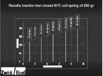
Fig. 2. Test to the dynamometer to quantify the practiced force in the distalization of the molar from the open coil spring of 5 mm in length when it is released by 2 mm.
Fig. 2. Test to the dynamometer to quantify the practiced force in
the distalization of the molar from the open coil spring of 5 mm in length when it is released by 2 mm.
21
Fig. 3. Test by the dynamometer to quantify the practiced force on the bicuspid, from the closed coil spring of 5 mm of length when it is activated by 2 mm.
Fig. 3. Test by the dynamometer to quantify the practiced force
on the bicuspid, from the closed coil spring of 5 mm of length when it is activated by 2 mm.
6 x 3 matrix, reflecting the sampling site (mesial or distal), the time points, and the 3 treatments. These 3 factors were used in a repeated measure 3-way ANOVA (50) to assess the data of GCF ALP activity. Furthermore, to test the simple main effect of each factor, 1-way repeated measures ANOVAs were performed to evaluate the significance of differences in ALP activity among the experimental groups at each time point and across times within each group in both mesial and distal sites. Bonferroni-corrected paired Student t tests were used as a
pair-wise comparison procedure when appropriate. The significance of differences in ALP activities between mesial and distal sites for the teeth categories at each time point were assessed with a paired Student t test as a post hoc procedure. ALP value less than 0.05 was accepted as being statistically significant.
Evaluation of the reaction force on the first bicuspid by photoelastic resin and a mathematical model. A molar and a bicuspid were put into the photoelastic resin. At different times, a non-compliance distalize appliance was inserted. Once a traditional appliance, and another time a 2FDA device. The purpose was to verify, by the change in coloration of the resin, the different load which involved the anchorage bicuspid submitted to stress.
The purpose was to understand how much reaction force arrived on the anchorage tooth. With 2FDA we were able to analyze the force produced by the open coil spring used to distalize the molar when this last was unloaded by 2 mm, and how much force produced the closed coil spring when it was activated by 2mm (Fig. 2, 3). Thus, knowing the forces practiced by the 2 coil springs and the quantity of deflection of each, it was possible to realize a mathematical model (Kpm Δxpm - Km Δxm / 2 where Kpm = coefficient of elasticity of the closed coil spring, Km = coefficient of elasticity of the open coil spring and Δx = variation of length of the coil spring) that allowed us to quantify the reaction force that arrived on the bicuspid anchorage tooth.
RESULTS
Evaluation of osteoblastic activity on mesial and distal
sites of the first molar and first bicuspid. The clinical
parameters had similar scores in both experimental
sites at baseline, without any statistically significant
differences. On day 45, all clinical parameters of the
TMs, CMs, TBs and CBs were significantly worse than
at the baseline; conversely, in the AMs, the parameters
did not show significant changes. At this point in the
experiment, the cross-sectional analysis also showed
a significant difference in the clinical parameters from
the 2 groups. The pair-wise comparison tests showed
that the significance was due to the differences in the
clinical data from the AMs compared with those from
the TMs, CMs, TBs and CBs. Within these TMs, CMs,
TBs and CBs group parameters there were no statistically
significant differences between the mesial and distal sites.
The means and SDs of GCF volume in microliters for
each experimental group were 0.14 ± 0.08 in the TMs,
0.14 ± 0.07 in the CMs, and 0.12 ± 0.06 in the AMs,
with a significant difference between the groups (1-way
ANOVA; P ≤ 0.01). GCF volume was significantly greater
in the TMs and TBs compared with the CMs and CBs (P
≤ 0.01 The 3-way repeated measures ANOVA reveals
that the subjects demonstrated significant differences in
GCF ALP activity levels between the time points (F-ratio
= 13.23; P ≤ 0.01), the treatments (F-ratio = 37.07; P ≤
0.01), and the sites (F-ratio = 11.46; P ≤ 0.01). In addition,
the interactions of treatments with times (F-ratio = 7.83;
P ≤ 0.01) and with sites (F-ratio = 6.43; P ≤ 0.02) were
22
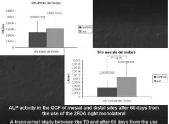
Fig. 4. ALP activity on mesial site (traction site) and on distal site (compression site).
Fig. 4. ALP activity on mesial site (traction site) and on distal
site (compression site).
23
Fig. 5. ALP activity on mesial site (compression site) and on distal site (traction site). The explanation for which it increases the osteoblastic activity on the site of compression is explained by the biomechanics of the appliance. In fact the direction of action of the closed coil spring is backwards. To the periodontal receptors of the mesial site (of compression), relative information is sent to an action of traction; vice versa for the distal site. The 2FDA are then able to turn the site of compression into one of traction inducing an osteoblastic production more important than that of osteoclastic. Therefore, the supposition that the bicuspid progress toward the mesial direction, losing anchorage, does not exist.
Fig. 5. ALP activity on mesial site (compression site) and
on distal site (traction site). The explanation for which it increases the osteoblastic activity on the site of compression is explained by the biomechanics of the appliance. In fact the direction of action of the closed coil spring is backwards. To the periodontal receptors of the mesial site (of compression), relative information is sent to an action of traction; vice versa for the distal site. The 2FDA are then able to turn the site of compression into one of traction inducing an osteoblastic production more important than that of osteoclastic. Therefore, the supposition that the bicuspid progress toward the mesial direction, losing anchorage, does not exist.
also significant.
One-way ANOVA showed a statistically significant
change in enzyme activity only in the TM and TB groups,
in mesial sites, among the repeated samplings during the
study period (Fig. 4, 5). Results of pair-wise comparisons
show a significantly greater enzymatic activity in mesial
sites from the TM group from day 1 to the end of the
experiment, as compared with the baseline. Conversely, in
the CM and CB group, over the study period no significant
statistical difference in ALP activities was seen from day
1 to the end of the experiment, as compared with the
baseline. At baseline and at 1 hour, in both distal and
mesial sites, ALP activity was similar in the all groups,
without significant differences (1-way ANOVA). In distal
sites, statistically significant differences both between the
TMs and CMs and between TBs and CBs were seen from
day 1 to the end of the experiment.
Evaluation of the reaction force on the first bicuspid
by photoelastic resin and a mathematical model. The
photoelastic resin showed that with the traditional
non-compliance distalize appliance the resin around all the
bicuspid root was completely discoloured. Therefore,
the majority of the reaction force was directed on the
tooth. Instead, with the 2FDA, the resin around the root
of the bicuspid remained absolutely transparent. No force
involved the root of the first bicuspid.
Meanwhile, on the molar, the load of the force was
more evident with the 2FDA than with a traditional
non-compliance distalize appliance. The explanation for
this can be found in the different quantity of force that
loads the molar and the bicuspid, as underlined by the
mathematical model previously described.
DISCUSSION
Evaluation of osteoblastic activity on mesial and distal
sites of the first molar and first bicuspid and evaluation
of the rate of the reaction force on the first bicuspid. The
evaluation of the osteoblastic activity by monitoring the
alkaline phosphatase (ALP), statistically underlines (p
≤ 0.05) a meaningful enzymatic increase at the site of
tension (mesial site) of the molar test (TM group) both
in comparison to the T0 and in comparison to the molar
control (CM group). This is in accord with the physiology
of the dental movement: in the bone remodelling in the
site of tension the osteoblastic activity has to prevail to
allow an apposition of the bone. Vice versa, in the site of
compression an osteoclastic activity prevails to allow the
tooth to move in the direction imposed by the orthodontic
force. Around the first bicuspid, tooth anchorage of the
appliance, the site of traction is the distal site, while that
of compression is the mesial one.
The statistic analysis of the enzymatic activity of
the ALP statistically underlines (p ≤ 0.05) a meaningful
increase of the enzyme on the compression site (mesial).
Such data results in contrast with the physiology of the
dental movement, for which the osteoblastic activity
has to be reduced in favour of an osteoclastic one in the
compression site. This results from the direction of the
application of the orthodontic force.
These results are justifiable according to the
biomechanics of the appliance. The presence of the closed
coil spring on the first bicuspid turns the mesial site of the
bicuspid from a compression site into a traction site. This
is possible because the coil spring once activated (open)
aims to return (because of its elasticity) to its initial
position producing stimuli of traction in the periodontal
receptors of the mesial site of the first bicuspid.
This data allows us to better understand the clinical
results related to the measurement of the loss of anchorage.
In fact, loading the site of compression on the anchorage
tooth leads to a prevalence of an osteoblastic activity or
an apposition of bone, otherwise it is not possible for the
same tooth to move mesially. It may induce movements
of rotation and/or of tipping but not of translation of the
tooth which represents the true loss of anchorage.
To better justify the 2FDA innovative philosophy, we
should underline that the quantity of the reaction force that
involves the first bicuspid is low. This data is confirmed
both by experimentation effected on photoelastic resin
and from the realization of a mathematical model which
made it possible for us to calculate the quantity of reaction
force that involves the anchorage tooth.
The 2FDA experimentation on the photoelastic resin
underlined that there is no discoloration and therefore
no load around the first bicuspid, unlike the traditional
non-compliance distalize system. This allows us to
deduce that if no force involves the tooth, it will not be
possible to move it. In order to clarify how much of the
force of reaction involved the anchorage tooth we used a
mathematical model (see Results.) which allowed us to
show that such force was equal to 8.34 grams. In relation
to the original 75 grams of the force of reaction, this value
represents 11% of all the reaction force. Such data seem
to explain both the clinical and enzymatic result related to
the loss of anchorage. In fact, 8.34 grams is equal to half
of the pressure of the capillary pressure of the periodontal
ligament (18 grams) (51), and is an insufficient force
to induce the secretion of the osteoclastic and
pre-osteoblastic vasoactive substances.
In conclusion, the Friction Free Distalize Appliance
(2FDA) is a fixed appliance designed to produce
distalization of maxillary first molars. This device
constitutes an effective and predictable method for the
correction of a Class II malocclusion for which no patient
co-operation is required.
The present study produced the following findings
regarding the use of the 2FDA appliance for the distal
movement of maxillary first molars during the correction
of Class II malocclusions.
1. Class II molar relationships were corrected to Class I
in about five months.
2. The distalizing force on the maxillary molar resulted
in 88.64% molar distalization and 11.36%
reciprocal anchorage loss measured at the maxillary
first premolar. This division is more favorable than
that reported in studies on other intraoral methods of
molar distalization
3. The maxillary first molars were moved distally an
average of 3.3 mm on side test. Net distalization was
less than that seen with the pendulum.
4. Anchorage loss, measured at the first premolars, was
0.43 mm one side test.
5. Evaluation of osteoblastic activity on mesial and
distal sites of the first molar and first bicuspid
showed a statistical change in enzyme ALP activity
only in the mesial site of the first molar and first
bicuspid of the test group.
It is important to note that the mesial site of the molar
is the tension site of a tooth subordinate to orthodontic
force, for this reason it is normal that the activity of this
enzyme is higher than the base-line.
Instead, is not normal that the ALP activity increases
at the mesial site of the first bicuspid because this site
represents a compression site of a tooth subordinate
to orthodontic force. The hypothesis of this event is
explained in the Discussion section.
ACKNOWLEDGEMENTS
We thank Prof Arcangelo Merla (ITAB – Institute of
Advanced Biomedical Technology) for the realization
of the mathematical model, and Mr Ugo Comparelli,
orthodontic technician of Foggia, Italy, who realized the
Friction Free Distalize Appliance (2FDA).
REFERENCES
1. Graber T.M. 1955. Extraoral force—facts and fallacies. Am. J. Orthod. 41:490.
2. Gould E. 1957. Mechanical principles in extraoral anchorage. Am. J. Orthod. 17:319.
3. Klein P.L. 1957. An evaluation of cervical traction on the maxilla and the upper first permanent molar. Angle Orthod. 27:61.
4. Newcomb M.R. 1958. Some observations on extraoral treatment. Angle Orthod. 28:131.
5. Kloehn S.J. 1961. Evaluation of cervical anchorage force in treatment. Angle Orthod. 31:91.
6. Greenspan R.A. 1970. Reference charts for the controlled extraoral force application to the maxillary molars. Am. J. Orthod. 58:486.
7. Hubbard G.W., R.S Nanda and G.F Currier. 1994.
A cephalometric evaluation of non-extraction cervical headgear treatment in Class II malocclusions. Angle Orthod. 64:359.
8. Tullouch J.F.C., C. Phillips and W.R. Proffit. 1997. The effect of early intervention on skeletal pattern in Class II malocclusion: a randomized clinical trial. Am. J. Orthod. Dentofacial Orthop. 111:391.
9. Cetlin N.M. and H.A. Ten.. 1983. Non-extraction treatment. J. Clin. Orthod. 17:396.
10. Muse D.S., M.J. Fillman, W.J. Emmerson and R.D.
Mitchell. 1993. Molar and incisor changes with the Wilson
rapid molar distalization. Am. J. Orthod. Dentofacial Orthop. 104:556.
11. Sinclair P.M.. 1994. The reader’s corner. J. Clin. Orthod. 28:361.
12. Behrents R.G. 1996. Iatrogenic problems associated with the clinical practice of orthodontics. In: Orthodontic treatment: the management of unfavorable sequelae. Craniofacial Growth Series 31. J.A. McNamara Jr, C. Trotman eds. Ann Arbor, Mich: Center for Human Growth and Development, The University of Michigan 1996, p. 1. 13. Koltun A. and G.C. Stone.. 1986. Past and current trends
in patient non-compliance research: focus on diseases, regimes-programs and provider-disciplines. J. Compl. Health Care 1:21.
14. Sahm G., A. Bartsch and E. Witt. 1990. Reliability of patient reports on compliance. Eur. J. Orthod. 2:438. 15. Nanda R.S. and M.J. Kieri. 1992. Prediction of
co-operation in orthodontic treatment. Am. J. Orthod. 102:15. 16. Cureton S.L., F.J. Regennitter and J.M. Yancy. 1993.
Clinical versus quantitative assessment of headgear compliance. Am. J. Orthod. Dentofacial Orthop. 104:277. 17. Sahm G., A. Bartsch and E. Witt. 1990. Micro-electronic
monitoring of functional appliance wear. Eur. J. Orthod. 12:297.
18. Gianelly A.A., A.S Vaitas, W.M. Thomas and D.G.
Berger. 1988. Distalization of molars with repelling
magnets. J. Clin. Orthod. 22:40.
19. Gianelly A.A.. 1998. Distal movements of maxillary molars. Am. J. Orthod. Dentofacial Orthop. 114:66. 20. Jones R.D. and M.J. White. 1992. Rapid Class II molar
correction with an open-coil jig. J. Clin. Orthod. 26:661. 21. Hilgers J.J. 1992. The pendulum appliance for Class II
non-compliance therapy. J. Clin. Orthod. 26:700.
22. Carano A. and M Testa. 1996. The distal jet for upper molar distalization. J. Clin. Orthod. 30:374.
23. Wilson W.I. 1978. Modular orthodontic system. J. Clin. Orthod. 12:259.
the bimetric “distalizing” arch. Semin. Orthod. 6:106. 25. Ghosh J. and R.S. Nanda. 1996. Evaluation of an
intraoral maxillary molar distalization technique. Am. J. Orthod. Dentofacial Orthop. 110:639.
26. Chaqués-Asensi J. and V. Kalra. 2001. Effects of the pendulum appliance on the dentofacial complex. J. Clin. Orthod. 35:254.
27. Bussick T.J. and J.A. McNamara Jr. 2000. Dentoalveolar and skeletal changes associated with the pendulum appliance. Am. J. Orthod. Dentofacial Orthop. 117:333. 28. Brickman C.D., P.K. Sinha and R.S. Nanda. 2000.
Evaluation of the Jones jig appliance for distal molar movement. Am. J. Orthod. Dentofacial Orthop. 118:526. 29. Ghosh J. and R.S. Nanda. 1996. Evaluation of an intraoral
maxillary molar distalization technique. Am. J.Orthod. Dentofacial Orthop. 110:639.
30. Chiu P.P.. 2001. A Comparison of Two Intraoral Molar Distalization Appliances:Distal Jet Versus Pendulum Appliance [unpublished master’s thesis]. Ann Arbor, Mich: Department of Orthodontics, University of Michigan, p. 1. 31. Chaque´s-Asensi J. and V. Kalra. 2001. Effects of the
pendulum appliance on the dentofacial complex. J. Clin. Orthod. 35:254.
32. Byloff F.K. and M.A. Darendeliler. 1997. Distal molar movement using the pendulum appliance. Part 1: clinical and radiological evaluation. Angle Orthod. 67(4):249. 33. Joseph A.A. and C.J. Butchart. 2000 An evaluation of the
pendulum ‘‘distalizing’’ appliance. Semin. Orthod. 6:129. 34. Haydar S. and O. Uner. 2000. Comparison of Jones jig
molar distalization appliance with extraoral traction. Am. J. Orthod. Dentofacial Orthop. 117:49.
35. Runge M.E., J.T. Martin and F. Bukai. 1998. Analysis of rapid maxillary molar distal movement without patient cooperation. Am. J. Orthod. Dentofacial Orthop. 115:153. 36. Gulati S., O.P. Kharbanda and H. Parkash. 1998. Dental
and skeletal changes after intraoral molar distalization with sectional jig assembly. Am. J. Orthod. Dentofacial Orthop. 114:319.
37. Bolla E., F. Muratore, A. Carano and S.J. Bowman. 2002. Evaluation of maxillary molar distalization with the distal jet: a comparison with other contemporary methods. Angle Orthod. 72(5):481.
38. Reitan K.. 1967. Clinical and histological observations on tooth movement during and after orthodontic treatment. Am. J. Orthod. 53:721.
39. Storey E. 1973. The nature of tooth movement. Am. J. Orthod. 63:292.
40. Robinson R. 1923. The possible significance of hexosephosphoric esters in ossification. Biochem. J. 17:286. 41. Perinetti G., M. Paolantonio, E. Serra, D. D’Archivio,
S. D’Ercole, F. Festa and G. Spoto. 2004. Longitudinal
monitoring of subgingival colonization by Actinobacillus actinomycetemcomitans, and crevicular alkaline phosphatase and aspartate aminotransferase activities around orthodontically treated teeth. J. Clin. Periodontol. 31:60. 42. Perinetti G., M. Paolantonio, M. D’Attilio, D.
D’Archivio, D. Tripodi, B. Femminella, F. Festa and G. Spoto. 2002. Alkaline phosphatase activity in gingival
crevicular fluid during human orthodontic tooth movement. Am. J. Orthod. Dentofacial Orthop. 122:548.
43. Grieve W.G. III, G.K. Johnson, R.N. Moore, R.A.
Reinhardt and L.M. DuBois. 1994. Prostaglandin E
(PGE) and interleukin-1 beta (IL-1 beta) levels in gingival crevicular fluid during human orthodontic tooth movement. Am. J. Orthod. Dentofacial Orthop. 105:369.
44. Lowney J.J., L.A. Norton, D.M. Shafer and E.F.
Rossomando. 1995. Orthodontic forces increase tumor
necrosis factor in the human gingival sulcus. Am. J. Orthod. Dentofacial Ortho.p 108:519.
45. Uematsu S., M. Mogi and T. Deguchi. 1996. Interleukin (IL)-1 beta, IL-6 tumor necrosis factor-alpha, epidermal growth factor, and beta 2-microglubulin levels are elevated in gingival crevicular fluid during human orthodontic tooth movement. J. Dent. Res. 75:562.
46. Griffiths G.S., A.M. Moulson, A. Petrie and I.T. James. 1988. Evaluation of osteocalcin and Peridinium crosslinks of bone collagen as markers of bone turnover in gingival crevicular fluid during different stages of orthodontic treatment. J. Clin. Periodontol. 25:492.
47. Paolantonio M., G. Di Placido, V. Tumini, M. Di
Stilio, A. Contento and G. Spoto. 2000. Aspartate
amonotransferase activity in crevicular fluid from dental implants. J. Periodontol. 71:1151.
48. Bowers G.N. Jr and R.B. McComb. 1966. A continuous spectrophotometric method for measuring the activity of serum alkaline phosphatase. Clin. Chem. 12:1
49. Glantz S.A. 1988. Primer of biostatistics. 2nd ed. New York: McGraw-Hill, p. 1.
50. Geisser S. and S.W. Greenhouse. 1958. An extension of Box’ s results on the use of the F distribution in multivariate analusis. Ann. Math. Sta.t 12:401.
51. Main J.H. and D. Adams. 1966. Experiments on the rat incisor into the cellular proliferation and blood-pressure theories of tooth eruption. Arch Oral Biol. 11:163.