Acute Cocaine Enhances Dopamine D
2R Recognition and Signaling
and Counteracts D
2R Internalization in Sigma1R-D
2R Heteroreceptor
Complexes
Dasiel O. Borroto-Escuela1,2,3&Manuel Narváez4&Wilber Romero-Fernández5&Luca Pinton1&Karolina Wydra6&
Malgorzata Filip6&Sarah Beggiato7&Sergio Tanganelli7&Luca Ferraro7&Kjell Fuxe1
Received: 4 December 2018 / Accepted: 20 March 2019 / Published online: 10 April 2019 # The Author(s) 2019
Abstract
The current study was performed to establish the actions of nanomolar concentrations of cocaine, not blocking the dopamine transporter, on dopamine D2 receptor (D2R)-sigma 1 receptor (δ1R) heteroreceptor complexes and the D2R protomer
recogni-tion, signaling and internalization in cellular models. We report the existence of D2R-δ1R heteroreceptor complexes in
subcor-tical limbic areas as well as the dorsal striatum, with different distribution patterns using the in situ proximity ligation assay. Also, through BRET, these heteromers were demonstrated in HEK293 cells. Furthermore, saturation binding assay demonstrated that in membrane preparations of HEK293 cells coexpressing D2R andδ1R, cocaine (1 nM) significantly increased the D2R Bmax
values over cells singly expressing D2R. CREB reporter luc-gene assay indicated that coexpressedδ1R significantly reduced the
potency of the D2R-like agonist quinpirole to inhibit via D2R activation the forskolin induced increase of the CREB signal. In
contrast, the addition of 100 nM cocaine was found to markedly increase the quinpirole potency to inhibit the forskolin-induced increase of the CREB signal in the D2R-δ1R cells. These events were associated with a marked reduction of cocaine-induced
internalization of D2R protomers in D2R-δ1R heteromer-containing cells vs D2R singly expressing cells as studied by means of
confocal analysis of D2R-δ1R trafficking and internalization. Overall, the formation of D2R-δ1R heteromers enhanced the ability
of cocaine to increase the D2R protomer function associated with a marked reduction of its internalization. The existence of
D2R-δ1R heteromers opens up a new understanding of the acute actions of cocaine.
Keywords Cocaine . Sigma 1 receptor . Dopamine D2 receptor . Heteroreceptor complexes . Oligomerization . Dimerization . Substance use disorder
Dasiel O. Borroto-Escuela and Manuel Narváez these authors have con-tributed equally to this work.
* Dasiel O. Borroto-Escuela [email protected] * Kjell Fuxe [email protected] Manuel Narváez [email protected] Wilber Romero-Fernández [email protected] Luca Pinton [email protected] Karolina Wydra [email protected] Malgorzata Filip [email protected] Sarah Beggiato [email protected] Sergio Tanganelli [email protected] Luca Ferraro [email protected]
Introduction
It recently became clear that cocaine in the nanomolar range can enhance dopamine D2 receptor (D2R) functions without
blocking the dopamine uptake mechanism as found in neuro-chemical and behavioral work [1,2]. These results indicated that cocaine can exert a direct or indirect positive allosteric modulation of the striatal D2R in line with the demonstration
of a cocaine-induced enhancement of Gi/o coupling at striatal D2R [2]. Such actions are likely of relevance for the rewarding
and relapse-induced effects of cocaine in view of the major role of accumbal D2R in mediating these actions of cocaine [3,
4]. The inhibitory D2R are enriched in the dorsal and ventral
striato-pallidal GABA neurons, the ventral component of which is an anti-reward system [5] which becomes inhibited by D2R activation [6].
It is known that sigma 1 receptors (δ1R) exist in substantial densities in large numbers of central neurons including accumbal neurons [7,8]. Cocaine was found to interact with δ1R and δ1R antagonists diminished cocaine actions [9–11]. Instead, reinforcing effects of cocaine self-administration were observed withδ1R agonists [12]. The molecular mech-anism of cocaine actions in the brain can probably involve δ1R-D1R and δ1R-D2R heteromerization [13–15]. Using
BRET,δ1R-D1R heteroreceptor complexes were
demonstrat-ed in cotransfectdemonstrat-ed cells [13]. The evidence suggested a direct involvement of δ1R in mediating the cocaine-induced (150μM) enhancement of D1R signaling over the
Gs-AC-PKA signaling pathway.
δ1R-D2R heteroreceptor complexes were also
demonstrat-ed both in cotransfectdemonstrat-ed cells and in striatum [14]. In cotransfected cells, cocaine in the micromolar range (30μM) was able to partially counteract the activation by the D2R agonist quinpirole of the Gi/o-mediated signaling of
the D2R. This inhibitory action of cocaine on D2R-mediated
Gi/o signaling was mediated via theδ1R protomer [14]. These results were in sharp contrast to results obtained with 100 nM of cocaine which demonstrated that cocaine enhanced the quinpirole-induced D2R signaling as studied on accumbal
ex-tracellular dopamine levels and on the efficacy of dopamine to stimulate binding of GTPγS to striatal D2-like receptors [2].
Furthermore, the locomotor actions of quinpirole were en-hanced by sub-threshold doses of cocaine. Recently, indica-tions were obtained that D2R-δ1R complexes exist on striatal
dopamine and glutamate nerve terminals. Nanomolar concen-trations of cocaine were found to enhance the Gi/o-mediated D2R signaling in such complexes [16]. The possible existence
of D2R-δ1R-N-type calcium channel heteroreceptor
com-plexes was proposed [16].
The current study was performed to establish the actions of nanomolar concentrations of cocaine, not blocking the dopa-mine transporter, onδ1R-D2LR andδ1R-D2SR heteroreceptor
complexes and the D2R protomer recognition, signaling, and
internalization in cellular models. Furthermore, the distribu-tion of theδ1R-D2R complexes in the ventral and dorsal
stri-atum was evaluated using the in situ proximity ligation assay.
Materials and Methods
Animals
All experiments were performed using male Sprague-Dawley rats (SD) (Scanbur, Sweden). The animals were group-housed under standard laboratory conditions (20–22 °C, 50–60% hu-midity). Food and water available ad libitum. All studies in-volving animals were performed in accordance with the Stockholm North Committee on Ethics of Animal Experimentation, the Swedish National Board for Laboratory Animal and European Communities Council Directive (2010/63/EU) guidelines for accommodation and care of Laboratory Animals.
Plasmid Constructs, Cell Culture, and Transfection
The constructs presented herein were made using standard molecular biology as described previously [17–19]. HEK293T cells were grown and transiently transfected as depicted in Borroto-Escuela et al. [20].
Brain Tissue Samples and their Preparation
First, animals were deeply anesthetized by an intraperitoneal (i.p.) injection of a high dose of pentobarbital (60 mg/ml, [0.1 ml/100 g]) and then perfused intracardially with 30– 50 mL ice-cold 4% paraformaldehyde (PFA) in 0.1 M phosphate-buffered saline (PBS, pH 7.4) solution. After per-fusion, brains were collected and transferred into well-labeled glass vials filled with 4% PFA fixative solution for 6–12 h. Then, the brains were placed in 10% and 30% sucrose (0.1 M PBS, pH 7.4) and incubated for 1 day (10% sucrose) and a number of days (30% sucrose) at 4 °C with several sucrose buffer changes, until freezing the brain. The brains were fro-zen with isopentane and then sectioned (10–30 μm-thick) using a cryostat. The brain’s slices were stored at − 20 °C on Hoffman solution.
In Situ Proximity Ligation Assay
To study the formation of the D2R-δ1R heteroreceptor
com-plexes, the in situ proximity ligation assay (in situ PLA) was performed as described previously [20–23]. Free-floating for-malin-fixed brain sections (30μm) at Bregma level (1.0 mm) from untreated Sprague–Dawley rats were employed using the following primary antibodies: mouse monoclonal anti-D2R (MABN53, 1:600, Millipore, Sweden) and rabbit monoclonal
anti-sigma1R (ab53852, 1:500, Abcam, Sweden). Control ex-periments employed only one primary antibody. The PLA sig-nal was visualized and quantified by using a confocal micro-scope Leica TCS-SL confocal micromicro-scope (Leica, USA) and the Duolink Image Tool software. Briefly, fixed free-floating rat brain sections (storage at− 20 °C in Hoffman solution) were washed four times with PBS and quenched with 10 mM glycine buffer (0.75 g glycine in 100 ml PBS), for 20 min at room temperature. Glycine buffer is used to reduce unspecific anti-bodies binding and brain tissue autofluorescence. Then, after three PBS washes, incubation took place with a perme-abilization buffer (10% fetal bovine serum (FBS) and 0.5% Triton X-100 or Tween 20 in Tris buffer saline (TBS), pH 7.4) for 30 min at room temperature. Again, the sections were washed twice, 5 min each, with PBS at room temperature and incubated with the blocking buffer (0.2% BSA in PBS) for 30 min at room temperature. The brain sections were then in-cubated with the primary antibodies diluted in a suitable con-centration in the blocking solution for 1–2 h at 37 °C or at 4 °C overnight. The day after, the sections were washed twice, and the proximity probe mixture was applied to the sample and incubated for 1 h at 37 °C in a humidity chamber. The unbound proximity probes were removed by washing the slides twice, 5 min each time, with blocking solution at room temperature under gentle agitation, and the sections were incubated with the hybridization-ligation solution (BSA (250 g/ml), T4 DNA li-gase (final concentration of 0.05 U/μl), Tween-20 (0.05%), NaCl 250 mM, ATP 1 mM, and the circularization or connector oligonucleotides (125–250 nM)) and incubated in a humidity chamber at 37 °C for 30 min. The excess of connector oligo-nucleotides was removed by washing twice, for 5 min each, with the washing buffer A (Sigma-Aldrich) Duolink Buffer A (8.8 g NaCl, 1.2 g Tris Base, 0.5 ml Tween 20 dissolved in 800 ml high purity water, pH to 7.4) at room temperature under gentle agitation, and the rolling circle amplification mixture was added to the slices and incubated in a humidity chamber for 100 min at 37 °C. Then, the sections were incubated with the detection solution in a humidity chamber at 37 °C for 30 min. In a last step, the sections were washed twice in the dark, for 10 min each, with the washing buffer B (Sigma-Aldrich, Duolink Buffer B (5.84 g NaCl, 4.24 g Tris Base, 26.0 g Tris-HCl. Dissolved in 500 ml high purity water, pH 7.5) at room temperature under gentle agitation. The free-floating sections were put on a microscope slide and a drop of appropriate mounting medium (e.g., VectaShield or Dako) was applied. The cover slip was placed on the section and sealed with nail polish. The sections were protected against light and stored for several days at− 20 °C before confocal microscope analysis.
BRET
2Saturation Assay
The BRET2 saturation experiment was performed as de-scribed previously, see [17, 24]. Forty-eight hours after
transfection, HEK293T cells, transiently transfected with con-stant (1μg) or increasing amounts (0.12–5 μg) of plasmids encoding for D2RRlucandδ1RGFP2, respectively, were rapidly washed twice in PBS, detached, and resuspended in the same buffer. Cell suspensions (20μg protein) were put in duplicates into the 96-well microplate black plates with a transparent bottom (Corning 3651) (Corning, Stockholm, Sweden) for fluorescence measurement or white plates with a white bottom (Corning 3600) for BRET determination. For BRET2 mea-surements, coelenterazine-400a also calledDeepBlue™C sub-strate (VWR, Sweden) was used at a final concentration of 5 μM. The readings were made 1 min after using the POLARstar Optima plate-reader (BMG Labtechnologies, Offenburg, Germany) that allows the sequential integration of the signals observed with two filter settings [410 nm (80 nm bandwidth) and 515 nm (30 nm bandwidth)]. The BRET2ratio is defined as previously described by Borroto-Escuela et al. [25]. Briefly, data are represented as a normal-ized BRET2 ratio, which is defined as the BRET ratio for coexpressed Rluc and GFP2 constructs normalized against the BRET ratio found for the Rluc expression construct alone in the same experiment: BRET2 ratio = [(GFP2emission at 515 ± 30 nm)/(Rluc emission 410 ± 80 nm)]– cf. The correc-tion factor, cf., corresponds to (emission at 515 ± 30 nm)/ (emission at 410 ± 80 nm) found with the Receptor-Rluc con-struct expressed alone in the same experiment.
BRET
2Competition Assay
Forty-eight hours after transfection, HEK293T cells transient-ly transfected with constant amounts (1 μg) of plasmids encoding for D2LRRlucandδ1RGFP2and increasing amounts
(0.1–8 μg) of plasmids encoding for wild-type D2LR, D2SR or
δ1R and the mock pcDNA3.1+, respectively. The energy transfer was determined as described for the BRET2saturation assay.
[
3H]-Raclopride Competition Binding Experiments
Competition experiments of quinpirole (0.3 nM-3 mM) versus the D2-likeR antagonist [3H]-raclopride (2 nM; specific activ-ity 78.1 Ci/mmol, PerkinElmer Life Sciences, Stockholm, Sweden) were carried out by membrane (20 μg per well) incubation at 30 °C for 90 min. Non-specific binding was defined by radioligand binding in the presence of 10 μM (+)-butaclamol (Sigma-Aldrich, Stockholm, Sweden). The in-cubation was terminated by rapid filtration through hydrophil-ic (LPB) Durapore ®Membrane (Millipore, Stockholm, Sweden) using a MultiScreen™ Vacuum Manifold 96-well (Millipore Corp, Bedford, MA), followed by five washes (200 μl per wash) with ice-cold washing buffer (50 mM Tris–HCl pH 7.4). The filters were dried, 4 ml of scintillation
cocktail was added, and the bound ligand was determined after 12 h by liquid scintillation spectrometry.
[
3H]-Raclopride Saturation Binding Experiments
Saturation binding experiments with the D2-likeR antagonist [3H]-raclopride (specific activity 82.8 Ci/mmol, PerkinElmer Life Sciences, Sweden) were performed in membrane prepa-rations from single and cotransfected HEK cells (100μg pro-tein/ml) incubated with increasing concentrations of [3H]-raclopride (ranging from 0.1 nM to 12 nM) in 250μl of incu-bation buffer (50 mM Tris–HCl, 100 mM NaCl, 7 mM MgCl2, 1 mM EDTA, 0.05% BSA and 1 mM dithiothreitol)
for 60 min at 30 °C in the presence or absence of cocaine (1 nM, 10 nM, and 100 nM) and the high affinity sigma 1 receptor antagonist PD144418 [26]. Non-specific binding was defined by radioligand binding in the presence of 10μM (+) butaclamol (Sigma Aldrich, Sweden). The incubation was ter-minated by rapid filtration Whatman GF/B filters (Millipore Corp, Sweden) using a MultiScreen™ Vacuum Manifold 96-well followed by three washes (~ 250μl per wash) with ice-cold washing buffer (50 mM Tris–HCl pH 7.4). The filters were dried, 5 ml of scintillation cocktail was added, and the bound ligand was determined after 12 h by liquid scintillation spectrometry.
CREB Luciferase Reporter Gene Assay
A dual luciferase reporter assay has been used to indirectly detect variations of cAMP levels in transiently transfected cell lines treated with different compounds in a range of concen-trations (typically 0.1 nM to 1μM). For luciferase assays, 24 h before transfection, cells were seeded at a density of 1 × 10⁶cells/well in 6-well dishes and transfected with Fugene (Promega, Stockholm, Sweden). Cells were co-transfected with plasmids corresponding to the four constructs as follows (per 6-well): 2μg firefly luciferase-encoding experimental plasmid (pGL4-CRE-luc2p; Promega, Stockholm, Sweden), 1μg of D2R plus σ1R expression vectors, and 0.5 μg Rluc-encoding internal control plasmid (phRG-B; Promega). Approximately, 46 h post-transfection, cells were incubated with appropriate ligands and harvested with passive lysis buff-er (Promega, Stockholm, Sweden). The lucifbuff-erase activity of cell extracts was determined using the Dual-Luciferase® Reporter (DLR™) Assay System according to the manufac-turer’s protocol Promega, Stockholm, Sweden) in a POLAR star Optima plate reader (BMG Labtechnologies, Offenburg, Germany) using a 535-nm filter with a 30-nm bandwidth. Firefly luciferase was measured as firefly luciferase lumines-cence over a 15-s reaction period. The luciferase values were normalized against Rluc luminescence values (Luc/Rluc ra-tio). Chemicals used for the gene reporter assays
(Raclopride, quinpirole, forskolin) were purchased from Tocris (UK) and (cocaine-HCL) Sigma Aldrich (Germany).
Receptor Internalization Analysis by Fluorescence
Confocal Microscopy
Internalization of D2R were evaluated by fluorescence
confo-cal microscopy using transiently single or cotransfected HEK293T cells with constant (1 μg) amounts of plasmids encoding for Sigma1R and D2LRYFP. Cells were incubated
with cocaine (100 nM) at different interval times or with a range of cocaine concentrations (1 nM–10 μM) during 30 min. Then, cells were fixed in 4% paraformaldehyde for 10 min, washed with PBS containing 20 mM glycine, and mounted in a Vectashield immunofluorescence medium (Vector Laboratories, UK). Microscope observations were performed with a × 63 oil immersion objective in a Leica TCS-SL confocal microscope (Leica, USA). The amounts of internalized D2RYFPare shown as a single z-scan image. The
rate of internalization was measured as the ratios (IntROI/ membROI). The memROI is obtained by measuring the D2RYFPfluorescence area detected at the cell surface
mem-brane depicted by phase contrast). The IntROI is instead ob-tained by measuring the entire D2RYFPfluorescence area
de-tected in the cytoplasm of the entire cell. The basal value is obtained from cells not exposed to cocaine.
Statistical Analysis
The number of samples (n) in each experimental condition is indicated in figure legends. Data from competition experi-ments were analyzed by nonlinear regression analysis using GraphPad Prism 5.0 (GraphPad Software Inc., San Diego, CA). The inhibition constants of the high and low affinity state of the receptor (pKiH, pKiL) from several independent
replica-tions were averaged allowing statistical comparisons using a one-way analysis of variance (ANOVA). Group differences after ANOVAs were measured by post hoc Turkey’s multiple comparison test. TheP value 0.05 and lower was considered significant. BRET2isotherms were fitted using a nonlinear regression equation assuming a single binding site, which provided BRETmax and BRET50 values.
Results
BRET
2Experiments on sigma1R and D2R
Cotransfected in HEK293 Cells
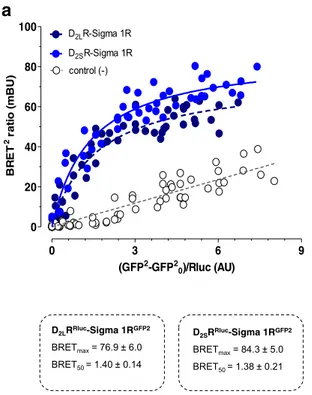
Saturation measurements were made with increasing amounts ofδ1RGFP2and a constant amount of D2LR and D2SR fused to
Renilla luciferase (Rluc). Similar saturable BRET2
curves were obtained with both D2R isoforms (Fig. 1a). The
BRETmax and BRET50 values did not differ between D2LR
and D2SR. Instead, a linear relationship was found between
acceptor and donor constructs in negative controls which consisted of a mixture of cells singly transfected withδ1R
GFP2
or D2LRRLuc(Fig.1a).
In competition experiments using increasing amounts of D2LR and D2SR cDNAs, similar displacement curves were
observed as seen from a 70% disappearance of the BRET2 signal in the δ1RGFP2-D2LRRluc heteroreceptor complex
using equal amounts of the two protomers (Fig.1b). In contrast, increasing amounts of theδ1R cDNA only caused a maximal 30% disappearance of the BRET2 signal (Fig.1b).
In Situ PLA Experiments on Distribution of D
2R-
δ1R
Heteroreceptor Complexes in Parts of the Rat
Forebrain
An overall high density of PLA-positive clusters was found in the dorsal striatum (Fig.2) based on the average number clusters per nucleus (in blue) per sample field. It was highly significantly increased vs values in negative controls and the myelinated bundles of the crus cerebri (CC) and the anterior limb of the anterior commissure (aca). In the nucleus accumbens shell, a medium density
of PLA-positive D2R-δ1R heteroreceptor complexes was found and in the nucleus accumbens core a low density (Fig. 2). They were both significantly different from the number of PLA-positive clusters in cc, aca, and negative controls regarded as background values.
Binding Experiments Using the D2 like Antagonist
3H-Raclopride in
δ1R and D
2LR Cotransfected
and D
2LR Singly Transfected HEK293 Cells
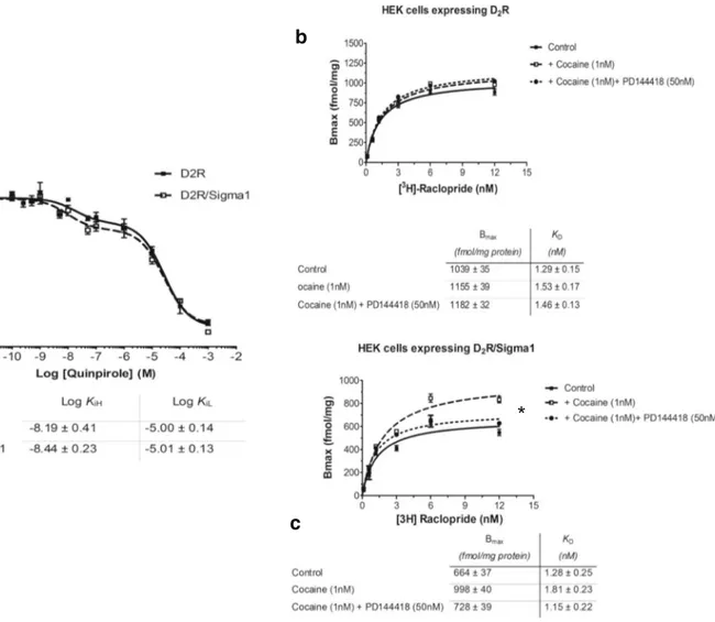
In competition experiments, the effects of quinpirole and co-caine were examined inδ1R and D2LR cotransfected HEK293
cells and in singly D2LR transfected cells. Quinpirole
pro-duced similar competition curves in the absence or presence ofδ1R (Fig.3a). The log values for the high affinity and low affinity D2 agonist binding sites (KiH and KiL) are found below Fig.3a. In saturation experiments, the effects of cocaine (1 nM, 10 nM, and 100 nM) were studied in cotransfected cells expressing both D2LR andδ1R and in singly transfected
cells only expressing D2LR. Cocaine 1 nM produced a
signif-icant increase in the Bmax values of the 3H-Raclopride bind-ing sites in the cotransfected cells but not in the D2LR singly
transfected cells (Fig.3b, c). Similar effects were also ob-served with cocaine 10 nM and 100 nM (data not shown). The Sigma-1R antagonist PD144418 (50 nM) blocked the
0 3 6 9 0 20 40 60 80 100 D2LR-Sigma 1R control (-) D2SR-Sigma 1R (GFP2-GFP2 0)/Rluc (AU) BR ET 2 ) U B m( oi t ar D2LRRluc-Sigma 1RGFP2 BRETmax= 76.9 ± 6.0 BRET50 = 1.40 ± 0.14 D2SRRluc-Sigma 1RGFP2 BRETmax= 84.3 ± 5.0 BRET50 = 1.38 ± 0.21
a
b
0 2 4 6 8 10 0 40 80 120 pcDNA3.1+ D2SR D2LR Sigma 1R ** *** *** *** *D2R - Sigma 1 BRET pair 1:1
* ** *** *** ** *** *** *** cDNA ( g) T E R B 2 l a n gi s ] e s n o p s er x a m f o %[
Fig. 1 a BRET2saturation experiments in HEK cells. Cotransfection of
D2LRlucor D2SRRlucand Sigma 1RGFP2form D2LRRluc-Sigma 1RGFP2
and D2SRRluc-Sigma 1RGFP2heteromers, respectively. The BRET signal
obtained gives similar BRETmax and BRET 50 values, for the two heteromers demonstrated. b BRET2competition experiments. D2LR and
D2SR receptor cDNAs were shown to produce similar competition curves
with marked and highly significant reductions of the BRET2signal.
Instead Sigma 1R cDNA in increasing concentrations only resulted in a small reduction of the BRET2 signal with a low statistical significance compared with the competition curves obtained with D2LR and D2SR
cDNAs. The data represent the means ± S.E.M. of three independent experiments performed in triplicate. Statistical analysis was performed by TWO-WAY ANOVA
action of cocaine in the cotransfected cells on the Bmax values. The KDvalues were not significantly altered by
co-caine (1 nM) neither in the absence or presence ofδ1R (Fig.3b, c).
Signaling Experiments Using CREB Luciferase
Reporter Gene Assay
Inδ1R and D2LR cotransfected HEK cells quinpirole had a
reduced potency to inhibit the CREB signal versus D2LR
sin-gly transfected cells as seen from the right shift of the compe-tition curve (Fig.4a).
However, the presence of cocaine (100 nM and 1μM) enhanced the potency of the D2R-like agonist to inhibit the
CREB signal in the cotransfected cells (Fig.4d). Furthermore, in the cotransfected cells, cocaine alone lacked effects on the forskolin-induced increase in the CREB signal and were sig-nificantly different from the quinpirole-treated groups with or without combined treatment with cocaine (Fig.4d).
In D2LR singly transfected HEK cells cocaine at 100 nM
and 10μM exerted no modulatory effects on the inhibitory actions of quinpirole to bring down the CREB signal (Fig.4b). Furthermore, in the D2LR singly transfected HEK cells
co-caine alone at 100 nM and 1μM again lacked effects on the increase of the CREB signal induced by forskolin 2μM (Fig.4b). Nor did cocaine in these concentrations modulate the inhibitory effects of quinpirole (100 nM) on the CREB signal.
Inδ1R singly transfected HEK cells cocaine alone at 100 nM and 1μM again failed to influence the increase in the CREB signal induced by forskolin 2μM (Fig.4c).
D
2LR
YFPTrafficking Experiments in
δ1R and D
2LR
Cotransfected and D
2LR Singly Transfected HEK293
Cells
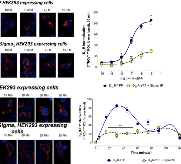
The internalization of D2LRYFPwas studied in the confocal
laser microscope after incubation for 30 min with different concentrations of cocaine from 1 nM to 10μM. The inter-nalization of D2LRYFP was measured by determining the
ratio of the total area of D2LRYFPfluorescence in the
cyto-plasm, and the total area of fluorescence in the plasma membrane taken in % of this ratio in the basal state. The images obtained with different cocaine concentrations (1 nM to 10uM) of cocaine are illustrated in Fig.5in which the D2LRYFP fluorescence is found in the plasma
mem-brane, and the cytosol at different ratios betweenδ1R and D2LR cotransfected and D2LR singly transfected HEK293
cells.
A highly significant reduction of the D2LR YFP
inter-n a l i z a t i o inter-n w a s o b s e r v e d i inter-n t h e δ1R and D2 LR
cotransfected versus D2LR singly transfected HEK293
cells after cocaine incubation. The inhibitory effects of cocaine started already at 10 nM and were clear-cut at 100 nM (Fig. 5).
The time-course of cocaine action over 90 min was also evaluated using a cocaine concentration of 100 nM with comparisons between D2LRYFPsingly expressing cells and
D2LRYFPandδ1R coexpressing cells as illustrated (Fig.5).
The cocaine-induced reduction of D2LRYFPinternalization
in the D2LR and Sigma 1R coexpressing cells was
ob-served during the first 60 min which was marked and high-ly significant. aca AcbC AcbSh CPu CC aca AcbCAcbSh CPu 0 1 2 3 + *** *** + *** s b ol b A L P f o r e b m u n e g ar ev A dl eif r e p s u el c u n r e p 0 3x 0 3 , dl eif el p m a s( m)
a
b
Fig. 2 a PLA experiments. D2R-δ1R heteroreceptor complexes are seen
as red clusters in nucleus accumbens core (AcbC), nucleus accumbens shell (AcbSh) and nucleus caudatus putamen (CPu), crus cerebri (cc) and anterior limb of the anterior commissure (aca). The few PLA clusters found in aca and negative controls are regarded as unspecific. Arrows point to some of the red PLA positive clusters. The length of the bar is
30μm. b The density (per nucleus per sampled field) of the PLA positive complexes in AcbC, AcbSh and CPu are highly significantly different from the density found in crus cerebri and aca (***P < 0.001). The density is also significantly higher in the AcbSh and CPu versus Acb core (+P < 0.05). Mean ± SEM, number of rats = 5. One-way ANOVA, Tukey post-test. For sampling fields at Bregma 1.00 mm
Discussion
Previous work demonstrated that cocaine in the nanomolar range (10 and 100 nM) can enhance the D2-like receptor function in the brain independent of its effects on the dopa-mine transporter and be related to direct and/or indirect allo-steric actions at the D2like receptor [1,2]. The current results in cellular models indicate that these enhancing actions of cocaine at the D2R in the nanomolar range are dependent on
the presence of D2R-δ1R heteroreceptor complexes which
were previously demonstrated [14,27,28]. Theδ1R protomer appears to mediate the enhancing actions of nanomolar con-centrations of cocaine on D2R recognition and signaling found
in the present article. The latter action involves also a clear-cut counteraction of cocaine-induced D2R internalization. These
observations underline the existence of an indirect positive allosteric modulation of D2R via the δ1R. The reported
cocaine-induced inhibition of D2R signaling over Gi/o-AC
in D2R-δ1R heteroreceptor complexes found in high
micro-molar concentrations [14] could not be observed with the cur-rent low concentrations used of cocaine.
The existence of D2LR-δ1R heteroreceptor complexes after
cotransfection of the two receptors in cell lines could be val-idated using BRET2. In addition, also D2 SR-δ1R
heteroreceptor complexes were demonstrated after cotransfections with this technique. In addition, it was found in competition experiments that increasing amounts of δ1R cDNA could only reduce the BRET2 signal from the D2R-δ1R heteroreceptor complex to a minor degree. These
observations can be explained by the assumption that the δ1R can interact with several domains of the D2R and not only with the D2R-δ1R interface. Thus, the δ1R may have multiple interactions with the D2R regions, increasing its
im-pact on D2R signaling.
a
b
c
*
Fig. 3 a 3H Raclopride competition curves with the D2likeR agonist quinpirole in D2R cDNA transfected HEK cells with or without
cotransfection withδ1R cDNA. The KiH and KiL values obtained were similar and not significantly different. b, c In saturation binding experiments with 3H Raclopride, 1 nM of cocaine lacked effects on the Bmax and KD values in the absence ofδ1R cotransfection (b), while a
significant increase of the Bmax values (*P < 0.05) was found with cocaine (1 nM) upon cotransfection with δ1R (c). The action by cocaine was significantly counteracted by the sigma 1R antagonist PD144418 (50 nM) (c). Data are presented as the mean ± s.e.m. from five independent experiments performed in triplicate. *P < 0.05 by Studentt test
The involvement ofδ1Rs in the cocaine action is also sup-ported by the observation that cocaine at 1 nM could signifi-cantly increase the Bmax values of the D2R antagonist
binding sites only in the presence of D2R-δ1R heteroreceptor
complexes. Furthermore, this action was blocked by theδ1R antagonist PD144418. It seems possible that this action of
Basal M Forskolin 2Qu in 10 0nM coca ine 1 00nM M coca ine 1 Qu in100 nM + coca ine 1 00nM M Quin 100 nM + co cain e 1 M Quin 100 nM + raclop ride 1 M M +r aclo p 1 Qui n100 nM + Coc1 0 40 80 120 160 [ D 2R alone ] *** +++ +++ && @@@ ^^ ### B E R C ) ni m 0 3 , K F e s n o p s er x a m %(
a
Basal M Forskolin 2coca ine 100 nM M coca ine 1 0 40 80 120 160 [ 1 receptor ] B E R C ) ni m 0 3 , K F e s n o p s er x a m %( -14 -12 -10 -8 -6 -4 0 50 100 150 D2R D2R - Sigma1R EC50 EC50 D2R + raclopride 1 M D2R-Sigma1R + raclopride 1 M Log [quinpirole](M) B E R C ) ni m 0 3 , K F e s n o p s er x a m %(d
c
b
Basal Forskolin 2mMQu in 10 0nM coca ine 1 00nM M coca ine 1 Qu in100 nM + coca ine 1 00nM M Qu in100 nM + coc aine 1 M Qu in100 nM + raclop ride 1 M M + rac lop 1 Quin 100 nM + Coc1 0 40 80 120 160 [ D 2R + Sigma1R] *** +++ +++ &&& @@@ ^^^ ### B E R C ) ni m 0 3 , K F e s n o p s er x a m %(Fig. 4 CREB signaling experiments. a In HEK cells cotransfected with D2LR andδ1R cDNAs vs cells singly transfected with D2LR cDNA, the
inhibition of CREB signaling obtained with increasing concentrations of quinpirole was shifted to the right upon cotransfection (D2R (LogIC50 =
−9.87 ± 0.17), D2R-δ1R (LogIC50 = −8.95 ± 0.22)). b In singly D2R
cDNA-transfected cells the inhibition of CREB by quinpirole after forskolin-induced activation of CREB was unaffected by cocaine (100 nM and 1μM) and cocaine alone in these concentrations lacked effects on the forskolin-induced increase in CREB signaling. The data represent the means ± S.E.M. of three independent experiments performed in triplicate. Statistical analysis was performed by one-way analysis of variance (ANOVA) followed by Tukey’s multiple comparison post-test. TheP value 0.05 and lower was considered significant. ***Significantly different compared to Forskolin 2μM (P < 0.001); +++Significantly different compared to quinpirole 100 nM (P < 0.001); &&Significantly different compared to cocaine 100 nM (P < 0.01); @@@Significantly different compared to cocaine 1 μM, (P < 0.001); ^^Significantly different compared to quinpirole 100 nM (P < 0.01); ###Significantly different compared to quinpirole 100 nM + cocaine 1μM (P < 0.001). c In cells singly transfected cells with Sigma1R, cocaine (100 nM and 1uM) again lacked effects on the forskolin (2μM) induced activation of CREB signaling. The data
represent the means ± S.E.M. of three independent experiments performed in triplicate. Statistical analysis was performed by one-way analysis of variance (ANOVA) followed by Tukey’s multiple comparison post-test. d In cells cotransfected with D2LR and δ1R
cDNAs, cocaine at 100 nM and 1μM was instead able to significantly enhance the inhibitory actions of quinpirole on forskolin-induced increase in CREB activity. By itself, cocaine in these concentrations failed to significantly influence the forskolin-induced activation of CREB activity. The data represent the means ± S.E.M. of three independent experiments performed in triplicate. Statistical analysis was performed by one-way analysis of variance (ANOVA) followed by Tukey’s multiple comparison post-test. TheP value 0.05 and lower was considered significant. ***Significantly different compared to Forskolin 2μM (P < 0.001); +++Significantly different compared to quinpirole 100 nM (P < 0.001); &&&Significantly different compared to cocaine 100 nM (P < 0.001); @@@Significantly different compared to cocaine 1 μM (P < 0.001). Δ and ΔΔΔSignificantly different compared to quinpirole 100 nM (P < 0.05 and P < 0.001); ^^Significantly different compared to quinpirole 100 nM (P < 0.0^01); ###Significantly different compared to quinpirole 100 nM + cocaine 1 μM (P < 0.001). Abbreviations; raclop: raclopride, coc: cocaine, quin: quinpirole
cocaine may be linked to the D2R-δ1R heteroreceptor
com-plex and involve counteraction of cocaine-induced D2R
protomer internalization (see below).
An allosteric D2R-δ1R receptor–receptor interaction also
appears to exist in the control of D2LR protomer signaling.
In the analysis of the Gi/o-AC-PKA-CREB pathway using the CREB luciferase reporter gene assay, the presence of a D2R-δ1R heteroreceptor complex led to a reduced potency
of quinpirole to inhibit the CREB signal. However, this neg-ative allosteric receptor–receptor interaction turned into a pos-itive interaction upon incubation with 100 nM of cocaine and restored the potency of the D2R agonist to inhibit this
path-way. These findings indicate that with these nanomolar con-centrations of cocaine, the Gi/o-mediated inhibition of the AC-PKA-CREB pathway is enhanced by cocaine not reduced as found with high 30μM concentrations of cocaine [14].
A remarkable new action of cocaine was discovered in the current experiments on D2R-δ1R heteroreceptor
com-plexes vs D2LR mono-homoreceptor complexes regarding
effects of cocaine in nanomolar concentrations on D2LR
internalization. Already at 10 and 100 nM cocaine pro-duced an increase in D2R internalization in singly D2LR
transfected cells, an action which saturated around 1– 10 μM. In D2R and δ1R cotransfected cells, this action
of cocaine was markedly counteracted at concentrations tested from 10 nM to 10 μM at the 30 min time-interval with a time course of inhibition lasting around 60 min. These results strongly indicate that cocaine already at nanomolar concentrations can reduce D2LR protomer
inter-nalization in D2R-δ1R heteroreceptor complexes. Through
the cocaine interaction with this receptor complex, alloste-ric receptor–receptor interactions are altered. This may
0 20 40 60 80 100 120 0 20 40 60 80 100
D2LR-YFP D2LR-YFP + Sigma 1R
*** Time (minute) D2L n oi t a zil a nr et ni P F Y-R ( In tRO I/ me mRO I, )l a s a b r e v o % -10 -9 -8 -7 -6 -5 -4 0 30 60 90 120 150
D2LR-YFP D2LR-YFP + Sigma 1R
Log [cocaine](M) D2L n oi t a zil a nr et ni R ( In tRO I/ me m ni m 0 3 , )l a s a b r e v o % , I O R
Fig. 5 Effects of cocaine on internalization of D2RYFPin HEK cells in the
absence or presence of coexpression of Sigma1R. In the upper part, a cocaine concentration response curve on D2RYFPinternalization is
illustrated in red. To the right, quantitation of the D2RYFPinternalization
is found. With increasing concentrations, cocaine produces marked internalization of D2RYFPwhich is counteracted by expression of
Sigma1R (D2R (LogEC50 =−7.04 ± 0.09), D2R-δ1R (LogEC50 =
−6.63 ± 0.40)). In the lower part, the time-course of the action of cocaine is found in the D2R cDNA-transfected cells in the absence or
presence of Sigma1R cDNA cotransfection. Cocaine (100 nM) induces a marked internalization of D2RYFPconstruct in the first hour. This action is
again counteracted by the coexpression of Sigma1R with a high significance. These results are illustrated in the left lower panels, and the quantitation is found in the right lower panel. The data represent the means ± S.E.M. of three independent experiments performed with a total of eight replicates each. Statistical analysis was performed by two-way ANOVA (***P < 0.001)
lead to reduced D2LR coupling to beta-arrestin which can
contribute to the markedly reduced internalization and pro-longation of the D2R protomer signaling in the D2R-δ1R
heteroreceptor complex at the plasma membrane.
Taken together, the current results in cellular models give evidence that the cocaine action at D2Rs in nanomolar
con-centrations is substantially altered by the D2R presence in
D2R-δ1R heteroreceptor complexes. The molecular
mecha-nisms still remain to be clarified, but cocaine actions by bind-ing to this heteroreceptor complex may alter its allosteric receptor–receptor interactions. This may lead to the demon-strated increase in Bmax values of the D2LR protomer and
increased D2LR signaling over Gi/o inhibiting the AC-PKA
system which reduced CREB phosphorylation. It was associ-ated with a markedly reduced D2LR protomer internalization
prolonging the duration of D2R activation. Such acute cocaine
actions in the nanomolar range at the D2R-δ1R heteroreceptor
complexes will likely contribute to understanding the role of δ1R in acute effects of cocaine on reward and seeking.
Acknowledgments The work was supported by the Swedish Medical Research Council (62X-00715-50-3) to KF, by ParkinsonFonden 2017 to KF and by Hjärnfonden (FO2017) and Karolinska Institutet Forskningsstiftelser (2016-2017) to D.O.B-E. D.O.B-E belongs to the “Academia de Biólogos Cubanos” group.
Authors Contributions We confirm and declare that all the authors meet the criteria for authorship according to the ICMJE, including approval of the final manuscript, and they take public responsibility for the work and have full confidence in the accuracy and integrity of the work of other group authors. They have substantially contributed to the conception or design of the work. Also, they have participated in the acquisition, anal-ysis, and interpretation of data for the current review version. They have also helped revising it critically for important intellectual content, and final approval of the version to be published. In addition, they have contributed in this last version of the manuscript in writing assistance, technical editing, and language editing. DOB-E, MN, WRF, LP, KW, MF, SB, LF, ST, and KF designed methods and experiments, carried out the laboratory experiments, analyzed the data, and interpreted the results. DOB-E, WRF, KW, MF, and KF co-designed and co-worked on the radioligand binding experiments. DOB-E, MN, WRF, LP, SB, LF, ST, and KF co-designed and co-worked on the immunolabeling experiments. DOB-E, MN, LP, SB, LF, ST, and KF co-designed, discussed analyses, interpretation, and presentation of all immunohistochemistry and in situ proximity ligation assay experiments. DOB-E, WRF, LP, and KF de-signed and directed its implementation of the quality assurance and con-trol of each used antibody. DOB-E, MN, and KF designed, analyzed the data, and interpreted all the BRET studies. DOB-E, MN, WRF, and KF designed, analyzed the data, and interpreted all the radioligand binding studies. DOB-E, MF, LF, ST, and KF wrote the paper. All the authors have contributed to, seen, and approved the manuscript.
Compliance with Ethical Standards
All studies involving animals were performed in accordance with the Stockholm North Committee on Ethics of Animal Experimentation, the Swedish National Board for Laboratory Animal and European Communities Council Directive (2010/63/EU) guidelines for accommo-dation and care of Laboratory Animals.
Conflict of Interest The authors declare that they have no competing interest.
Open Access This article is distributed under the terms of the Creative C o m m o n s A t t r i b u t i o n 4 . 0 I n t e r n a t i o n a l L i c e n s e ( h t t p : / / creativecommons.org/licenses/by/4.0/), which permits unrestricted use, distribution, and reproduction in any medium, provided you give appro-priate credit to the original author(s) and the source, provide a link to the Creative Commons license, and indicate if changes were made.
References
1. Ferraro L, Beggiato S, Marcellino D, Frankowska M, Filip M, Agnati LF, Antonelli T, Tomasini MC et al (2010) Nanomolar con-centrations of cocaine enhance D2-like agonist-induced inhibition of the K+-evoked [3H]-dopamine efflux from rat striatal synapto-somes: a novel action of cocaine. J Neural Transm 117(5):593–597.
https://doi.org/10.1007/s00702-010-0389-4
2. Ferraro L, Frankowska M, Marcellino D, Zaniewska M, Beggiato S, Filip M, Tomasini MC, Antonelli T et al (2012) A novel mech-anism of cocaine to enhance dopamine d2-like receptor mediated neurochemical and behavioral effects. An in vivo and in vitro study. Neuropsychopharmacology 37(8):1856–1866.https://doi.org/10. 1038/npp.2012.33
3. Rouge-Pont F, Usiello A, Benoit-Marand M, Gonon F, Piazza PV, Borrelli E (2002) Changes in extracellular dopamine induced by morphine and cocaine: crucial control by D2 receptors. J Neurosci 22(8):3293–3301
4. Dalley JW, Everitt BJ (2009) Dopamine receptors in the learning, memory and drug reward circuitry. Semin Cell Dev Biol 20(4): 403–410.https://doi.org/10.1016/j.semcdb.2009.01.002
5. Koob GF, Le Moal M (2008) Addiction and the brain antireward system. Annu Rev Psychol 59:29–53.https://doi.org/10.1146/ annurev.psych.59.103006.093548
6. Durieux PF, Bearzatto B, Guiducci S, Buch T, Waisman A, Zoli M, Schiffmann SN, de Kerchove d'Exaerde A (2009) D2R striatopallidal neurons inhibit both locomotor and drug reward processes. Nat Neurosci 12(4):393–395.https://doi.org/10.1038/nn.2286
7. Alonso G, Phan V, Guillemain I, Saunier M, Legrand A, Anoal M, Maurice T (2000) Immunocytochemical localization of the sig-ma(1) receptor in the adult rat central nervous system. Neuroscience 97(1):155–170
8. Hayashi T, Su T (2005) The sigma receptor: evolution of the concept in neuropsychopharmacology. Curr Neuropharmacol 3(4):267–280 9. Matsumoto RR, Liu Y, Lerner M, Howard EW, Brackett DJ (2003)
Sigma receptors: potential medications development target for anti-cocaine agents. Eur J Pharmacol 469(1–3):1–12
10. Kourrich S, Su TP, Fujimoto M, Bonci A (2012) The sigma-1 receptor: roles in neuronal plasticity and disease. Trends Neurosci 35(12):762–771.https://doi.org/10.1016/j.tins.2012.09.007
11. Romieu P, Phan VL, Martin-Fardon R, Maurice T (2002) Involvement of the sigma(1) receptor in cocaine-induced condi-tioned place preference: possible dependence on dopamine uptake blockade. Neuropsychopharmacology 26(4):444–455.https://doi. org/10.1016/S0893-133X(01)00391-8
12. Hiranita T, Soto PL, Tanda G, Katz JL (2010) Reinforcing effects of sigma-receptor agonists in rats trained to self-administer cocaine. J Pharmacol Exp Ther 332(2):515–524.https://doi.org/10.1124/jpet. 109.159236
13. Navarro G, Moreno E, Aymerich M, Marcellino D, McCormick PJ, Mallol J, Cortes A, Casado V et al (2010) Direct involvement of sigma-1 receptors in the dopamine D1 receptor-mediated effects of
cocaine. Proc Natl Acad Sci U S A 107(43):18676–18681.https:// doi.org/10.1073/pnas.1008911107
14. Navarro G, Moreno E, Bonaventura J, Brugarolas M, Farre D, Aguinaga D, Mallol J, Cortes A et al (2013) Cocaine inhibits dopamine D2 receptor signaling via sigma-1-D2 receptor heteromers. PLoS One 8(4):e61245.https://doi.org/10.1371/journal.pone.0061245
15. Borroto-Escuela DO, Brito I, Romero-Fernandez W, Di Palma M, Oflijan J, Skieterska K, Duchou J, Van Craenenbroeck K et al (2014) The G protein-coupled receptor heterodimer network (GPCR-HetNet) and its hub components. Int J Mol Sci 15(5): 8570–8590.https://doi.org/10.3390/ijms15058570
16. Beggiato S, Borelli AC, Borroto-Escuela D, Corbucci I, Tomasini MC, Marti M, Antonelli T, Tanganelli S et al (2017) Cocaine mod-ulates allosteric D2-sigma1 receptor-receptor interactions on dopa-mine and glutamate nerve terminals from rat striatum. Cell Signal 40:116–124.https://doi.org/10.1016/j.cellsig.2017.09.007
17. Borroto-Escuela DO, Marcellino D, Narvaez M, Flajolet M, Heintz N, Agnati L, Ciruela F, Fuxe K (2010) A serine point mutation in the adenosine A2AR C-terminal tail reduces receptor heteromerization and allosteric modulation of the dopamine D2R. Biochem Biophys Res Commun 394(1):222–227.https://doi.org/ 10.1016/j.bbrc.2010.02.168
18. Borroto-Escuela DO, Romero-Fernandez W, Tarakanov AO, Marcellino D, Ciruela F, Agnati LF, Fuxe K (2010) Dopamine D2 and 5-hydroxytryptamine 5-HT((2)a) receptors assemble into function-ally interacting heteromers. Biochem Biophys Res Commun 401(4): 605–610.https://doi.org/10.1016/j.bbrc.2010.09.110
19. Borroto-Escuela DO, Van Craenenbroeck K, Romero-Fernandez W, Guidolin D, Woods AS, Rivera A, Haegeman G, Agnati LF et al (2011) Dopamine D2 and D4 receptor heteromerization and its alloste-ric receptor-receptor interactions. Biochem Biophys Res Commun 404(4):928–934.https://doi.org/10.1016/j.bbrc.2010.12.083
20. Borroto-Escuela DO, Romero-Fernandez W, Mudo G, Perez-Alea M, Ciruela F, Tarakanov AO, Narvaez M, Di Liberto V et al (2012) Fibroblast growth factor receptor 1- 5-hydroxytryptamine 1A heteroreceptor complexes and their enhancement of hippocampal plasticity. Biol Psychiatry 71(1):84–91.https://doi.org/10.1016/j. biopsych.2011.09.012
21. Borroto-Escuela DO, Narvaez M, Perez-Alea M, Tarakanov AO, Jimenez-Beristain A, Mudo G, Agnati LF, Ciruela F et al (2015) Evidence for the existence of FGFR1-5-HT1A heteroreceptor com-plexes in the midbrain raphe 5-HT system. Biochem Biophys Res Commun 456(1):489–493.https://doi.org/10.1016/j.bbrc.2014.11.112
22. Borroto-Escuela DO, Romero-Fernandez W, Garriga P, Ciruela F, Narvaez M, Tarakanov AO, Palkovits M, Agnati LF et al (2013) G protein-coupled receptor heterodimerization in the brain. Methods Enzymol 521:281–294. https://doi.org/10.1016/B978-0-12-391862-8.00015-6
23. Borroto-Escuela DO, Hagman B, Woolfenden M, Pinton L, Jiménez-Beristain A, Oflijan J, Narvaez M, Di Palma M et al (2016) In situ proximity ligation assay to study and understand the distribution and balance of GPCR homo- and heteroreceptor complexes in the brain. In: Lujan R, Ciruela F (eds) Receptor and Ion Channel detection in the brain, vol 110. Neuromethods. Springer, Berlin, pp. 109–126.https:// doi.org/10.1515/revneuro-2015-0024
24. Borroto-Escuela DO, Narvaez M, Di Palma M, Calvo F, Rodriguez D, Millon C, Carlsson J, Agnati LF et al (2014) Preferential activa-tion by galanin 1-15 fragment of the GalR1 protomer of a GalR1-GalR2 heteroreceptor complex. Biochem Biophys Res Commun 452(3):347–353.https://doi.org/10.1016/j.bbrc.2014.08.061
25. Borroto-Escuela DO, Flajolet M, Agnati LF, Greengard P, Fuxe K (2013) Bioluminescence resonance energy transfer methods to study G protein-coupled receptor-receptor tyrosine kinase heteroreceptor complexes. Methods Cell Biol 117:141–164.
https://doi.org/10.1016/B978-0-12-408143-7.00008-6
26. Lever JR, Miller DK, Fergason-Cantrell EA, Green CL, Watkinson LD, Carmack TL, Lever SZ (2014) Relationship between cerebral sigma-1 receptor occupancy and attenuation of cocaine’s motor stimulatory effects in mice by PD144418. J Pharmacol Exp Ther 351(1):153–163.https://doi.org/10.1124/jpet.114.216671
27. Borroto-Escuela DO, Narvaez M, Wydra K, Pintsuk J, Pinton L, Jimenez-Beristain A, Di Palma M, Jastrzebska J et al (2017) Cocaine self-administration specifically increases A2AR-D2R and D2R-sigma1R heteroreceptor complexes in the rat nucleus accum-bens shell. Relevance for cocaine use disorder. Pharmacol Biochem Behav 155:24–31.https://doi.org/10.1016/j.pbb.2017.03.003
28. Feltmann K, Borroto-Escuela DO, Ruegg J, Pinton L, de Oliveira Sergio T, Narvaez M, Jimenez-Beristain A, Ekstrom TJ et al (2018) Effects of long-term alcohol drinking on the dopamine D2 receptor: gene expression and heteroreceptor complexes in the striatum in rats. Alcohol Clin Exp Res 42(2):338–351.https://doi.org/10.1111/acer. 13568
Publisher’s Note Springer Nature remains neutral with regard to jurisdictional claims in published maps and institutional affiliations.
Affiliations
Dasiel O. Borroto-Escuela1,2,3&Manuel Narváez4&Wilber Romero-Fernández5&Luca Pinton1&Karolina Wydra6&
Malgorzata Filip6&Sarah Beggiato7&Sergio Tanganelli7&Luca Ferraro7&Kjell Fuxe1 1
Department of Neuroscience, Karolinska Institutet, Biomedicum (B0851). Solnavägen 9, 171 77 Stockholm, Sweden
2
Department of Biomolecular Science, Section of Physiology, University of Urbino, Campus Scientifico Enrico Mattei, via Ca’ le Suore 2, 610 29 Urbino, Italy
3 Observatorio Cubano de Neurociencias, Grupo Bohío-Estudio,
Zayas 50, 62100 Yaguajay, Cuba
4
Instituto de Investigación Biomédica de Málaga, Facultad de Medicina, Universidad de Málaga, Málaga, Spain
5
Science for Life Laboratory, Department of Cell and Molecular Biology, Uppsala University, BMC, Box 596, 751
24 Uppsala, Sweden
6
Institute of Pharmacology, Department of Drug Addiction Pharmacology, Polish Academy of Sciences, 12 Smetna Street, 31-343 Kraków, Poland
7 Department of Life Sciences and Biotechnology (SVEB), University