Università degli Studi di
Ferrara
DOTTORATO DI RICERCA IN
"SCIENZE e TECNOLOGIE per l’ARCHEOLOGIA e i BENI
CULTURALI"
CICLO XXVI
COORDINATORE Prof. Carlo Peretto
Study on Modern and Contemporary works of art through non invasive integrated physical techniques
Settore Scientifico Disciplinare FIS/07
Dottorando Tutore
Dott. Tisato Flavia Prof. Petrucci Ferruccio C.
Anni 2011/2014
Corso di Dottorato in convenzione con
1
Study on Modern and Contemporary works of art through
non invasive integrated physical techniques
Table of contents
Introduction
Pag. 51. Investigations on Contemporary art Pag. 7
1.1 Diagnostic survey (2011-2013) on six paintings at Casa Museo
Remo Brindisi: Pag. 7
1.1.1 G. De Chirico, Ettore e Andromaca. The underdrawing Pag. 12
1.1.2 J. Mirò, Composizione con figure. Differentiation of
materials Pag. 14
1.1.3 G. Turcato, Senza titolo. The polimatericity Pag. 17
1.1.4 E. Vedova, Senza titolo. The diversity of expressive
media Pag. 21
1.1.5 U. Boccioni, Senza titolo. The brush stroke becomes a
protagonist Pag. 24
1.1.6 M. Sironi, Composizione. What the eye does not see Pag. 26
1.2 Artworks from private collections Pag. 29
1.2.1 G. Boldini, Donna in lettura sul letto e anziano Pag. 29
1.2.2 F. De Pisis, Natura morta and Fiori Pag. 37
1.2.3 F. Patruno, Omaggio a De Pisis Pag. 44
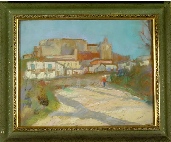
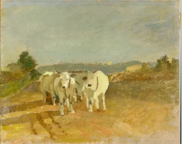
1.2.4 E. Pazzini, Lungo la strada a Padulli con donna e anitre, Paesaggio di San Marino, San Leo, Buoi,
2
Chapter 2
Applications of non invasive techniques to ancient artworks
Pag. 592.1 Giovanni Da Mel, Madonna con il Bambino tra i santi Rocco e
Sebastiano (XVI century) Pag. 60
2.2 Marco Da Mel. Samples from a fresco in Saint Bartolomeus
Church, Villapiana di Lentiai, Belluno (XVI century) Pag. 69
2.3 Anonymous, Portrait of Giacomo Masino Pag. 73
2.4 Anonymous, Desco da parto Pag. 82
2.5 Artworks on paper (XVII century) Pag. 89
2.6 Characterization of polychrome ceramics from Iranian
artworks (XVII century) Pag. 93
Chapter 3
Non invasive techniques for materials and artistic
techniques
Pag. 1033.1 Study on materials: Evaluation of the optical properties of
consolidants for restoration Pag. 105
3.2 Monitoring of contemporary painting layers. Test painting:
Image spectroscopy and PCA to monitor modern materials Pag. 116
3.3 Colour and texture characterization of a sample (Training at
the Centre for Fine Print Research, Bristol) Pag. 123
3.3.1 The BRDF (Bi-Directional Reflection Functions) Pag. 123
3.3.2 The practical approach: construction of a system for
the observation of prints, through a microscope Pag. 124
3.3.3 Photographs, at different magnifications, of samples
3
Chapter 4
Equipment
Pag. 1334.1 Non standard instruments Pag. 133
4.1.1 Wide band infrared reflectography scanner Pag. 133
4.1.2 Digital X-Ray radiography scanner Pag. 136
4.1.3 K-edge differential radiography Pag. 138
4.1.4 Image spectroscopy Pag. 141
4.2 Commercial instruments Pag. 144
4.2.1 X Ray fluorescence (XRF) Pag. 144
4.2.2 VIS Spectrophotometer Pag. 146
4.2.3 Wood lamp for ultraviolet fluorescence Pag. 148
4.2.4 Digital camera for infrared reflectography Pag. 149
Conclusions
Pag. 151Bibliography
Pag. 1535
Introduction
Every work of art, regardless whether ancient or modern, is not eternal and is thus subject to unavoidable alterations over time.
In the recent decades, the study of contemporary painting materials, used both for realization and for restoration of works of art, has acquired great importance. Many of those materials were originally created for different purposes than for the artistic and conservative aims, and designed for different needs than those asked by Cultural Heritage. Therefore, they may – or may not – had to be adapted to new demands.
The study presented in this thesis has been articulated around two themes.
The first one is focused on works of art, contemporary in particular but also but not exclusively, the second one relates to materials, used for realization and restoration of such objects.
The study of works of art, discussed in Chapters 1 and 2, was carried out by making use of different diagnostic techniques available in the Archaeometry laboratory, taking advantage of a variety of methodologies. In particular, frequently used image techniques include photography and macro photography in diffuse light and specular and raking light, ultraviolet fluorescence, wide band infrared reflectography, digital radiography and K-edge radiography. To get as much information as possible, to be properly integrated with other data, other, specific diagnostic techniques have been used, such as reflectance spectrophotometry, colorimetry and X Ray fluorescence (XRF).
All these techniques were exploited in a complementary way, in order to meet the different requirements, and to answer to the questions of knowledge requested, each time, by the various artifacts. Just this integrated mode in exploiting the diagnostic techniques, represents the innovative aspect of the research, being all the techniques already developed, and possibly already commercial.
The main goal of the study on art materials was, instead, to monitor their behaviour over time and, if possible, to predict their degradation in advance. This last objective has been achieved on materials applied on a support (Chapter 3). On the other hand, on materials as they are, investigations allowed their characterization, mainly from the visual point of view, and hence for some of their optical properties.
6 The same section also includes the experience carried out during a three-month internship at the Centre for Fine Print Research, University of the West of England (Bristol).
In this context, I’ve been working with an empirical approach to the realization of a prototype of dome, provided with an electrical circuit, with which to equip a stereo microscope. Constant conditions, relative to the illumination and observation geometry, was thus provided while examining artifacts (mainly on paper) in raking light, at different magnifications, allowing their qualitative characterization.
Finally, diagnostic instruments and techniques used to investigate both works of art and materials, are described in detail in Chapter 4.
Overall, this thesis presents the main results of diagnostic activities carried out on different works of art, mainly of contemporary art, but also from other times.
The main aspect of the work, that links all the artworks presented here, ancient and contemporary ones, is to detect the artistic technique used by artists, and their ways to proceed.
Conservation and valorisation of works of art and collections are the primary aspects of the whole work, and have been reached through the use of different diagnostic techniques. In all of the investigated case studies, the work of art decided, each time, the techniques to be used. Such methodologies, already developed and eventually commercial, have thus been significantly integrated together.
As a consequence of that, the part devoted to instruments is simply a description, taking on the task of obtaining a greater completeness of the whole thesis.
7
Chapter 1
Investigations on Contemporary art
Documentation and conservation are very important issues to deal with, as they can be the initial start point to embark on a restoration. But they are not end in themselves, as a close investigation on artworks is enlightening to go back to painting techniques. Moreover, the aspect of valorization plays an important role in the dissemination of knowledge about works of art, achieved by means of various diagnostic techniques.
1.1 Diagnostic survey (2011-2013) on six paintings at Casa Museo Remo
Brindisi
In this section, results of a diagnostic survey performed at Casa Museo Remo Brindisi in Lido di Spina, Comacchio, Italy, are presented.
Six contemporary paintings, all belonging to the twentieth century and characterized by different pictorial techniques, were analyzed, to suggest the wide range of possible case studies. Results presented are proposed as examples of integrated image diagnostics, to study both the material characteristics of the works of art, and the artists’ executive techniques. The activity was also conducted to try to develop a protocol, devoted to the study of the XX century paintings on the basis of materials and artistic techniques. The diagnostic curriculum, which is well known for ancient artworks, has never found an updated definition for contemporary artworks, because of the wide availability of artistic materials and the fragmentation of their use, limited to the individual artist. The definition of a sequence of non invasive investigations, in the near future, will have multiple purposes, such as the study and the conservation of the artwork. In this regard, Casa Museo Remo Brindisi, with its huge and diversified content of works of art and materials, is an ideal context where to start a systematic study by means of non invasive image diagnostics.
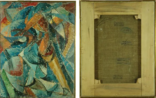
The building (fig. 1), which is the result of the collaboration between Remo Brindisi and the architect and designer Nanda Vigo, was created primarily to house the art collection of Brindisi. It is at the same time the summer house and the workshop of the artist, a place for exchange and comparison, where to express creativities and to discuss. The house consists of both a public and a private part. The first one includes an entrance hall, the living room on the ground floor and a large central cylinder, which develops along the entire height of
8 the building. Around it, private parts are arranged on four floors: the bedrooms and the study of the artist. Outside there is a big garden (fig. 2), an evocative place which poses serious problems of conservation to the metal sculptures, there erected.
Fig.1 Entrance of the Casa Museo Remo Brindisi. Fig. 2 Casa Museo Remo Brindisi, the outdoor
garden.
Fig. 3 Interior of the Casa Museo, with Remo
Brindisi’s profile.
Fig. 4 G. Pomodoro, Radiale. Fiberglass (1965),
9 Overall, the Casa Museo was set up as a manifesto of the “integration of arts”, an open space for everybody, without physical, conceptual interruptions between architecture and furniture, design and works of art. The space in here, goes beyond the division of the arts, according to the research of Nanda Vigo, and also fits in with the latest trends of the time (figs. 3, 4) (Piraccini, 2005).
During the collaboration with the Casa Museo Remo Brindisi, the use of image diagnostics to analyze six works of art from the collection, proved to be a useful and non invasive tool to document and study the paintings (figs. 5-8). On six of them, presented hereafter, different aspects concerning their study will be highlighted.
While diagnostics were in progress, such different issues, concerning the study of the artifacts, variously emerged, depending on the investigated work of art.
As an example, on the painting by Giulio Turcato, the materic appearance of the creation made his pictorial technique the predominant issue to deal with, giving the opportunity to speculate about his way to proceed.
The conservative issue was, instead, the starting point that moved the observation in raking light on Mario Sironi’s painting, which was submitted to a conservative intervention after a first diagnostic survey.
Works such as those by Emilio Vedova and Joan Mirò gave good pretexts to highlight some technical issues, like the diversity of materials used.
On the painting by Giorgio De Chirico, again, the preparatory study was the starting point for deeper investigations by means of infrared radiations.
The surfaces of all the paintings were investigated by selecting appropriate spectral bands, allowing thus to get useful and various information about them. In particular, pictures taken in raking light revealed the morphological features of the surface, while infrared reflectography showed the presence of preparatory drawings and pentimenti, underneath. Trans-illumination proved to be helpful in order to get more information about the conservation of the support, and on the thickness and homogeneity of the paint layers. Finally, induced ultraviolet fluorescence emphasized both the varnish and the diversity of pictorial materials on the surface(Albertin, 2012 (a)).
10 Fig. 5 Image diagnostic survey at Casa Muse Remo Brindisi, 2011-2013.
Nonetheless, valorisation of works of art resulted to be one of the main aspects of the entire project developed at Casa Museo, as an issue that connected all the different aspects emphasized in each case. As a matter of fact, paintings and artworks in the Casa Museo, are nowadays not so well known, and should thus be more valorised.
During the exhibition L’invito del Maestro, held in the summer of 2011 at the Casa Museo, an educational exhibition, based on image diagnostics performed on the artworks, was prepared (fig. 6).
Fig. 6 Dissemination of the results obtained.
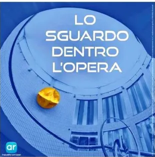
11 An innovative museum itinerary was also inaugurated on October 5th 2013, with the aim to reveal new details on works of art, by means of devices for Augmented Reality. The project Lo sguardo dentro l’opera - A look inside the work of art, was originated by the synergy between two research groups focused on Equipment, materials and techniques for museography and exhibition design and Diagnostics and preservation (TekneHub Laboratory, University of Ferrara), just with the purpose to promote and enhance the collection at the Casa Museo.
In such a realization, some short videos, illustrating image diagnostics and other contents, are linked to works by Turcato, Boccioni, Vedova, Sironi and others. The painting technique of each artist was thus highlighted by image diagnostics, which becomes, in this context, a tool for enhancement and enjoyment (figs. 7, 8).
Fig. 7 Poster of the event The look inside the work
of art (Opening: 5 October 2013). Implementation of additional content through Fig. 8 The look inside the work of art. augmented reality.
12
1.1.1 G. De Chirico, Ettore e Andromaca. The underdrawing
The first investigated painting is an oil on canvas, by Giorgio De Chirico. The subject, typically painted in the metaphysical manner, depicts Hector and Andromache. In this case, the use of infrared radiation revealed itself as illuminating, in discovering invisible details under the paint layer.
First images acquired in raking light allowed to study the three dimensionality of the diversified surface. A diffuse craquelure was highlighted, due to a non homogeneous tension of the canvas (figs. 11, 12).
Thanks to infrared reflectography, traces of a preparatory drawing, made with a fine-tip tool, probably a pencil, were unveiled. It was also possible to observe some pentimenti between the preliminary drawings and the final painting, for instance in the decorations (twirls and swirls) on the chest of the dummy on the left, and in correspondence with its thigh, as well as on some elements that make up the scenography. Changes from the final version were also detected by trans-irradiation, that is, by collecting the transmitted infrared radiation through the canvas.
Such a technique allowed to better appreciate the underdrawing and the pentimento, referred to the mantle of the figure on the right (fig. 13), as well as the trend of craquelure.
Fig. 9 Ettore e Andromaca. Total in diffuse light, recto.
Fig. 10 Ettore e Andromaca. Total in diffuse light,
13 Shadows and shades of colour were set by pencil during the execution of the preparatory drawing (fig. 14).
Fig. 11 Raking light photograph. Folds in
corners and diffuse craquelures, due to uneven tension of the canvas.
Fig. 12 Craquelure, in raking light.
Fig. 13 Pentimento on the
mantle of the figure on the right, in trans-irradiation.
14
1.1.2 J. Mirò, Composition with figures. Differentiation of materials
At a first sight, the choice of vivid colours and the contrast of complementary colours in the composition, stand out. What immediately emerges is the variety of hues used by Mirò (figs. 15, 16). In such a work, ultraviolet fluorescence was particularly functional in differentiating materials having the same hue at a simple visual inspection.
Fig. 15 Composition of figures. Total in diffuse light, recto.
15 Afterwards, going more in depth, it is interesting to hypothesize how Mirò originally thought the composition. In this regard, infrared image of both whole and details of the painting, allowed to find out traces of a preparatory drawing, disappeared under the pictorial layer (figs. 17-19) and related to the creative phase of the drawing.
Ultraviolet fluorescence played an important role in the differentiation of materials, by returning the painting with different colours, depending on the pigment used (fig. 24). Photographs in raking light emphasized the rough texture of the canvas support and the strokes of the secure boundaries. The author signature, observed in raking light, showed the roughness of the canvas, while the reproposal of the same signature in the infrared reflectography, at a close distance, allowed to fully appreciate the paint layer (figs. 20, 21). The features of canvas, colour and brush strokes, are well appreciable in the photographs taken in raking light (figs. 22, 23).
On the verso of the painting, finally, the structure of the canvas was revealed, due to the white preparation passing through it, as well as some writings, which must still be interpreted.
Fig. 17 Total in infrared reflectography, recto.
16 Fig. 18 Detail in infrared reflectography.
A pentimento pertains to the position of the dress. emphasizing a different, previous version. Fig. 19 Detail in infrared reflectography,
Fig. 20 Detail of the signature in raking light. Fig. 21 Detail of the signature in infrared
reflectography.
Fig. 22 Detail in raking light of the figure’s head.
Features of the rough canvas are well emphasized. The colour, pretty liquid, has dampened the fibers of the Fig. 23 Detail in raking light of the figure’s dress. canvas.
Fig. 24 Total in ultraviolet fluorescence, recto.
17
1.1.3 G. Turcato, Senza titolo. The polimatericity
While craft artistic materials are progressively replaced by equivalent commercial products and synthetic compounds, unusual pigment associations are also used, as well represented in the case of Turcato’s creation (figs. 25, 26). In this “painting”, raking light technique proved to be particularly useful in amplifying the three-dimensional components, and try to reconstruct the technique used by the artist.
Fig. 25 Untitled. Total in diffuse light, recto.
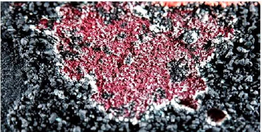
18 All the gathered observations on this particular work of art (fig. 27), made it possible to hypothesize the artist’s way of painting. As shown in fig. 28, the artist probably firstly prepared the canvas surface (reasonably, a canvas with industrial preparation) with various colours. Some black, granular material (fig. 29) has thus been overlaid, in all likelihood after applying an adhesive substance. Then, he glued, in various agglomerations, a material of variable grain size (maybe sand), which he subsequently scraped in some places, to make room for strokes of full-bodied red-orange pigment (fig. 30).
The observation in raking light facilitates the study of the painting technique, enhancing in here the polimatericity of the work. The colour laids in a thick impasto, highlighting the features of the brush stroke: relief, direction, width and curvature. Sand and earthy material concurred to create suggestive and varied effects on the pictorial surface (figs. 29, 32). Images obtained in trans-illumination provided information about the thickness of the paint layers and their homogeneity. The light selectively passed through the material, where the brush strokes were thinner (fig. 31).
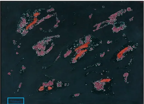
The multi-material aspect of the work is also confirmed by the images obtained by ultraviolet fluorescence (fig. 33), which differentiated the various materials used by the artist.
On the background, dark in the inspection with ultraviolet radiation, the fluorescent pink and red portions stood out, presenting, respectively, purple and red-orange colours.
Fig. 27 Overall vision of the painting without frame. Picture taken in diffuse light does not highlight the
19 Fig. 28 Detail in diffuse light, showing the
presence of a coloured preparation, under the upper layers.
Fig. 29 Detail in raking light of the black, granular
material, applied on some areas of the surface.
Fig. 30 Detail in raking light of a part where
various layers followed one another. Fig. 31 Detail in trans-illumination of the same detail. Light passes through the canvas where less material is present on the support.
20 Fig. 33 Total in ultraviolet fluorescence, verso. Differentiation of pictorial materials is due to their behaviour
to the radiation.
The painting by Turcato is a prime example of how it is possible to reconstruct the painting technique of an artist. Observations result from the application of image techniques and, therefore, do not provide the contact with the work of art.
21
1.1.4 E. Vedova, Senza titolo. The diversity of expressive media
The work by Emilio Vedova is a painting on paper, transferred to a canvas to give greater support to the paper itself (fig. 34). It appears to be quite significant as an example of the capability, by radiations different than visible light, to differentiate materials.
The painting by Vedova is characterized by spontaneity and immediacy of the artistic gesture. Rapid brush strokes were directly applied on the support, without an underlying preparation, and colours were chosen from a wide range of materials.
Diagnostic imaging was a valuable aid, both for analyzing the conservative state of the painting, as in the case of ultraviolet fluorescence, and to make preliminary investigations on the materials used by the artist, differentiating between them.
Fig. 34 Untitled. Total in diffuse light, recto.
Initial photographic documentation in diffuse and, particularly, in raking light, provided interesting information about the artist’s way to proceed and the tools he used, highlighting the flatness of the stretch obtained with pastel, in contrast to the materic, protruding stroke, applied with brush (figs. 35, 36). Looking at the same details in raking light, it can also be
22 assumed the use of another tool, probably wax pastels, apart from brush. A first sketch was probably executed in pastel, while brush strokes in different colours were later applied.
Illumination with ultraviolet light stimulated fluorescence of the support, which appeared clear in the infrared image, contrasting whit the lack of fluorescence on the black pigment (fig. 37a-b).
The red pigment disappeared in the infrared image, being transparent to this radiation, whereas an evident red fluorescence resulted when observing the painting under ultraviolet light (fig. 38a-b). Yellow pigments presented, in some areas, a characteristic yellow fluorescence (fig. 39a-b).
Furthermore, particularly interesting is the comparison of the behavior of painting materials to the solicitation provided by ultraviolet and infrared radiations. Both the red and the yellow pigment, for example, disappear in the infrared image, being transparent to this type of radiation. Instead, when observed under ultraviolet radiation, the same materials exhibit fluorescence, red (for red pigment) and, only in some parts, yellow (for yellow pigment). The presence of fluorescence in the yellow pigment only in some parts of the painting is probably related to the use of the two materials (having the same yellow hue) used for the realization.
Figs. 35-36 Raking light pictures, showing both materic and thin brush strokes, and the different materials
23 Fig. 37a Detail of the painting, with a large portion
of the paper support, uncovered by pictorial material.
Fig. 37b The paper support appears white when
inspected with ultraviolet radiation.
Fig. 38a Red pigments, detail in diffuse visible
light. Fig. 38b Red pigments, detail inspected with ultraviolet radiation. The red material presents a dark red fluorescence.
Fig. 39a Yellow pigments, detail in diffuse visible
light.
Fig. 39b Yellow pigments, detail inspected with
ultraviolet radiation.
Yellow material presents, only in some areas, a characteristic yellow fluorescence.
24
1.1.5 U. Boccioni, Senza titolo. The brush stroke becomes a protagonist
The application of a simple and immediate technique, such as raking light, can sometimes result the most suitable, in highlighting the most important features of a work of art.
This is the case of the painting by U. Boccioni.
Fig 40 Untitled. Total in diffuse light, recto. Fig 41 Untitled. Total in diffuse light, verso.
Many ink stamps are present.
Through the painting technique, the futurist painter expressed the dynamism of the subject (figs. 40, 41), and the brush stroke became thus a protagonist when observing the painting in raking light.
Such a technique, as a matter of fact, highlighted the materiality of the brush stroke, in contrast to the black, flat signs, which separate the coloured areas (fig. 42). Some horizontal lines are due to irregularities on the edge of the canvas. The fluid brush strokes, sometimes more dense, were used to create highlights, concurring to the delineation of shapes. On the contrary, thinner glazes revealed the texture of the canvas (fig. 43). The dark contours, below the painting, determined the direction of the brush stroke.
25 Fig 42 Raking light photo highlighting the materiality of the brush stroke, in contrast to the black, flat signs,
which separate the coloured areas.
26
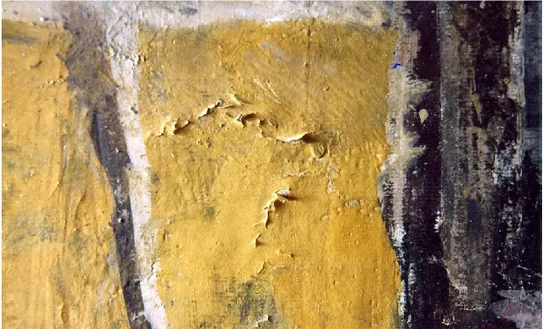
1.1.6 M. Sironi, Composizione. What the eye does not see
Imaging techniques, even when simple, like diffuse and semi-raking light, can be useful for conservation purposes, as shown in the case of Mario Sironi’s painting (figs. 44, 45). On this work, first applied documentation techniques, revealed adhesion problems on the pictorial film, on the yellow background field.
Fig 44 Composition. Total in diffuse light, recto.
Fig 45 Composition. Total in diffuse light, verso.
Several inscriptions and labels are visible, regarding the title of the painting, the date of its execution and its conservative history.
27 On the verso of the artwork, several inscriptions are documented, giving the opportunity to trace back to vicissitudes, changes in ownership, and exhibition venues.
Photographic documentation in visible light, both on the recto and on the verso of the painting, enabled to gain more information about its lifetime. On the recto, a raising of the pictorial film was documented before and after the restoration (fig. 46), aimed at its levelling, while on the verso some inscriptions relate to its conservative history (fig. 47).
Fig 46 Detail in raking light of a detachment of the pictorial layer.
Fig 47 The same detail in raking light, after the conservative intervention, aimed at ensure that the paint film
28
Conclusions
Overall, the main contribution to an enhanced knowledge about techniques and artistic methods adopted by various artists, has been achieved by means of different techniques. Such techniques, considering the uniqueness of every artwork, were differently adopted and mixed, allowing in many case to speculate about the succession of work phases.
The most evident case is represented by the work of Turcato, where raking light transillumination, and UV fluorescence altogether, played a crucial role in recognizing and differentiating the various materials used, according to their different materiality and appearances.
On the artwork by Sironi, the aspect that most emerges, is the conservative one, as a simple observation of the painting in raking light allowed to recognize a lift in the pictorial film, which therefore could be conveniently cured with a minimum intervention.
On the artwork by De Chirico, instead, the discovery of the underdrawing under the pictorial film, traced back to the different phases of the work, highlighting the value of diagnostic techniques in studying the creative processes.
29
1.2 Artworks from private collections
Diagnostic techniques were carried out on contemporary works of art from other private collections, aimed at understanding the painting techniques and at identifying their constituent materials for authentication purposes. Information obtained were also helpful to a deepening of the peculiarities in the artists’ way to proceed, outlining a richer cognitive framework, about materials used in contemporary times and on their methods of use. Considering the wide variety of pigments available since the nineteenth century, it is not surprising the large number of elements identified. Moreover, it makes it difficult to distinguish and recognize such materials.
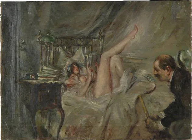
1.2.1 G. Boldini, Donna in lettura sul letto e anziano
The painting, part of a private collection (figs. 48, 49), was firstly submitted to image diagnostics, to better understand the general behaviour of the surface, and to address subsequent deeper and punctual analysis, such as X Ray fluorescence. On this artwork, data collected were aimed to find out more information, in order to support or not, the assumption that materials and techniques used were actually compatible with a painting by Boldini. Data obtained were subject of study for a bachelor thesis (D’Avossa, 2012).
As a first approach, photographs in raking light were acquired, hightlighting the diffuse craquelures, mainly caused by contraction of the different constituents of the painting: support, preparation and pictorial layers (fig. 50).
Image obtained in ultraviolet fluorescence let guess that no interventions have been performed on the painting after its execution, as no trace of alterations, fillings, re-varnishings or new added elements were visible. A general, homogeneous behaviour is shown on the entire surface. The varnish showed a greenish fluorescence, which increased where tiny droplets of material resulted spread on the whole surface of the work.
Also the figure of the elderly seemed to be coeval with the rest of the work. Such a finding could help in solving some doubts about the addition of the man in a second version of the work. As a matter of fact, a particular issue pertains to this figure, that appears in other attested works by Boldini. On the other hand, the overall workmanship raises some doubts about the authenticity of the painting, still not solved.
30 Fig 48 Donna in lettura sul letto e anziano. Total in diffuse light, recto.
31 Fig 50 Raking light, details. The old man, on the left edge of the painting, near the frame (left), and the
young woman on the bed (right). Both dense brush strokes and craquelures are well emphasized.
Fig 51 Ultraviolet fluorescence, total of the verso. Differentiation of materials due to their behaviour: bluish
fluorescence of paper, pale blue light related to the glue, and no fluorescence of the coloured preparation passed through the canvas.
As well as on the front, UV fluorescence distinguished different materials also on the back of the painting. As an example, the paper presented a bluish fluorescence (fig. 51), while the pale blue fluorescence related to the glue. Halos due to moisture, and a coloured preparation with no fluorescence, passed through the canvas, are also observed (D’Avossa, 2012).
32 Infrared reflectography showed no preparatory drawing (fig. 52), but the thick paint layers could also have a role in the difficulty to detect anything below. The signature disappeared, only leaving a trace of its presence (fig. 53a-b). An additional element explaining the obstacle in detecting any preparatory drawings, is the hypothesis of a dark pigmentation of the background layer, that appeared to be confirmed by X Ray fluorescence analysis. On the edge of the painting, a red coloration was observed under the green-brown pictorial film, in correspondence with the background of the painting. This was analyzed by X Ray fluorescence (fig. 55), showing a strong presence of mercury. The distribution of such a coloration, on the whole surface, although not uniformly, is confirmed by the presence of white spots in digital radiography (fig. 54).
Fig 52 Infrared reflectography, total of the recto (contrasted image).
Differentiation of materials due to their behaviour: bluish fluorescence of paper, pale blue light related to the glue and no fluorescence of the coloured preparation passed through the canvas.
Fig 53a Diffuse light, detail of the signature, on the
left, low corner of the painting.
Fig 53b Infrared reflectography, detail of the
signature. Only the capital letter (B) can slightly be distinguished.
33 Fig 54 Digital radiography, total of the recto. On the edges, white portions correspond to the nails.
White spots, diffused on the whole surface of the painting, are related to the presence of mercury and lead, respectively contained in Cinnabar and Lead white.
XRF data in the table below present results as peak integrals.
On some points on the back of the painting, in correspondence with points on the front, XRF detected increased counts of mercury and, sometimes, lead. Higher counts of calcium on the front were thus probably due to a preparation based on gypsum, animal glue and earths (due to the presence of iron). The presence on the back of mercury and lead, highly radiopaque elements, could instead justify the white spots in radiography. It can also be assumed that the canvas used for the painting is a reused painting, roughly cleaned from a preparation based on lead and mercury.
In all the other points, XRF investigation did not allow to clarify the composition in terms of pigments used on the painting. Spectra of the investigated points showed the presence of the same elements in many areas. The large amount of chemical elements found, and their coexistence (often likely to be indistinguishable), did thus not allow to attribute only one pigment to specific areas. Such a finding confirmed the non-use of pure colours as directly released from the tube, but more likely mixed together, to modulate colour tones.
34 copper, cobalt, manganese, chrome, cadmium, tungsten and, probably, sulfur, potassium, silicon, aluminum and sodium (fig. 56). According to the hues that characterize the different areas, such elements can be related to lots of pigments, as indicated below for each different colour.
On points 9 and 26, there is a lot of calcium, as these areas are in correspondence with the preparation of the painting, probably constituted by gypsum (hydrated calcium sulfate). In addition, the coexistence of iron together with calcium, zinc, lead and barium, explained the dark coloration of the preparation.
On white points, elements such as lead, zinc and calcium are present in high amounts, suggesting Lead white, Zinc white, Lithopone and Gypsum as the most probable pigments. In correspondence with the yellow points, elements found (cadmium, strontium, chrome, barium, iron, zinc, potassium and silicon) left several possibilities open, regarding the pigments used. These include: Cadmium Yellow, Strontium yellow, Chromium yellow, Barium yellow, Mars yellow, Litharge, Zinc yellow, Yellow ochre, Burnt Siena earth, Smalt yellow. It is also not possible to exclude the presence of organic pigments, such as Spincervino yellow.
For red pigments, instead, Cadmium Red, Minium, Chrome Red, Natural red ocher, Mars red, Red earth, Ultramarine red, Vermilion and Garanza lake were hypothesised, based on the detected elements (cadmium, selenium, sulphur, lead, iron, chrome, mercury, sodium). Among the hypothesized blue pigments, according to the elements found in the related areas (tungsten, iron, sodium, aluminum, manganese), there are: Wolfram blue, Prussian blue, Manganese Blue, Ultramarine artificial blue, Artificial Indigo.
The most probable green pigments were Transparent green Chrome oxide, Lamoriniere green, Chrome green, Malachite, Verdigris, Verditer, Smalt Green, Ultramarine green, due to the relative amounts of chrome, copper, iron, chloride, silicon, aluminium and sodium.
Finally, among the possible browns, Natural Siena earth, Burnt Siena earth, Natural umber, Burnt umber, Green burnt earth, Iron brown, Mars brown, Prussian brown, Cassel brown, Bistro and Bitumen brown were suggested, being iron, manganese, aluminium and silicon the majoritarian elements (Moioli, 2002).
35 Fig 55 Measuring points analyzed by XRF.
Fig 56 Table with XRF results (cps: counts per second). Points 35, 36 and 37 correspond, on the back of the
painting, to points 32, 33 and 34. (*): indicates counts per second referred to L lines. If not specified, cps refer to K lines.
36
Conclusions
The X ray fluorescence is a non invasive technique, which did not need to take any sample from the painting surface. It detected many possible pigments, despite not being able to identify the single formulations. Regarding the investigated painting, the advantageous use of not absolutely invasive techniques, did not detect any anachronism in found elements. However, it proved impossible to determine most of the pigments.
Radiography allowed to obtain a map relating to the distribution areas of the elements, according to their different densities. In correspondence of many bright areas, the although slight presence of mercury, identified by XRF, led to hypothesize the widespread presence, perhaps in preparation level, of a pigment such as Cinnabar. Such a finding would lead back to the particularity of a dark preparation.
Overall, scientific investigations performed on the painting did not dispel the doubts about the figure of the elderly. Whether it can be attributed - or not - to Boldini, remains a stylistic issue, still not solved.
37
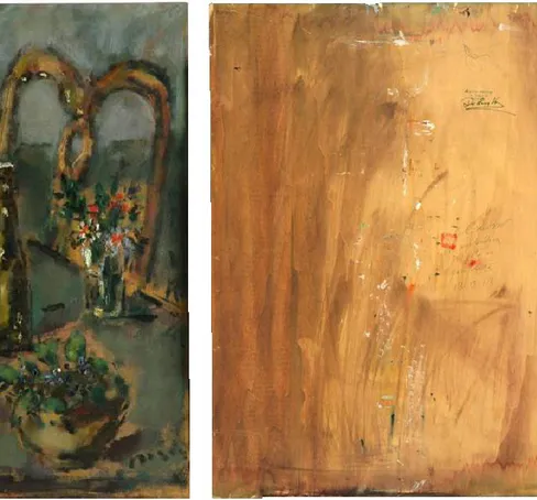
1.2.2 F. De Pisis, Natura morta and Fiori
Both paintings, signed De Pisis, come from private collections, and represent a floreal subject. What differs, is the support and the pictorial technique: Natura morta (figs. 57, 58) is a oil on a (thin) wood panel, while the other artwork, representing a flowerpot, is made by watercolours. Despite different materials have been used, some common features emerge, constituting a peculiarity.
As a matter of fact, De Pisis painting technique is considered to be full of pathos and emotivity. In this perspective, the choice of the palette, to the materiality and density of the brush strokes, is dictated by the artist’s emotions. Moods that De Pisis blocks on the canvas with a few, instantaneous and immediate gestures (Briganti, 1991).
Fig 57 Natura morta. Total in diffuse light, recto. Fig 58 Natura morta. Total in diffuse light, verso.
The conservative history of the artworks did not probably know any problematic moment, as their conservative state appears to be quite good. A different amount of information, instead, was obtained from the two works of art, according to the painting technique and the materials used. Just due to the greater complexity and richness in terms of materials, the oil on wood returned the most interesting data.
38 On this (latter) painting, as usual, investigations started with a general photographic documentation in visible light.
In particular, raking light highlighted the different thicknesses that characterize the pictorial layer. Thin brush strokes are present on the background, where the support, without any preparation, often emerges. Protruding, more materic brush strokes are evident, instead, on some details, as shown in figs. 59 and 60.
Investigations with specular reflected light was also useful, facilitating the observation of the most reflective areas of the painting, although the lack of varnish (fig. 61). On the contrary, the support (with its matt appearance) emerges on non-painted areas.
Images of the ultraviolet fluorescence returned diversified colours, occasionally even in similar backgrounds (in visible light). A low, general fluorescence, also pertaining to the lack of paint or protective materials, is evident (fig. 62).
Reflectographic images brought to light, instead, some traces underlying the final version of the painting. Lines related to a preparatory drawing, about setting up, emerge, as well as some details, designed in an earlier version and later covered by overlying pictorial layers (figs. 63, 64). Composition sketches delineate the lines of the table and of a book above it, subsequently hidden by brush strokes. Rounded lines seem instead to trace the outline of some grapes, also disappeared in the final version.
Fig 59 Detail in raking light.
39 Fig 61 Detail in specular reflected light.
Highlighting of the most reflective areas of the painting.
Fig 62 Total of the painting in ultraviolet
fluorescence. Differentiation of pictorial materials. Stronger UV fluorescence of white pigments.
Fig 63 Detail in infrared reflectograhy.
Composition sketches refer to a preparatory drawing, highlighting some details, later covered by overlying
pictorial layers.
40 X Ray fluorescence was performed on several points on the surface, to deepen the knowledge about pigments used. Hypothesis about artist’s palette include some modern pigments such as Prussian blue, Ultramarine blue, Zinc white, Cadmium red, Lead and tin yellow, Chrome green and Iron brown. For all of them, the compatibility with the period of use can be asserted.
Points investigated by XRF analysis are variously located on the pictorial surface, concentrated in areas where colour is more saturated (fig. 65). The majority of them is characterized by thick and full-bodied brush strokes. Only three points (12, 13, 14) are distinguished for the absence or the thinness of the pictorial layers.
Overall, the pigments used would appear to be modern materials, compatible with the period of use and of the work’s realization (first half of 1900).
In particular, on all the investigated points, the presence of zinc is constant, followed by others elements such as barium, lead, and calcium associated with strontium.
As an example, the two white points investigated (1, 6) are certainly based on a zinc compound, due to the important presence of this element.
For the other points, it is difficult to identify the various pigments used by the artist. As a matter of fact, a simultaneous presence, in the same small area, of elements attributable to different hues, was found. It seems therefore probable that, in the realization of the painting (characterized by spontaneous and impulsive strokes), the artist applied more layers of different colours, also in mixture between them.
Nevertheless, it is still possible to distinguish some of the pigments primarily responsible for the various colorations. As an example, the simultaneous presence on both the red points (3, 8) of cadmium and selenium, leads back to the use of Cadmium red (Cadmium sulphoselenide), probably one of the red pigments most commonly used at the time. That is also for the availability of various shades of the hue (from light red to reddish brown), depending on the proportion between sulphur and selenium present in it (Adrover Gracia, 2010).
The presence of lead, tin, barium, chromium and cadmium on the yellow point (5) suggested a mixture of Tin and Lead yellow, or Cadmium Yellow, Barium Yellow , Chrome Yellow, Strontium yellow.
The greenish yellow coloration on point 4, where the only element potentially responsible for the pigmentation is iron, can be attributed to many pigments containing this element, possibly in combination with an organic pigment.
Also with regard to the green points (2, 9), the presence of chromium suggested the use of Chromium oxide green, Guignet Green, or Lamoriniere Green. The additional presence of
41 iron betrayed, however, the mixture with one or more of the following pigments: Green lake, Cinnabar Green, Zinc Green.
The coexistence, on the blue point (7), of iron and cobalt, is not foreseen in any pigment. If Cobalt Blue and Prussian Blue (which contains iron) are the most probable candidates, just the presence of iron and cobalt leaned toward the hypothesis that it is a mixture.
The violet point (11), because of the presence of cadmium, selenium and iron, would be likely to have been obtained by mixing the same pigments used for the red and blue points. On the brown point (10), the detection of iron left open a wide range of possible pigments. These include: Mars Brown, Iron Brown, Prussia Brown, Caput Mortum, Sepia Brown, Earth of Kassel, Burnt Umber, Burnt Sienna Earth, Green Earth. There is also the possibility of an organic brown pigment.
On the points at the bottom of the painting (12, 13, 14), characterized by the almost total absence of pictorial material, the elements zinc, lead, calcium, barium are present in varying amounts. On the same points, the lighter elements detected could be linked instead to the organic nature of the support (Moioli, 2002).
42 On the other painting (a watercolour on paper) whose title could be Fiori (fig. 66), imaging techniques did not allow to reach significative information, only enabling to distinguish its different materials, as in the case of ultraviolet fluorescence (fig. 67), or to emphasize, by means of infrared reflectography, the squaring lines, evidently stretched with a pencil (fig. 68).
Fig 66 Fiori. Total of the painting in diffuse light.
Fig 67 Ultraviolet florescence, highlighting the
strong fluorescence of the paper support.
Fig 68 Infrared reflectography, emphasizing the
43
Conclusions
In the case of the oil on panel, the material is thick and full-bodied, laid in rather materic layers.
The main observations are therefore related to the work realized with oil colours, which is more interesting for the painting technique used. In particular, this presents a strong contrast between full bodied and materic brush strokes, and areas where the color is lying in very thin layers.
The extreme variety of pigments, chosen from those available during the twentieth century, is perhaps the main feature of the oil painting. Here, moreover, the distinction and the recognition of the materials, are complicated by the presence of many of these pigments, on various points of the surface.
This way of painting can be considered usual for many artists of the twentieth century. Another example of the variety of pigments available for contemporary artists is reported for the next painting.
44
1.2.3 F. Patruno, Omaggio a De Pisis
The painting was realized by F. Patruno with pastel and acrylic colours on canvassed cardboard, during a restoration course, held at the University of Ferrara (figs. 69, 70). The work of art was never restored, and the results of the diagnostics performed certified the good general condition of the painting. Nevertheless, the entire painting results extremely warped.
Fig 69 Omaggio a De Pisis. Total of the painting
(recto) in diffuse light. Violet frames refer to details shown in following figures.
Fig 70 Omaggio a De Pisis. Total of the painting
(verso) in diffuse light.
To a first simple visual inspection, the paint film presents a certain heterogeneity of colours (in terms of hues) and thicknesses, as shown by raking light photos (figs. 71, 72). To achieve this effect, the unconventional choice to side at the brush other work instruments such as pastels and fingers, as a tool of execution, gives a contribution. On the back, there are a red spot, a fingerprint of the artist and an ink stamp, showing the dimensions of the support (fig. 73-75) (Petitta, 2012).
45 If images in raking light showed the materiality of the brush strokes and the realization of layers with various tools and mediums, infrared reflectography did not show any preparatory drawing below. The painting was thus probably realized directly on the canvas. Nonetheless, the same reflectography clearly emphasized the diversity of materials used, showing different properties of transparency, absorption and reflectance of the pictorial materials to infrared radiation (fig. 76a-b). In particular, white and yellow appear opaque and reflective, blacks and dark blue are opaque but absorbent, while red, pink and blue light are transparent to infrared radiation.
A solicitation with ultraviolet radiation, finally, differentiates non fluorescent pastels from acrylic colours, which present some fluorescence (figs. 77, 78), which is obviously different according to the materials. As an example, thick brush strokes of white acrylic pigment returned a very weak fluorescence, while red and yellow fluorescence characterized red and yellow pigment. Just the yellows, showed two different behaviours to the radiation, probably due to the use of two kinds of pigments.
X-Rays fluorescence analysis was also performed, in order to better understand the compositional nature of pigments used, assuming that they were contemporary pigments. As a start point, the presence of zinc on the canvas, and not on white points, was found, suggesting an industrially prepared canvas based on zinc, probably Zinc White.
On white points, instead, titanium, strontium and barium were the predominant elements, suggesting the presence of Titanium white.
Iron detected on yellow points can only be justified by pigments such as yellow ocher and Mars yellow.
Similarly, due to the presence of iron, red ocher and Mars red appear to be the pigments used with greater probability. However, the use of organic materials, such as red lake, can not be excluded.
Furthermore, in one of the pink points, the presence of lead (at least unusual for that period) suggested the use of Lead white, in mixture with an iron-based red (Red ocher or Mars red).
Iron would also appear the element responsible for coloration of blue points, which could thus be obtained with a Prussian blue or Ultramarine blue.
Finally, more difficult was to identify the pigment present used for black point, where the coexistence of several elements lets open (too) many possibilities.
46 Fig 71a Detail, in diffuse light. Fig 71b The same detail, in raking light, highlighting
the cracking and detachment of the pictorial material.
Fig 72a Diffuse light, detail of a white brush stroke. Fig 72b The same detail, in raking light, emphasizing
the thickness of the materic brush stroke.
Fig 73 Verso, diffuse light. Detail
of the ink stamp showing the dimensions of the support.
Fig 74 Verso, diffuse light. Detail
of the red spot.
Fig 75 Verso, diffuse light. Detail
47 Fig 76a Diffuse light, detail of the central part.
The green frame refers to the following detail in ultraviolet fluorescence.
Fig 76b Infrared reflectography, detail of the central
part showing the differentiation of materials and colours, according to their behaviour to near infrared
radiation. Coloured frames individuate the different hues.
Fig 77 Ultraviolet fluorescence, detail of the previous
picture (in the green frame).
Differentiation between non fluorescent pastels (dark appearance) and fluorescent red and yellow acrylic
colours.
Fig 78 (a) Infrared reflectography,
detail of the signature.
(b) Ultraviolet florescence, detail of the signature.
A flack of different material, beneath, is emphasized.
(a)
48
Conclusions
The painting, on the whole, shows a variety of painting techniques and ways to use the paint materials, included by fingers. Colours were applied on canvas just as they came out of the tube, that is, without mixing them. Such a features made it easier to recognize some of the pigments used, by means of XRF.
In addition, image diagnostics applied, like ultraviolet fluorescence and infrared reflectography, emphasized the different characteristics of contemporary materials, differentiating them according to their hue and type (acrylic or pastel).
To sum up, the study of this work of art represents a typical example of how contemporary materials behave.
It can thus be a valuable reference for subsequent investigations of contemporary works of art, both regarding materials and pictorial technique.
49
1.2.4 E. Pazzini
Edoardo Pazzini was a painter from Romagna (Verucchio (Rimini) 1897- Rimini 1967). His production, focused on reproducing landscapes around his birthplace, includes various types of support and painting techniques. In order to better understand these latter, wide band infrared reflectography acted, in this case, as a useful tool in the detection of underdrawing and, even more, to deepen the executive technique. Some features about his way to proceed can thus be followed over time, outlining some peculiarities in the style and pictorial habits.
As an example, Lungo la strada a Padulli con donna e anitre (1943) is an oil on cardboard (fig. 79). On the front of the painting, surely due to the thickness of the brush strokes, infrared reflectography couldn’t notice any trace of a preparatory drawing. On the back of the artwork, instead, reflectography clearly showed numerous inscriptions. These contribute to reconstruct part of the history of the painting, referring to the title, the outdoor stages, noted with sharp precision (12/11 “terminato prima seduta” – 12/12/1943), type of paper used (Fabriano) and the squaring lines under the framing tape (fig. 80). The study of painting materials, takes advantage of results arising from the use of many diagnostic techniques, both imaging and punctual, integrated with each other. The inspection with ultraviolet radiation, for instance, showed a strong red fluorescence in correspondence with the yellow brush strokes (fig. 81). These are present on the sky, distributed in the foliage, and in correspondence with the human figures on the path. X Ray fluorescence analysis, performed on the yellow point in the sky, revealed the presence of cadmium, suggesting Cadmium Yellow, as a pigment used.
On another work of art, Paesaggio di San Marino (1926), realized on paper with pastels, the painter chose to create a preparatory drawing by setting the essential lines of the composition before applying the colour (figs. 82, 83). As a proof of this assertion, infrared reflectography performed with a InGaAs scanner (Section 4.1.1) returned decisive and dark lines of the drawing below in the image (fig. 84), moreover already visible to the naked eye. In particular, on the left side of the painting, various signs are clearly noticeable, conducted with a rather rough stretch and, moreover, not always well correlated to the final version. Also the date of realization (1926) is more clearly visible, thanks to the solicitation with infrared radiation.
50 In the case of San Leo, an oil on plywood attributable to the 50s of the twentieth century (fig. 85), the detection of a possible underdrawing was not possible, due to the superimposition of a fluid and materic brush stroke, whose trend and direction was well highlighted by infrared reflectography (fig. 86). On the contrary, the same technique emphasized a different kind of stroke, short and “dry”, that characterizes another oil on cardboard, entitled Buoi, also attributable to the same years (figs. 87, 88). In both cases, a notation about the painting technique of the artist can be highlighted, like the practice of tracing the squaring lines, constantly maintained even in the years of his maturity. Such a habit is well shown on the painting Buoi, whose upper edge is interested by the presence of some sketches, delineating the scene of the landscape. The same outlines, to setup the scene, are shown in L’azzurra vision di S. Marino (figs. 89, 90), where tiny strokes just sketched, are visible on the bottom of the painting, through the use of infrared radiation.
X Ray fluorescence was also applied on both San Leo and on Buoi, in order to gain some more information about pigments used by the artist. On San Leo, detection of both selenium and cadmium on some yellow and red points, suggested the presence of Cadmium yellow and of Cadmium red (Cadmium solphoselenide), which were common material in the twentieth century and, thus, compatible with the artist’s palette. Such a finding was already encountered in the painting Lungo la strada a Padulli con donna e anitre, but although suggesting certain use of this pigment, does not exclude the use of other materials and techniques. As an example, on the painting entitled Buoi, the low amount of cadmium detected was strangely not related to a red point, but to a green one. In general, the co-presence, on the same points, of several elements, relatable with a variety of different pigments (and different hues), made it impossible to proceed to a precise characterization of the pigments used. Just their simultaneous presence on various points of different hues, represented an indication about the artist’s painting technique, consisting in mixing a variety of colours on his palette.
Conclusions
Overall, all these findings reveal the utility of imaging techniques, such as wide band infrared reflectography, sometimes with the support of punctual techniques like XRF, in deepening the painting technique of the artist. This becomes particularly important when considering several works of art of the same artist, regarding different periods of his production.
51 Fig 79 Lungo la strada a Padulli con donna e anitre, oil on cardboard, 1943. Total in diffuse light (recto).
Fig 80 Lungo la strada a Padulli con donna e anitre, verso.
Infrared reflectography (strongly contrasted image), showing various inscriptions and, at the bottom, vertical traces of squaring lines.
52 Fig 81 Paesaggio di S. Marino (recto). Total in visible light, inside the frame and with the glass.
53 Fig 82 Paesaggio di S. Marino. Total in visible light, inside the frame and with the glass (recto).
54 Fig 84 Paesaggio di S. Marino (recto).
55 Fig 85 S. Leo, oil on cardboard, 1943. Total in diffuse light (recto).
56 Fig 87 Buoi. Total in diffuse light (recto).
57 Fig 89 L’Azzurra vision di S. Marino, oil on cardboard. Total in diffuse light in (recto).
Fig 90 L’Azzurra vision di S. Marino, oil on cardboard.
59
Chapter 2
Applications of non invasive techniques to ancient artworks
During my PhD, some ancient and diversified works of art were investigated, too.
Indeed, also the numerous issues raised by non contemporary artworks, can be very different, as they are associated to the different nature of the objects examined.
Despite this, for ancient works of art, the use of materials and painting techniques is, at least, well established (Maltese, 2005). Unlike contemporary art, many texts and treatises about Italian and European painting are known and available. The most representative is The Book of Art by Cennino Cennini, containing information about pigments, brushes and painting techniques, and telling some tips and “tricks” of the trade (Frezzato, 2009).
The case studies presented below, concern exactly six artworks of different nature. Each of these was bringing questions, which often dictated the most appropriate investigations.
The first one is a large altarpiece, whose great dimensions and weight were important issus to deal with, both while moving and investigating on it (Section 3.1).
The second case study is represented by three samples taken from a fresco, and subjected to XRF analyses to formulate assumptions about the pigments used (Section 3.2).
Then, a painting on canvas was investigated, soon revealing itself as an interesting case of reuse of a support (Section 3.3).
Another peculiar artwork, a desco da parto, was studied, arising some doubts about the effective attribution to the Renaissance period (Section 3.4).
Two artworks on paper, ascribed to the seventeenth century, were investigated by means of a microscope, allowing to appreciate the smallest details of the ink strokes (Section 3.5).
Finally, some glazed ceramics of Iranian provenance were examined, to characterize their optical features (Section 3.6).
60
2.1 Giovanni Da Mel. Madonna con il Bambino tra i santi Rocco e Sebastiano (XVI century)
An artwork on wood panel, attributed to the second half of the sixteenth century, has been examined in the Larix laboratory in March 2011 (figs. 91, 92). With the priority of identifying the original paint layers, a radiographic image was required. As a matter of fact, the pictorial layer, raised in various points and then temporarily repaired with tissue paper, showed extensive repaintings.
The painting was digitally radiographed with the 2D scanner of the Department of Physics and Earth Sciences, University of Ferrara. Due to the size of the detector, which is 11 x 3.3 centimeters, 1500 images were totally acquired, and subsequently merged, in order to obtain the total image (fig. 93).
Fig. 91 The painting during the acquisition of radiographic images with the 2D radiographic scanner.
Some observations concern the support, starting with the three crosspieces, and three long reinforcements, which were intended to give greater strength to the structure during its movement. Some long nails are also evident, two different manufactures, on the top and on
61 the right edge. Six (butterfly shaped) joints, arranged on both ends, join the four axes which constitute the support.
The pictorial layer, corresponding to the portion of the sky (at the right of the Virgin), is non-uniform and fragmented, thus differentiating the original paint layer from subsequent repaintings.
Fig. 92 Madonna con il Bambino tra i santi Rocco e
Sebastiano. Total in diffuse light (recto). Fig. 93 Total digital radiography, showing the internal structure of the painting.
The cleaning test on the face of St. Sebastiano allowed a direct reference to the part of the original paint layer which is still present. At the same time, it enabled to observe the radiographic appearance, compared to that of the subsequent repaintings (fig. 94, 95). On the radiographic image, the appearance of original portions, rugged, minimal and irregular, is lighter than the background. These fragments are characterized by the presence of a
62 preparation, that X Ray fluorescence confirmed to be based on gypsum, probably with a superimposed imprimitura of White Lead. Where in the paint layers, additional amounts of lead or other radiopaque elements are present, the tone of the radiographic image appears clearer. The edge, thickened for the prominence of the colour applied by brush, is visible on the mantle of the Virgin (fig. 96, 97). On the musician angels, the radiographic image offers a further indication of the painting technique of Giovanni Da Mel. The strings of the instruments have been traced and retracted in some places, probably with the aid of a ruler (fig. 98).
Fig. 94 Cleaning test on the face of St. Sebastian. Fig. 95 Radiography of the area around the cleaning
test.
Fig. 96 Cleaning test on the mantle of the Virgin. Fig. 97 Radiography of the area around the cleaning