Review
ATP2C1 gene mutations in Hailey–Hailey disease and
possible roles of SPCA1 isoforms in membrane
trafficking
M Micaroni*
,1, G Giacchetti
2,3, R Plebani
2,4, GG Xiao
1and L Federici
2,4ATP2C1 gene codes for the secretory pathway Ca
2+/Mn
2+-ATPase pump type 1 (SPCA1) localizing at the golgi apparatus. Mutations
on the human ATP2C1 gene, causing decreased levels of the SPCA1 expression, have been identified as the cause of the Hailey–
Hailey disease, a rare skin disorder. In the last few years, several mutations have been described, and here we summarize how they
are distributed along the gene and how missense mutations affect protein expression. SPCA1 is expressed in four different
isoforms through alternative splicing of the ATP2C1 gene and none of these isoforms is differentially affected by any of these
mutations. However, a better understanding of the tissue specific expression of the isoforms, their localization along the secretory
pathway, their specific binding partners and the role of the C-terminal tail making isoforms different from each other, will be future
goals of the research in this field.
Cell Death and Disease (2016) 7, e2259; doi:10.1038/cddis.2016.147; published online 9 June 2016
Facts
! Mutations occurring on the ATP2C1 gene clearly have no
hotspots, although some mutations are redundant and
the majority of missense mutations are spotted on
specific exons.
! The C-terminal tails of two out of four SPCA1 isoforms
display a sequence motif recognized by PDZ domains,
potentially used to interact with different pools of protein and
involved in different signaling pathways.
! SPCA1 has important roles in regulating membrane
trafficking, not only as a Ca
2+pump able to trigger the
Ca
2+influx into the lumen of the golgi apparatus (and of
consequence the cytosolic peri-golgi Ca
2+concentration/
signaling), but it also has a direct role in organizing cargo
maturation/delivery from the golgi apparatus, which is
imbalanced in cancer and other diseases.
Open Questions
! Why mutations on the ATP2C1 gene cause a different
etiology between human and mouse? Does the overlap of
ATP2C1 gene with ASTE1 gene have a role in regulating
the SPCA1 expression in a different manner between
species?
! Identification of proteins interacting with the C-terminal tails
of the SPCA1 isoforms, and their possible role in mediating
the function and the sub-organellar redistribution of the
different SPCA1 isoforms in different cell types.
! Although SPCA1 is ubiquitously expressed in all the
tissues, why mutations occurring on the ATP2C1 gene are
mostly affecting the skin?
The study of intracellular membrane trafficking is important for
the understanding of cellular structure and organelle function,
and the coordinated cellular activities within complex
organ-isms. The intracellular transport can be divided into different
phases, which include the synthesis of lipids and proteins in
the endoplasmic reticulum (ER), their folding and quality
control, transport from ER-to-golgi apparatus and across
the golgi apparatus, and delivery of cargoes to their final
destinations. The golgi apparatus also participates in the
post-translational modifications (mostly glycosylation) of many
proteins and lipids during their transport, and it is the central
station of the intracellular secretory pathway.
1,2The physiology of the secretory pathway and the golgi
apparatus is finely regulated and maintained by pumps and
channels that maintain the luminal pH/ion levels, making each
sub-compartment of the golgi unique (i.e., the cis-side of
the golgi apparatus is different from the trans-side).
3The
secretory pathway Ca
2+-ATPase pump type 1 (SPCA1)
regulates the golgi luminal Ca
2+homeostasis, it is distributed
1School of Pharmaceutical Science and Technology, Dalian University of Technology, Dalian 116024, China;
2Aging Research Center (Ce.S.I.), University ‘G. D’Annunzio’ of
Chieti-Pescara, Chieti 66100, Italy;
3Department of Neuroscience, Imaging and Clinical Sciences, University ‘G. D’Annunzio’ of Chieti-Pescara, Chieti 66100, Italy and
4Department of Medical Oral and Biotechnological Sciences, School of Medicine and Health Sciences, University ‘G. D’Annunzio’ of Chieti-Pescara, Chieti 66100, Italy
*Corresponding author: M Micaroni, School of Pharmaceutical Science and Technology, Dalian University of Technology, West Campus, No. 2 Linggong Road, Dalian
116024, China. Fax: +86 411 8498 6473; E-mail: [email protected]
Received 20.11.15; revised 17.4.16; accepted 28.4.16; Edited by E Candi
Abbreviations: CASQ, calsequestrin; COPI, coat protein complex I; HHD, Hailey–Hailey disease; IGF1R, insulin-like growth factor receptor; ER, endoplasmic reticulum;
PMR1, the yeast homolog of SPCA1; SERCA, ER ATPase Ca
2+pump type 2; SPCA1, secretory pathway Ca
2+-ATPase pump type 1; TGN, trans-Golgi network
along the secretory pathway membranes
4and ubiquitously
expressed in all tissues.
5The human ATP2C1 gene encoding for SPCA1 is located on
chromosome 3q21 and consists of 28 exons.
6,7Alternative
processing at the 3
′-end of the human ATP2C1 pre-mRNA
produces four distinct ATP2C1 splice variants (corresponding
to SPCA1a-d proteins; Figure 1a), namely (i) SPCA1a from the
splicing of exon 26 to exon 27 with the translation stop codon
located in exon 27 producing a protein of 919 amino acids; (ii)
SPCA1b which contains 939 amino acids and results from
splicing of exons 27 to 28 following activation of the internal
5′-splice donor site D1; (iii) splicing of exons 26–28 gives rise
to SPCA1c, which has 888 amino acids; (iv) splicing at internal
site D2 in exon 27 to exon 28 gives rise to SPCA1d, which is
the largest variant with 949 amino acids (Figure 1a
′).
The resulting four SPCA1 protein isoforms differ in their
C-terminal cytosolic tails and are organized in actuator domain
(A), phosphorylation domain (P), nucleotide-binding domain
(N), 5 stalk helices (S) in the cytoplasm, and 10
transmem-brane helices (M) (Figure 1b).
8The C-terminal tails of each
SPCA1 isoforms, unique to each alternatively spliced product
(Figure 1c), present characteristics for potential specific
functions that we will discuss later. The alternative splicing is
not present in other species where we have a single SPCA1
(Figure 1c).
A schematic representation of the ATP2C1 gene sequence
and the relative encoded SPCA1 protein sequences is
reported in Figure 2. Here, we show where the exons start/
finish (codons highlighted in yellow), which is the
correspond-ing cytosolic (purple), transmembrane (gray) and luminal
(blue) portion of SPCA1, as well as the C-terminal tail (azure).
Hailey–Hailey Disease
Mutations of the ATP2C1 gene have been reported in Hailey–
Hailey disease (HHD) patients,
6,7originally described by the
Hailey brothers (Hugh Edward and William Howard) in 1939.
9HHD is also known as familial benign chronic pemphigus
10or
familial benign pemphigus.
11The prevalence of HHD is
estimated to be 1:50 000.
12HHD is a monoallelic genetic
disorder inherited in an autosomal dominant pattern,
6,7meaning that one copy of the altered gene is sufficient to
cause the disorder. Comparison between genotype and
phenotype failed to show a clear correlation between the
nature of the mutation and the clinical features of HHD (age of
onset, severity, progression). Extensive inter-familial and
intra-familial variation was noted in clinical features, as well as
between families sharing the same mutation.
13Mutations in
the ATP2C1 gene reduce the amount of functional SPCA1.
This abnormality impairs cells ability to store Ca
2+normally.
For unknown reasons, this abnormal Ca
2+storage affects
keratinocytes more than other cells types. The abnormal
regulation of Ca
2+impairs many cell functions, including cell
adhesion. As a result, keratinocytes do not stick tightly to one
another, which causes the epidermis to become fragile and
less resistant to minor trauma. Because the skin is easily
damaged, it develops raw, blistered areas, particularly in skin
folds where there is moisture and friction.
Recent observations shed new light on a possible role
for altered Ca
2+homeostasis and responsiveness in HHD
keratinocytes in creating an early defect in differentiation
process due to a reduced production of involucrin.
14Keratin
expression was also delayed in acantholytic epidermal
segments of HHD skin.
15Because of decreased Ca
2+stored
in the lumen of the golgi apparatus of HHD keratinocytes,
involucrin mRNA degraded in response to abnormally raised
cytosolic Ca
2+.
16From dermatological perspectives, the HHD
patients have been treated with topical steroid preparations to
help outbreaks. Several drugs (antibiotics, antifungals,
corti-costeroids, etc.) keep under control the progression of the
disease but are ineffective for severe chronic or relapsing
forms. Some HHD patients found relief in laser resurfacing that
burns off the top layer of the epidermis, allowing healthy
non-affected skin to regrow in its place.
Mutations on the Human ATP2C1 Gene
Here, we review the literature about the mutations occurring
on the ATP2C1 gene, and give a comprehensive view of where
these mutations fall along the gene sequence. Furthermore,
we focus our attention on why some missense mutations
affect SPCA1 efficiency without affecting the protein levels
and address our interest to amino acidic residues that could
probably have a role in the correct functioning of the protein
and are not yet fully characterized. Finally, we focus our
attention on the possible role of three different cytosolic tails of
the SPCA1a, SPCA1b, and SPCA1d isoforms, and their
potential for binding different partners.
Several mutations have been reported on the ATP2C1 gene
in HHD patients, some of which were redundant even if no
evident hotspots have been recognized so far. In Table 1 we
summarize all the mutations reported so far, mentioning their
localization along the gene sequence, the kind of mutation as
well as the resultant change in the amino acid sequence. All
the mutations generating truncated protein, which supposedly
do not have a complete functionality or precise sub-organellar
localization or are destabilized and therefore arguably
destined for degradation, are indicated as premature
termina-tion codon (PTC).
After careful reviewing of literature using PubMed and the
Chinese Biological Medicine Database (http://www.sinomed.
ac.cn/zh/), we listed a total of 166 unique mutations on the
ATP2C1 gene that have been reported in HHD to date. Of
them, ~ 55% lead to PTC, supporting the possibility that
haploinsufficiency of ATP2C1 is a prevalent mechanism for the
dominant inheritance of HHD. Twenty-four (~14%) were
nonsense mutations, fifty-nine (~36%) were deletion/insertion
mutations (of which five (~4%) are in-frame deletion and
insertion mutations), thirty-four (~20%) were splice-site
mutations, and forty-nine (~30%) were missense mutations
(Table 1). Thus, many mutations predict the absence, or a
marked reduction of the mutated ATP2C1 product via
nonsense-mediated mRNA decay. Non-conservative amino
acid changes in functional domains of the molecule are highly
conserved between golgi Ca
2+pumps from different species,
and between other Ca
2+-ATPases. In fact, some of these
mutations occur at amino acid residues conserved between
SPCA1 and SERCA1 (the ER ATPase Ca
2+pump type 1);
27
1
a
1
b
28
D1
26
1
c
28
26
1
d
28
27
26
26
27
a b d
d
b
c
D1 D2
26
27
28
1
C-Ter tail
Hs-SPCA1a
DLLFLLGLTSSVCIVAEIIKKVERSREKIQKHVSSTSS--SFLEV
Hs-SPCA1b
DLLFLLGLTSSVCIVAEIIKKVERSREKIQKHV---WLWERSGQQLVEIHPHLETGLPLTEDVSCV
Hs-SPCA1c
--- GLALGEEWTAAG
Hs-SPCA1d
DLLFLLGLTSSVCIVAEIIKKVERSREKIQKHVSSTSS--SFLEVWLWERSGQQLVEIHPHLETGLPLTEDVSCV
Pt-SPCA1
DLLFLLGLTSSVCIVAEIIKKVERSREKIQKHVSSTSS--SFLEVWLWERSGQQLVEIHPHLETGLPLTEDVSCV
Cl-SPCA1
DLLFLVGLTSSVCVVAEIIKKVERSREKIQKHVSSTSS--SFLEVWFRERSGQQLIEIHPHLQTALPLTEDVSCV
Mm-SPCA1
DLLFLLGLTSSVCIVSEIIKKVERSREKVQKNAGSASS--SFLEV
Rn-SPCA1
DLLFLLGLTSSVCIVSEIIKKVERSREKTQKNATSTPS—-SFLEV
Gg-SPCA1
DLLFLLGLTSSVCIVTEIIKKFERSKEKIQKHGSSSSSTSSFLDVWLSERTAEQLIEIQSHLKTALPLAKAMCSI
Dr-SPCA1
DLLLLFGLTSSVCVVSEIIKKIERLRCGSEKMSDMDS---EFHDV
N-ter
Golgi
membrane
cytosol
Golgi lumen
s2
s5
l1
l2
l3
l4
l5
s1
s6
s7
10
1
2
3
4
5
6
7
8
9
M
C-ter
D2
Figure 1
Representation of the ATP2C1 gene alternatively spliced and the molecular structure of encoded SPCA1. (a) The ATP2C1 gene consists of twenty-eight exons (represented by boxes), which are alternatively spliced as indicate by the internal 5′ donor splice sites, D1 and D2 generating four different mRNA. Diagonal lines illustrate the slicing patterns generating splice variants ATP2C1a-d. (a′) The ATP2C1a-d splice variants are schematically represented. (a) and (a′) are modified from Micaroni and Malquori.50 (b) Actuator domain (A), phosphorylation domain (P), nucleotide-binding domain (N) and 5 stalk helices (S) in the cytoplasm, and 10 transmembrane helices (M). This figure was adapted from Matsuda et al.8(c) In gray is the exon 26, in yellow the exon 27, in green the exon 28. According to the present literature, the isoform SPCA1c seems not be coded in a protein. This isoform is missing the exon 27 coding for the transmembrane 10 (M10). Furthermore, this isoform is missing the possibility to have a cytosolic C-terminal tail where potential binding sites for other proteins is present, reinforcing the idea that this isoform is not functional. Hs-SPCA1a (NP_055197); Hs-SPCA1b (NP_001001487.1); Hs-SPCA1c (NP_001001485.2); Hs-SPCA1d (NP_001001486.1); Pt-SPCA1 (XP_001145788.1); Cl-SPCA1 (XP_534262.2); Mm-SPCA1 (NP_778190.3); Rn-SPCA1 (NP_571982.2); Gg-SPCA1 (XP_015137243.1); Dr-SPCA1 (XP_003200287). Hs: Homo sapiens; Pt: Pan troglodytes; Cl: Canis lupus; Mm: Mus musculus; Rt: Rattus norvegicus; Gg: Gallus gallus; Dr: Danio reriomutagenesis and the relevant mutations were shown to
abrogate SERCA1 function.
17Mutations are scattered along the ATP2C1 gene without
apparent clustering, showing a substantial allelic heterogeneity,
and are distributed all over the encoded sequence
(Supple-mentary Figure 1), as well as in the intron splice sites generating
alternative splicing and/or truncated proteins. Mutations affect
all domains of the resulting protein. Rarely, mutations seem to
1 2
1 ATG aag gtt gca cgt ttt caa aaa ata cct aat ggt gaa aat gag aca atg att 1 M K V A R F Q K I P N G E N E T M I 55 cct gta ttg aca tca aaa aaa gca agt gaa tta cca gtc agt gaa gtt gca agc 19 K A S E L P V S E V A S P V L T S K
3
109 att ctc caa gct gat ctt cag aat ggt cta aac aaa tgt gaa gtt agt cat agg 37 I L Q A D L Q N G L N K C E V S H R 163 cga gcc ttt cat ggc tgg aat aag ttt gat att agt gaa gat gag cca ctg tg g 55 R A F H G W N K F D I S E D E P L W
4
217 aag aag tat att tct cag ttt aaa aat ccc ctt att atg ctg ctt ctg gct tct 73 K K Y I S Q F K N P L I M L L L A S 271 gca gtc atc agt gtt tta atg cat cag ttt gat gat gcc gtc agt atc act gt g
91 A V I S V L M H Q F D D A V S I T V
5 6
325 gca ata ctt atc gtt gtt aca gtt gcc ttt gtt cag gaa tat cgt tca gaa aaa 109 A I L I V V T V A F V Q E Y R S E K
7
379 tct ctt gaa gaa ttg agt aaa ctt gtg cca cca gaa tgc cat t gt gtg cgt gaa 127 K L V P P E C H C V R E S L E E L S 433 gga aaa ttg gag cat aca ctt gcc cga gac ttg gt t cca ggt gat aca gtt tgc 145 G K L E H T L A R D L V P G D T V C
8
487 ctt tct gtt ggg gat aga gtt cct gct gac tta cgc ttg ttt gag gct gtg gat 163 L S V G D R V P A D L R L F E A V D 541 ctt tcc att gat gag tcc agc ttg aca ggt gag ac a acg cct tgt tct aag gtg 181 L S I D E S S L T G E T T P C S K V 595 aca gct cct cag cca gct gca act aat gga gat ctt gca tcg aga agt aac at t 199 T A P Q P A A T N G D L A S R S N I
9
649 gcc ttt atg gga aca ctg gtc aga tgt ggc aaa gc a aag ggt gtt gtc att gga 217 A F M G T L V R C G K A K G V V I G 703 aca gga gaa aat tct gaa ttt ggg gag gtt ttt aaa atg atg caa gca gaa ga g 235 T G E N S E F G E V F K M M Q A E E
10
757 gca cca aaa acc cct ctg cag aag agc atg gac ct c tta gga aaa caa ctt tcc 253 A P K T P L Q K S M D L L G K Q L S
11
811 ttt tac tcc ttt ggt ata ata gga atc atc atg ttg gtt ggc tgg tta ctg gga 271 F Y S F G I I G I I M L V G W L L G
12
865 aaa gat atc ctg gaa atg ttt act att agt gta a gt ttg gct gta gca gca att
289 K D I L E M F T I S V S L A V A A I
919 cct gaa ggt ctc ccc att gtg gtc aca gtg acg cta gct ctt ggt gtt atg aga 307 P E G L P I V V T V T L A L G V M R
13 973 atg gtg aag aaa agg gcc att gtg aaa aag ctg cct att gtt gaa act ctg ggc 325 M V K K R A I V K K L P I V E T L G 1027 tgc tgt aat gtg att tgt tca gat aaa act gga aca ctg acg aag aat gaa atg 343 C C N V I C S D K T G T L T K N E M
14
1081 act gtt act cac ata ttt act tca gat ggt ctg cat gct gag gtt act gga gtt 361 T V T H I F T S D G L H A E V T G V 1135 ggc tat aat caa ttt ggg gaa gtg att gtt gat ggt gat gtt gtt cat gga tt c 379 G Y N Q F G E V I V D G D V V H G F
15
1189 tat aac cca gct gtt agc aga att gtt gag gcg ggc tgt gtg tgc aat gat gct 397 Y N P A V S R I V E A G C V C N D A
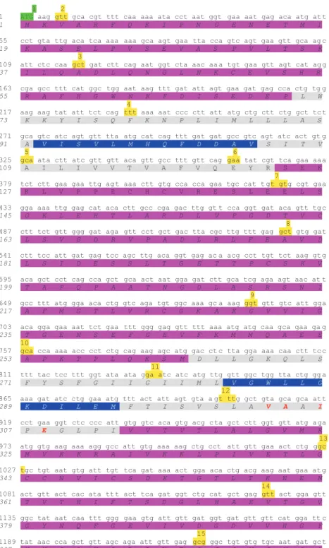
Figure 2
Identification of transmembrane domains vs cytosolic or luminal domains of SPCA1. For simplicity, SPCA1a (NP_055197) has been represented; other isoforms have identical sequence until the C-terminal tail. The highlighted codons in green and in red are the starting and stop codon, respectively. Highlighted in yellow is the first codon of each corresponding numbered exon. Highlighted in gray is the transmembrane domain. The highlighted amino acids in purple are cytoplasmic, whereas those highlighted in blue are luminal. The highlighted amino acids in azure represent the cytosolic C-terminal tail (underlined is the KXD/E motif putative COPI-binding site). The bold typed red and blue amino acids are the residues directly involved in the binding of Ca2+/Mn2+and Mg2+, respectively. Of note, the putative COPI-binding motif KXD/E is just below the M10 domain. The prediction of these domains/residues was solved by similarity (UniProt database)be addressed to the 3
′ end of the gene (namely the last 3 exons/
introns), where the alternative splicing generates the four
different isoforms of SPCA1.
Few nonsense mutations were frequently reported in
different families worldwide (reports with at least three
cases): 115C4T causing R39X;
6,18,19457C4T causing
R153X;
7,20–251402C4T causing R468X;
7,26–281516C4T
causing Q506X;
29,30and 2395C4T causing R799X.
18,25,31–34The above single residues represent ‘hotspots’ for mutations,
and notably, they all occur at CpG sites where the possibility
of mutations is higher.
35The fact that the majority of the
mutations cause PTC through frameshift or single-base-pair
substitution points to haploinsufficiency as the prevalent
mechanism for the dominant inheritance of HHD.
6,7However,
the possibility that at least some mutations cause the
disease through a dominant negative mechanism cannot be
excluded.
13Interestingly, only one mutation was found in the most
3
′ region (exons 27–28) of the gene where differential splicing
generates the four different transcripts.
22The single reported
1243 gta att aga aac aat act cta atg ggg aag cca aca gaa ggg gcc tta att gc t 415 V I R N N T L M G K P T E G A L I A
16
1297 ctt gca atg aag atg ggt ctt gat gga ctt caa caa gac tac atc aga aaa gct 433 L A M K M G L D G L Q Q D Y I R K A 1351 gaa tac cct ttt agc tct gag caa aag tgg atg gct gtt aag tgt gta cac cg a 451 E Y P F S S E Q K W M A V K C V H R
17
1405 aca cag cag gac aga cca gag att tgt ttt atg aaa ggt gct tac gaa caa gta 469 T Q Q D R P E I C F M K G A Y E Q V 1459 att aag tac tgt act aca tac cag agc aaa ggg cag acc ttg aca ctt act ca g 487 I K Y C T T Y Q S K G Q T L T L T Q 1513 cag cag aga gat gtg tac caa caa gag aag gca cgc atg ggc tca gcg gga ctc 505 Q Q R D V Y Q Q E K A R M G S A G L
18
1567 aga gtt ctt gct ttg gct tct ggt cct gaa ctg gga cag ctg aca ttt ctt gg c 523 R V L A L A S G P E L G Q L T F L G 1621 ttg gtg gga atc att gat cca cct aga act ggt gtg aaa gaa gct gtt aca aca 541 L V G I I D P P R T G V K E A V T T 1675 ctc att gcc tca gga gta tca ata aaa atg att act gga gat tca cag gag act 559 L I A S G V S I K M I T G D S Q E T
19
1729 gca gtt gca atc gcc agt cgt ctg gga ttg tat tcc aaa act tcc cag tca gtc 577 A V A I A S R L G L Y S K T S Q S V 1783 tca gga gaa gaa ata gat gca atg gat gtt cag cag ctt tca caa ata gta cc a 595 S G E E I D A M D V Q Q L S Q I V P
20
1837 aag gtt gca gta ttt tac aga gct agc cca agg cac aag atg aaa att att aag 613 K V A V F Y R A S P R H K M K I I K
21
1891 tcg cta cag aag aac ggt tca gtt gta gcc atg aca gga gat gga gta aat ga t 631 S L Q K N G S V V A M T G D G V N D 1945 gca gtt gct ctg aag gct gca gac att gga gtt gcg atg ggc cag act ggt aca 649 A V A L K A A D I G V A M G Q T G T 1999 gat gtt tgc aaa gag gca gca gac atg atc cta gtg gat gat gat ttt caa ac c 667 D V C K E A A D M I L V D D D F Q T
22
2053 ata atg tct gca atc gaa gag ggt aaa ggg att tat aat aac att aaa aat ttc 685 I M S A I E E G K G I Y N N I K N F
23
2107 gtt aga ttc cag ctg agc acg agt ata gca gca tta act tta atc tca ttg gc t 703 V R F Q L S T S I A A L T L I S L A
2161 aca tta atg aac ttt cct aat cct ctc aat gcc atg cag att ttg tgg atc aat
721 T L M N F P N P L N A M Q I L W I N
24
2215 att att atg gat gga ccc cca gct cag agc ctt gga gta gaa cca gtg gat aaa 739 I I M D G P P A Q S L G V E P V D K 2269 gat gtc att cgt aaa cct cct cgc aac tgg aaa gac agc att ttg act aaa aac 757 D V I R K P P R N W K D S I L T K N 2323 ttg ata ctt aaa ata ctt gtt tca tca ata atc att gtt tgt ggg act ttg ttt 775 L I L K I L V S S I I I V C G T L F
25
2377 gtc ttc tgg cgt gag cta cga gac aat gtg att aca cct cga gac aca aca atg
793 V F W R E L R D N V I T P R D T T M
2431 acc ttc aca tgc ttt gtg ttt ttt gac atg ttc aat gca cta agt tcc aga tcc 811 T F T C F V F F D M F N A L S S R S
mutation, located in exon 27, was a nonsense mutation
(2660C4A), which results in a stop codon (S887X) on the last
transmembrane domain (M10). The mutation was found in a
patient with a family history of HHD and suffering from
classical HHD symptoms.
22Notably, this mutation caused the
loss of the C termini of the SPCA1a, SPCA1b and SPCA1d
isoforms but not of SPCA1c that does not include exon 27.
Thus, SPCA1c, the shortest of the isoforms, misses the M10,
and therefore, its stability and consequent ability to function
as a Ca
2+pump is rather questionable.
36Nevertheless, if
SPCA1c was effectively expressed, we can conclude that it is
not functional enough to overcome the loss of the other three
isoforms at least in the skin of the patient presenting the
mentioned S887X mutation.
22Remarkably, this observation
suggests that the mRNA encoding for SPCA1c is probably not
translated as previously suggested.
37Furthermore, the mutation on exon 27 causes a reduction in
SPCA1a, SPCA1b and SPCA1d of 33, 52 and 63 amino acids,
respectively, resulting in a partial truncation of the M10 and of
the full cytoplasmic tail, similarly to what happens in SPCA1c
(888 amino acids). Of consequence, this mutation is predicted
to determine an incomplete expression and degradation of
these SPCA1 isoforms thus resembling SPCA1c. Hereafter,
we will no more consider SPCA1c in this discussion.
Splice variants differ in their C-terminal tail sequence
(Figure 1c) and this is likely to be important for the functionality
of the pump. Indeed, the C-terminal tail could have a role in
mediating interactions with cytoplasmic effectors for
intracel-lular signaling or for targeting the single isoforms to specific
sub-organellar localization. Presently, no cytoplasmic
inter-acting proteins have been found for the SPCA1 C-terminal
domain. However, specific interactions of different SPCA1
tails with proteins involved in membrane trafficking (i.e., golgi
matrix, Ca
2+-binding proteins, Arf/Rab family members,
cytoskeleton, etc.) could support a specific role for C-terminal
tail in the differential distribution of the protein at
sub-organellar level. This would in turn make them unique not
only in triggering the Ca
2+influx into the golgi apparatus, but
also in mediating different cytoplasmic signaling, thus
orches-trating membrane trafficking at different levels along the
secretory pathway. Obviously, these speculations need to be
experimentally proven.
No further mutations have been discovered in the 3
′-end of
the ATP2C1 gene. In other words, once the ATP2C1 gene
is mutated (independently if missense, nonsense, insertion
and/or duplication or part of the primary sequence) the
functionality of all the isoforms is equally compromised in
HHD patients, and at least in the skin, levels of SPCA1 appear
to be not sufficient. However, up-regulation of SERCA2
(the ER ATPase Ca
2+pump type 2) has been recently shown
to (at least partly) compensate for the decreased levels of
SPCA1 in HHD.
38Therefore, although SPCA1 is ubiquitously expressed, is
there differential expression of the isoforms? Is the
compen-satory role for non-functional or depleted SPCA1 by other
pumps tissue specific? Is there a general mechanism? If so,
how does it work? Even if some tentative explanations have
been given,
38,39these open questions need answers to fully
understand the role and distribution of the SPCA1 isoforms.
Missense Mutations on the ATP2C1 Gene
Missense mutations generate single amino acid substitution,
not leading to a PTC. A quick view on where the missense
mutations occur along the ATP2C1 gene sequence gives an
idea of their preferential distribution in few exons. In particular,
almost two-thirds (63%) of the missense mutations (31/49)
localize in only five exons (exon 12, 13, 18, 21, and 23;
Table 1). These exons encode for M4, P, ATP and M5/M6
domains, respectively (Figure 3). Amino acids located on M4,
M5 and M6 (together with M8) transmembrane domains are
critical for Ca
2+/Mn
2+binding. It is not surprising that affecting
residues in these regions, either directly involved in
Ca
2+-binding or compromising the stability or structure of the
protein, may cause a severe effect on SPCA1 functionality,
also without a reduction in its levels. Looking at the
conservation across species we found high level of homology
of these exons and conservativeness of the resulting amino
acid coded by the codons where missense mutations occur
(Figure 3), confirming their critical role.
Several residues have been reported to have a critical
and direct role in Ca
2+/Mn
2+binding, which are conserved
throughout species
40and also in SPCA2, the homolog of
SPCA1.
41Missense mutation D778A in the yeast homolog
(PMR1) resulted in a loss of function mutant apparently
defective for the transport of both Ca
2+and Mn
2+, whereas
mutant Q783A displayed a differential sensitivity consistent
with the selective loss of Mn
2+transport.
42In a similar study,
26
2485 cag acc aag tct gtg ttt gag att gga ctc tgc agt aat aga atg ttt tgc tat 829 Q T K S V F E I G L C S N R M F C Y 2539 gca gtt ctt gga tcc atc atg gga caa tta cta gtt att tac ttt cct ccg ct t 847 A V L G S I M G Q L L V I Y F P P L
27
2593 cag aag gtt ttt cag act gag agc cta agc ata ctg gat ctg ttg ttt ctt ttg
865 Q K V F Q T E S L S I L D L L F L L
2647 ggt ctc acc tca tca gtg tgc ata gtg gca gaa att ata aag aag gtt gaa agg 883 G L T S S V C I V A E I I K K V E R 2701 agc agg gaa aag atc cag aag cat gtt agt tcg aca tca tca tct ttt ctt gaa 901 S R E K I Q K H V S S T S S S F L E 2755 gta tga
919 V STOP
Table 1 Mutations on the ATP2C1 gene reported in the literature
Exon/Intron Nucleotide change Mutation Numbera Codonb Effect Domainc References
Exon 2 28delG/ins24bpd Deletion/insertion 1 PTC N-ter 6,23 d
Exon 2d 115C4T Nonsense 4 R39X PTC N-ter/s1 6,18,19
Intron 2 117+2T4G Donor splice 1 PTC(?) N-ter/s1 95
Intron 2 118-2A4G Acceptor splice 1 PTC N-ter/s1 96
Intron 2 118-1G4A Acceptor splice 3 N-ter/s1 22 e,23,97 d, f
Exon 3d 134delG Deletion 1 45GfsX1 PTC N-ter/s1 98
Exon 3 163C4T Nonsense 3 R55X PTC N-ter/s1 31,97 d, f,99
Exon 3 168delC Deletion 1 ? PTC N-ter/s1 31
Exon 3 180G4Ad Nonsense 1 W60X PTC N-ter/s1 97
Exon 3 185delAGTT Deletion 1 62KfsX34 PTC N-ter/s1 28
Exon 3 212delT Deletion 1 71LfsX26 PTC M1 100g
Intron 3 235-2A4G Acceptor splice 2 M1 101,102
Exon 5 335delT Deletion 1 111LfsX19 PTC M2 23
Intron 5 360+1G4A Donor splice 1 M2 33
Intron 5 360+1G4C Donor splice 1 Skip exon 5 M2 24
Intron 5 360+2T4A Donor splice 2 Skip exon 5 M2 103
Intron 5 361–1G4Ad Acceptor splice 1 PTC/skip exon 6 M2 19
Intron 5d 361-2A4Gd Acceptor splice 1 PTC/loss exon 6 M2 104
Exon 6 366T4A Nonsense 1 Y122X PTC M2 105
Exon 7 457C4T Nonsense 8 R153X PTC A 7,20,21,23–25 e,106
Exon 7 490delT Deletion 1 163LfsX24 PTC A 13
Exon 7 519insA Insertion 2 173LfsX3 PTC A 13,33
Exon 7 520delC Deletion 1 174RfsX14 PTC A 24
Intron 7 531+2T4Ad Donor splice 1 A 18
Exon 8 602C4Th Missense 2 P201Lh A 6 d,13
Exon 8d 635C4A Nonsense 1 S212X PTC A 96
Exon 8 661A4Cd Missense 1 T221P A 87
Exon 8 681dupA Insertion 1 227KfsX13 PTC A 24
Intron 8 688-1G4A Acceptor splice 1 A 13
Exon 9 689G4A Missense 1 G230D A 107
Exon 9 705delA Deletion 1 235TfsX12 PTC A 108
Exon 9 745C4T Nonsense 1 Q249X PTC S3 13
Exon 10 767insCCCT Insertion 1 256TfsX42 PTC S3 7
Exon 10 775C4T Nonsense 1 Q259X PTC S3 105
Exon 10 806T4G Missense 1 L269R M3 105
Exon 10 832G4Ai Missense/
insertion 2 278GfsX22 M3
91
Intron 10 832+3A4T Donor splice 2 M3 13
Intron 10 832+2T4C Donor splice 1 Skip exon 10 M3 20
Intron 10d 833-1G4Ad Acceptor splice 1 M3 6
Exon 11 836insT Insertion 1 279IfsX19 PTC M3 7
Exon 11 854G4A Nonsense 1 W285X PTC l2 95
Intron 11 899+1G4T Donor splice 1 PTC M4 24
Intron 11 899+1G4C Donor splice 2 PTC M4 109
Exon 12 910G4T Missense 2 A304S M4 7 j,20
Exon 12 920C4T Missense 1 P307L M4 104
Exon 12 920C4A Missense 1 P307H M4 36
Exon 12 923delAAG Deletion 1 308delEk M4 28
Exon 12 925G4T Missense 1 G309C M4 13
Exon 12 932del21bpd Deletion 1 311deld M4 18
Exon 12 935T4C Missense 1 I312T M4 110
Exon 12 950del9bp/ins24bpl Deletion/insertion 1 318-320del/insl S4 20
Exon 12 953T4C Missense 1 L318P S4 7
Exon 12 956delC Deletion 1 319AfsX3 PTC S4 24
Exon 12 1001delA Deletion 1 333KfsX12 PTC S4 13
Exon 12 1004T4C Missense 1 L335P S4 111
Exon 12 1022T4C Missense 1 L341P S4 13
Intron 12 1024+1G4Ad Donor splice 1 PTC/skip exon 12 S4 21
Exon 13 1031G4Ad, h Missense 1 C344Yd, h P 6
Exon 13 1042T4C Missense 1 C348R P 105
Exon 13 1045delT Deletion 1 348CfsX6 PTC P 13
Exon 13 1049A4T Missense 3 D350V P 28
Exon 13 1055C4Td Missense 1 T352I P 112
Exon 13 1058G4Td Missense 1 G353V P 30
Exon 13d 1067delC Deletion 1 356TfsX3 PTC P 96
Exon 13 1068del16bpm Deletion 1 356TfsX60 PTC P 113
Exon 13 1085insA Insertion 1 363TfsX11 PTC P 29
Exon 13 1087A4G Missense 1 T363A P 114
Exon 13 1089delTCAC Deletion 4 363TfsX21 PTC P 13,23,28,115
Exon 14 1218G4Cd Missense 1 E406D Skip exon 14 P 19
Exon 15 1231T4C Missense 1 C411R P 13
Exon 15d 1250G4Ad Missense 3 R417K P 32
Intron 15 1308+1G4A Donor splice 1 P 36
Intron 15 1309-1G4A Acceptor splice 1 P 13
Intron 15 1309-4a4t/1309-2a4g Acceptor splice 1 Skip exon 16 P 7
Exon 16 1327C4Td Nonsense 1 Q443Xd PTC P 6
Table 1 (Continued )
Exon/Intron Nucleotide change Mutation Numbera Codonb Effect Domainc References
Exon 16 1402C4T Nonsense 4 R468X PTC P 7,26–28
Exon 16 1413G4C Missense 1 Q471H ? 110
Intron 16 1415-2A4C Acceptor splice 1 PTC/skip exon 17 ? 104
Exon 17 1413del28bpn Deletion 2 472DfsX14 PTC ? 116
Exon 17 1431T4A Nonsense 1 C477X PTC ? 117
Exon 17 1455delAd Deletion 1 485QfsX1 PTC N? 118
Exon 17 1462deld,o Deletion 1 488delo N 104
Exon 17 1469G4T Missense 1 C490F N 31
Exon 17 1508delCTCAd Deletion 1 503TfsX32 PTC N 18
Exon 17 1510C4T Nonsense 1 Q504X PTC N 36
Exon 17 1516C4T Nonsense 4 Q506X PTC N 29,30 d
Exon 17 1523delAT Deletion 2 508DfsX23 PTC N 97,104
Exon 17 1566delCA Deletion 1 522LfsX9 PTC N 7
Intron 17 1570+2T4C Donor splice 1 PTC N 24
Exon 18 1582G4C Missense 1 A528P N 23
Exon 18 1588G4C Missense 1 G530R N 95
Exon 18 1685C4G Nonsense 3 S562X PTC N 6 d,13,28
Exon 18 1709C4Td, h Missense 2 T570Id, h N 6
Exon 18 1723delG Deletion 1 574QfsX24 PTC N 13
Exon 18 1738A4G Missense 2 I580V N 13,31
Intron 18 1694-1G4A Acceptor splice 1 N 6
Exon 19 1751T4C Missense 1 L584P N 21
Exon 19 1782delAGTC Deletion 1 593SfsX5 PTC N 119
Exon 19 1816C4T Nonsense 1 Q606X PTC N 13
Intron 19 1839+2insTd Donor splice 1 PTC N 23
Intorn 19 1840-1G4C Acceptor splice 1 N 98
Exon 20 1854G4Ad Missense 1 R619K N 87
Exon 20 1869delG Deletion 1 623RfsX2 PTC N 33
Exon 20 1874delA Deletion 1 M626X PTC N 120
Exon 20 1875delG Deletion 1 M626X PTC s5 7
Intron 20 1890+1delGTGAG/ins Donor splice 1 s5 22 e
Intron 20 1891-1G4T Acceptor splice 1 s5 121
Exon 21 1897C4T Nonsense 1 Q633X PTC s5 122
Exon 21 1914del/insd Deletion/insertion 1 638Vfs10X PTC s5 123 d
Exon 21 1922T4G Missense 1 M641R s5 7
Exon 21d 1931A4G Missense 1 D644G s5 33
Exon 21 1933G4A Missense 1 G645R s5 7
Exon 21 1934G4Td Missense 1 G645V s5 124
Exon 21 1942G4T Missense 1 D648Y s5 104
Exon 21 1952C4A Missense 1 A651D s5 105
Exon 21 1982T4G Missense 1 M661R s5 113
Exon 21 1983delG Deletion 1 661MfsX14 PTC s5 7
Exon 21 2023delAd Deletion 1 675MfsX PTC s5 18
Exon 21 2025delG Deletion 1 675MfsX2 PTC s5 105
Intron 21d 2058(-17C4T)d Acceptor splice 1 PTC s5 97
Intron 21d 2058-1G4Cd Acceptor splice 1 s5 28
Intron 21 2058-1G4A Acceptor splice 1 Skip exon 22 s5 7
Exon 22 2068G4T Nonsense 1 E690X PTC s5 110
Exon 22 2111insA Insertion 1 704RfsX23 PTC M5 20
Exon 22 2126C4T Missense 3 T709M M5 7,28,107
Intron 22 2127+1G4Ad Donor splice 2 Skip exon 23 (?) M5 28 d,125 d
Intron 22d 2126(+5G4A)d Donor splice 1 PTC M5 97
Exon 23 2132T4G Missense 1 I711R M5 31
Exon 23 2141T4A Nonsense 1 L714X PTC M5 23
Exon 23 2164insACAT Insertion 1 722LfsX6 PTC l3 122
Exon 23 2198A4G Missense 1 Q733R M6 31
Exon 23 2215delATT Deletion 1 739delI M6 20
Exon 23 2224G4T Missense 1 D742Y M6 13
Exon 23 2227delGd Deletion 1 743GfsX8 PTC M6 6
Exon 23 2231C4G Missense 2 P744R M6 7 p,20
Exon 23 2235insC Insertion 1 746AfsX10 PTC M6 107
Exon 23 2236G4Ad Missense 1 A746Td M6 18
Exon 23 2237C4Td Missense 1 A746V M6 34
Intron 23 2243+2T4C Donor splice 1 PTC M6 104
Exon 24 2246T4G Missense 1 L749R M6 20
Exon 24 2251delGT Deletion 1 751VfsX5 PTC s6 126
Exon 24 2264delA Deletion 2 755DfsX17 PTC s6 25,33
Exon 24 2303delAC Deletion 2 768DfsX4 PTC s6 7,20
Exon 24 2339delTTGTd Deletion 1 780LfsX3 PTC M7 19
Exon 24 2357delTT Deletion 1 786IfsX10 PTC M7 7
Exon 24 2365G4A Missense 1 G789R M7 13
Exon 24 2371delTTGT Deletion 4 791LfsX12 PTC M7 7,20,127 d
Exon 24 2374delTTTG Deletion 10 792FfsX10 PTC M7 6 d,7,13,23,31,117,128,129 d,130
Exon 24 2375delTTGT Deletion 2 792FfsX4 PTC M7 102,104
Exon 24 2384G4A Nonsense 1 W795X PTC l4 22 e
Exon 25 2395C4T Nonsense 14 R799X PTC l4 16 d,25,31–34 d
Exon 25d 2412delT Deletion 1 803IfsX7 PTC l4 102
Table 1 (Continued )
Exon/Intron Nucleotide change Mutation Numbera Codonb Effect Domainc References
Exon 25 2422delAC Deletion 2 808TfsX10 PTC l4 33
Exon 25 2445del10bp Deletion 3 814CfsX7 PTC M8 131
Exon 25 2454delT Deletion 1 818FfsX6 PTC M8 31
Exon 25 2454dupT Insertion 1 D819X PTC M8 28
Exon 25 2460delG Deletion 1 820MfsX4 PTC M8 21
Exon 25 2468A4Cd Missense 2 A823E M8 97
Exon 26 2529delGT Deletion 1 843MfsX27 PTC M9 7
Exon 26 2558del10bpq Deletion 1 853MfsX17 PTC M9 31
Exon 26 2593C4Td Nonsense 2 Q865Xd PTC l5 6,112
Exon 26d 2597A4C Missense 1 K866T l5 96
Intron 26 2630-1delG Acceptor splice 2 M10 6 d,13
Exon 27 2660C4A Nonsense 1 S887X PTC M10 22e
Appendix 1 Exon 23 2130T4Cd Polymorphism 1 S710Sd M5 87,88 Appendix 2 Number Mutation % PTC % 59 Deletion/insertion 35.54 55 59.78 24 Nonsense 14.46 24 26.09 49 Missense 29.52 0 0.00 34 Acceptor/donor splice 20.48 13 14.13 166 92 55.42 Appendix 3 Normal p.309 G G P I V V T V T L A L p.320
c.925 GGT CTC CCC ATT GTG GTC ACA GTG ACG CTA GCT CTT c.960
c.925 GGT CTC CTA GCT CTT c.939
p.309 G L L A L p.313
Mutant
Human ATP2C1 gene mutations were summarized. The mutations are grouped for deletion/insertion (azure), nonsense (orange), missense (yellow), acceptor/donor splice (green). Nucleotides are reported in italic along the table as well as in the figure legend and along the full manuscript. We found some reported mutations were inaccurate or not unified. Therefore, we revised or collated some descriptions according to the reported cDNA reference sequence (GenBank accession No. NM_AF181120).7
Appendix 1: A polymorphism was wrongly reported to be a new mutation 2323C4T generating Y711H.87After careful check with the correct reading frame this was not a mutation but a polymorphism.88
Appendix 2: A resuming panel graphs the amount and relative percentage of each kind of mutations and PTC.
Appendix 3: The 884–904delCCATTGTGGTCACAGTGACGC mutation and consequent amino acid 296-302delIVVTVTL was incorrectly reported,18while it referred to as a 21bp deletion located at 932-952 and amino acid 311-317delPIVVTVT. This mutation did not generate the reported missense mutation P295V;18the first ‘C’ of the codon 311 (encoding for a proline, P) recombined with ‘TA’ of codon 318 (leucine, L) generating the codon CTA which encoded for a leucine.
aNumber of reported cases of patients presenting the mutation.
bMissense mutations causing an amino acid substitution in extremely conserved residue through all the ATPases and in different species are highlighted in light pink.23 cPutative protein domain prediction is based on the position of the equivalent residue within the structure of ATP2A1 (SERCA1).
dUsing the running correct coding sequence and relative reading frame of the ATP2C1 gene (Ref. NG_007379.1) we unified the position of the mutation site, protein change, exon/intron location all over the reported mutations. In doing this we found few mutations published as new which were already known.
eThe same authors published their findings in two identical papers on different journals.22,89
fDue to incorrect interpretation of discovered mutations the authors reported as new previously reported mutations.90
gIn their manuscript, the authors reported a previously described mutation. A mistake on referring to this mutation was recently reported (see erratum in Acta Derm Venereol 2015; 95: 1040).
hP201L, C344Y, and T570I, respectively, represent mutations P185L, C328Y, and T554I, originally reported by Sudbrak et al.6Mutation nomenclature has now been updated with respect to the 5'-end sequence published by Hu et al7and the results of 5' RACE-PCR experiments from Fairclough et al.36
iThe missense mutation 832G4A causing the nucleotide change G278R generated an aberrant splicing with a resulting insertion of the first 11 bp (GTAAGAGAAGA) from intron 10 between the mutated exon10 and the exon 11 (see Figure 5 in Chao et al)91causing a PTC.
jThis mutation was incorrectly reported as A304T by Hu et al7as previously reported.20 kE308, and not G309 (which are both Ca2+-binding site residue), was deleted.
l950delCGCTAGCTCTT4CT4insCCACAATGTGTTGGTGTTATGAGAAT (underlined are the deleted/inserted nucleotides) generates the in frame 318delLAL/ insTMCWCYEN.
m1068delAAGAATGAAATGACTG.
nThe delGACAGACCAGAGATTTGTTTTATGAAAG cause a frame shift and PTC. oIn frame delAAGTACTGTACTACATACCAGAGC with amino acid delKYCTTYQS. pThis mutation was incorrectly reported as P724R by Hu et al7as previously reported.20 qThe deleted sequence is TGGGACAATT.
Homo sapiens Pan troglodites Canis lupus Danio rerio Pan troglodites Mus musculus
_M4____________
s4________________________
LAVAAIPEGLPIVVTVTLALGVMRMVKKRAIVKKLPIVETL
LAVAAIPEGLPIVVTVTLALGVMRMVKKRAIVKKLPIVETL
LAVAAIPEGLPIVVTVTLALGVMRMVKKRAIVKKLPIVETL
LAVAAIPEGLPIVVTVTLALGVMRMVKKRAIVKKLPIVETL
LAVAAIPEGLPIVVTVTLALGVMRMVKKRAIVKKLPIVETL
LAVAAIPEGLPIVVTVTLALGVMRMVKKRAIVKKLPIVETL
---
-
--
-
-
-
--
-
---
-
---
-
---
-___P________________________________________________________
GCCNVICSDKTGTLTKNEMTVTHIFTSDGLHAEVTGVGYNQFGEVIVDGDVVHGFYNPAV
GCCNVICSDKTGTLTKNEMTVTHIFTSDGLHAEVTGVGYNQFGEVIVDGDVVHGFYNPAV
GCCNVICSDKTGTLTKNEMTVTHIFTSDGLHAEVTGVGYNPFGEVIVDGDVVHGFYNPAV
GCCNVICSDKTGTLTKNEMTVTHI
L
TSDGLHAEVTGVGYNQFGEVIVDGDVVHGFYNPAV
GCCNVICSDKTGTLTKNEMTVTHI
L
TSDGLHAEVTGVGYNQFGEVIVDGDVVHGFYNPAV
S
CCNVICSDKTGTLTKNEMTVTH
L
FTSDG
Q
HAEVTG
I
GYN
NS
GE
ILM
DG
E
VVHG
ISNTSI
*-
-
---
-
-
-
-
--
---
-
-**----*---*---**--***--*----******
_________________________________________________
SRIVEAGCVCNDAVIRNNTLMGKPTEGALIALAMKMGLDGLQQDYIRKA
SRIVEAGCVCNDAVIRNNTLMGKPTEGALIALAMKMGLDGLQQDYIRKA
SRIVEAGCVCNDAVIRNNTLMGKPTEGALIALAMKMGLDGLQQDY
V
RKA
SRIVEAGCVCNDAVIRNNTLMGKPTEGALIALAMKMGLDGLQQDYIRKA
SRIVEAGCVCNDAVIRNNTLMGKPTEGALIALAMKMGLDGLQQDYIRKA
S
K
IVE
V
GCVCNDA
M
IRHNTLMG
R
PTEGALIALAMKMGL
E
GLQ
HEFV
R
LE
-*--
-
*---
-
---*-
-
---*---*---****-**
___N_____________________________________________________
VLALASGPELGQLTFLGLVGIIDPPRTGVKEAVTTLIASGVSIKMITGDSQETAVAI
VLALASGPELGQLTFLGLVGIIDPPRTGVKEAVTTLIASGVSIKMITGDSQETAVAI
VLALASGPELGQLTFLGLVGIIDPPRTGVKEAVTTLIASGVSIKMITGDSQETAVAI
VLALASGPELGQLTFLGLVGIIDPPRTGVKEAVTTLIASGVSIKMITGDSQETA
I
AI
VLALASGP
D
LGQLTFLGLVGIIDPPRTGVKEAVTTLIASGVSIKMITGDSQETA
I
AI
VLALASG
S
E
M
G
S
LTFLGLVGIIDPPR
A
GVKEAV
A
TLI
S
SGV
AV
KM
V
TGDS
E
ETAVAI
----
-
-
-
***-*---*---*---*---**--*
-
---*---*-
-___S5___________________________________________________
SLQKNGSVVAMTGDGVNDAVALKAADIGVAMGQTGTDVCKEAADMILVDDDFQTIM
SLQKNGSVVAMTGDGVNDAVALKAADIGVAMGQTGTDVCKEAADMILVDDDFQTIM
SLQKNGSVVAMTGDGVNDAVALKAADIGVAMGQTGTDVCKEAADMILVDDDFQTIM
SLQKNG
A
VVAMTGDGVNDAVALKAADIGVAMGQTGTDVCKEAADMILVDDDFQTIM
SLQKNGSVVAMTGDGVNDAVALKAADIGVAMGQTGTDVCKEAADMILVDDDFQTIM
SLQ
NI
G
A
VVAMTGDGVNDAVALKAADIGVAMGQTGTDVCKEAADMILVDDDFQTI
L
---**-*---
-
--
--
--
-
--
-
---
-
---*
__ M5__
_____l3____
___M6_________________
SIAALTLISLATLMNFPNPLNAMQILWINIIMDGPPAQSL
SIAALTLISLATLMNFPNPLNAMQILWINIIMDGPPAQSL
SIAALTLISLATLMNFPNPLNAMQILWINIIMDGPPAQSL
SIAALTLISLATLMNFPNPLNAMQILWINIIMDGPPAQSL
SIAALTLISLATLMNFPNPLNAMQILWINIIMDGPPAQSL
SIAALTLISLATL
L
NFPNPLNAMQILWINIIMDGPPAQSL
-
-
---*---
-
---
-
-
-
-
-
---Homo sapiens Pan troglodites Mus musculus Danio rerio Canis lupus Rattus norvegicus Homo sapiens Pan troglodites Mus musculus Danio rerio Canis lupus Rattus norvegicus Homo sapiens Pan troglodites Mus musculus Danio rerio Canis lupus Rattus norvegicus Homo sapiens Pan troglodites Mus musculus Danio rerio Canis lupus Rattus norvegicus Homo sapiens Pan troglodites Mus musculus Danio rerio Canis lupus Rattus norvegicusFigure 3
Missense mutations mostly affect critical SPCA1 domains. Most of the missense mutations localize on exons 12, 13–16, 18, 21, and 23 of ATP2C1 gene coding for the indicated protein domains (M4, P, N, S5, M5-M6) crucial for the protein enzymatic activity and Ca2+/Mn2+binding. In human HHD patients the missense mutations cause the indicated amino acid changes, supporting a crucial role for them in functionality of the SPCA1. Of note, the mutations localize on highly conserved residue/codon between the considered species, highlighting the importance of those encoded amino acids. In blue are indicated the different amino acids compared to the human sequence, in red where the mutations occur. Alignments were obtained from Ensembl database as described in Figure 1G783 and V335 in PMR1 showed conformation-sensitive
packing at the cytoplasmic interface, suggesting that these
residues are important in organizing the entry gate for Mn
2+ions.
43Missense mutation can also cause a change in protein
conformation affecting possible interaction of SPCA1 with
other proteins, like cofilin, which has been found to be crucial
in recruiting actin to mediate the cargo sorting that interact
directly with SPCA1 nucleotide domain through an interaction
mediated by Ca
2+ions.
44Remarkably, these examples highlight how a possible
missense mutation can affect SPCA1 resulting not only in a
less efficient pump, but also accounting for its diminished
ability to interact with other proteins. In some HHD patients this
scenario results in a severe etiology, comparable to the
situation where mutations on ATP2C1 gene cause PTC, but in
the presence of unaffected levels of total protein expression.
ATP2C1 Gene: Human Versus Murine
Another aspect to highlight is the different etiology occurring
between humans and mice as a consequence of ATP2C1
mutations. In humans, HHD rarely degenerates in skin cancer
although squamous cell carcinoma and basal cell carcinoma
arising in lesions of HHD have been described in the
literature.
45–47However, which is the relationship, if any,
between skin cancer development and HHD has not been
deeply investigated. To our knowledge, there are no reports of
melanoma or non-cutaneous malignant neoplasms
asso-ciated with HHD.
On the other side, heterozygotic mice exhibit no evidence
of skin lesions indicative of HHD, meanwhile generated
skin cancer in adult.
48,49Null mutant embryos exhibited
growth retardation, failure of neural tube closure, but normal
hematopoiesis and cardiovascular development, resulting in
embryonic lethality.
48At subcellular level, golgi membranes of
Spca1
− / −embryos were dilated, had fewer stacked leaflets,
and were expanded in amount, consistent with increased golgi
biogenesis.
48Increased golgi-associated vesicles and a
marked reduction of ribosomes on rough ER were also
observed.
48Coated pits, junctional complexes, desmosomes,
and basement membranes appeared normal in mutant
embryos, indicating that processing and trafficking of proteins
in the secretory pathway was not massively impaired.
48However, apoptosis was increased, possibly the result of
secretory pathway stress, and a large increase in cytoplasmic
lipid was observed in mutant embryos, consistent with
impaired handling of lipids by the golgi.
48Possibly, reduction
of the proper levels of Spca1 resulted in alteration in the
activity of the glycosyl-transferases due to a decreased
accumulation of Mn
2+in the golgi lumen, which could
contribute to the secretory pathway stress as well as apoptosis
due to a decreased of Ca
2+handling.
Recently, an important difference between human and
murine genomes that could explain the differential outcome of
mutations in the two species has been reported.
50The Atp2c1
gene in mice expresses only one isoform of the gene, not
presenting alternative splicing events at the 3
′-end of the gene
like the human counterpart. The last exon of the ATP2C1
human gene overlapped in the antisense direction with the last
exon of the ASTE1 (also named HT001) gene, which codes for
a protein of unknown function.
50ATP2C1/ASTE1 overlap (not
present in the murine genome) could have a role in regulating
isoform expression, or more largely, the full SPCA1
expression.
50This sort of regulation could have in turn a
protective role that is imbalanced once one of the two alleles is
mutated in humans, whereas not in mice.
50Putative Interacting Domains in the C-terminal Cytosolic
Tails of SPCA1 Isoforms
As suggested above, different SPCA1 isoforms due to their
unique C termini could have a different pattern of distribution
and functional roles. The primary sequence of the C-terminal
tail of each isoform is reported in Figure 1c. An intriguing
possibility is that the uniqueness of their function is a
consequence of the interaction with different protein partners.
It is well known that many resident proteins display in their
tails, motifs like sorting signals responsible for retrieval or
retention. For instance, the C-terminal KKXX motif, identified
in many ER-localized type I membrane proteins, can function
as an ER-retention signal to retrieve ER-resident membrane
proteins from the golgi via a direct interaction with coat protein
complex I (COPI) coatomer.
51–53Another example is the
semi-conserved (F/L)-(L/V)-(S/T) motif, which has been shown to
function as a golgi-retention signal by interacting with COPI
vesicles via Vps74p and thereby maintaining the steady golgi
localization of glycosyl-transferases in yeast.
54Recently, a
conserved retention function and COPI-binding ability for the
KXD/E motif has been shown in the golgi apparatus through
the evolution in different species and in many endomembrane
proteins.
55,56Using bioinformatics tools (see Figure 4 legend), we
analyzed the SPCA1 sequences in the areas corresponding
to the terminal exons 26–28 in terms of secondary structure
propensity (Figure 4a), predicted transmembrane helices
topology (Figure 4b), and interaction with putative protein
partners (Figure 4c). We found that the KXD/E motif is present
in isoforms SPCA1a, SPCA1b, and SPCA1d, in the cytosolic
tail immediately after the M10 (Figure 4c) in a region that is
predicted to be a helical extension of M10 in all three isoforms
(compare Figures 4a and b). This motif is also highly
conserved throughout different species suggesting functional
importance (Figure 4d).
Therefore, SPCA1 is potentially involved in the interaction
with COPI. This reinforces the idea that SPCA1 could be
involved in regulating local fusion/fission of cargo domain/
vesicles carrying proteins along the secretory pathway,
triggering the cytosolic Ca
2+level in the peri-golgi membranes,
where Ca
2+-binding proteins are temporarily recruited to
organize and regulate the membrane fusion/fission, a
funda-mental process for cargo progression.
4,57The analysis of the potential of C-terminal tail residues
to interact with protein partners also highlighted additional
sequences, which are differentially present in the four isoforms
(Figure 4c). In particular, the isoforms SPCA1b and SPCA1d
both end with the C-terminal sequence EDVSCV, which is
predicted to be a protein interaction site (Figure 4c). By
inspecting the literature we realized that this motif perfectly
reproduces the structural features that are necessary
for recognition by class I PDZ domains: (i) being at the
C-terminal of a protein and (ii) matching the consensus for the
last three residues S/T-X-Φ-COO
− ,
58where X is any residue
and Φ is a hydrophobic residue (often a valine). Thus, this motif
is strongly predicted to interact with the PDZ domains
of other proteins.
59Several proteins containing the PDZ
domain can potentially interact with the tail of SPCA1b
and SPCA1d modulating the function of the pump.
Interest-ingly, evidences that the C-terminal tail of SPCA1 could
interact with the golgi matrix arise from a recent observation of
the interaction of C-terminal valine-bearing cargoes moving
through the golgi apparatus by binding to GRASP55
and GRASP65, two golgi matrix proteins that contain PDZ
domains.
60PDZ domain recognition of the C-terminal
tail is involved in golgi maintenance and in cargo
progres-sion.
60,61The SPCA1 isoforms present a valine residue as
last amino acid residue in the C-terminal position (Figures
1c and 4c).
Also the FLEV motif present at the C-terminal end of
SPCA1a and in the cytoplasmic tail of SPCA1d is predicted to
be a protein binding motif (Figure 4c). Interestingly, this motif is
also present in rat calsequestrin (CASQ; AAA75480). CASQ
regulates Ca
2+-binding in the sarcoplasmic reticulum in
SPCA1a
SPCA1b
SPCA1c
SPCA1d
Pan troglodytes Mus musculus CONSENSUS Canis lupus Rattus norvegicus