Università degli Studi di Ferrara
DOTTORATO DI RICERCA IN
"BIOCHIMICA, BIOLOGIA MOLECOLARE E BIOTECNOLOGIE"
CICLO XXVICOORDINATORE Prof. Francesco Bernardi
Molecular mechanisms
impairing biosynthesis and function of
hemostatic proteins
Settore Scientifico Disciplinare BIO/11
Dottorando
Tutore
Dott. Matteo Campioni
Prof. Francesco Bernardi
Co-tutore
Prof. Mirko Pinotti
3
Contents
Summary
Abstract ... 7 Abbreviations ... 8 Introduction ... 9 Hemostasis ... 9 Blood coagulation ... 10 Initiation phase ... 11 Amplification phase ... 12 Propagation phase ... 12 Termination phase ... 13Macromolecular complexes in blood coagulation ... 15
FIX ... 17
F9 gene ... 17
FIX synthesis and maturation ... 18
FIX structure and activation ... 18
Role of coagulation factor IX in the coagulation cascade. ... 19
Molecular basis of Hemophilia B ... 19
Conventional treatment strategies hemophilia B ... 21
Von Willebrand Factor ... 21
Von Willebrand Factor in hemostasis ... 21
Von Willebrand Factor structure and biosynthesis... 22
VWF domains ... 23
Von Willebrand Diseases ... 24
Aim of the PhD thesis... 26
Ribosome Readthrough Over Nonsense Mutations influences the Residual FIX expression ... 27
1.1 Ribosome Readthrough Accounts for Secreted Full-Length Factor IX in Hemophilia B Patients with Nonsense Mutations ... 27
4
Translation termination ... 27
Nonsense mutations ... 28
Readthrough ... 30
Readthrough in human disease ... 31
Aim of the study ... 32
Patients’ data and analysis ... 32
Evidence for the occurrence of readthrough in vivo ... 34
Evidence for the occurrence of readthrough in vitro ... 35
Conclusion ... 38
1.2 Induction of Ribosome Readthrough Over F9 Nonsense Mutations by Aminoglycosides ... 39
The state of art of ribosome readthough induction ... 39
Rationale and Aim of the study ... 41
Investigation of readthrough induced by G418 in vitro ... 42
FIX Tyr450Cys mutation ... 44
Replacement of the Y450 (c234) phenyl ring in the carboxyl-terminal region of coagulation factor IX causes pleiotropic effects on secretion and enzyme activity. ... 44
Missense mutations ... 44
In Vitro Expression of the rFIX-450Cys variant ... 44
Investigation of the role of the Tyr450 side-chain ... 46
Conclusions ... 51
Molecular mechanisms in VWD ... 52
Dominant negative effects in Von Willebrand Factor biosynthesis ... 52
The dominant negative effect ... 52
Previous knowledge ... 52
In vivo P1105_C1926delinsR ... 54
“Double mutant” strategy ... 56
in vitro investigation of the DEL+C2773R variant ... 57
Western blotting analysis of media ... 59
Multimer analysis of conditioned media ... 60
Investigation of DEL+C2773R variant in mouse model ... 60
5
General Methods ... 65
Transformation of bacteria ... 65
Preparation of bacterial competent cells ... 65
Plasmid DNA purification ... 65
Direct sequencing ... 65
Gel electrophoresis ... 66
Polymerase chain reaction (PCR) ... 66
Retro trasciption reaction (RT) ... 67
Plasmids ... 67
Site specific mutagenesis ... 68
Transfection protocol ... 68
Western blot analysis ... 68
ELISA ... 68
Factor IX activity ... 69
Hydrodynamic injection ... 69
Multimer analisys ... 70
Bibliography ... 72
Non-viral transfection systems for nucleic acids ... 78
long-chain cationic derivatives of PTA (1,3,5-triaza-7-phosphaadamantane) as new components of potential non-viral vectors ... 79
Introduction ... 79
Materials and Methods ... 80
Characterization of CP-SLN: size, potential and morphology ... 80
Analysis of the electrophoretic mobility of complexes between CP-SLN and DNA ... 81
DNA stability studies ... 81
Effect of CP-SLN on cell proliferation ... 81
Transfection studies ... 82
Results and Discussion ... 82
Binding migration studies of CP-SLN ... 83
Stability studies ... 84
Gene transfection experiments ... 84
6
Acknowledgements ... 85
Cationic lipid nanosystems as carriers for nucleic acids ... 93
Introduction ... 93
Materials and methods ... 94
Transfection studies ... 96
Statistical analyses ... 96
Results and Discussion ... 96
Morphological analysis ... 97
Cytotoxicity studies ... 97
Binding migration studies... 98
Transfection experiments ... 98
Conclusions ... 100
Acknowledgements ... 101
References ... 107
7
Abstract
The gene mutations leading to hemorrhagic disorders provide peculiar models to elucidate molecular mechanisms underlying protein biosynthesis, and the relationship between the structure and function.
The research activity has been focused on the molecular defects leading to severe deficiency of Factor IX (FIX), a serine protease with a key role in the intrinsic pathway of blood coagulation, and of von Willebrand factor (VWF), a large multimeric protein essential for the primary hemostasis.
In particular, I investigated mechanisms due to three different molecular defects such as nonsense and missense mutations in F9 gene, associated with type I hemophilia B, and an in-frame deletion in the VWF gene, displaying a dominant-negative effect.
Results from investigations in patients’ plasma and the expression of nonsense FIX variants in eukaryotic cells led to the demonstration of trace levels of full-length FIX molecules even in the presence of nonsense mutations through a mechanism of ribosome read-through. The efficiency was dependent of the specific nonsense mutation and on it sequence context. Moreover, I investigated the susceptibility of a panel of nonsense FIX mutations to the induction of readthrough by aminoglycosides. The data suggested a direct relationship between the spontaneous and the drug-induced readthrough. Overall data indicated that not all nonsense mutations can be considered truly “null-mutations”, a finding that have pathophysiological implications.
The severe p.Tyr450Cys mutation in the carboxyl-terminal region of coagulation FIX was chosen as model to study the interplay between impaired protein biosynthesis and/or function caused by missense mutations in relation to specific protein regions, which has been poorly investigated. Results from the expression of a panel of recombinant variants demonstrated the key role of the tyrosine phenyl group for both FIX secretion and coagulant activity. Comparison among highly homologous coagulation serine proteases indicate that additive or compensatory pleiotropic effects on secretion and function by carboxyl-terminus mutations produce life-threatening or mild phenotypes in the presence of similarly reduced protein amounts.
Finally I contributed to the characterization of the dominant inheritance in VWD, due to two essential process in VWF dimerization and multimerization VWD can express dominant-negative features. Previous study characterized and demonstrate d the modulation of this dominant effect. In our study we reproduced in vivo the effect of dominance and through a creation of an artificial mutation, we demonstrated in vitro an in vivo the key role of interaction between wild type and mutant protein monomers during dimerization and or multimerization, We believe that our finding have general implication for the dominant forms of VWD, the most frequent inherited bleeding disorders in humans.
8
Abbreviations
The standard abbreviations used in this thesis follow IUPAC rules. All the abbreviations are defined also in the text when they are introduced for the first time.
bp: base pais
cDNA: Complementary DNA DNA: DeoxyriboNucleic acid
dNTPs: Deoxynucleoside triphosphate (A, C, G and T) ELISA: Enzyme-Linked Immuno Sorbent Assay
FIX: Factor IX FVII: Factor VII FX: Factor X Kb: Kilobase kDa: Kilodalton
NMD: Non sense Mediated Decay nt: Nucleotides
PBS: Phosphate bufer saline
PTC: Premature Termination Codon PTC: Premature termination codon RNA: RiboNucleic Acid
SDS: N-lauroylsarcosine sodium salt TF: Tissue Factor
VWF: Von Willebrand Factor WB: Western Blotting Assay WT: Wild-type
9
Introduction
Hemostasis
It is known since long time that hemostasis is a dynamic process whereby blood is maintained fluid under normal life conditions, but is allowed to rapidly clot in case of trauma to prevent excessive blood loss and death. This mechanism is carefully regulated and it involves various cellular and molecular components.
Blood coagulation is part of this important organism defense mechanism. In resting state the endothelial cell inhibit the platelet adherence and thus the activation of the blood coagulation, and are responsible of synthesis of prostacyclin, heparin-like substances and hold protein complexes (thrombin-thrombomodulin), leading to generation of anticoagulant proteins (activated protein C), which prevent clot formation in heath normal blood vessels (Furie and Furie 1992).
After vessel injury, damaged endothelial cells expose negatively charged phospholipids and platelets can adhere to macromolecules in subendothelial tissues and then aggregate to form the primary hemostatic plug that temporary blocks blood loss.
The interaction between platelets and the damaged endothelium requires von Willebrand Factor (VWF), a large multimeric plasma protein which acts as a bridge by binding exposed collagen in the sub-endothelium and a specific receptor on platelet surface Glicoprotein Ib. The phospholipids composition of the platelet membrane changes, resulting in the exposure of negatively charged phopshatidylserine on the outer leaflet of platelet membrane (Bevers, Comfurius et al. 1982, Bevers, Comfurius et al. 1983). The activation of platelets by thrombin, ADP, thromboxane A2 or epinephrine triggers characteristic morphological and biochemical alterations in the platelet(Furie and Furie 1992).
Activated platelets secrete -granules, containing fibrinogen, Factor V (FV), Factor VIII (FVIII), vWF and other proteins involved in haemostasis, and -granules, containing calcium ions and ADP, and aggregate at the site of injury, forming a sort of plug that provisionally blocks blood loss. The expression on the platelet surface of a receptor (glycoprotein IIb-IIIa) for plasma proteins (fibrinogen) mediates platelet aggregation (Dahlback 2005).
These events are followed by inflammation and repair reactions. Thrombin plays a key role in these processes by chemotactically drawing leukocytes to the site of injury and by stimulating tissue remodelling and mitogenesis. P-selectin expressed on the platelet membrane in the haemostatic plug acts as a receptor for monocytes and neutrophils which, in addition to providing ideal membrane surfaces for blood coagulation, sustain the inflammatory response. During wound healing, the fibrin clot is degraded by the serine protease plasmin, a process known as fibrinolysis (Collen 1999, Riddel, Aouizerat et al. 2007).
10
Blood coagulation
In a classical view coagulation is represented as a “cascade” or “waterfall” model, divided into two pathways: an “intrinsic pathway”, so named because all the components are present in blood, and an “extrinsic pathway”, in which the subendothelial cell membrane protein tissue factor (TF) is required in addition to circulating components. The initiation of both pathways resulted in activation of Factor X (FX) and the eventual generation of a fibrin clot through a common pathway (Luchtman-Jones and Broze 1995). Although these concepts represented a significant advance in the understanding of coagulation and served for many years as a useful model, more recent clinical and experimental observations (Kleinschnitz, Stoll et al. 2006) explain how the cascade hypothesis does not fully and completely reflect the events of hemostasis in vivo.
A cell-based model of coagulation explain, in a more physiological way, how coagulation cascade evolves in consequence of a vascular injury, underlying the roles of cellular elements. Several cells play different roles in the coagulation process, due to their procoagulant and anticoagulant properties.
A cell-based model of coagulation explain, in a more physiological way, how coagulation cascade evolves in consequence of a vascular injury, underlying the roles of cellular elements. The cascade model included the recognition of negative charged phospholipids, principally phosphatidylserine, as a requirement for the assembly and the full function of coagulation complexes, but the role of cells, especially platelets, was thought to be primarily to provide anionic phospholipids and not to be actively involved in the process.
Several cells play different roles in the coagulation process, due to their procoagulant and anticoagulant properties. Blood platelets and TF-bearing microparticles (MPs) play a major role in supporting procoagulant reactions, supplying negatively charged phospholipids essential for the correct assembly of molecular complexes. Microparticles are vesicles that carry a cytoskeleton surrounded by a membrane consisting of a phospholipid bilayer which shows a high density of negative charged phospholipids, particularly phosphatidylserine, on its outer membrane layer (Lechner and Weltermann 2008). Among the various hypothesis functions of MPs and one of the most studied is their possible role in hemostasis and thrombosis, and the capacity by monocytes of shedding microparticles selectively enriched in TF has been observed (Del Conde, Shrimpton et al. 2005). Vascular endothelial cells play a key role in maintaining the anticoagulant properties of the vasculature; thus the process of coagulation is prevented, at least in part, by keeping the two cell types (platelets and endothelial cells) apart until an injury makes activation of the coagulation system indispensable.
Coagulation pathway proceeds as a sequence of events localized on the site of vessel injury, essentially the whole process can be condensed in four different phases: initiation, amplification, propagation and last but not least termination.
11
Initiation phase
The process of blood coagulation starts by the exposure of TF-expressing cells to flowing blood. TF is expressed constitutively on cells such as smooth muscle cells and fibroblasts but not on resting endothelium. TF is also expressed in several other districts that constitute an hemostatic envelope normally not in contact with blood (Dahlback 2005). Disruption of the endothelium walls or activation of endothelial cells or monocytes results in the exposure of TF on blood flow. Stronger evidence suggests that TF also circulates in blood exposed on the surface of MPs; this TF derive from various cell types: white blood cells, endothelial cells, and platelets, and might play important roles in development of pathological thrombosis (Osterud and Bjorklid 2006).
Figure 1: FVIIa bound to TF activates FX and FIX. FXa formed binds to FVa on that cell and converts prothrombin to thrombin.
Upon an injury, FVIIa, present at trace levels in plasma, binds tightly to TF and forms the TF/FVIIa complex that activates small amounts of FX and Factor IX (FIX). Activated FX (FXa) associates with its cofactor, activated Factor V (FVa), and forms the prothrombinase complex on the surface of the TF-bearing cells (Monroe and Hoffman 2006), leading to the conversion of small amounts of circulating Prothrombin (II) to Thrombin (IIa). The active form of FV derives from one of several sources. The adhesion process to components as collagen partially activates platelets and promotes secretion of partially activated FV from their α-granules. Zymogen FV can also be converted to FVa by thrombin, FXa (Monkovic and Tracy 1990).
The localization to the cell surface make FXa relatively protected from inactivation mediated by protease inhibitors. However, FXa molecules that dissociate from TF-bearing cells are
12
rapidly inhibited in the fluid phase by Tissue Factor Pathway Inhibitor (TFPI) and Antithrombin (AT). Thus, the presence of inhibitors localizes FXa activity to the surface on which it was converted to the active enzyme form. Contrarily, FIXa can move from TF-bearing cells to the platelet surface since it is not inhibited by TFPI and more slowly inhibited by AT than FXa.
The coagulation proteins leave the vasculature, percolate through the tissues, and are found in the lymph roughly in proportion to their molecular size (Miller, Howarth et al. 2000); thus FVII is probably bound to extravascular TF even in the absence of an, and the extravascular FX and FIX can be activated as they pass through the tissues. This idea is consistent with the finding that low levels of the activation peptides from coagulation factors are present in the blood of normal individuals injury (Monroe and Hoffman 2006).
This process does not lead to clot formation under normal circumstances, because the really large components of the coagulation process, platelets and Factor VIII (FVIII)/ VWF complex, are kept sequestered in the vascular space. Coagulation only proceeds when damage to the vasculature allows platelets and Factor VIII/ VWF exposure into the extravascular tissues.
Amplification phase
The small amount of thrombin generated on the TF-bearing cell in the initiation phase, has several important functions; one of that is activation of platelets resulting in an increase in phosphatidylserine exposure on the membrane outer leaflet, thus serving as a surface for assembly and activity of the coagulation complexes. Although platelets have already adhered at the site of injury and become partially activated, the addition of thrombin can induce a higher level of procoagulant activity than adhesive interactions alone (Alberio and Dale 1999). As a result platelets release partially activated forms of FV onto their surfaces. Another function of thrombin formed during the initiation phase is the activation of the cofactors FV and FVIII on the activated platelet surface. In this process, the FVIII/ VWF complex is dissociated, permitting VWF to mediate additional platelet adhesion and aggregation at the site of injury
Thrombin also activates Factor XI (FXI), activated by the prekallekrein – kininogen – Factor XII cascade in the classic “intrinsic parhway”, which acts as a “booster” of thrombin generation on the platelet surface (Alberio and Dale 1999). This finding also strengthen the hypothesis that the intrinsic mechanism gives no contribute to in vivo coagulation process. By the end of the amplification phase, successive stage is a production of great amount of thrombin, called propagation phase.
Propagation phase
Now activated-Factor IX (FIXa) surface of activated platelets generated during the initiation phase can now bind to its cofactor, activated-Factor VIII (FVIIIa), on the platelet surface, assembling in the so called “tenase-complex”, additional FIXa is supplied by platelet-bound activated-FactorXI (XIa). Because FXa cannot move effectively from the TF-bearing cell to the activated platelet, FXa must be provided directly on the platelet surface by FIXa/FVIIIa
13
complex. Then FXa rapidly associates with FVa bound to the platelet during the amplification phase, producing a powerful increase in thrombin generation to provide rapidly to clot (Dahlback 2005, Monroe and Hoffman 2006). Due to the burst of production during propagation phase more than 95% of the total thrombin generated in a single event of clotting (Mann, Brummel et al. 2003).
The burst of thrombin generated on the platelet surface produces a stable clot structure, indeed, thrombin does additional actions responsible for clot stabilization: activation the fibrin stabilizing factor Factor XIII (FXIII) (Lorand 2001); cleavage of a receptor that contributes to the full activation of platelets the protease-activated receptor-4 (PAR-4)31]; and (Ofosu 2003) activation of thrombin activable fibrinolysis inhibitor (Bajzar, Manuel et al. 1995). Thrombin activable fibrinolysis inhibitor (TAFI) is a carboxypeptidase that removes terminal lysine residues from fibrin, thereby removing potential binding sites for fibrinolytic enzymes and enhancing clot resistance to fibrinolysis (Nesheim 1998) the failure in TAFI activation can contribute significantly to the bleeding tendency in hemophilia (Mosnier, Lisman et al. 2001).
Figure 2: Cascade reaction and signal amplification during propagation phase.
Termination phase
Once a fibrin platelet clot is formed over a damaged area, the clotting process must be limited to avoid thrombotic occlusion in other normal areas of the vasculature (Hoffman 2003).
14
The TF/FVIIa activity is inhibited by the Kunitz-type inhibitor TFPI, secreted by endothelium. TFPI binds to FXa forming a quaternary complex with TF/FVIIa that quickly limits coagulation (Broze, Girard et al. 1990).
The serine protease inhibitor Antithrombin (AT) inhibit the enzymes of the coagulation system, its physiological role is to protect the circulation from free enzymes and limit the coagulation process to sites of vascular damage; AT is known as the major thrombin-inactivating protein (Beresford and Owen 1990). Circulating AT is a relatively inefficient, but its activity is stimulated by heparin and presumably by heparin-like molecules such as sulphated glycosaminoglycans that normally are synthesized and expressed by endothelial cells (Weitz 2003). This increased efficiency of AT by heparin is the molecular basis for the use of heparin as a therapeutic anticoagulant and coating for medical devices.
While TF-bearing cells and platelets have procoagulant functions, vascular endothelial cells have anti-coagulant features.
The protein C (PC) anticoagulant system inhibits the procoagulant functions of FVIIIa and FVa, the cofactors involved the tenase (FIXa/FVIIIa) and prothrombinase (FXa/FVa) complexes, respectively (Dahlback 2005). The key component in this system is PC, a vitamin K-dependent zymogen (pro-enzyme) and it is activated by thrombin bound to the membrane protein thrombomodulin (TM), that acting as a receptor for thrombin on the surface of intact endothelial cells. Upon binding to TM, the specificity of thrombin is changed, becoming more effective at activating PC than clotting fibrinogen or activating platelets (Ye, Esmon et al. 1991) changing its activity from a pro-coagulant to an anti-coagulant effect depending of its localization.
Activated PC (APC) cleaves just few peptide bonds in each of the phospholipid membrane-bound cofactors FVa and FVIIIa, resulting in the inactivation of the cofactors, moreover APC can also cleave the intact form of FV (Dahlback 2004). APC-mediated cleavage of factor V results in generation of anticoagulant FV that works in synergy with protein S as APC cofactor in the degradation of FVIIIa; indeed, FV can act as a procoagulant and as anticoagulant cofactor too, in fact procoagulant factor Va being formed by limited proteolysis by thrombin or factor Xa, anticoagulant FV activity is reached after proteolysis by APC (Dahlback 2000).
APC activity is enhanced by protein S (PS) another vitamin K-dependent inhibitory cofactor; normally in humans, protein S can be found for the ≈30% as of circulating free protein in plasma and the remaining ≈70% is bound to the complement regulatory protein C4b-binding protein, however only the free form of PS can works as a cofactor to APC.
In addition to TM and heperan-like glycosaminoglycans on surface of endothelial cells is present a cell-surface ADPase (CD39) that metabolizes ADP that is normally released from activated platelets, CD39 catalysis block aggregation of platelets in proximity to healthy endothelium to avoid uncontrolled aggregation and thrombus formation.
15
Figure 3: The protein C pathway on the left inactivation of FVa by APC and PS, on the rigth Inactivation of FVIIIa by APC and PS
Macromolecular complexes in blood coagulation
Activation and activity of clotting factors does not occur in the solution phase, but in macromolecular complexes on the membrane surfaces; each complex include a vitamin K-dependent serine protease, a non-enzymatic protein cofactor and a zymogen substrate, as well as Ca2+ ions (Mann, Nesheim et al. 1990).
Complex Enzyme Cofactor Substrate
Initiation complex FVIIa TF FIX, FX
Prothrombinase FXa FVa PT
Intrinsic tenase FIXa FVIIIa FX
Protein C-ase Thrombin TM PC
Table 1: Macromolecular complexes in blood coagulation.
The protein-phospholipids and protein-protein interactions within the macromolecular complexes enhance reaction rates by several orders of magnitude, by affecting both the affinity constant (KM) for the substrate and the turnover (kcat) of the enzyme. Moreover,
localization of different enzyme complexes on the same nearby membrane surfaces allows to canalize successive reaction, like industrial assembly line, this circumstance also protects activated factors from inactivation by circulating inhibitors (Neurath 1984).
Localizated on membrane and associated as macromolecular complexes, coagulation reactions are finely regulated. In fact complex assembly requires a number of simultaneous events: the conversion of a zymogen to the active serine protease, the activation of a procofactor to the active cofactor and the availability of negatively charged phospholipid membranes. All these different condition guarantees the confinement of the coagulation process to the site of injury escaping to the risk of uncontrolled thrombisis (Mann, Nesheim et al. 1990).
The catalytic domains of the coagulation serine proteases are highly homologous(Neurath 1984) but despite the similarities, the coagulation proteases act on their substrates with precision and distinctive specificity (Neurath 1984). It is assumed that the substrate specificity arises from specific interactions between the enzyme active site and distinctive
16
sequences surrounding the scissile bond. In the case of FVIIa-TF, this assumption is supported by similarities between the residues preceding the scissile bonds in FIX and FX. A series of studies have suggested a role for extended interactions between FX and surfaces in both FVIIa and TF during FX activation and evidences support a direct interaction between the N-terminal Gla domain in the FX light chain and regions of the FVIIa-TF complex near the membrane surface (Ruf, Kalnik et al. 1991). Since the activation peptide at the N-terminus of the heavy chain of FX is released upon cleavage, it is possible that the interactions between the substrate and the FVIIa-TF complex involve structural determinants common to FX and FXa, suggesting the usage of a common region of TF in a dual role, as cofactor for FXa-mediated FVII activation and as cofactor for FVIIa-FXa-mediated FX activation moreover same TF domain would also contact FIX during its FVIIa-mediated activation (Kirchhofer, Lipari et al. 2000).
17
FIX
F9 gene
Coagulation factor IX gene (F9, MIM#300746, Genebank accession number K02402.1) is located on the long arm the X chromosome, more towards the centromere at Xq28, in region q27.1-q27.2.
Figure 4: Localization of FIX gene on human X chromosome
The gene were cloned and sequenced by Kurachi e Davie in 1985; is approximately 34 kb in length and contains only eight exons named from a to h, the largest of which is only 1935 bp. The 95% of the transcript is composed by introns and the coding sequence is only 2803 bases in length and comprises a short 5’ UTR (29 bp), an open reading frame plus stop codon (1383 bp) and a 3’ UTR (1390 bp).
18
FIX synthesis and maturation
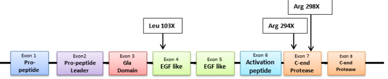
The open reading frame encodes a pre-pro-protein of 461 amino acid. The first two exons (A, B) encode for pre-pro-peptide sequence (28 amino acid) that directs FIX to the endoplasmic reticulum for subsequent post-translational modification and secretion. The pro-peptide (18 amino acid) provides a binding domain for vitamin K dependent carboxylase, which carboxylates certain glutamic acid residues in the adjacent Gla domain (Anson, Choo et al. 1984). Part of exon B and exon C encode for the Gla domain which contains 12 residues of glutamic acid, that undergo ɣ-carboxylation. Exon D and E for 2 consecutive epidermal growth factor-like domains (EGF1 and EGF2) (Colman, Hirsh et al. 2001). Exon F encodes for the activation domain containing the cleavage sites for the FVIIa/TF complex leading to zymogen conversion into FIXa (Bowen 2002). The last two exons G and H encode for heavy chain where is located the serine protease domain, where take place the catalytic domain which is responsible for proteolysis and activation of FX to FXa.
FIX structure and activation
Factor IX also called Christmas factor or anti-haemophilic factor B, is a single chain protein synthetized in hepatocytes, secreted and freely circulating in blood flow as inactive zymogen. The biological relevance of this protein is glaring, in fact it has long been know that absence or low level activity of FIX causes haemophilia B (Biggs, Douglas et al. 1952).
Mature protein is 414 amino acid long with a molecular weight of 57 kDa, starting from N-end of the protein, first domain is extN-ended for 46 amino acids and constitute Gla domain with 12 residues of ɣ-carboxylated glutamic acid this post translational modification guarantee correct protein folding and functionality of calcium binding domain (Li, Darden et al. 1997). Residues from 47 to 127 compose two EGF-Like domains, in second EGF-like domain we find a cysteine essential to keep joined FIX after activation, indeed from amino acid 128 and 195 is localized activation peptide that is removed after activation. Last 220 aminoacid compose the catalytic domain, where inside an hollow serine 365, histidine 221 and aspartic acid 269 compose the catalytic triad. Moreover is important to remind other important modification: asparagine 157 and 159, serine 61 and threonine 169 and 172 were glycosylated, partial hydroxylation of aspartic acid 64, sulphatation of tyrosine 155 and phosphorylation of serine 158, last but not least before secretion also pre-pro-peptide sequence were removed (Taran 1997, Colman, Hirsh et al. 2001).
19
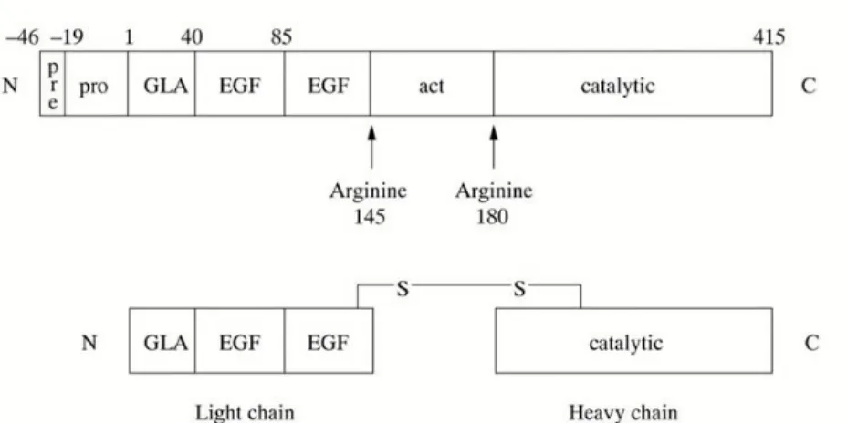
Figure 6: On top: FIX protein domains, on bottom: activated Factor IX comprising a N-terminal light chain and a C-terminal
heavy chain held together by a disulphide bond between cysteine residue 132 and 279. Legend Gla: Gla domain; EGF: epidermal growth factor-like domain; Act: activation peptide released after proteolitic cleavage; catalytic: the serine
protease domain.
Activation of factor IX involves cleavage of two peptide bonds, one on the C-terminal side of arginine 145 (α-cleavage) the other on the C-terminal side of arginine 180 (β-cleavage). These cleavages are caused by activated factor XI generated through the intrinsic pathway or via tissue factor/activated factor VII complex of the extrinsic pathway (Di Scipio, Kurachi et al. 1978). The activation cleavages generate an N-terminal light chain and a C-terminal heavy chain, held together by a disulphide bond between cysteine residues 132 and 279 (Bowen 2002).
Role of coagulation factor IX in the coagulation cascade.
Coagulation factors VIII and IX, whose deficiency are known to cause haemophilia A and B respectively, circulate as inactive precursors that are activated at the time of haemostatic challenge, via the intrinsic or extrinsic pathways (Zdziarska, Undas et al. 2009). As decribed before FVIII is a cofactor with no enzymatic activity per se; FIX is a serine protease with an absolute requirement for FVIII as cofactor. Upon activation, and in presence of calcium ions and phospholipid surfaces, FVIII and FIX form an active complex, which activates factor X. Subsequent stages of the cascade then proceed, culminating in the deposition of fibrin, the structural polymer of the blood clot (Bowen 2002).
Molecular basis of Hemophilia B
Hemophilia B (or Christmas disease) is a X-linked coagulopathy (Biggs, Douglas et al. 1952) caused by mutations in the F9 gene. Generally, only male are symptomatic (incidence of 1:35000 live male births) due to the presence of two alleles in female subjects. Based on FIX levels (antigen and/or protein activity), patients experience severe hemorrhagic symptoms, not rarely life−threatening (central nervous system and gastrointestinal bleeds), or causing substantial handicap (hemarthrosis, muscle hematoma). Based on FIX levels on plasma, the phonotype of patients is classified as mild, moderate or severe:
20
Moderate, if FIX level is in 1-5% range (or 0,01-0,05 IU/mL)
Severe , when the FIX level is below 1% or <0,01 IU/mL
Generally mild and moderate patients do not suffer of spontaneous hemorrhage, even if they can experience life threatening hemorrhage during surgery (even dentary surgery) if not properly treated. The mutations causing haemophilia B have been localized and characterized in several hundreds of patients. Based on the enormous number of mutations that have been elucidated it is clear now that the molecular bases of haemophilia are extremely diverse. Among all mutations, missense mutations are the most common (68%), followed by non-sense mutations (14%). Mutations altering splicing sequences have been found in 9% of all patients, with frame-shift mutation, promoter located and in frame deletion mutations ranging in 5%, 3% and 1% respectively. Point mutations (single nucleotide substitutions) are the most common gene defect and are present in approximately 90% of patients. Deletions are the second most common gene defects are present in approximately 5-10% of patients. Insertions and other rearrangements are quite rare within the haemophilia B population (Bowen 2002). The point mutations that occur in haemophilia B comprise missense mutation (these change a codon so that a different aminoacid is encoded), nonsense point mutation (these change an aminoacid codon into a translation stop codon), and mRNA splice site point mutations (these corrupt a true mRNA splice site, or create a novel one) (Koeberl, Bottema et al. 1990, Ketterling, Drost et al. 1999). In particular mutations that destroy or create mRNA splice sites are associated with variable severity of haemophilia: this depends on whether some correct transcripts can be processed (mild to moderate disease) or whether there is a complete loss of correct mRNA processing (severe disease). Exon skipping is a possible consequence of a mutation affecting splicing: the outcome depends on whether the skip is in frame or results in a frame shift (Tavassoli, Eigel et al. 1998, Tavassoli, Eigel et al. 1998).
In haemophilia approximately 30% of mutations involves a CpG site; the remaining 70% of distinct point mutations do not occur a CpG sites and may arise, for example, as a result of nucleotide misincorporation during DNA replication (Bowen 2002).
In general nonsense mutations are associated with severe forms of haemophilia; exon skipping is a further possibility arising from a nonsense mutation and it is also extremely detrimental: an in frame skip will result in a protein lacking the aminoacids encoded by skipped exon, an out of frame skip will result in a frame shift (Dietz, Valle et al. 1993, Ketterling, Drost et al. 1999).
Deletions of F9 gene include whole gene deletions, partial gene deletions at 5’ or 3’ end or within the gene, and microdeletions of one to several base pairs. A deletion, in general, has a high probability of destroying genetic function, removing domains of a protein, or introducing a frame shift, all of which are extremely detrimental. Therefore is not surprisingly that deletion are associated with severe forms of the disease (Cooper 1991, Giannelli and Green 1996).
21
Conventional treatment strategies hemophilia B
The current treatment is based on the intravenous administration of FIX (replacement therapy), either plasma derived or recombinant (Carcao and Aledort 2004, Pipe, High et al. 2008). However, there are still limitations that encourage research towards alternative therapeutic approaches. Moreover, the administration of a protein that the body is not able to synthetize by itself results, in a long term therapy, in the development of neutralizing FIX antibodies. The costs of substitutive treatments (≈50000 euro/year/person with severe disease) are prohibitive for the majority of the world hemophilia populations, so the demand for alternative therapies is growing up.
Enormous efforts have been pushed on substitutive gene therapy, and very recently it has been demonstrated that intravenous infusion of a AAV vector encoding FIX in HB patients resulted in FIX expression ranging from 1% to 6% for periods of 2 years, with amelioration of bleeding phenotypes (Nathwani, Tuddenham et al. 2011). DNA editing by using zinc finger nucleases has been also exploited in HB mouse models (Wang, Louboutin et al. 2011).
Von Willebrand Factor
Von Willebrand Factor in hemostasis
Von Willebrand Factor (VWF) is multimeric glycoprotein that play a major role in hemostasis; in normal condition it circulates inactive in plasma in a globular state, but in case of vascular injury, the shear stress and subendothelial matrix convert VWF in its active state, allowing platelet adhesion and aggregation; its function as a bridge is essential for the platelet plug formation because platelets are unable to bind directly matrix components; Moreover VWF is the plasmatic carrier of FVIII 8-10; preventing FVIII premature clearance from circulation (Arnout, Hoylaerts et al. 2006).
VWF plasmatic level is around 10 μg/ml, with 12 h of half-life but its levels are very variable into the population; plasmatic levels are genetically determined for the 66% and of this percentage, ≈30% of can be explained by ABO blood group, in fact people carrying blood group O usually have form 25% to 35% VWF antigen below the average of other groups (Gill, Endres-Brooks et al. 1987).
22
Figure 7: schematic representation of VWF interaction during endothelium damage.
Von Willebrand Factor structure and biosynthesis
VWF gene (178 kb) is located on the short arm of chromosome 12 (12p13.3), also present in human genome a pseudogenic fragments, which contain exons 23-28 (Marchetti, Patracchini et al. 1991). The funtional transcript is normally expressed only in endothelial cells and megakaryocytes, it is 9kb long and it contains 52 exons (Jaffe, Hoyer et al. 1974).
The protein is a single chain of 2813 amino acids and include a 22 residues of signal peptide, 741 residues of pro-peptide and finally the mature subunit of 2050 residues (FIGURE).
All the protein is composed by an highly repetitive structure with four mains domains, these domains have different function in maturation or activity of the mature protein (Springer 2011).
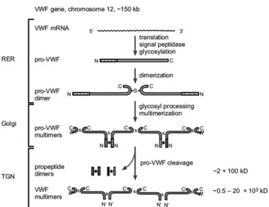
Pre-pro-VWF is translocated to the endoplasmic reticulum (ER) where it is extensively glycosylated but most important is the dimerization of “Cysteine Knot” (CK) domain (Katsumi, Kojima et al. 1998).
Dimerization occurs by disulfide bond formation between two C-terminal CK domain of two molecules of VWF; after dimerization proVWF dimers transported into the Golgi where the protein is N and O-linked glycosylated (Samor, Michalski et al. 1989). When VWF move to trans Golgi, Furin cut the D1-D2 domains of pro-peptide cleavage occurs and the process called multimerization will start (Sadler 2009); two D’D3 amino-terminal regions of align and multiple cysteine bond were formed (Springer 2011). VWF Multimers, which can rise above 20000 kDa and usually they were constitutively secreted (95%) of stored in Weibel-Palade body (WPB) in endothelial cells and α-granules in platelets (Michaux and Cutler 2004).
23
Figure 8:Biosynthesis steps of von Willebrand factor. *:on-covalent interaction; s:disulphide bonds. (Verweij 1988)
WPBs appear as tubules surrounded by a membrane and their formation is the direct consequence of multimerization. Release of WPB usually happens at sites of vascular damage where thrombogenic molecules are needed or after stimuli (Reininger 2008). Once secreted in blood flow, VWF size is well regulated by the enzymatic activity of a metallo-protease named ADAMTS-13 (Fujikawa, Suzuki et al. 2001).
Figure 9: Weibel-Palade bodies formation and assembly, adapted from (Springer 2011)
VWF domains
After prepeptide first two domains D1-D2 contains pro-peptide which dispalys has signal and maturation features (Sadler 2009), then in D’ and D3 domain we can find FVIII and heparin binding site, moreover it may contain the site of interaction for P-selectin which appears to anchor released ultra-large multimers to the surface of activated endothelial cells and contribute to present ADAMTS-13 cleavage site (Pimanda and Hogg 2002).
24
Following domains are A1-2 domains that contains the binding site for: platelet receptor GPIbα, heparin, botrocetin and probably also a binding site for collagen (Ruggeri 2007); moreover in A2 domain is present ADAMTS-13 cleavage site and in A3 domain are present the binding sites for type I and III collagens. C1 domain is known to host the RGD sequence for Integrin αIIβ3 binding. (Schneppenheim and Budde 2011). In the carboxyl terminal domain of the protein is present CK domain that as mentioned before it contains the essential cysteine for dimers formation (Schneppenheim, Budde et al. 2001, Tjernberg, Vos et al. 2004).
Figure 10:pre-pro-VWF domain organization (Springer 2011)
Von Willebrand Diseases
Von Willebrand Diseases (VWD) are a cluster of genetic inhered disorder characterized by deficient or defective VWF; VWD are considerate one of the most common inherited coagulation disorder in humans, in fact estimation shown that about the 1.3% of the population have reduced levels of VWF but only 1/1000 displays symptom of the disease (Keeney and Cumming 2001).
VWD were descrived for the first time in 1926 (Willebrand 1999 (1926)) but only in 1950 we isoled the "plasma factor” actually called VWF and actually 701 unique variant of this gene were reported (on line database http://www.vwf.group.shef.ac.uk)
Due to VWF peculiar characteristic and functions VWD displays a wide heterogeneity of symptoms that can be associated to qualitative and or quantitative defects. (Schneppenheim, Budde et al. 2001, Lillicrap 2007)
Von Willebrand disease was classified in three major categories:
Type 1: it is the most frequent groups with around the 80% of total cases of VWD, it is characterized with quantitative deficiencies, patients shown reduced VWF levels from 40 to 1% of normal plasma levels, also factor VIII may be reduced;
Type 2 collect all qualitative deficiencies of VWF, this class due to heterogeneity of defects is divided in 4 subgroups 2A, 2B, 2M and 2N;
Type 3 is the most rare with the approximate prevalence of one on one million, it is characterized by the total absence of VWF and also FVIII levels are strongly impaired.
Platelet-type or pseudo VWD is similar to Type 2B, but in this type the affected element is the platelets receptor GpIb.
25
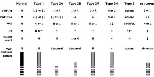
Type 1 VWD show mild to moderate quantitative deficiency of VWF affecting multimers of all sizes, but VWF functionality is normal. Inheritance of this type is typically dominant with highly variable penetrance. Due to its variability diagnosis is difficult and the borderline between low VWF and type 1 still controversial.
Type 2 is a group that gather all qualitative defects; subclasses 2A, 2B, 2M displays common features, showing dominant inheritance, low VWF antigen, impaired collagen binding and increased bleeding time. Type 2N usually displays recessive trait and distinctive characteristics.
Type 2 A show the peculiar signature of VWF:RCo – VWF:Ag ratio <0,6; moreover this subclass show abnormal multimers pattern, normal or decreased FVIII antigen and it appear to be more susceptible to ADAMS-13 cleavage. This class is also divided in subtypes IIC, IID and IIE/F depending of the different mechanism of HMWMs impairment.
Type 2 B is characterized by gain-of-function of platelet binding, resulting in HMWM abnormal multimers pattern and platelets reduction.
Type 2 M is a sort of other face of the coin of type 2 B, it is characterized by defective binding to GpIb but with normal multimers pattern.
Type 2 N is different from all other types, it is characterized by decreased FVIII levels in the circulation due to inability of VWF to bind FVIII results in increased clearance of unbound FVIII; clinical symptoms are more similar of those of Haemophilia A.
Type 3 VWD is a recessive form of this disease and usually this type of VWD is caused by two null mutations; due of low or none circulating VWF also FVIII is impaired with plasmatic levels below 10% of PNP.
Figure 11: L, 30—50 IU/dL; ↓, ↓↓, ↓↓↓, relative decrease; ↑, ↑↑, ↑↑↑, relative increase; BT, bleeding time; FVIII, factor VIII activity; N, normal; VWF:Ag, VWF antigen; VWF:RCo, VWF ristocetin cofactor activity. (Montgomery RR; http://www.nhlbi.nih.gov/guidelines/vwd/3_diagnosisandevaluation.htm)
26
Aim of the PhD thesis
My PhD research activity has been focused on the molecular mechanisms leading to severe deficiency of two proteins having key roles in the coagulation cascade (Factor IX) or in the primary Hemostasis (von Willebrand factor).
In particular, I investigated mechanisms due to three different molecular defects such as nonsense and missense mutations in F9 gene, associated with type I hemophilia B, and an in-frame deletion in the VWF gene, displaying a dominant-negative effect.
Hemophilia B is a rare disorder with a incidence of 1:35000 live male births that is associated with hemorrhagic tendency. Current treatment is based on the infusion of FIX. The replacement therapy either plasma derived or recombinant still have limitations that encourage research towards alternative therapeutic approaches. Moreover, the administration of a foreign protein, in a long term therapy, can result in the development of neutralizing FIX antibodies.
The study of FIX nonsense mutations, was triggered by the observation that nonsense mutations are associated with a risk of developing inhibitors lower that deletion. We therefore studies whether certain nonsense mutations, depending on the sequence context, undergo ribosome readthrough and are associated with trace levels of full-length FIX molecules. Moreover, we studied whether the readthrough can be induced by aminoglycosides, an approach that can have therapeutic implications.
The study of the FIX Tyr450Cys missense mutation was aimed at investigating the interplay between mechanisms impairing protein biosynthesis and function that is poorly defined, particularly in relation to specific protein regions.
Von Willebrand Factor is multimeric glycoprotein that play a major role in hemostasis. Von Willebrand Diseases are a cluster of genetic inhered disorder characterized by deficient or defective VWF and are considerate one of the most common inherited coagulation disorder in humans (1.3%) however only around 1:1000 displays disease symptoms. Dimerization and multimerization are distinct property of VWF biosynthesis both necessary to guarantee VWF production, activity and function.
In last part of the PhD, I focused my attention to the elucidation of the dominant-negative mechanisms leading to a VWF disease form caused by the P1105_C1926delinsR mutation in heterozygous condition. The approach was based on the evaluation of the VWF expression and maturation, in the presence of the mutation alone or in combination with a missense change impairing dimerization, both in cellular and mouse models.
27 Chapter 1
Ribosome Readthrough Over Nonsense
Mutations influences the Residual FIX
expression
1.1 Ribosome Readthrough Accounts for Secreted Full-Length
Factor IX in Hemophilia B Patients with Nonsense Mutations
Mirko Pinotti, Pierpaolo Caruso, Alessandro Canella, Matteo Campioni, Giuseppe Tagariello, Giancarlo Castaman, Sofia Giacomelli, Donata Belvini, Francesco Bernardi
Based on: Hum Mutat. 2012 Sep;33(9):1373-6
Introduction
The mechanism through which nonsense mutations impair gene expression and cause human genetic disease (Mort, Ivanov et al. 2008) consists of premature translation termination, and synthesis of truncated proteins, with loss-of-function features. Moreover, these mutations can trigger nonsense-mediated mRNA decay (NMD) (Khajavi, Inoue et al. 2006). With this as background, they are commonly believed as responsible for null genetic conditions.
However, the mechanism of ribosome readthrough, consisting of mis-recognition of the premature stop codon by an aminoacyl-tRNA instead of the termination factors (Rospert, Rakwalska et al. 2005), could restore translation impaired by nonsense mutations. Even if this process is expected to occur at low rate, it might account for minimal full-length protein biosynthesis.
Translation termination
It has long been known that termination of translation is an essential process in cell physiology since it permits the release from the translational machinery of the neo-synthetized protein of the proper size.
The translation termination is encoded on mRNA by codons called stop or nonsense codons (Brenner et al. 1965, 1967), most part of species on earth use three codons to mediate the signal of end of translation: UAA, UAG and UGA. In eukaryotes all three stop codon are recognized by eRF1 (class I release factor), this protein play the key role to recognize with high fidelity the stop codon and recruit eRF3 (class II release factor), a GTPase, and together these two factors collaborate to hydrolyze the bound between the nascent protein and the t-RNA present in the site P of the ribosome(Alkalaeva, Pisarev et al. 2006), releasing the protein and the whole translational machinery guaranteeing the recycle of all different components from ribosome to mRNA (Dever and Green 2012).
28
Nonsense mutations
Inspection of the Human Gene Mutation Database strongly highlights the relevance of nonsense mutations, which account for ≈20% of the pathogenic single point mutation (www.hgmd.org).
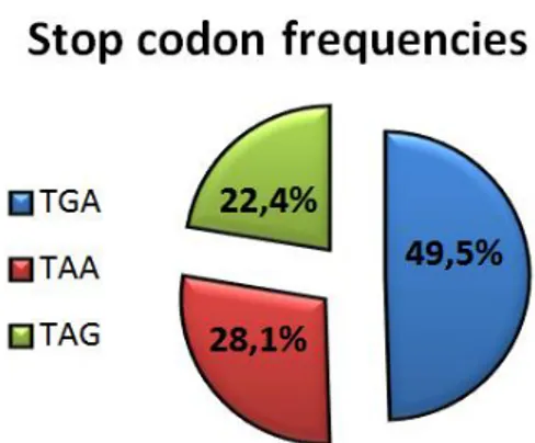
A bio-statistical analysis of human genes shows that the three different stop codons occur with different frequency, the most frequent is the codon TGA (49,5% of genes) followed by TAA and TAG with 28,1% and 22,4% respectively (Sun, Chen et al. 2005) (Jacobs, Rackham et al. 2002) (figure 12).
Figure 12: Stop codon frequencies in human transcripts. Data from: Sun, Chen et al. 2005.
When disease-causing stop codons are compared with authentic stop codons it results that the proportion of TAG is significantly higher (40,4 %, p<0,0001) and TGA and TAA proportions are lower (38,5% and 21,1% respectively) (Mort, Ivanov et al. 2008). These discrepancy can be explained in the light of the frequency of most common transition (CT) that is responsible of 46% of nonsense mutations. As a matter of fact, only 18 codon (10 amino acids) can be directly converted into stop codon; moreover the confirmation of these findings are the two most frequent nonsense mutated codons: CGA (Arg) and CAG (Gln) (Mort, Ivanov et al. 2008) (table 2).
Codon
Amino acid normally encoded
Mutating to: Codon
Amino acid normally encoded
Mutating to:
CGA Arginine TGA TTG Leucine TAG
AGA Arginine TGA TTA Leucine TGA / TAA
TGT Cysteine TGA AAG Lysine TAG
TGC Cysteine TGA AAA Lysine TAA
CAG Glutamine TAG TCG Serine TAG
CAA Glutamine TAA TCA Serine TGA / TAA
29
GAA Glutamic acid TAA TAT Tyrosine TAG / TAA
GGA Glycine TGA TAC Tyrosine TAG / TAA
Table 2: Normally occurring amino acids codons that can be converted into stop codon by single nucleotide mutagenesis. Adapted from : (Mort, Ivanov et al. 2008)
When a mutation produces a premature termination codon (PTC) on the genomic DNA, the resulting mRNA can run into multiple different consequences: the first and simplest is the translation into a truncated protein, this protein usually is unable fold and for this reason is retained into the cell and then degraded by the unfolded protein response (UPR). Moreover, the mRNA can undergo degradation by the nonsense-mediated decay process (NMD) (Khajavi, Inoue et al. 2006).
The NMD is a mRNA surveillance system that can lead to degradation of mRNAs harboring a premature termination codon (PTC). NMD allow to eliminate aberrant and abnormal transcripts inside the cell, this process take place outside of the nucleus, during the first translation, called “pioneer round”(Ishigaki, Li et al. 2001). During pioneer round, in normal condition, the ribosome remove all the exon junction protein (EJP), these proteins are left on the junction of the exons by the splicing process, these proteins in fact work as marker of the exons and they play a critical role in PTC recognition process. The ribosome in first translation by simply steric displacement remove EJP proteins till the end of the mRNA and also removing other proteins called Upf which are essential trigger of NMD. If a PTC is present in the mRNA sequence, the ribosome pauses on it and the releasing factors eRF1 and eRF3 associate with the ribosome to start disassembling, then Upf 1-2-3 proteins which are bounded to EJP, take contact with the termination complex triggering NMD and consequently mRNA degradation (Lejeune and Maquat 2005).
Nevertheless, some transcripts can escape the NMD, indeed also in normal transcripts a stop codon is present, normally in the last exon, and for this reason NMD cannot distinguish PTCs present less than 55bp from the last exon junction. Probably this distance is derived from the dimension of ribosome that displace the EJP before recognize the stop codon (figure 13) (Le Hir, Izaurralde et al. 2000).
30
Not only PTC activate NMD. Indeed, another source of PTC are the errors due to high processivity of the ribosome that can slip on mRNA and result in frame-shift, out-of-phase or “drop off” PTC (Dong and Kurland 1995).
Having this as background, the NMD process is extremely critical and it is important to remind how NMD protect limits the synthesis of dominant-negative truncated proteins, or of gain-of-function dangerous proteins and, last but not least, the accumulation of protein aggregates and cell death due to UPR.
Readthrough
Notwithstanding the conservation along the evolutive tree and the critically role of this process, the termination of translation can misrecognizes the stop codon through a process called “ribosome readthrough”. Although known since 1974, this phenomenon is still poorly explored. As a matter of fact, the first explicative paper on this topic was written in 2000 from Manuvakhova.
The readthrough process consists in the misrecognition of the stop codon by an aminoacyl-tRNA instead of eRF1, this event entail the skipping of the stop codon and the prosecution of the translation till the successive stop codon. This event occurs when a cognate tRNA successfully competes with the termination factors, and an amino acid is incorporated into the protein sequence. This type of readthrough hits all three stop codons and is important to discern this kind of readthrough from the selective readthrough, resulted from an aa-tRNA that decode a stop codon when only one termination codon is suppressed (Stansfield and Tuite 1994).
The three different stop codons (TGA, TAA and TAG) show strong differences from the point of view of the readthrough efficiency. The different readthrough efficiency observable among the literature, is due to the impact of type of stop codon and the surrounding sequence. As a first step just observing the three stop codon we can appreciate differences of readthrough from 0,2% to 3,8% (see table 3 for details) (Manuvakhova, Keeling et al. 2000). Stop codon Spontaneous readthrough UGA 0,6 - 3,8 % UAG 0,8 - 1,6 % UAA 0,2 - 0,5%
Table 3:Spontaneous readthrough of different stop codon (Manuvakhova, Keeling et al. 2000)
These strong differences (obtained by reporter gene system) underline the complexity of this scenario, and pose questions concerning the cause of this differences; in the same study, analysis of the first base after the stop codon reveals how much just a single base can influence the readthrough process (table 3).
31
Codon Readthrough Codon Readthrough Codon Readthrough
UGA A 1,0 % UAG A 0,9 % UAA A 0,2 %
UGA C 3,8 % UAG C 1,1 % UAA C 0,5 %
UGA G 0,6 % UAG G 0,8 % UAA G 0,4 %
UGA U 0,7 % UAG U 1,6 % UAA U 0,2 %
Table 4: Spontaneous readthrough of different stop codon and the sequence context (Manuvakhova, Keeling et al. 2000)
Several studies indicated that the differences on readthrough depend on the nonsense triplet and its sequence context, and demonstrated that the downstream sequence context can influence the “score of readthough” of each stop codon. In yeast, the presence of a peculiar consensus sequence appears to promote more than 5% of readthrough (figure 14) (Namy, Hatin et al. 2001).
Figure 14: Downstream sequence for high readthough levels in yeast (sequence from: Namy, Hatin et al. 2001)
The readthrough is directly connected to the translational fidelity, for this reason everything that impairs fidelity can change readthrough occurrence. The signs of this double linkage, appear clearly events that involve readthrough and cytoskeleton; indeed actin mutants shown increased readthrough of stop codon (Kandl, Munshi et al. 2002) and moreover low levels of eRF3 increase readthrough and impairs the cytoskeleton (Valouev, Kushnirov et al. 2002); despite different studies on this field, significance of these interconnections still unclear.
Moreover, readthrough efficiency can be influenced by drugs impairing ribosome fidelity such as the antibiotic class of aminoglycosides. Eukaryotic cells are enough resistant to the toxic effect of aminoglycosides due to the 20-50-fold lower affinity of this family of drugs with human ribosome than bacteria ribosome, nevertheless our ribosome still susceptible to the misreading effects of these drugs (Chattoo, Palmer et al. 1979) and this opportunity opened the studies on readthrough induction (see below).
Readthrough in human disease
As previously described the readthrough process physiologically occurs at low rate (10-4), thus providing the rationale for defining the nonsense mutations as associated “null genetic condition”. However, by investigating nonsense mutations in LAMA3 gene, it has been recently demonstrated that the phenomenon of readthrough can occur in vivo and produce appreciable amounts of full-length protein, thus potentially influencing the clinical phenotype of patients (Pacho, Zambruno et al. 2011). In this study, a child was found to be compound heterozygous for two different nonsense mutation R943X and R1159X, but manifested a very mild form of Junctional Epidermolysis Bullosa. It turned out that the ribosome readthrough accounted for the presence of full-length laminin-332 α3 chain and accumulation of lamini-332. The evidence of readthrough in human is easy to demonstrate
32
for protein like LAMA3, which is a structural protein (laminin-332 α3 chain) accumulating over time in cells. While this feature magnifies the readthrough effects, it does not favor the proper assessment of mutation-dependent readthrough rate. So far, no data have been provided on secreted proteins with limited half-life such as coagulation factors (from few hours to few days) (Furie and Furie 1992), which would better reflect the spontaneous ribosome readthrough efficacy in vivo.
Aim of the study
Among the molecular defects responsible for FIX deficiency, single point mutations leading to missense and nonsense mutations represent the 85,0% of all pathological mutations (figure 8) (Rallapalli, Kemball-Cook et al. 2013).
Figure 15: Whole FIX mutation present in global database. Total cases reported: 3580
Aim of the study was to investigate the occurrence of ribosome readthrough over nonsense mutations in F9 gene and the presence of trace levels of full-length FIX protein in HB patients. We planned to investigate three nonsense mutations in F9 gene (MIM# 300746; GenBank accession number K02402.1) differing in position and sequence context, which are candidate determinants of ribosome readthrough. Studies in plasma from HB patients and in
vitro were undertaken to evaluate their differential impact on mRNA and protein biology.
Figure 16:Patients' scheme of position of different FIX stop mutation in this study
Patients’ data and analysis
Plasma samples from severely affected HB patients bearing the Leucine 103 Stop (P-103X), Arginine 294 Stop (P-294X) and Arginine 298 Stop (P-298X) nonsense mutations (Belvini, Salviato et al. 2005), and also reported in the Human Gene Mutation Database,
33
http://www.hgmd.cf.ac.uk] were exploited to investigate the impact of ribosome readthrough in vivo (table 5).
Patient P-103X P-294X P298X
Mutation Leu 103 Stop Arg 294 Stop Arg 298 Stop
Sequence context TGTTAAAA AAGTGAAA ATTTGAAT
Table 5: Patients' data, nonsense mutation and sequence context
Plasma samples of patients P-103X and P294X were collected after a wash-out period of one week, plasma of patient P-298X upon a 70 hour wash-out period. The half-life of the infused recombinant FIX (BeneFix, Wyeth, Taplow, UK) is around 20 hours, thus not allowing us to ruled out the presence of confounding residual FIX protein levels in P-298X plasma.
Patient P-103X P-294X P-298X
Mutation Leu 103 Stop Arg 294 Stop Arg 298 Stop
Sequence context TGTTAAAA AAGTGAAA ATTTGAAT
FIX:Ag <1% <1% 1%
FIXc <1% <1% 1.5%
Table 6: Patients' antigen and activity levels.
Antigen and activity were undetectable in plasma of patient P-103X and P-294X. In plasma from P-298X we revealed traces of circulating FIX antigen (1.5% of PNP) that were associated with measurable FIX activity (1%) (table 6).
To verify the presence of the transcript or the activation of NMD we investigated the presence of the FIX mRNA in patients, trough analysis of leukocyte mRNA by reverse transcription followed by polymerase chain reaction (RT-PCR) (Pinotti, Toso et al. 1998). While the housekeeping GAPDH mRNA was clearly detected in all samples, the correct amplified FIX fragments were identified in P-294X and P-298X only (Figure 17).
34
Moreover, RT-PCR and sequencing was exploited for a semi-quantitative estimation of the mutant versus normal FIX mRNA forms in female mutation carriers. The expression of the mutated FIX mRNA was appreciable in carriers of the P-294X and P-298X mutations, but not in the P-103X carrier, these findings suggest that being L103X mutation an early stop codon, was expected to trigger major NMD (Figure 17); opposite the data obtained at the ectopic mRNA level, do not support major NMD for the P-294X and P-298X mutations.
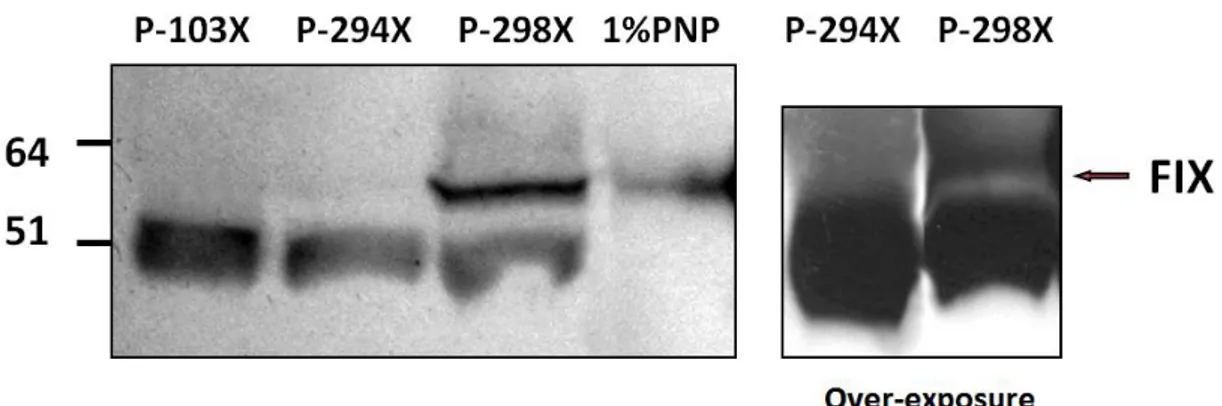
Subsequently we analyzed patients’ plasma trough western blotting, optimized to investigate low levels of FIX molecules in plasma, undetectable with other methods.
Evidence for the occurrence of readthrough in vivo
Patients’ plasma samples were diluted 1:30 in PBS buffer, and pooled normal plasma (PNP) diluted 1:100 were used as internal reference. Plasma patient’s P-103X, in which NMD prevents the synthesis of FIX, was used as negative control. Commercially available FIX deficient plasma was also used as negative control in setup experiments but, being immuno-depleted, it might contain traces of FIX protein that could have confounding effects in our assays aimed at evaluating very low protein amounts.
Western blotting analysis (figure 18) clearly shown a band corresponding to full-length FIX (57kD) in P-298X plasma, and upon film over-exposure, in P-294X plasma. This band was undetectable in P-103X’s plasma, which as mentioned before provided us with an internal negative control and validated the specificity of the antibody used. We have no interpretation of lighter bands (≈50kDa) present in samples but not in PNP. However this band is also present in patient P-103X in which we have demonstrated activation of NMD. This led us to hypothesize that the extra-band is not FIX-related.
124 184 B
*
*
wt wt mut wt mut P-103X P-294X P-298X FIX GAP D H FIX GAP D HFigure 17:RT-PCR products and chromatograms of mRNA from leukocytes of female carrier. B:Blank
35
Figure 18: Western blotting of patients' plasma; left panel 1 h exposure, right over-night exposure.
Evidence for the occurrence of readthrough in vitro
To validate our results on plasma samples we conducted expression studies in eukaryotic cells to investigate the occurrence of readthrough over the different nonsense triplets, and its rate.
As mentioned before several studies have been conducted in vitro with different intents and exploiting reporter genes. In our study, we inserted the nonsense mutations into the full-length human FIX cDNA and investigated the proteins secreted in medium, thus favoring the comparison of truncated and full-length molecules, and therefore the evaluation of readthrough effects in the proper nucleotide (table 7) and protein context (figure 19).
Mutation Leu 103 Stop Arg 162 Stop Arg 294 Stop Arg 298 Stop
Sequence
context TGTTAAAA TATTGACTT AAGTGAAA ATTTGAAT
Predicted Readthrough
<0,5 3-4% 1% 1%
Table 7: Mutation, sequence context and predicted readthrough of different mutations
Ee expressed these four nonsense FIX mutants and analyzed the secreted FIX protein in media through ELISA and Western Blotting
Figure 19: Position of different FIX nonsense mutations
In this experimental model, the recombinant rFIX-294X and rFIX-298X molecules were secreted at appreciable level respectively the 3.1±1.1% and 2.5±0.7% of rFIX-wt, whereas
36
not surprisingly rFIX-103X antigen was undetectable in medium (figure 20). However antigen levels obtained through ELISA cannot discriminate between truncated and full-length form of the protein, for these reason western blotting analysis is a forced analysis to verify the presence of full-length protein.
Figure 20:Media antigen levels of recombinant FIX variants expressed as percentage of rFIX-WT
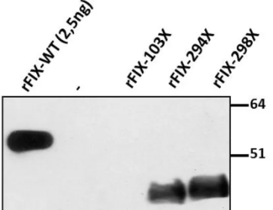
Western blotting of medium from the rFIX-294X and rFIX-298X expressing cells revealed a large proportion of truncated FIX forms (figure 21), which were not detected in patients’ plasma probably because of removal from circulation of this partial forms of the protein. As expected, FIX was not detected in medium from cells expressing the rFIX-103X.
Figure 21: Western blotting analysis of conditioned medium from recominant FIX variants. “-“ :Negative control
Comparison of results from ELISA and Western blot indicated that the truncated variants were secreted with lower efficiency than rFIX-wt, which is consistent with the essential role of the FIX carboxyl-terminal region for secretion (Kurachi, Pantazatos et al. 1997).
Noticeably, overexposure of films (figure 22) showed a form compatible with full-length FIX, with an intensity of approximately 0.2% of rFIX-WT in rFIX-294X and rFIX-298X media.
Moreover the band corresponding to full-length FIX was absent, even upon overexposure, in rFIX-103X and in the empty pCMV5, thus emphasizing the specificity of the signal detected. This differential readthrough efficiency might have contributed to appreciate mutated mRNA in carriers (figure 17).