UC Irvine Previously Published Works
Title
Role of the satiety factor oleoylethanolamide in alcoholism
Permalink
https://escholarship.org/uc/item/4hp5w7nz
Authors
Bilbao, A
Serrano, A
Cippitelli, A
et al.
Publication Date
2015
DOI
10.1111/adb.12276
License
CC BY 4.0
Peer reviewed
Role of the satiety factor oleoylethanolamide
in alcoholism
Ainhoa Bilbao
1,2*, Antonia Serrano
1,3*, Andrea Cippitelli
4*, Francisco J. Pavón
1,3,
Andrea Giuffrida
5, Juan Suárez
1, Nuria García-Marchena
1,6, Elena Baixeras
1,
Raquel Gómez de Heras
6, Laura Orio
6, Francisco Alén
6, Roberto Ciccocioppo
4,
Benjamin F. Cravatt
7, Loren H. Parsons
3, Daniele Piomelli
8,9&
Fernando Rodríguez de Fonseca
1,6Unidad de Gestión Clínica de Salud Mental, Instituto IBIMA-Hospital Regional Universitario de Málaga, Spain1, Institute of Psychopharmacology, Central Institute
of Mental Health, Medical Faculty of Mannheim, University of Heidelberg, Germany2
, Committee on the Neurobiology of Addictive Disorders3
, Department of Chemical Physiology7, The Scripps Research Institute, La Jolla, CA, USA, School of Pharmacy, Pharmacology Unit, University of Camerino, Italy4, Department of
Pharmacology, University of Texas Health Science Center at San Antonio, San Antonio, TX, USA5, Departamento de Psicobiología, Facultad de Psicología,
Universidad Complutense de Madrid, Spain6, Departments of Anatomy and Neurobiology, Pharmacology, and Biological Chemistry, University of California, Irvine,
CA, USA8
and Department of Drug Discovery and Development, Istituto Italiano di Tecnologia, Italy9
ABSTRACT
Oleoylethanolamide (OEA) is a satiety factor that controls motivational responses to dietary fat. Here we show that alcohol administration causes the release of OEA in rodents, which in turn reduces alcohol consumption by engaging peroxisome proliferator-activated receptor-alpha (PPAR-α). This effect appears to rely on peripheral signaling mecha-nisms as alcohol self-administration is unaltered by intracerebral PPAR-α agonist administration, and the lesion of sensory afferent fibers (by capsaicin) abrogates the effect of systemically administered OEA on alcohol intake. Addi-tionally, OEA is shown to block cue-induced reinstatement of alcohol-seeking behavior (an animal model of relapse) and reduce the severity of somatic withdrawal symptoms in alcohol-dependent animals. Collectively, these findings demonstrate a homeostatic role for OEA signaling in the behavioral effects of alcohol exposure and highlight OEA as a novel therapeutic target for alcohol use disorders and alcoholism.
Keywords Alcohol self-administration, alcoholism, oleoylethanolamide, PPAR-α, relapse.
Correspondence to: Fernando Rodriguez de Fonseca, Laboratorio de Medicina Regenerativa, Instituto IBIMA-Hospital Regional Universitario de Málaga,
Avenida Carlos Haya 82, Málaga 29010, Spain. E-mail: [email protected]; Antonia Serrano, Laboratorio de Medicina Regenerativa, Instituto IBIMA-Hospital Regional Universitario de Málaga, Avenida Carlos Haya 82, Málaga 29010, Spain. E-mail: [email protected]
INTRODUCTION
Oleoylethanolamide (OEA) is a bioactive lipid mediator that belongs to the fatty acid ethanolamide (FAE) family.
Although OEA is a structural analogue of the
endocannabinoid arachidonoyl ethanolamide (AEA or anandamide), it neither binds to nor activates cannabi-noid receptors (Rodríguez de Fonseca et al. 2001; Fu et al. 2003). Indeed, OEA exerts a number of pharmacological effects, including the induction of satiety, the reduction of body weight gain and the stimulation of lipolysis, through the activation of the peroxisome proliferator-activated receptor-alpha (PPAR-α) (Rodríguez de Fonseca et al. 2001; Fu et al. 2003; Guzmán et al. 2004). OEA binds to
this nuclear receptor with high affinity and the majority of its effects are absent in mice lacking PPAR-α (Fu et al. 2003; Guzmán et al. 2004).
There is a growing body of evidence supporting the idea that OEA might serve as a homeostatic signal that controls multiple aspects of the intake and metabolism of highly caloric fat-containing foods (for review, see Piomelli 2003). OEA regulates energy expenditure and fat utilization primarily through its involvement in the modulation of fat appetite (Schwartz et al. 2008), lipid and glucose metabolism (Fu et al. 2003; González-Yanes et al. 2005; Serrano et al. 2006, 2011; Martínez de Ubago et al. 2009) and the reward associated with high-calorie intake (Tellez et al. 2013). OEA is generated in the
*These authors contributed equally to this work.
doi:10.1111/adb.12276 PRECLINICAL STUDY
intestine upon the arrival of fat-containing food through a process controlled by the sympathetic nervous system (Fu et al. 2007, 2011). By engaging PPAR-α, the OEA formed in the intestine activates peripheral sensory systems located in the vagus nerve that convey informa-tion to the nucleus of the solitary tract and gut peptides that are released into the blood stream to regulate the hypothalamic integrative networks that control energy expenditure (Gaetani et al. 2010; Serrano et al. 2011). Additionally, in vitro and in vivo studies have demon-strated that OEA stimulates lipolysis and fatty acid oxida-tion through a mechanism that involves PPAR-α (Guzmán et al. 2004; Suárez et al. 2014).
In addition to its role as a homeostatic signal that controls energy expenditure, growing evidence suggests that OEA might influence motivational processes through its ability to modulate reward systems in the brain. Recent reports (Tellez et al. 2013) have shown that, by modulating gut sensory nerve terminals of the vagus nerve, intestinal OEA affects the dopaminergic activity that is induced by infusion of either low- or high-calorie emulsions. Indeed, exogenously administered OEA might restore the dopaminergic deficiency that is associated with high-fat diets, which indicates that, in addition to the control of appetite, OEA might serve as a homeostatic signal that is associated with the motiva-tional drives involved in food selection (Tellez et al. 2013). This finding aligns with the recent description of the ability of OEA to modulate the dopaminergic responses associated with nicotine self-administration (Melis et al. 2008; Mascia et al. 2011). Indeed, OEA not only modu-lates mesocorticolimbic dopaminergic neurons but also blocks both induced reward and nicotine-induced nicotine self-administration through a PPAR- α-dependent mechanism (Mascia et al. 2011). Considering the presence of the OEA-signaling machinery in the brain, particularly in the hippocampus (Rivera et al. 2014) and the description of OEA’s ability to modulate memory consolidation (Campolongo et al. 2009), the homeostatic role of this bioactive lipid extends to both motivational and cognitive processes that are associated with the hedonic valence of actions.
Given that ethanol is both a high-calorie food and a drug of abuse that is capable of modulating reward, we hypothesized that OEA might interfere with the endog-enous signals activated by alcohol. There is compelling evidence that lipid mediators of the FAE family are involved in alcohol dependence (Rodríguez de Fonseca et al. 2005). Studies have shown that acute administra-tion of intoxicating doses of ethanol inhibits the in vivo formation of AEA in the rat brain (Ferrer et al. 2007), whereas low to moderate doses enhance its release in vivo and in vitro (Basavarajappa, Ninan & Arancio 2008; Ceccarini et al. 2013). Chronic ethanol exposure is
asso-ciated with increases in the formation of AEA and FAE precursors. Although AEA and CB1receptors have been
shown to mediate some of the pharmacological and behavioral aspects of alcohol use (Basavarajappa & Hungund 2002; Rodríguez de Fonseca et al. 2005; Ferrer et al. 2007), the effects of non-cannabinoid FAEs such as OEA have been poorly characterized, and direct experi-mental evidence regarding the behavioral significance of the OEA–PPAR-α interaction on ethanol-related behaviors is still lacking. To fill this knowledge gap, we evaluated the potential roles of OEA and PPAR-α in alcohol-seeking behavior (ethanol self-administration and relapse) and monitored the changes in OEA produc-tion during acute and chronic ethanol exposure.
MATERIALS AND METHODS Animals
The experiments were performed on male Wistar rats (Charles River, Barcelona, Spain). In vivo microdialysis studies were performed on adult C57Bl/6 male mice (Jackson Laboratory, Bar Harbor, ME, USA). All of the procedures involving the care and use of animals were conducted in adherence with the European Directive 2010/63/EU on the protection of animals used for scien-tific purposes and Spanish regulations (Real Decreto 53/2013 and 178/2004, Ley 32/2007 and 9/2003 and Decreto 320/2010) for the care and use of laboratory animals.
Drugs
OEA was synthesized as previously described (Rodríguez de Fonseca et al. 2001). OEA was dissolved in a vehicle of 5 percent Tween-80 (Sigma Aldrich Co., St. Louis, MO, USA) and 95 percent sterile saline and administered i.p. in a volume of 1 ml/kg body weight. The following doses of OEA were used: (1) 1, 5 and 20 mg/kg for the self-administration and reinstatement studies; (2) 1 and 5 mg/kg for the ethanol deprivation study; and (3) 5 mg/kg for the evaluation of alcohol withdrawal signs. Wy-14643 (Tocris Bioscience, Bristol, UK) was mixed in a vehicle of 10 percent DMSO, 10 percent Tween-80 and 80 percent sterile saline. Wy-14643 was adminis-tered i.p. in a volume of 1 ml/kg body weight and by i.c.v. injection in a total volume of 5μl. The following doses were used: (1) 5, 20 and 40 mg/kg for the self-administration and reinstatement studies; (2) 20 mg/kg for the ethanol deprivation study; (3) 5 and 20 mg/kg for the locomotor and anxiety tests; and (4) 1 and 10μg for central administrations.
The PPAR-α antagonist GW6471 (Tocris Bioscience) was dissolved in a vehicle of 5 percent Tween-80 and 95 percent sterile saline and administered i.p. at a dose of 1 mg/kg in a volume of 1 ml/kg body weight.
The orphan G-protein coupled receptor 119 (GPR119) antagonist PSN375963 (Tocris Bioscience) was dissolved in a vehicle of 5 percent Tween-80 and 95 percent sterile saline and administered i.p. at doses of 0.1 and 10 mg/kg in a volume of 1 ml/kg body weight.
The cannabinoid CB1receptor antagonist SR141716A
(rimonabant) was purchased from Sanofi-Aventis (Sanofi S.A., Paris, France). Rimonabant was suspended with two to three drops of Tween-80 in sterile saline as the vehicle. Rimonabant was administered i.p. at a dose of 3 mg/kg in a volume of 1 ml/kg body weight.
The neurotoxin capsaicin was purchased from Sigma-Aldrich Co. and dissolved in 5 percent Tween-80, 5 percent propylene glycol and 90 percent sterile saline.
Deafferentation
Capsaicin was administered subcutaneously (12.5 mg/ ml) (Kaneko, Kaunitz & Tache 1998) to rats that were anesthetized with ethyl ether. The total dose of capsaicin (125 mg/kg) was divided into three injections (25 mg/kg in the morning, 50 mg/kg in the afternoon and 50 mg/kg on the following day). The control rats received vehicle injections. The experiments were performed 10 days after capsaicin treatment in rats that had lost the corneal chemosensory reflex (i.e. eye wiping for 1–3 minutes after the application of 0.1 percent ammonium hydroxide into one eye).
Intracerebral infusion cannulas and intra-NAc OEA testing
For the intracerebral infusions, stainless steel guide can-nulas aimed at the NAc were implanted in the rats. Animals that had previously been trained to self-administer 10 percent ethanol were anesthetized with an isoflurane/oxygen vapor mixture (1.5–2.0 percent) and implanted with bilateral microinfusion guide cannulas (12 mm, 22 gauge, stainless steel) that terminated 3 mm above the ventral surface of the NAc [from bregma: anteroposterior (AP), +1.6 mm; mediolateral (ML),±2.0 mm; and dorsoventral (DV), −5.0 mm from dura]. These coordinates provided injector placements at the interface between the shell and core of the NAc. A minimum of 5 days of post-operative recovery was allowed before the evaluation of the effect of the local administration of OEA on ethanol self-administration. After the establishment of stable self-administration behavior, the animals received an initial microinjector insertion (no liquid infusion) immediately before self-administration to acclimate them to the procedure and to produce the initial tissue damage from the insertion of the injector. Subsequently, vehicle or OEA infusions were made via bilateral 33-gauge microinjectors that extended 2 mm beyond the tips of the guide cannulas. Infusions of
1μl per side were made over a 2-minute period, and the removal of the injector was delayed by an additional 1 minute to allow for drug diffusion. The stylets were then replaced in the guide cannulas, and the rats were allowed immediate access to the ethanol self-administration para-digm. The effects produced by vehicle or 1μg OEA per side were evaluated.
Surgery procedure and i.c.v. drug injection
For the i.c.v. injections, stainless steel guide cannulas aimed at the lateral ventricle were implanted in the rats. The animals were anesthetized with an isoflurane/ oxygen vapor mixture (1.5–2.0 percent) and placed on a stereotaxic apparatus (David Kopf Instruments, Tujunga, CA, USA) with the incisor bar set at 5 mm above the interaural line. A guide cannula (7 mm, 23 gauge) was secured to the skull with two stainless steel screws and dental cement and was closed with 30-gauge obturators. The implantation coordinates calculated relative to bregma as follows: AP +0.6 mm, ML ±2.0 mm and DV −3.2 mm from the surface of the skull. These coordinates placed the cannula 1 mm above the ventricle. After 7 days of a post-surgical recovery period, cannula patency was confirmed by the gravity-fed flow of isotonic saline through a stainless steel injector (8 mm, 30 gauge) that was inserted within the guide to 1 mm beyond its tip. This procedure allowed the animals to become familiar with the injection technique.
For the i.c.v. administration of Wy-14643, the stylet was removed from the guide cannula of each rat and an injector connected to 70 cm of calibrated polyethylene-10 tubing was lowered into the ventricle. The tubing was then raised until the flow began, and 5μl of drug solution at doses of 1 and 10μg was infused over a 30- to 60-second period. The injector was left in the guide cannula for an additional 30 seconds and then removed. The stylet was immediately replaced. The animals were tested 5 minutes after injection. The i.c.v. cannula placements were evalu-ated after each experiment via dye injection. Only rats with proper i.c.v. placements were included in the data analysis.
Acute ethanol administration: blood and tissue sampling
Ethanol was dissolved in sterile 0.9 percent (w/v) saline and administered i.p. at a dose of 4 g/kg.
For the blood and visceral sampling, a group of animals was anaesthetized with methoxyflurane at 0, 45, 90 or 240 minutes after the injection of ethanol. Blood (2 ml) was collected from the heart of the animal with a syringe filled with 1 ml of Krebs–Tris buffer (136 mM NaCl, 6 mM KCl, 1.2 mM MgCl2, 2.5 mM CaCl2, 10 mM
samples were centrifuged in Accuspin tubes (Sigma-Aldrich Co.) at 800× g for 10 minutes at 22°C. The samples were obtained from the small intestine and the liver, and were frozen immediately on solid CO2.
The brains were collected from a second group of animals after rapid decapitation and frozen immediately on solid CO2. The frozen brains were placed in acrylic rat
brain matrices, and 2-mm thick slices were obtained using brain matrix razor blades. The NAc and cerebellum were dissected in 2-mm thick frozen coronal slices that corresponded to plates 9–13 (NAc) and 56–65 (cerebel-lum) of the atlas of Paxinos & Watson (1998).
Chronic ethanol administration and ethanol withdrawal: collection of rat plasma
The regular chow diet was removed and replaced with a liquid diet consisting of chocolate-flavored boost liquid nutritional supplement fortified with vitamins and min-erals. The rats were separated into two groups: the ethanol group received a diet containing 10 percent (w/v) ethanol and the control group received an ethanol-free diet supplemented with sucrose to equalize the caloric intake across the two groups. The diet was available 24 hours for 21 days. On the final liquid diet day, the animals were maintained in their home cage with access to the regular chow diet and water. Blood samples were drawn at 0, 6 and 12 hours after the removal of the liquid diet.
Ethanol withdrawal behaviors
Beginning 6 hours after the diet with ethanol was with-drawn, the animals were evaluated at hourly intervals. The rats were observed for 5 minutes, and ethanol with-drawal signs, including general activity, shakes, tremors, mobility, spontaneous convulsive seizures and rigidity, were scored.
Plasma ethanol levels
The plasma ethanol levels were enzymatically measured using a commercial kit (Sigma-Aldrich Co.). The assays were run according to the manufacturer’s instructions.
High-performance liquid chromatography–mass spectrometry (HPLC-MS) analyses of the tissue content of OEA
OEA was extracted with methanol/chloroform (1:2, v/v) from the plasma, peripheral tissues (small intestine and liver) and brain areas (NAc and cerebellum). Samples were injected into an HP 1100 Series HPLC-MS system equipped with a Hewlett-Packard OctaDecyl Silica Hypersil column (100× 4.6 mm i.d., 5 μm). The esti-mated recovery of OEA was 99.7± 0.3 percent. Reversed
phase separations were performed using linear increases of methanol in water at a flow rate of 0.5 ml/min as previously described (Giuffrida, Rodríguez de Fonseca & Piomelli 2000). The MS analyses were performed in the positive ionization mode with an electrospray ion source. For the quantitative analyses, diagnostic fragments cor-responding to the protonated molecules [(M+ H)+] and the sodium adducts of the molecular ions [(M+ Na)+] were followed in the selected ion monitoring mode. System control and data evaluation were conducted using an on-line system software (HP Chemstation Software, Hewlett-Packard, Palo Alto, CA, USA). In vivo microdialysis studies
Mice received i.p. injections of ethanol (2 g/kg) every other day over 2 weeks (see the experimental design in the Supporting Information). The day before the last injec-tion, the mice were each implanted with a single microdialysis probe into the NAc that was secured to the skull with dental cement. The stereotaxic coordinates
from bregma were +1.5 mm AP, ±0.8 mm ML and
−5.0 mm DV (from the skull) (Paxinos & Watson 1998), and the microdialysis probes employed in 1-mm length active membranes (polyethyl sulfone membrane, 15 kDa cut off from SciPro Inc., Sanborn, NY, USA).
The probes were perfused with artificial cerebrospinal fluid, which was delivered at a flow rate of 0.6μL/min and was composed of the following (in mM): 149 NaCl, 2.8 KCl, 1.2 CaCl2, 1.2 MgCl2, 0.25 ascorbic acid,
5.4 D-glucose and 30 percent (w/v) hydroxypropyl- β-cyclodextrin (HP-β-CD). The inclusion of the HP-β-CD in the perfusate provided a substantial increase in the dialy-sis recovery of OEA. Approximately 12–16 hours after probe implantation, dialysate samples were collected at 10-minute intervals. Following a baseline dialysate col-lection (60 minutes), mice were injected with ethanol (2 g/kg; i.p.) and dialysates collected for 120 minutes. Dialysate samples were frozen on dry ice and stored at −80°C until analysis for OEA content using liquid chro-matography coupled with mass spectrometry (Caillé et al. 2007).
Proper placement of the active dialysis membrane within the NAc was histologically verified for each experi-ment. The brains were sliced (20μm), and the place-ments of the microdialysis probes were verified using the atlas of Paxinos (Paxinos & Watson 1998).
Operant training for liquid reinforcers
Training and testing were conducted in standard operant chambers that were located in sound-attenuating, venti-lated environmental cubicles. Each chamber was equipped with a drinking reservoir (volume capacity: 0.10 ml) positioned 4 cm above the grid floor in the
center of the front panel of the chamber, and two retract-able levers were located 3 cm to the right and left of the drinking receptacle. Auditory and visual stimuli were presented via a speaker and a light located on the front panel. A microcomputer controlled the delivery of fluids, presentation of auditory and visual stimuli, and record-ing of the behavioral data. The rats were trained to self-administer 10 percent (v/v) ethanol, 10 percent sucrose or water in 30-minute daily sessions on a fixed ratio 1 schedule of reinforcement in which each response resulted in the delivery of 0.1 ml of fluid as previously described (Weiss et al. 1993). Briefly, for the first 3 days of training, water availability in the home cage was restricted to 2 hours per day to facilitate the acquisition of operant responding for a liquid reinforcer. During this time, lever pressing that was reinforced with a 0.2 percent (w/v) saccharin or a 10 percent (w/v) sucrose solution was established. At this point, water was made freely available, and saccharin/sucrose self-administration training continued until the animals reached stable baseline responding. The rats in the saccharin-trained group were then trained to self-administer ethanol using a modification of the
sucrose-fading procedure (Samson 1986) that employed
saccharin rather than sucrose (Weiss et al. 1993). During the first 6 days of this ethanol initiation phase, a 5 percent (w/v) ethanol solution containing 0.2 percent saccharin (w/v) was available to the rats. Beginning on day 7, the concentration of ethanol gradually increased from 5 to 8 percent and finally to 10 percent (w/v), whereas the concentration of saccharin correspondingly decreased to 0 percent. At the beginning of the saccharin-fading procedure, a second, inactive lever was introduced. During all training and testing phases, the responses on this lever were recorded as a measure of non-specific behavioral activation, but they had no pro-grammed consequences (Cippitelli et al. 2005).
Sucrose self-administration
Following completion of the sucrose training, Wistar rats were used to study the effects of OEA (0, 1, 5 and 20 mg/ kg) when administered 30 minutes prior to a self-administration session. These experiments were conducted every fourth day using a Latin square coun-terbalanced design. Responding at the inactive lever was recorded throughout the experiment to monitor non-specific behavioral effects.
Ethanol self-administration
Following the completion of the saccharin-fading proce-dure, Wistar rats were trained in sessions of 30 minutes/ day to lever press for 10 percent ethanol (0.1 ml per
response) until stable baseline responding was reached. We studied the effects of OEA (0, 1, 5 and 20 mg/kg) or Wy-14643 (0, 5, 20 and 40 mg/kg) when administered 30 minutes prior to the self-administration sessions. These experiments were conducted every fourth day using a Latin square counterbalanced design. Respond-ing at the inactive lever was recorded throughout the experiment to monitor non-specific behavioral effects.
Alcohol deprivation
Rats were trained to self-administer 10 percent ethanol. Following 3 days of baseline responding sessions, the rats were deprived of ethanol for five consecutive days (no operant sessions). At the end of the fifth day of the dep-rivation phase, the rats were distributed into four groups that were treated with vehicle, OEA (1 and 5 mg/kg) or Wy-14643 (20 mg/kg) 30 minutes before the test session.
Reinstatement of ethanol-seeking behavior
Conditioning phase
At the completion of the fading procedure, the animals were trained to discriminate between 10 percent ethanol and water in 30-minute daily sessions. Beginning with self-administration training at the 10 percent ethanol concentration, discriminative stimuli that were predictive of ethanol or water availability were presented during the ethanol and water self-administration sessions, respec-tively. The discriminative stimulus for ethanol consisted of an orange extract odor (S+), and the water availability (i.e. no reward) was signaled with an anise extract (S−). The olfactory stimuli were generated by placing six to eight drops of the respective extract into the bedding of the operant chamber. Additionally, each lever press resulting in the delivery of ethanol was paired with the illumina-tion of the chamber’s house light for 5 seconds (CS+). The corresponding cue during the water sessions was a 5-second tone (70 dB; CS−). During the presentation of these stimuli, a 5-second time out period was in effect; during this period responses were recorded but not rein-forced. The olfactory stimuli that served as the S+or S−for ethanol or water availability were introduced 1 minute before the extension of the levers and remained present throughout the 30-minute sessions. The bedding of the chamber was changed, and the bedding trays were cleaned between sessions. The rats were only given ethanol sessions during the first 3 days of the condition-ing phase. Subsequently, ethanol and water sessions were conducted in a random order across training days with the constraint that all of the rats received totals of 10 ethanol and 10 water sessions.
Extinction phase
After the last conditioning day, the rats were subjected to 30-minute extinction sessions for 15 consecutive days. During this phase, the sessions began with the extension of the levers without the presentation of the discrimina-tive stimuli. Responses at the lever activated the delivery mechanism but did not result in the delivery of liquids or the presentation of the response-contingent cues (i.e. the house light or tone).
Reinstatement testing
The reinstatement tests began the day after the last extinction session. These tests lasted 30 minutes, and the conditions were identical to those of the conditioning phase with the exception that the alcohol and water were not available. The sessions were initiated by the extension of both levers and the presentation of either the ethanol (S+)- or water (S−)-paired stimulus. The respective dis-criminative stimulus remained present during the entire session, and responses at the previously active lever were followed by the activation of the delivery mechanism and a 5-second presentation of the CS+in the S+condition or the CS−in the S−condition. The animals were tested under in the S+/CS+condition on day 1 and in the S−/CS− condi-tion on day 2. Subsequently, reinstatement experiments were conducted every fourth day (on days 6, 10 and 14), and OEA or Wy-14643 was administered 30 minutes prior to these sessions. Responding at the inactive lever was constantly recorded to monitor possible non-specific behavioral effects.
Two-bottle choice test
The two-bottle choice method was used to determine the voluntary ethanol consumption of rats. The animals were individually housed in standard cages and allowed to acclimate for 1 week. Each rat had continuous access to two identical bottles, one of which always contained tap water. Food was available ad libitum. After 4 days of water consumption (both bottles), a choice between 10 percent (v/v) ethanol and water was offered for several days until reaching a stable baseline. The bottle positions were changed every day to avoid position preferences. On the test day, animals received an administration of vehicle, PSN375963, OEA and GW6471 or a combina-tion of OEA and GW6471 30 minutes prior to ethanol and water access, and consumption was recorded over 24 hours. Both ethanol and water intake (g/kg) were calcu-lated at different time intervals.
Statistical analyses
All data in the graphs are expressed as the mean± SEM. The different experiments included 6–10 animals per treatment according to the assay. Statistical analyses of
the results were performed with one- and two-way ANOVA followed by multiple comparison tests (Dunnett’s test). A P-value below 0.05 was considered statistically significant. All analyses were performed with the com-puter program GraphPad Prism version 5.04 (GraphPad Software Inc., San Diego, CA, USA).
RESULTS
Effects of ethanol administration on plasma and tissue levels of OEA
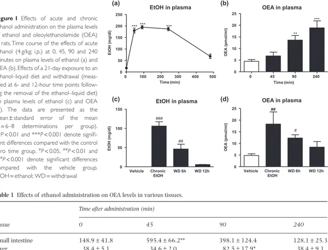
To determine whether ethanol influenced OEA mobiliza-tion, we evaluated the time course of the effects of acute ethanol administration on the OEA levels in the plasma and various peripheral tissues and brain areas over a period of 240 minutes. Single ethanol injections (4 g/kg; i.p.) resulted in rapid and marked elevations in plasma ethanol levels (F4,50= 131.6, P < 0.001) that were
signifi-cant at 45, 90 and 240 minutes after administration com-pared with the zero time control group (***P< 0.001) (Fig. 1a). Decreases in plasma ethanol concentrations were evident 8 hours after the administrations, and the control levels were re-established within the first 24 hours (data not shown). Because plasma ethanol levels remained stable for 240 minutes, we selected the times of 0, 45, 90 and 240 minutes for the analyses of OEA in the plasma and tissues. A one-way ANOVA detected a signifi-cant effect of the plasma OEA response to ethanol admin-istration (F3,41= 11.55, P < 0.001) due to significant
increases at 90 (**P< 0.01) and 240 minutes (***P< 0.001) (Fig. 1b).
Additionally, we evaluated whether OEA influenced ethanol levels. We found similar plasma ethanol concen-trations after a single ethanol administration (4 g/kg; i.p.) to rats pre-treated with vehicle or OEA (5 mg/kg) (Sup-porting Information Fig. S1), indicating that OEA admin-istration does not result in alterations in ethanol pharmacokinetics in the 0–240 minutes post-injection interval.
As shown in Table 1, acute ethanol also led to eleva-tions in the OEA levels in the peripheral tissues and brain areas (small intestine: F3,41= 6.65, P = 0.001; liver: F3,41= 4.29, P = 0.011; NAc: F3,41= 3.21, P = 0.034;
and cerebellum: F3,41= 9.44, P < 0.001). In the small
intestine, the OEA increase was apparent at 45 minutes (**P< 0.01) after ethanol administration, and the OEA levels returned to the control value at 240 minutes. In the liver, OEA remained at control levels for the first 45 minutes, peaked at 90 minutes (*P< 0.05) and returned to normal levels at 240 minutes. Similarly, ethanol resulted in increases in OEA in the NAc at 45 and 90 minutes after its administration (*P< 0.05) and in the cerebellum at 90 minutes (***P< 0.001). These results
suggest that the intestine and NAc were the tissues in which the ethanol-associated increases in OEA occurred most quickly.
To evaluate the effects of chronic ethanol exposure and withdrawal on plasma OEA levels, Wistar rats were maintained on an ethanol-containing liquid diet for 21 days. The animals displayed a significant increase in plasma ethanol concentration at the end of the liquid diet exposure (###P< 0.001) (Fig. 1c). This rise remained
evident 6 hours into withdrawal (##
P< 0.01). Regarding the plasma OEA levels, long-term ethanol exposure pro-duced a significant increase (##P< 0.01) (Fig. 1d). In the
plasma, this elevation was also evident following 6 and 12 hours of withdrawal, but was only significantly differ-ent from the control group at 6 hours (#P< 0.05).
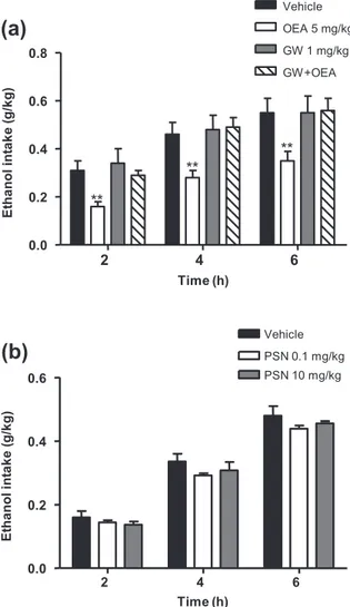
Effects of OEA on ethanol consumption
To examine whether OEA affects the ethanol consump-tion, we used the two-bottle choice paradigm. OEA
(5 mg/kg) administration significantly decreased ethanol intake relative to the vehicle group (F1,60= 26.50, P< 0.001), and significant decreases were observed at all time points (**P< 0.01) (Fig. 2a). Next, because OEA is a PPAR-α agonist, we tested whether the PPAR-α antago-nist GW6471 was able to reverse the decrease in ethanol consumption caused by OEA. As shown in Fig. 2a, GW6471 antagonized the decrease in ethanol consump-tion that was produced by OEA (F3,21= 2.81, P = 0.025).
Because GPR119 has been reported as a novel OEA target, we also evaluated whether the GPR119 receptor agonist PSN375963 produced similar effects than OEA on ethanol consumption. However, as shown in Fig. 2b, PSN375963 (0.1 and 10 mg/kg) did not affect ethanol intake at all time points evaluated. Similarly, this com-pound had no effect on ethanol self-administration (Sup-porting Information Fig. S2).
In addition, water consumption was not affected by none of the treatments tested (Supporting Information Fig. S3). 0 45 90 240 0 5 10 15 20 25 ** *** ** OEA in plasma Time (min) OE A ( pm ol /m l) Vehicle WD 6h WD 12h 0 5 10 15 20 25 ## OEA in plasma Chronic EtOH ## # OE A ( pm ol /m l) (a) (b) (c) (d) EtOH in plasma 0 100 200 300 400 500 0 50 100 150 200 250 *** *** *** Time (min) Et O H ( m g /d l) Vehicle WD 6h WD 12h 0 50 100 150 ### EtOH in plasma ## Chronic EtOH E tO H ( m g/ dl )
Figure 1 Effects of acute and chronic
ethanol administration on the plasma levels of ethanol and oleoylethanolamide (OEA) in rats. Time course of the effects of acute ethanol (4 g/kg; i.p.) at 0, 45, 90 and 240 minutes on plasma levels of ethanol (a) and OEA (b). Effects of a 21-day exposure to an ethanol–liquid diet and withdrawal (meas-ured at 6- and 12-hour time points follow-ing the removal of the ethanol–liquid diet) on plasma levels of ethanol (c) and OEA (d). The data are presented as the mean± standard error of the mean (n= 6–8 determinations per group). **P< 0.01 and ***P < 0.001 denote signifi-cant differences compared with the control zero time group.#P< 0.05,##P< 0.01 and ###P< 0.001 denote significant differences
compared with the vehicle group. EtOH= ethanol; WD = withdrawal
Table 1 Effects of ethanol administration on OEA levels in various tissues.
Tissue
Time after administration (min)
0 45 90 240
Small intestine 148.9± 41.8 595.4± 66.2** 398.1± 124.4 128.1± 25.3
Liver 38.4± 5.1 34.6± 2.0 82.5± 17.9* 38.4± 9.1
Nucleus accumbens 128.1± 67.4 610.7± 108.8* 582.6± 209.4* 265.7± 21.6 Cerebellum 248.2± 19.4 420.7± 75.5 610.8± 96.6*** 164.4± 17.9 Effects of acute intraperitoneal administration of ethanol (4 g/kg) on OEA levels (pmol/g of tissue) in small intestine, liver and brain sections (nucleus accumbens and cerebellum) were measured at 0, 45, 90 and 240 minutes after injection. Data are means± standard error of the mean (six to eight determinations per group). *P< 0.05, **P < 0.01 and ***P < 0.001 denote significant differences compared with zero time group. OEA= oleoylethanolamide.
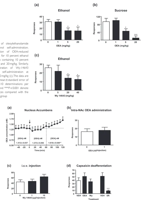
Effects of OEA on ethanol self-administration
Next, we examined whether OEA affects ethanol self-administration. Wistar rats trained to self-administer 10 percent ethanol received injection of various doses of OEA (1, 5 and 20 mg/kg). OEA administration significantly decreased ethanol self-administration relative to the control condition (F3,31= 3.42, P = 0.031), and
signifi-cant decreases were observed after the administration of both 5 and 20 mg/kg of OEA (*P< 0.05) (Fig. 3a). These effects were also observed for sucrose (F3,31= 10.71, P< 0.001) after the administration of both 5 (*P < 0.05) and 20 mg/kg of OEA (***P< 0.001) (Fig. 3b). Similarly,
systemic administration of Wy-14643 reduced ethanol self-administration at doses of 20 and 40 mg/kg (*P< 0.05) (Fig. 3c).
Additionally, Wy-14643 treatment was evaluated in behavioral tests and doses of 5 and 20 mg/kg did not affect locomotion and did not induce anxiety-like behaviors (Supporting Information Fig. S4).
Because OEA reduces food intake through a peripheral mechanism, but reports exists about its ability to modulate central dopaminergic pathways, we studied the effects of OEA when injected centrally and in animals with chemi-cal sensory deafferentation. Because NAc was found to display rapid variations in OEA levels following ethanol injection, we first evaluated the effects of repeated ethanol administration on the OEA response to ethanol challenge. The effects of repeated ethanol treatment (2 g/kg; i.p., and every other day for 14 days) on NAc OEA levels are shown in Fig. 4a. During baseline sample collection dialysate OEA levels were 1.412± 0.037 nM, and no trend toward an increase or decrease in levels was observed during this period. Relative to these baseline levels, dialysate OEA content was significantly enhanced after an acute ethanol challenge (2 g/kg; i.p.) (F12,43= 2.04, P = 0.025). This
increase began approximately 60 minutes after ethanol challenge and progressively increased over the course of the subsequent 60 minutes. During the final hour of sam-pling, OEA levels were significantly higher as compared with baseline (**P< 0.01), reaching a maximum of 134 percent of baseline 120 minutes after injection.
Next, we evaluated the effects of the injection of OEA into NAc on ethanol self-administration. As shown in Fig. 4b, the injection of OEA into the NAc did not modify ethanol self-administration. Similarly, central infusions into the lateral ventricles of different doses of Wy-14643
(1 and 10μg) had no effects on ethanol
self-administration (Fig 4c). However, sensory deafferentation with the neurotoxin capsaicin abolished both OEA- and Wy14643-induced reduction of operant responding for ethanol but not the reduced responding induced by the cannabinoid receptor antagonist/inverse agonist rimonabant (Fig. 4d) that acts centrally to reduce alcohol self-administration.
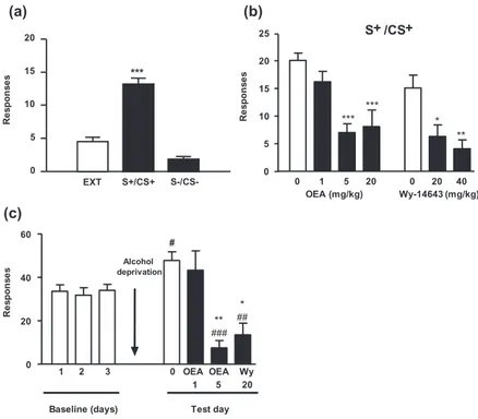
Effects of OEA on conditioned reinstatement and alcohol deprivation
We next examined the effects of OEA and Wy-14643 as modulators of the operant responses elicited by the con-textual stimuli associated with ethanol. Reinstatement was induced by presenting cues that were associated with ethanol delivery during training. The operant responses induced by the ethanol-associated stimuli (S+/CS+) were robustly increased to 19.4± 2.6 presses per session, which was significantly different from the behaviors elic-ited on the last day of extinction (4.5± 0.7 responses;
(a)
2 4 6 0.0 0.2 0.4 0.6 0.8 Vehicle OEA 5 mg/kg GW 1 mg/kg GW+OEA ** ** ** Time (h) E tha nol int a k e (g /k g )(b)
2 4 6 0.0 0.2 0.4 0.6 PSN 0.1 mg/kg PSN 10 mg/kg Vehicle Time (h) E tha nol in ta k e (g /k g)Figure 2 The effects of oleoylethanolamide (OEA) on alcohol
intake are mediated by peroxisome proliferator-activated receptor-alpha (PPAR-α).The effects of OEA (5 mg/kg) on voluntary alcohol intake in a two-bottle choice paradigm were prevented by pre-treatment with the synthetic PPAR-α antagonist GW6471 (1 mg/kg) (a). The GPR119 receptor agonist PSN375963 (0.1 and 10 mg/kg) had no effect on voluntary alcohol intake (b).The data are presented as the mean± standard error of the mean (n = 8–10 determinations per group). **P< 0.01 denotes significant differences compared with the control group. GW= GW6471; PSN = PSN375963
(a) (b) (c) 0 1 5 20 0 10 20 30 40 * Ethanol * OEA (mg/kg) Re s pons e s 0 1 5 20 0 40 80 120 Sucrose * *** OEA (mg/kg) Re s pons e s 0 5 20 40 0 10 20 30 Ethanol * * Wy 14643 (mg/kg) R e sp o n se s
Figure 3 Effects of oleoylethanolamide
(OEA) on ethanol self-administration. Acute i.p. injection of OEA-reduced operant responses for 10 percent ethanol (a) and a solution containing 10 percent sucrose (b) at 5 and 20 mg/kg. Similarly, systemic administration of Wy-14643 reduced ethanol self-administration at doses of 20 and 40 mg/kg (c). The data are presented as the mean± standard error of the mean (n= 8–10 determinations per group). *P< 0.05 and ***P < 0.001 denote significant differences compared with the respective control group
(b)
(c)
Intra-NAc OEA administration
0 1 0 10 20 30 OEA (µg/injection) Re s pons e s
VEH OEA Wy VEH SR 0 10 20 30 40 50 60 70 Capsaicin deafferentation *** Treatment Re s pons e s 0 1 10 0 20 40 60 80 i.c.v. injection Wy 14643 ( Re s pons e s µg/injection) (d) (a) Nucleus Accumbens [OEA] nM 1.412 ± 0.037 [OEA] nM 1.373 ± 0.050 [OEA] nM 1.618 ± 0.040** -40 -20 0 20 40 60 80 100 120 0.50 0.75 1.00 1.25 1.50 1.75 2.00 Time (min) OE A c onc e nt ra ti on (nM )
Figure 4 Site of action of oleoylethanolamide (OEA) on alcohol self-administration. Dialysate OEA content was significantly enhanced after
an acute ethanol challenge (2 g/kg; i.p.). In NAc of mice previously exposed to repeated ethanol treatment (2 g/kg every other day for 14 days). Ethanol injection is indicated by arrow at time point zero (a). Acute intra-accumbens injection of OEA had no effect on ethanol self-administration (b). This lack of a central effect was also observed when the synthetic peroxisome proliferator-activated receptor-alpha agonist Wy-14643 was injected into the lateral ventricles (c). The effect of OEA on ethanol self-administration was abolished by the deafferentation of the vagal terminals with capsaicin (d). Capsaicin pre-treatment abolished the effects of OEA and Wy-14643 on ethanol self-administration but did not affect the actions of the CB1cannabinoid receptor antagonist rimonabant (SR141716A), which acts centrally
to reduce alcohol self-administration.The data are presented as the mean± standard error of the mean (n = 8–10 determinations per group). **P< 0.01 denotes significant differences compared with baseline OEA concentration; ***P < 0.001 denotes significant differences compared with the control group. SR= rimonabant; VEH = vehicle; Wy = Wy-14643
***P< 0.001) and under the S−/CS−condition (4.4± 1.1 responses; ***P< 0.001, Fig. 5a). As shown in Fig. 5b, OEA pre-treatment produced a dose-dependent reduction in S+-induced ethanol seeking relative to the control group (F3,31= 9.54, P < 0.001); significant reductions
were observed at OEA doses of 5 and 20 mg/kg (***P< 0.001). Similarly, Wy-14643 pre-treatment pro-duced a dose-dependent reduction in S+-induced ethanol seeking compared with the controls (F2,23= 8.29, P= 0.002), and significant reductions were observed at Wy-14643 doses of 20 (*P< 0.05) and 40 mg/kg (**P< 0.01).
We also evaluated the effects of PPAR-α agonists in an alcohol deprivation model (Fig. 5c). During the final three self-administration sessions (baseline) before ethanol deprivation, the animals obtained an average of 33.1± 3.0 ethanol reinforcers per session. Ethanol dep-rivation increased the operant responses for ethanol to 47.8± 4.0. OEA administration decreased ethanol self-administration following the ethanol deprivation phase (F2,23= 13.79, P < 0.001), and significant decreases were
observed after the administration of 5 mg/kg OEA (**P< 0.01). Similar results were observed after the administration of 20 mg/kg Wy-14643 (*P< 0.05). The numbers of operant responses observed after either treat-ment were even lower than those observed in the baseline condition (OEA:###P< 0.001; Wy-14643:##P< 0.01).
Effects of OEA on ethanol withdrawal symptoms
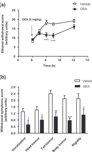
To further explore the effects of OEA on ethanol-related behaviors, several signs of withdrawal were scored in
ethanol-dependent rats after the withdrawal of ethanol. As shown in Fig. 6a, OEA treatment had a significant effect on ethanol withdrawal symptoms (F1,70= 34.13, P< 0.001). This analysis also revealed a significant interaction between treatment and time (F4,70= 6.73, P< 0.001).Thus, OEA administration induced significant decreases in the total scores for ethanol withdrawal signs at 8 and 9 hours into withdrawal (***P< 0.001) com-pared with the vehicle-treated animals. Indeed, the administration of OEA produced significant decreases in each of the withdrawal symptoms that were evaluated; i.e. vocalization, head tremor and rigidity (*P< 0.05); tail tremor (**P< 0.01); and body tremor (***P < 0.001) (Fig. 6b).
DISCUSSION
The present results identified a prominent role for OEA as a homeostatic signal that controls multiple aspects of the physiological adaptations to alcohol exposure. OEA pro-duction is triggered by alcohol administration and con-tributes to the regulation of alcohol intake, the acute motivational response to alcohol and the onset of with-drawal symptoms after cessation of chronic alcohol con-sumption. Because OEA has been identified as an intestinal satiety signal that controls hedonic value of food (mainly fat), we cannot consider the observed response as specific for ethanol. Moreover, OEA has been identified already as an important mediator of the rein-forcing properties of nicotine (Mascia et al. 2011) sup-porting this general role of control of the hedonic homeostasis. (b) (a) 0 1 5 20 0 20 40 0 5 10 15 20 25 S+ /CS+ *** *** * ** OEA (mg/kg) Wy-14643 (mg/kg) R e sp o n se s 0 5 10 15 20 S+/CS+ S-/CS-R e sp o n se s *** EXT (c) 0 20 40 60 1 2 3 0 OEA OEA Wy 1 5 20 ** ### * ## Alcohol deprivation
Baseline (days) Test day
Re s pons e s #
Figure 5 Effects of oleoylethanolamide
(OEA) on conditioned reinstatement and the effects of alcohol deprivation. Condi-tioned responses to ethanol-associated cues (a) were reduced by the administra-tion of both OEA (1, 5 and 20 mg/kg) and the peroxisome proliferator-activated receptor-alpha receptor agonist Wy-14643 (20 or 40 mg/kg). (b) i.p. administration of OEA or Wy-14643 reduced the enhance-ment of the operant ethanol responses that were observed after a 5-day ethanol dep-rivation period (c). The data are presented as the mean± standard error of the mean (n= 8–10 determinations per group). *P< 0.05, **P < 0.01 and ***P < 0.001 denote significant differences compared with the respective control group.#P< 0.05, ##P< 0.01 and###P< 0.001 denote
signifi-cant differences compared with the last day of baseline period
The first interesting finding of the present study was the induction of OEA mobilization following acute expo-sure to alcohol. This effect was observed in different tissues and was detected in plasma samples. The intensi-ties and time courses of alcohol-induced OEA production were time and tissue dependent, and the small intestine (jejunum) and the NAc were the first tissues in which OEA accumulation was detected. In the NAc, the time course of OEA release (Fig. 4a) paralleled that of classical transmitters activated by ethanol. These transmitters can activate receptors capable of triggering OEA formation through the NAPE-PLD pathway. Alcohol might recruit either catecholaminergic, cholinergic or glutamatergic neurons (Spanagel 2009), which in turn might trigger receptor-dependent OEA activation (Stella & Piomelli 2001). A contribution of a modification of FAAH activity induced by the presence of ethanol was discarded in
pre-vious studies (Ferrer et al. 2007). The mechanisms by which alcohol triggers OEA mobilization in the periphery might be related to the dependence of OEA production on sympathetic activity (Fu et al. 2011). Alcohol is an acti-vator of the sympathetic nervous system (for review see Spanagel 2009) and sympathetic activation has been found to increase OEA production not only in the intes-tine but also in metabolically relevant organs such as adipose tissue (Guzmán et al. 2004; LoVerme et al. 2006; Fu et al. 2011). Thus, alcohol-enhanced activation of sympathetic output might be responsible for the observed increases in OEA. Because the plasma and liver OEA elevations were delayed relative to the intestinal accumu-lation, we infer that this effect is secondary to the activa-tion of primary targets of sympathetic innervaactiva-tion. In the brain, the rapid response of the NAc, which was observed by monitoring tissue contents, is suggestive of a direct action of alcohol on the brain. In this organ, alcohol might recruit either catecholaminergic, cholinergic or glutamatergic neurons (Spanagel 2009), which in turn might trigger receptor-dependent OEA acti-vation (Stella & Piomelli 2001). This hypothesis needs to be tested in future studies. Finally, plasma OEA levels were found to be constantly elevated during chronic alcohol consumption. The removal of alcohol from the diet induced a decrease in OEA levels that paralleled the decrease in alcohol level, thus indicating a tight associa-tion between OEA formaassocia-tion and the presence of alcohol in the body. As we discuss later, the disappearance of OEA is related to overt alcohol withdrawal symptoms, which supports the adaptive nature of OEA as an alcohol-driven homeostatic signal that is necessary to adapt the body to the presence of ethanol.
OEA has been linked to physiological responses that are associated with high-calorie intake, including the control of motivational aspects of eating and metabolic adaptations to high-calorie foods (Piomelli 2003; Schwartz et al. 2008). Based on this hypothesis, we studied whether alcohol-induced OEA regulated alcohol intake. Confirming this hypothesis, we found that the administration of either OEA or PPAR-α agonists reduced alcohol intake and self-administration. The action of OEA effect was dependent on the presence of PPAR-α, because it was blocked by a selective PPAR-α antagonist. This effect was extended to sucrose and was not related to the activation of an additional OEA target, the GPR119 receptor (Overton et al. 2006).
Overall, these findings indicate that, as has been described for fats and high-calorie foods (Rodríguez de Fonseca et al. 2001; Fu et al. 2003, 2007; Piomelli 2013), OEA is capable of modulating both the consump-tion and motivaconsump-tional responses to alcohol, which is also highly caloric. These motivational aspects extend to the control of the effects of contextual memories associated
(b)
(a)
4 6 8 10 12 14 0 5 10 15 20 25 Vehicle OEA OEA (5 mg/kg) *** *** Time (h) E tha nol w it hdr a w a l s c or e (a rb it ra ry u n it s ) 0.0 0.4 0.8 1.2 1.6 2.0 2.4 2.8 Vehicle OEA * * ** *** * W it hdr a w a l s y m pt om s s c or e (a rb it ra ry u n it s )Figure 6 Effects of oleoylethanolamide (OEA) on ethanol
with-drawal symptoms. Acute administration of OEA reduced the global ethanol withdrawal scores that were measured between 6 and 12 hours into withdrawal (a). This effect was observed on each of the symptoms that were evaluated (b). The data are presented as the mean± standard error of the mean (n = 8–10 determinations per group). *P< 0.05, **P < 0.01 and ***P < 0.001 denote significant dif-ferences compared with the vehicle-treated group
with alcohol consumption in alcohol relapse. Interest-ingly, both OEA and PPAR-α agonists reduced cue-induced reinstatement of alcohol consumption, which suggests that, following extinction, OEA has the ability to curb motivational memories that are associated with the effects of alcohol. Although OEA has been demonstrated to strengthen memory consolidation (Campolongo et al. 2009), the present results suggest that it can also modu-late the impact of a recalled memory on motivational drives. However, this hypothesis needs to be confirmed as a peripheral-driven satiety/anhedonic response derived of the activation of the vagus nerve by OEA might counter-act the motivational contextual responses associated with alcohol intake.
Following the strategy used to determine the site of action of FAEs in the suppression of food intake (Rodríguez de Fonseca et al. 2001; Gómez et al. 2002), we searched for the site of action of OEA on alcohol self-administration. As described for food intake, the effects of OEA on alcohol self-administration and cue-induced reinstatement were found to be dependent on the integ-rity of the peripheral sensory system. Capsaicin-induced deafferentation of the small intestine abrogated these effects and left the centrally mediated effects of com-pounds, such as the CB1receptor antagonist rimonabant
(which reduces alcohol self-administration by targeting the prefrontal cortex) intact (Hansson et al. 2007). This finding was confirmed by the lack of effects of i.c.v. injec-tions of PPAR-α agonist into alcohol self-administering animals. Thus, we hypothesize that alcohol, as has been described for high-calorie foods, might activate ascending sensory pathways that ultimately inhibit the motiva-tional aspects of alcohol intake by releasing OEA in the intestine (Rodríguez de Fonseca et al. 2001; Piomelli 2003). The participation of the nucleus of the solitary tract and its connections with the oxytocinergic (Gaetani et al. 2010) and histaminergic (Provensi et al. 2014) systems that further mediate OEA-induced feeding inhi-bition, will be addressed in future studies.
Following the observation of the decrease in plasma OEA with ethanol withdrawal, we observed that this decline paralleled the onset of alcohol withdrawal symp-toms. OEA injection at the beginning of withdrawal (i.e. the time at which OEA levels dropped) induced a clear reduction in the severity of the behavioral symptoms associated with withdrawal. At the present time, we do not know whether this finding reflects an intrinsic ability of OEA to reduce the hyper-excitability associated with ethanol withdrawal. Although OEA has been found to interact with ion channels to regulate excitability, we cannot rule out further actions in targets that are recruited by alcohol withdrawal including the peptidergic and classical neurotransmitter systems. However, the reductions of the motivational effects of
ethanol-associated cues and reductions of the severities of alcohol withdrawal symptoms by the single molecule OEA provide a unique profile for the future design of therapies for alcoholism.
In conclusion, our results revealed that OEA is an endogenous signal that participates in the homeostatic adaption to alcohol. The actions of OEA cover multiple physiological aspects and open a potential new path for the development of effective therapies for alcoholism.
Acknowledgements
The present study has been supported by the Ministerio de Economía y Competitividad (MEC) and Instituto de Salud Carlos III (III) (project PI13/02261); MEC, ISC-III and Red de Trastornos Adictivos UE-FEDER/EU-ERDF 2012 (RD12/0028); Ministerio de Sanidad, Servicios Sociales e Igualdad and Plan Nacional Sobre Drogas (pro-jects 049/2009 and 049/2013); Junta de Andalucía and Plan Andaluz de Investigación, Desarrollo e Innovación UE-FEDER/EU-ERDF (CTS-433); Junta de Andalucía and Consejería de Economía, Innovación y Ciencia (project PI45403); Junta de Andalucía and Consejería de Igualdad, Salud y Políticas Sociales (projects PI0823-2012 and PI0228-2013). A.S., F.J.P. and J.S. hold a ’Miguel Servet’ research contract from ISC-III (CP14/ 00212, CP14/00173 and CP12/03109, respectively).
DISCLOSURES/CONFLICT OF INTEREST
The authors state no competing financial interests or other interests that might be perceived to influence the results and discussion reported in this article.
Authors Contribution
AB performed operant self-administration experiment as well as i.c.v. administration, ethanol treatments and open field and elevated plus-maze studies. AS did the two-bottle choice experiments, the intra-NAc infusion studies and wrote the manuscript. AC was responsible of operant self-administration and reinstatement of ethanol-seeking experiments. FJP did the in vivo microdialysis, the mass spectrometry measures and ran all statistical analyses. AG measured OEA in both tissues and plasma samples. JS and EB synthesized the OEA. RGH and NG-M were responsible for ethanol withdrawal studies. LO and FA did the GPR119 experiments. RC, BFC and LHP helped with experimental design and provided methodological support for relapse studies. DP contributed to experimen-tal design and supervision and corrected the manuscript. FRF designed the study, coordinated the experiments and wrote the manuscript.
References
Basavarajappa BS, Hungund BL (2002) Neuromodulatory role of the endocannabinoid signaling system in alcoholism: an overview. Prostaglandins Leukot Essent Fatty Acids 66:287– 299.
Basavarajappa BS, Ninan I, Arancio O (2008) Acute ethanol suppresses glutamatergic neurotransmission through endocannabinoids in hippocampal neurons. J Neurochem 107:1001–1013.
Caillé S, Álvarez-Jaimes L, Polis I, Stouffer DG, Parsons LH (2007) Specific alterations of extracellular endocannabinoid levels in the nucleus accumbens by ethanol, heroin, and cocaine self-administration. J Neurosci 27:3695–3702. Campolongo P, Roozendaal B, Trezza V, Cuomo V, Astarita G, Fu
J, McGaugh JL, Piomelli D (2009) Fat-induced satiety factor oleoylethanolamide enhances memory consolidation. Proc Natl Acad Sci U S A 106:8027–8031.
Ceccarini J, Casteels C, Koole M, Bormans G, Van Laere K (2013) Transient changes in the endocannabinoid system after acute and chronic ethanol exposure and abstinence in the rat: a combined PET and microdialysis study. Eur J Nucl Med Mol Imaging 40:1582–1594.
Cippitelli A, Bilbao A, Hansson AC, del Arco I, Sommer W, Heilig M, Massi M, Bermúdez-Silva FJ, Navarro M, Ciccocioppo R, Rodríguez de Fonseca F (2005) Cannabinoid CB1 receptor antagonism reduces conditioned reinstatement of ethanol-seeking behavior in rats. Eur J Neurosci 21:2243–2251. Ferrer B, Bermúdez-Silva FJ, Bilbao A, Álvarez-Jaimes L,
Sánchez-Vera I, Giuffrida A, Serrano A, Baixeras E, Khaturia S, Navarro M, Parsons LH, Piomelli D, Rodríguez de Fonseca F (2007) Regulation of brain anandamide by acute administra-tion of ethanol. Biochem J 404:97–104.
Fu J, Astarita G, Gaetani S, Kim J, Cravatt BF, Mackie K, Piomelli D (2007) Food intake regulates oleoylethanolamide formation and degradation in the proximal small intestine. J Biol Chem 282:1518–1528.
Fu J, Dipatrizio NV, Guijarro A, Schwartz GJ, Li X, Gaetani S, Astarita G, Piomelli D (2011) Sympathetic activity controls fat-induced oleoylethanolamide signaling in small intestine. J Neurosci 31:5730–5736.
Fu J, Gaetani S, Oveisi F, Lo Verme J, Serrano A, Rodríguez de Fonseca F, Rosengarth A, Luecke H, Di Giacomo B, Tarzia G, Piomelli D (2003) Oleylethanolamide regulates feeding and body weight through activation of the nuclear receptor PPAR-alpha. Nature 425:90–93.
Gaetani S, Fu J, Cassano T, Dipasquale P, Romano A, Righetti L, Cianci S, Laconca L, Giannini E, Scaccianoce S, Mairesse J, Cuomo V, Piomelli D (2010) The fat-induced satiety factor oleoylethanolamide suppresses feeding through central release of oxytocin. J Neurosci 30:8096–8101.
Giuffrida A, Rodríguez de Fonseca F, Piomelli D (2000) Quanti-fication of bioactive acylethanolamides in rat plasma by electrospray mass spectrometry. Anal Biochem 280:87–93. Gómez R, Navarro M, Ferrer B, Trigo JM, Bilbao A, del Arco I,
Cippitelli A, Nava F, Piomelli D, Rodríguez de Fonseca F (2002) A peripheral mechanism for CB1 cannabinoid receptor-dependent modulation of feeding. J Neurosci 22:9612–9617. González-Yanes C, Serrano A, Bermúdez-Silva FJ, Hernández-Domínguez M, Páez-Ochoa MA, Rodríguez de Fonseca F, Sánchez-Margalet V (2005) Oleoylethanolamide impairs glucose tolerance and inhibits insulin-stimulated glucose uptake in rat adipocytes through p38 and JNK MAPK path-ways. Am J Physiol Endocrinol Metab 289:E923–E929.
Guzmán M, Lo Verme J, Fu J, Oveisi F, Blázquez C, Piomelli D (2004) Oleoylethanolamide stimulates lipolysis by activat-ing the nuclear receptor peroxisome proliferator-activated receptor alpha (PPAR-alpha). J Biol Chem 279:27849– 27854.
Hansson AC, Bermúdez-Silva FJ, Malinen H, Hyytia P, Sánchez-Vera I, Rimondini R, Rodríguez de Fonseca F, Kunos G, Sommer WH, Heilig M (2007) Genetic impairment of frontocortical endocannabinoid degradation and high alcohol preference. Neuropsychopharmacology 32:117–126. Kaneko H, Kaunitz J, Tache Y (1998) Vagal mechanisms
under-lying gastric protection induced by chemical activation of raphe pallidus in rats. Am J Physiol 275:G1056–G1062. LoVerme J, Guzman M, Gaetani S, Piomelli D (2006) Cold
exposure stimulates synthesis of the bioactive lipid oleoylethanolamide in rat adipose tissue. J Biol Chem 281:22815–22818.
Martínez de Ubago M, García-Oya I, Pérez-Pérez A, Canfran-Duque A, Quintana-Portillo R, Rodríguez de Fonseca F, González-Yanes C, Sánchez-Margalet V (2009) Oleoylethanol-amide, a natural ligand for PPAR-alpha, inhibits insulin recep-tor signalling in HTC rat hepatoma cells. Biochim Biophys Acta 1791:740–745.
Mascia P, Pistis M, Justinova Z, Panlilio LV, Luchicchi A, Lecca S, Scherma M, Fratta W, Fadda P, Barnes C, Redhi GH, Yasar S, Le Foll B, Tanda G, Piomelli D, Goldberg SR (2011) Blockade of nicotine reward and reinstatement by activation of alpha-type peroxisome proliferator-activated receptors. Biol Psychiatry 69:633–641.
Melis M, Pillolla G, Luchicchi A, Muntoni AL, Yasar S, Goldberg SR, Pistis M (2008) Endogenous fatty acid ethanolamides sup-press nicotine-induced activation of mesolimbic dopamine neurons through nuclear receptors. J Neurosci 28:13985– 13994.
Overton HA, Babbs AJ, Doel SM, Fyfe MC, Gardner LS, Griffin G, Jackson HC, Procter MJ, Rasamison CM, Tang-Christensen M, Widdowson PS, Williams GM, Reynet C (2006) Deor-phanization of a G protein-coupled receptor for oleoylethanolamide and its use in the discovery of small-molecule hypophagic agents. Cell Metab 3:167–175. Paxinos G, Watson C (1998) The Rat Brain in Stereotaxic
Coor-dinates, New York: Academic Press, Spiral Bound.
Piomelli D (2003) The molecular logic of endocannabinoid sig-nalling. Nat Rev Neurosci 4:873–884.
Piomelli D (2013) A fatty gut feeling. Trends Endocrinol Metab 24:332–341.
Provensi G, Coccurello R, Umehara H, Munari L, Giacovazzo G, Galeotti N, Nosi D, Gaetani S, Romano A, Moles A, Blandina P, Passani MB (2014) Satiety factor oleoylethanolamide recruits the brain histaminergic system to inhibit food intake. Proc Natl Acad Sci U S A 111:11527–11532.
Rivera P, Arrabal S, Cifuentes M, Grondona JM, Pérez-Martín M, Rubio L, Vargas A, Serrano A, Pavón FJ, Suárez J, Rodríguez de Fonseca F (2014) Localization of the cannabinoid CB1 receptor and the 2-AG synthesizing (DAGLalpha) and degrading (MAGL, FAAH) enzymes in cells expressing the Ca(2+)-binding proteins calbindin, calretinin, and parvalbumin in the adult rat hippocampus. Front Neuroanat 8:56.
Rodríguez de Fonseca F, del Arco I, Bermúdez-Silva FJ, Bilbao A, Cippitelli A, Navarro M (2005) The endocannabinoid system: physiology and pharmacology. Alcohol Alcohol 40:2–14. Rodríguez de Fonseca F, Navarro M, Gómez R, Escuredo L, Nava
S, Kathuria S, Gall C, Piomelli D (2001) An anorexic lipid mediator regulated by feeding. Nature 414:209–212. Samson HH (1986) Initiation of ethanol reinforcement using a
sucrose-substitution procedure in food- and water-sated rats. Alcohol Clin Exp Res 10:436–442.
Schwartz GJ, Fu J, Astarita G, Li X, Gaetani S, Campolongo P, Cuomo V, Piomelli D (2008) The lipid messenger OEA links dietary fat intake to satiety. Cell Metab 8:281–288. Serrano A, Pavón FJ, del Arco I, Rojo-Martínez G, Valenzuela M,
Bermúdez-Silva FJ, Soriguer F, Rodríguez de Fonseca F (2006) Oleoylethanolamide reverses changes in both fatty acid com-position and desaturase mRNA expression in a new model of liver steatosis. Obe Metab 2:155–164.
Serrano A, Pavón FJ, Tovar S, Casanueva F, Señaris R, Diéguez C, Rodríguez de Fonseca F (2011) Oleoylethanolamide: effects on hypothalamic transmitters and gut peptides regulating food intake. Neuropharmacology 60:593–601.
Spanagel R (2009) Alcoholism: a systems approach from molecular physiology to addictive behavior. Physiol Rev 89:649–705.
Stella N, Piomelli D (2001) Receptor-dependent formation of endogenous cannabinoids in cortical neurons. Eur J Pharmacol 425:189–196.
Suárez J, Rivera P, Arrabal S, Crespillo A, Serrano A, Baixeras E, Pavón FJ, Cifuentes M, Nogueiras R, Ballesteros J, Diéguez C, Rodríguez de Fonseca F (2014) Oleoylethanolamide enhances beta-adrenergic-mediated thermogenesis and white-to-brown
adipocyte phenotype in epididymal white adipose tissue in rat. Dis Model Mech 7:129–141.
Tellez LA, Medina S, Han W, Ferreira JG, Licona-Limon P, Ren X, Lam TT, Schwartz GJ, de Araujo IE (2013) A gut lipid messen-ger links excess dietary fat to dopamine deficiency. Science 341:800–802.
Weiss F, Lorang MT, Bloom FE, Koob GF (1993) Oral alcohol self-administration stimulates dopamine release in the rat nucleus accumbens: genetic and motivational determinants. J Pharmacol Exp Ther 267:250–258.
SUPPORTING INFORMATION
Additional Supporting Information may be found in the online version of this article at the publisher’s web-site:
Figure S1 Effects of OEA pre-treatment on the plasma levels of ethanol
Figure S2 Effects of the GPR119 receptor agonist on ethanol self-administration
Figure S3 Effects of OEA and synthetic ligands on water intake
Figure S4 Effects of Wy-14643 on locomotion and anxiety-like behaviors