Endoscopic findings, microbiological and cytological
evaluation of tracheal aspirates in a population
of Standardbred horses with poor performances
F. Laus, A.R. Attili, M. Cerquetella, A. Spaterna, B. Tesei, V. Cuteri
Department of Veterinary Science, University of Camerino, Matelica, ItalyABSTRACT: Sixty-two Standardbred horses housed at the same racetrack, with history of reduced exercise toler-ance, cough lasting for at least two weeks and/or prolonged recovery time were clinically examined. An endoscopic examination of the nasopharynx, larynx and trachea to the level of the carina was performed, amount of mucus in trachea was registered and samples of tracheal wash for cytological and microbiological examinations was col-lected. A strong statistical association between amount of mucus in trachea and neutrophils percentage in tracheal wash was found. Bacteria isolated included Streptococcus equi subsp. zooepidemicus (14 horses), Streptococcus
pneumoniae (four horses), S. mutans (four horses), S. equinus (four horses) and Burkholderia cepacia (10 horses). S. zooepidemicus and S. pneumoniae were associated with elevated amount of mucus and increased neutrophilic
percentage. B. cepacia was associated with cytological evidence of haemosiderophages but its role in racehorses needs further investigations. Mycoplasma spp. and Pasteurella spp. have not been isolated, suggesting that, as for the other putative causes of inflammatory airway diseases, infection could have a regional distribution among horse populations. This study shows that various types of airway inflammations exist in the examined population and that S. zooepidemicus and S. pneumoniae could play an important role in etiopathogenesis of airway inflam-mation in some horses. Particulate matter, pro-inflammatory agents or noxious gases present in the stables or on the track matter, could be the cause of inflammation in non infected horses.
Keywords: horse; tracheal wash; mucus; cytology; bacteriology; Streptococci
Poor performances and chronic cough are among the most usual complains reported by own-ers, trainers or drivers. RAO (Recurrent Airway Obstruction) or “heaves” is a well know allergic condition of mature to old horses (Couetil and-Ward, 2003) and clinical signs include episode of severe respiratory distress, coughing and increased abdominal effort (Robinson et al., 1996).
IAD (Inflammatory Airway disease) is a syndrome that can be differentiated from RAO on the basis of aetiology, epidemiology and symptoms.
IAD can affect horses of any age (Couetil et al., 2007) but is more common in young racehorse in training and incidence decreases with age (Wood et al., 2005b). Clinical signs are subtle and include decreased performance in racing and training, mild exercise intolerance and chronic cough with
nasal discharge in few cases (MacNamara et al., 1990; Burrell et al., 1996; Christley et al., 2001; Robinson, 2003). Classically, affected horses do not show respiratory effort at rest, are not febrile and do not have any haematological abnormalities (Couetil et al., 2007). Endoscopic evaluation reveals variable amount of mucus in trachea containing increased proportion of neutrophils (Burrell et al., 1996; Moore et al., 1997; Christley et al., 2001; Robinson, 2003).
Independently from definition, several studies have been performed to better understand the causes of respiratory inflammation in young race-horses and a multifactorial aetiology has been hy-pothesized (Burrell et al., 1996).
Viral, bacterial (Burrell et al., 1996; Christley et al., 2001) and environmental agents (Burrell et al.,
1996; Holcombe et al., 2001; Couetil et al., 2007) are supposed to be involved, even though the ex-act aetiology and pathogenesis are not completely understood.
A statistical association between IAD and tra-cheal infection with Streptococcus pneumoniae (capsule type 3), Streptococcus equi subsp. zooepi-demicus (S. zooepizooepi-demicus), Actinobacillus spp. and Mycoplasma equirhinis in British Thoroughbred has been found (Wood et al., 2005b). Since the prev-alence and incidence of IAD, of S. zooepidemicus and S. pneumoniae decreased with age, it was sup-posed that these bacteria could play an important role in the pathogenesis of IAD in young horses. Specific association between upper and lower air-way disease and S. zooepidemicus was highlighted also by other Authors (Burrell et al., 1996; Laus et al., 2007; Newton et al., 2008). On the other hand, several horses with IAD may result negative for culture of respiratory secretion (Chapman et al., 2000) and potentially pathogenic bacteria can be isolated from healthy horses (Sweeney et al., 1985). If the presence of bacteria in the trachea of affected horses indicates a primary infection or a secondary decreased clearance is therefore still unclear.
IAD is probably a broad spectrum definition in-cluding a wide range of respiratory disorders with uncertain diagnosis but characterized by airways inflammation.
Such a mild forms of airway inflammation are very common among Standardbred horse in Italy and are often cause of withdrawal from training and racing for weeks or months. The aim of this study was to verify the association among mucus accumulation, tracheal neutrophylia and presence of bacteria in poor performing Standardbred horses in a racetrack with high prevalence (49%) of chroni-cally coughing horses.
MATERIAL AND METHODS Population
The total population at the racetrack included 350 horses and about 200 of them were less than four years old. All owners and/or drivers and the resident veterinaries were interviewed for poor performances in association with respiratory symp-toms in young horses. History, clinical examination and application of re-breathing bag (to elicite cough reflex) of these horses resulted in a prevalence of
poor performing and chronically coughing young horses of 49% (97 horses). A pretty large numbers of histories correlated the beginning of coughing with the introduction of the horse in the racetrack. Animals
Sixty-two Standardbred horses in training with history of reduced exercise tolerance and/or pro-longed recovery time and cough lasting from at least two weeks were selected. All horses examined were housed at the same racetrack, permanently indoor and in conventional stable environment (bedded on straw and fed hay). All animals were regularly vaccinated, treated for gastrointestinal parasite and trained on the same track surfaces, made of a plastic polymer. The horses’ age ranged from 2–4 years. Animals presenting systemic signs of diseases like fever or depression or showing increased respira-tory effort at rest, were excluded from the study. Both clinical and endoscopical evaluations were carried out in an examination room belonging to the racetrack complex.
Clinical and endoscopical evaluation of horses
Horses were clinically examined for upper and lower respiratory tract diseases.
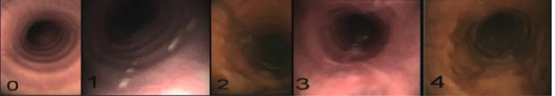
An endoscopical examination of the nasophar-ynx, larynx and trachea to the level of the carina was performed (Mercury Endoscopia Italiana, L = 320 cm, ø = 0.98 cm, Italy) and animals with evi-dence of upper airway dysfunction were excluded from the study. Horses were restrained with the only use of a twichnose. Mucus accumulation in the trachea was classified using the following scoring system suggested by Gerber et al. (2004):
Score 0: no mucus
Score 1: small, singular drops of mucus Score 2: larger, confluent drops of mucus Score 3: streams of mucus
Score 4: large streams or pools of mucus covering more than Ľ of tracheal diameter (Figure 1).
To perform tracheal wash (TW), the endoscope was retracted to the mid-tracheal level and 40 ml of sterile and prewarmed saline solution infused via a sterile catheter passed in the accessory channel of the endoscope. Advancing caudally in the trachea the pool of saline solution was than individuated,
collected using a 50 ml sterile syringe and trans-ferred in sterile tubes stored on ice. All samples were brought to the laboratories within two hours from collection.
The endoscope was cleaned with an iodopovi-done solution, rinsed with sterile saline solution and, lastly, with sterile distilled water before the following horse examination.
Bacteriological examination
An aliquot (100 μl) of TW fluid was cultured on Columbia blood Agar with and without Streptococcus Selective Supplement, MacConkey Agar, Baird-Parker Agar and Plate Count Agar (Oxoid, Milan, Italy). Plates were incubated in aero-bic and anaeroaero-bic conditions at 37°C for 48 h. The colonies were subcultured and identified by using biochemical gallery (RapID™ System, Oxoid, Milan,
Italy), serological (Streptex®, Oxoid, Milan, Italy)
or PCR techniques (Preziuso et al., 2007).
An equal amount, 100 μl, of TW fluid was used for Mycoplasma isolation using Mycoplasma Broth with Mycoplasma Selective Supplement – G (Oxoid, Milan, Italy) incubated at 4°C for 24h. After cen-trifugation at 2 000 × g for 20 min, the superna-tant was added to Mycoplasma broth with 20% of equine sterile serum and incubated at 37°C for 72 h. Finally, 100 μl of supernatant were inoculated into Mycoplasma Agar (Oxoid, Milan, Italy) with 20% of equine serum and plates were incubated for 10 days at 37°C in humid chamber with 10% CO2.
Cytology
After cytocentrifugation, the TW samples were smeared, air-dried, stained with May-Grundwald-Giemsa and observed under a microscope using a 100× magnification. The numbers of neutrophils were estimated by counting 300 cells and the per-centages higher than 30% were considered sign of inflammation.
Statistical analysis
Results were elaborated by χ2 test with Yates
cor-rection factor. The level of significance was estab-lished at P < 0.05 for all the tests.
RESULTS
There were no significant differences among symptoms observed apart from little variability concerning the rate of cough.
Mucus scores detected at endoscopic examina-tion and their correlaexamina-tions with neutrophilic in-flammation and total bacterial count are reported in Table 1.
The previous table shows as 20 out of 24 horses (83.3%) with mucous score > 1 had an high neu-trophils percentage in TW, revealing a strong sta-tistical association between amount of mucous in trachea and neutrophils percentage in TW (P < 0.05). Furthermore, the increasing of mucous score Figure 1. Different amount of mucus in trachea and relative scores
Table 1. Mucus score correlated with neutrophilic inflammation and total bacterial load
Mucus score Number of horses Horses with neutrophilic inflammation Total bacterial load mean (CFU/ml)
0 18 0 149 × 102 1 20 4 394 × 102 2 18 14 1 194 × 102 3 2 2 4 940 × 102 4 4 4 3 417 × 102 Total 62 24
is pretty consistent with the increasing of total bac-terial count (P < 0.05).
The association between neutrophilic inflamma-tion and total bacterial load is reported in Table 2.
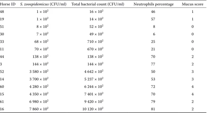
Among β-haemolytic Streptococci, S. zooepidem-icus has been isolated from TW fluid of 14 horses. The correlation between these results and other parameters of the same horses are summarized in Table 3.
There was not a statistical correlation for S. zoo- epidemicus neither with mucus score nor with neu-trophils percentage.
Four horses were positives for Streptococcus pneu-moniae and the association with mucus amount and neutrophil percentage is showed in Table 4.
S. zooepidemicus and S. pneumoniae were never isolated together in the same horse.
Other α-haemolytic streptococci isolated were S. mutans (from four horses) and S. equinus (from four horses).
From ten horses Burkholderia cepacea, without association with neutrophil percentage or mucus score, has been isolated. Two samples resulted sterile.
Table 2. Association between neutrophils percentage and total bacterial load
Number of horses Neutrophilic inflammation Total bacterial load mean (CFU/ml)
37 no 309 × 102
25 yes 1 666 × 102
Table 3. Neutrophils percentage and mucus score in horses positive for S. zooepidemicus
Horse ID S. zooepidemicus (CFU/ml) Total bacterial count (CFU/ml) Neutrophils percentage Mucus score
48 1 × 10² 16 × 102 46 1 19 1 × 10² 14 × 102 57 1 51 8 × 10² 52 × 102 8 0 30 7 × 10² 49 × 102 6 0 33 68 × 102 710 × 102 25 0 11 70 × 102 670 × 102 21 0 44 138 × 102 138 × 102 70 2 3 144 × 102 144 × 102 77 2 52 3 580 × 102 4 642 × 102 50 3 14 3 700 × 102 5 237 × 102 53 3 60 4 280 × 102 6 244 × 102 72 4 15 4 350 × 102 7 401 × 102 70 4 61 6 980 × 102 9 420 × 102 79 2 16 7 860 × 102 10 120 × 102 81 2
Table 4. Neutrophils percentage and mucus score in horses positive for S. pneumonia
Horse ID S. pneumoniae (CFU/ml) Total bacterial count (CFU/ml) Neutrophils percentage Mucus score
32 890 × 102 1 012 × 102 21 2
59 560 × 102 815 × 102 29 0
39 1 × 10² 3 × 10² 56 4
From none of the horses other potentially patho-gen bacteria, included Mycoplasma spp., have been isolated, and no haematological abnormalities were found. Results about isolation of normal flora has not been reported.
DISCUSSION
The 62 examined horses represent the 64% of the total young coughing horses (97) and can be con-sidered representative of the coughing population. Therefore, it is possible to carried out both general and specific considerations.
The results of this study confirm a strong statisti-cal association between neutrophilic inflammation and accumulation of mucus in trachea. Increased number of neutrophils could be the cause of mu-cus production but infectious agents and some environmental contaminants can directly trigger it (Robinson, 2003; Gerber et al., 2004). Has been demonstrated that mucus in trachea is associ-ated with decreased performances (Holcombe et al., 2006) but it is a matter of concern if strepto-cocci or other bacteria could be a causative agent of the increasing in tracheal secretions in airways inflammation affected horses. The result of this study shows a fairly agreement between the mucus score and quantitative analysis for S. zooepidemi-cus and these results could indicate that muzooepidemi-cus in trachea is elicited by a specific bacteria. On the other hand, a similar correlation between mucus score and total bacterial load was found. Therefore, it could be possible that the decreased clearance in lower and upper airway consequent to inflam-mation, leaded to a bacterial accumulation in the airways. In this case, mucus accumulation would be the cause and not the effect of bacteria colonisa-tion of the airways. Nevertheless, some observa-tions should be done: although a large number of horses having high mucus score and/or neutrophil percentage were negative for S. zooepidemicus in trachea, among the 14 positive horses, the higher S. zooepidemicus load belong to the horses with mucus score > 2. Furthermore, 10 out of 14 positive horses had cytological evidences of inflammation with the higher neutrophil percentage (81%) asso-ciated at the highest bacterial load (7 860 × 102) in
horse number 16. Similar findings in thoroughbred were also reported (Chapman et al., 2000). In horse number 3, with evident endoscopical and cytologi-cal signs of inflammation, S. zooepidemicus was the
only bacteria isolated. Based on this findings, even though a statistical association were not found, we can not exclude a role of S. zooepidemicus in the pathogenesis of IAD, as a causative or, more prob-ably, as a perpetuating agent of inflammation in Standardbred horses.
Tracheal infection with S. pneumoniae has been statistically associated with IAD in British Racehorse (Wood et al., 2005a). In horses of the present work, S. pneumoniae was associated with increased mucus amount (Table 4) but, unexpect-edly, bacterial count showed almost a reversed cor-relation with neutrophilic inflammation and mucus. Mycoplasma felis and Mycoplasma equirhinis are frequently isolated in British Thoroughbred with clinical respiratory disease (Newton et al., 2003; Wood et al., 2005a), but has never been isolated in Standardbred horses of this study. The different etiological forms of IAD, depending on feeding, housing, preventive medicine practice and, most important, different distribution of infectious agents (Couetil et al., 2007) could be an explana-tion for variability of these results.
Burkholderia cepacia is the major pathogens that colonize the airway surface and cause pro-gressive respiratory failure and high mortality in human being, especially in cystic fibrosis patients (Treerat et al., 2008) and usually is not isolated from healthy people (Hyde and Humphreys, 1997).
Differently from Burkholderia mallei, its role is unknown in horses. B. cepacia was isolated from 10 horses, without association with neutrophil percentage or mucus score. Recent study showed that Burkholderia cenocepacia, a member of the B. cepacia complex, require a source of iron to colo-nize the lung and to satisfy its nutritional require-ments and that is the only one able to extract iron from ferritin (Whitby et al., 2006). None of the five horses had history or clinical signs of exercise induced pulmonary haemorrhage (EPEI) but some haemosiderophages (macrophages containing a fer-ritin polymer called hemosiderin) in their tracheal secretions were found at cytological examination. This is a normal finding also in healthy racehorses but could be interesting to establish the epidemi-ology of B. cepacia among horses and to further investigate the relationship between B. cepacia and respiratory horse diseases.
A load of pathogenic bacteria greater than 102
CFU/ml, in the presence of neutrophilic inflamma-tion, is supposed to be a cut-off value to warrant administration of antibiotic therapy (Christley and
Rush, 2007). All horses positive for S. zooepidemi-cus or S. pneumoniae with cytological signs of in-flammation had a bacterial load ≥ 102 CFU/ml and
can be considered affected by so called bacterial IAD. Nevertheless, from this point of view, this study failed in found a common or, at least, a more diffuse causes for so high prevalence of coughing horses. Eighteen horses did not show any mucus in trachea in spite of the presence of cough and poor performances. These horses could be affected by a deeper problem in respiratory tract, like bron-chioles inflammation and bronchospasmus, with-out involvement of trachea. This cases and other horses with various grades of mucus and/or signs of cytological inflammation without signs of infec-tion, could be affected by the other subcategory, called non-bacterial IAD. Inhalation of particulate matter, pro-inflammatory agents or noxious gases are reasonable causes for non infectious coughing horse. To test airborne endotoxin concentration in the stables where horses are housed and in the racetrack matter where horses are trained, will be the second step of the present study.
REFERENCES
Burrell M.H., Wood J.L., Whitwell K.E., Chanter N., Mackintosh M.E., Munford J.A. (1996): Respiratory disease in thoroughbred horses in training: the rela-tionships between disease and viruses, bacteria and environment. The Veterinary Record, 139, 308–313. Chapman P.S., Green C., Main J.P., Taylor P.M.,
Cun-ningham F.M., Cook A.J., Marr C.M. (2000): Retro-spective study of the relationships between age, inflammation and the isolation of bacteria from the lower respiratory tract of thoroughbred horses. The Veterinary Record, 146, 91–95.
Christley R., Rush B.R. (2007): Inflammatory airway disease, 591–600. In: Mc Gorum B.C., Dixon P.M., Robinson N.E., Schumacher J. (eds.): Equine Respira-tory Medicine and Surgery. Saunders, Philadelphia. 705 pp.
Christley R.M., Hodgson D.R., Rose R.J., Wood J.L., Reids S.W., Whitear K.G., Hodgson J.L. (2001): A case-control study of respiratory disease in Thoroughbred racehorses in Sydney, Australia. Equine Veterinary Journal, 33, 256–264.
Couetil L.L., Ward M.P. (2003): Analysis of risk factors for recurrent airway obstruction in North American horses: 1 444 cases (1990–1999). Journal of the Amer-ican Veterinary Medical Association, 223, 1645–1650.
Couetil L.L., Hoffman A.M., Hodgson J., Buechner-Maxwell V., Viel L., Wood J.L.N., Lavoie J.P. (2007): Inflammatory airway disease of horses. Journal of Vet-erinary Internal Medicine, 21, 356–361.
Gerber V., Straub R., Marti E., Hauptman J., Herholz C., King M., Imhof A., Tahon L., Robinson N.E. (2004): Endoscopic scoring of mucus quantity and quality: observer and horse variance and relationship to in-flammation, mucus viscoelasticity and volume. Equine Veterinary Journal, 36, 576–582.
Holcombe S.J., Jackson C., Gerber V., Jefcoat A., Berney C., Eberhardt S., Robinson N.E. (2001): Stabling is as-sociated with airway inflammation in young Arabian horses. Equine Veterinary Journal, 33, 244–249. Holcombe S.J., Robinson N.E., Derksen F.J., Bertold B.,
Genovese R., Miller R., de Feiter Rupp H., Carr E.A., Eberhart S.W., Boruta D., Kaneene J.B. (2006): Effect of tracheal mucus and tracheal cytology on racing performance in Thoroughbred racehorses. Equine Veterinary Journal, 38, 300–304.
Hyde J., Humphreys H. (1997): Absence of Burkholderia
cepacia from the respiratory tract of non-cystic
fibro-sis patients. European journal of clinical microbiology & infectious diseases, 16, 253–254.
Laus F., Preziuso S., Spaterna A., Beribe F., Tesei B., Cu-teri V. (2007): Clinical and epidemiological investiga-tion of chronic upper respiratory diseases caused by beta-hemolytic Streptococci in horses. Comparative immunology, microbiology and infectious diseases, 30, 247–260.
MacNamara B., Bauer S., Iafe J. (1990): Endoscopic evaluation of exercise-induced pulmonary hemorrhage and chronic obstructive pulmonary disease in asso-ciation with poor performance in racing Standard-breds. Journal of the American Veterinary Medical Association, 196, 443–445.
Moore B.R., Krakowka S., Mcvey D.S., Cummins J.M., Robertson J.T. (1997): Inflammatory markers in bron-choalveolar lavage fluid of standardbred racehorses with inflammatory airway disease: response to interferon-alpha. Equine Veterinary Journal, 29, 142–147. Newton J.R., Wood J.L., Chanter N. (2003): A case
con-trol study of factors and infections associated with clinically apparent respiratory disease in UK Thor-oughbred racehorses. Preventive Veterinary Medicine, 60, 107–132.
Newton J.R., Laxton R., Wood J.L., Chanter N. (2008): Molecular epidemiology of Streptococcus
zooepidem-icus infection in naturally occurring equine respiratory
disease. Veterinary journal, 175, 338–345.
Preziuso S., Bastianini L., Laus F., Cuteri V. (2007): Pre-liminary evaluation of a PCR protocol to directly
de-Corresponding Author:
Prof. Vincenzo Cuteri, University of Camerino, Department of Veterinary Science, Via Circonvallazione, 93/95-62024 Matelica (MC), Italy
Tel. +39 0737 404007, E-mail: [email protected] tect Streptococcus equi subsp. zooepidemicus in equine specimens. International Journal of Antimicrobial Agents, 29, 398.
Robinson N.E. (2003): Inflammatory airway diseases: defining the syndrome. Conclusion of the Havemeyer Workshop. Equine Veterinary Education, 15, 61–63. Robinson N.E., Derksen F.J., Olszewski M.A.,
Buechner-Maxwell V.A. (1996): The pathogenesis of chronic obstructive pulmonary disease of horses. The British Veterinary Journal, 152, 283–306.
Sweeney C.R., Beech J., Roby K.A. (1985): Bacterial iso-lates from tracheobronchial aspirates of healthy horses. American Journal of Veterinary Research, 46, 2562– 2565.
Treerat P., Widmer F., Middleton P.G., Iredell J., George A.M. (2008): In vitro interactions of tobramycin with
various nonantibiotics against Pseudomonas
aerugi-nosa and Burkholderia cenocepacia. FEMS
Microbiol-ogy Letters,285, 40–50.
Whitby P.W., Vanwagoner T.M., Springer J.M., Morton D.J., Seale T.W., Stull T.L. (2006): Burkholderia
ceno-cepacia utilizes ferritin as an iron source. Journal of
medical microbiology, 55, 661–668.
Wood J.L., Newton J.R., Chanter N., Mumford J.A. (2005a): Association between respiratory disease and bacterial and viral infections in British racehorses. Journal of Medical Microbiology, 43, 120–126. Wood J.L., Newton J.R., Chanter N., Mumford J.A.
(2005b): Inflammatory airway disease, nasal discharge and respiratory infections in young British racehorses. Equine Veterinary Journal, 37, 236–242.
Received: 2009–01–09 Accepted: 2009–09–24