DOI: 10.14601/Phytopathol_Mediterr-15008
Corresponding author: S.O. Cacciola E-mail: [email protected]
RESEARCH PAPERS
Decline of jackfruit (Artocarpus heterophyllus) incited by Phytophthora
palmivora in Vietnam
Mai Van TRi1, nguyen Van HOa1, nguyen MinH CHau1, anTOnella Pane2, RObeRTO FaeDDa2, alessanDRO De
PaTRiZiO2, leOnaRDO sCHena3, CHRisTeR H. b. OlssOn4, sanDRa a. i. WRigHT5, MauRiTZ RaMsTeDT6 and sanTa
Olga CaCCiOla2
1 Southern Horticultural Research Institute, Box 203, My Tho, Tien Giang, Vietnam
2 Department of Agriculture, Food and Environment, University of Catania, 95123 Catania, Italy
3 Department of Agraria, University Mediterranea of Reggio Calabria, Località Feo di Vito, 89124 Reggio Calabria, Italy 4 Department of Biological &Environmental Sciences, University of Gothenburg, 40530 Gothenburg, Sweden
5 Department of Occupational and Public Health Sciences, University of Gävle,80176 Gävle, Sweden
6 Department of Forest Mycology and Plant Pathology, Swedish University of Agricultural Sciences (SLU), 75007 Uppsala,
Sweden
Summary. A new disease of jackfruit (Artocarpus heterophyllus Lam.) was observed in the south- eastern region of South Vietnam. Symptoms included root rot, cankers and gummosis of trunks, chlorosis, wilt, blight of leaves, defoliation, fruit brown rot, and tree death. The disease was found in 10% of surveyed farms with an incidence varying from 2% to nearly 60% of the trees. A Phytophthora species, identified as P. palmivora (Butler) Butler, using the ITS1-5.8S-ITS2 region of the rDNA as a barcode gene and morphological and cultural features, was consist-ently isolated from symptomatic roots, fruits, trunk cankers and leaves. Koch’s postulates were fulfilled using pathogenicity tests on seedlings, leaves and detached fruits of jackfruit. To our knowledge, this is the first report of P. palmivora on jackfruit in Vietnam.
Key words: Oomycetes, South Vietnam, ITS regions, A1 mating type, Koch’s postulates.
Introduction
Jackfruit (Artocarpus heterophyllus Lam., family Moraceae) is an important multi-purpose fruit crop in tropical and subtropical regions, providing food, timber, fuel, fodder, medicinal and industrial prod-ucts. Native to the south-western rain forests of In-dia, A. heterophyllus is now widely grown in many Asian countries, especially Bangladesh, Myanmar, Nepal, Sri Lanka, Thailand, Vietnam, Malaysia, In-donesia, India and the Philippines (Elevitch and Manner, 2006). From Asia it spread to tropical Af-rican countries, including Zanzibar, Kenya, Ugan-da, Madagascar and Mauritius. From the mid-17th
century to the late 19th century, the species spread further to tropical and subtropical America and Aus-tralia (Haq, 2006).
In Vietnam, jackfruit is widespread but grown mainly in the south eastern region, traditionally in household gardens, forest plantations or small farms, usually intercropped with other fruit crops, such as mango, longan, durian, rambutan, guava, water ap-ple, pineapap-ple, breadfruit or industrial crops such as coffee, cashew nut, black pepper, rubber tree, or co-coa (Sidhu, 2012). Jackfruit can generate an income for small farmers through the sale of fruits and is considered a good feed resource for cattle and pigs (Elevitch and Manner, 2006). Areas under cultivation have been rapidly expanding in the last 10 years, and it is estimated to cover approx. 50,000 ha with an av-erage fruit yield of about 30 tons ha-1. The crops have
developed from for small-scale household jackfruit production to important cash crops (Mai Van Tri and Nguyen Van Hoa, 2014). Approximately 60% of the production is for processing and the rest is for fresh consumption. Dried jackfruit as chips is the major processed product for local markets and export (Mai Van Tri and Nguyen Van Hoa, 2014).
In 2012 and 2013, in four provinces of South Vi-etnam (Ba Ria-Vung Tau, Binh Duong, Binh Phuoc and Dong Nai) a decline disease was observed in approximately 10% of surveyed jackfruit plantings, with an incidence varying from 2% up to approx. 60% of the trees in each planting. Affected trees showed symptoms of leaf blight and chlorosis, wilt-ing, defoliation, trunk cankers with resin exudates and root rot. Sometimes, symptoms of brown rot on fruits hanging from the trees or fallen to the ground were also observed. Leaf blight began as small, wa-ter soaked flecks that, in a few days, turned brown and expanded rapidly into large necrotic lesions. In wet conditions, white velvety mycelium was visible at the edges of lesions on leaves and fruits. Stem can-kers appeared firstly as wet lesions on the bark sur-faces, often close to the insertion of large branches, but more frequently at trunk bases. A reddish-brown resin oozed from cracks in the bark. The wood tis-sues under the lesions showed cream to reddish brown discoloration. The infected areas enlarged, girdling the stems and causing severe decline of the trees. Decline was always associated with the rot of fine roots. In some cases, the whole tree died. Symp-toms of decline were severe mostly in areas prone to flooding or with poor drainage. Leaf blight and fruit brown rot were observed after periods of heavy rain-fall and when the relative humidity was high.
The involvement of a Phytophthora species as causal agent of the disease was suspected on the ba-sis of symptoms. As a consequence, analyses were focused on Oomycetes and the causal agent of jack-fruit disease observed in South Vietnam was identi-fied and characterized.
Materials and methods
Isolation and morphological characterization of isolates
Isolations were made from trees with symptoms of decline. Affected necrotic tissues were thorough-ly washed with tap water to remove soil and plant debris. Small sections (2–5 mm) from symptomatic
fine roots and the edge of rotted tissues of stems, fruits and leaves were blotted dry on sterile paper towels and transferred onto BNPRAH selective me-dium for Phytophthora (Masago et al., 1977) in Petri dishes, where they were incubated for 5 d at 24–25°C in the dark. Hyphal tips of Phytophthora colonies, after several transfers on BNPRAH medium, were sub-cultured on potato dextrose agar (PDA, Oxoid Ltd) to obtain pure cultures. Isolates were grown in Petri dishes (9 cm diam.) containing 20 mL of either PDA or V8 vegetable juice agar (V8A; Campbell) at 24±1°C for 14 d in darkness to determine the colony morphology. For cardinal growth temperature as-says, a 5-mm diam. mycelial plugs were transferred to PDA and incubated at different temperatures (at 3°C intervals from 3°C to 39°C). Growth rates were determined on PDA at 27 ± 1°C for a maximum of 7 d. Mating type for isolates was determined on V8A in dual culture with A1 and A2 reference isolates of Phy-tophthora palmivora and P. nicotianae, as described by Brasier et al. (2003). Two representative isolates, MD5 obtained from fine roots and MD6 from leaves, were selected for DNA analysis and pathogenicity tests. Living cultures of isolates MD5 and MD6 are pre-served both at the CABI Genetic Resource Collection, United Kingdom (IMI 503890 and IMI 503891) and in the collection of the Department of Agriculture, Food and Environment, University of Catania, Italy.
Sequencing and analysis of ITS regions
DNA was extracted from 10 mg of fresh mycelium of 7-d-old colonies of isolates MD5 and MD6 grown in Petri dishes on V8A at 24°C, using DNeasy Plant Mini Kit according to the manufacturer’s instruc-tions (Qiagen GmbH). DNA samples were stored at -20°C prior to PCR amplification. The quality and quantity of extracted DNA samples were evaluated using a DNA Quant-it assay kit (Molecular Probes) and by electrophoresis in 1% agarose gels contain-ing SYBR® Safe (Invitrogen Life Technologies) DNA gel stain. Amplification and sequencing of the inter-nal transcribed spacer (ITS) region of rDNA were performed according to the protocol described by Cooke et al. (2000), using 20 ng of genomic DNA as a template and ITS6 and ITS4 primers. Amplicons were analyzed by electrophoresis and using a DNA Quant-it assay kit (Molecular Probes) as described above, purified using the ExoSAP-ITkit for PCR Product Cleanup (Affymetrix) and sequenced in
both directions using an external sequencing service (BMR-genomic). The identity of isolates was estab-lished according to the level of similarity of their ITS sequences with those of reliable reference-sequences.
Pathogenicity tests
Pathogenicity trials were performed on detached fruits as well as on leaves, stems and roots of potted 6-month-old A. heterophyllus plants using the isolates MD5 and MD6.
Detached immature jackfruit fruits (five fruits per isolate) were superficially disinfected with 1% NaClO, rinsed with sterile distilled water and dried with pa-per tissues. A hole (5 mm diam.) was made in each fruit with a cork borer and a disc of tissue from the fruit surface was removed; the wound was filled with a 5 mm agar plug containing actively growing myce-lium, and was then sealed with adhesive tape. Sterile agar plugs were inserted into control fruits. The fruits were singly incubated in sealed plastic bags at room temperature and inspected daily for symptoms.
The leaves of jackfruit plants (ten seedlings per isolate) were sprayed with 70% ethanol and allowed to air dry. A water suspension of each isolate was obtained by flooding a 12-d-old culture on V8A in a Petri dish with sterile distilled water (s.d.w.) and superficially scraping the mycelium with a sterile scalpel. The concentration was adjusted to 1×10⁴ spo-rangia mL-1 and the suspension was sprayed on the leaves. Ten seedlings sprayed with s.d.w. served as experimental controls. The plants were covered with large, transparent plastic bags and s.d.w. was nebu-lized into the bags to maintain high relative humidity. In a third experiment, jackfruit plants (10 for each isolate) were wound-inoculated on the stems. A small strip of bark (3–4 mm in length) was cut aseptically from the stem of each plant with a scal-pel and 3 mm agar plugs from the edge of actively growing cultures were placed into the wound. Ster-ile agar plugs were used for control seedlings. The bark strips were replaced on wounds and the stems were tightly sealed with Parafilm®.
In a fourth experiment, jackfruit plants (ten for each isolate) were transplanted into pots (12 cm diam.) containing a mixture of 1:1 (v:v) steam-ster-ilized sandy loam soil with 4% inoculum produced on autoclaved wheat kernels, according to Pane et al. (2009) and Faedda et al. (2013). Ten control plants were transplanted into pots containing non-infested
soil. All plants were then grown for 3 weeks and wa-tered to field capacity once a week.
The last three experiments described above were carried out in a greenhouse, with natural light con-ditions and temperatures ranging from 24 to 28 °C. Plants were inspected daily for symptoms for 30 d after inoculation.
Results
Isolation and morphological and cultural characterization
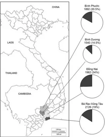
The results of the survey are shown in Figure 1, where the proportion of symptomatic jackfruit trees relative to the number of trees surveyed in each province (Ba Ria-Vung Tau, Binh Duong, Binh Phuoc and Dong Nai) is outlined. The total numbers of surveyed trees and proportions of symptomatic trees per province was: 2126 trees in Ba Ria-Vung Tau province with 19% of symptomatic trees; 1540 in the Binh Duong province with 14.5% of symptomatic trees; 1892 in the Binh Phuoc province with 20.5% of symptomatic trees and 1962 in the Dong Nai prov-ince with 34% of symptomatic trees. A Phytophthora species was consistently recovered from all symp-tomatic trees on BNPRAH selective medium, with frequencies of 90% from fruits, 83% from leaves, 71% from roots and 54% from trunk bark samples. All isolates had identical morphological and cultural features and closely matched those of P. palmivora (E.J. Butler) E.J. Butler (Stamps et al., 1990; Erwin and Ribeiro; 1996; Gallegly and Hong, 2008). On PDA, colonies had stoloniferous mycelium, whereas on V8A they showed stellate patterns. On V8A, all isolates produced elliptical to ovoid, conspicuously papillate sporangia with a mean length/breadth ra-tio of 1.8. Sporangia were caducous with short pedi-cels (mean pedicel length = 5 µm) and conspicuous basal plugs at the insertion of the pedicels. They were formed on sympodially branched sporangiophores. Minimum and maximum temperatures for growth on PDA were, respectively, 9°C and 36°C, with the optimum at 27°C and a growth rate of 7 mm d-1 at this temperature. All isolates were heterothallic and were of A1 type, producing amphigynous antherid-ia, oogonantherid-ia, and oospores after pairing with the A2 reference isolates of P. nicotianae and P. palmivora.
The origins of the isolates and the numbers of plantations where they were sourced are shown in Table 1.
Molecular identification of isolates
The analysis of the ITS1-5.8S-ITS2 sequences from the representative isolates MD5 and MD6 (accession nos. KF823978 and KF823979) revealed 100% similar-ity with several reference isolates of P. palmivora (e.g., accession nos. HQ643305, GQ398157 and KC415917) and with a sequence (AF266781) deposited as P. arecae (Grünwald et al., 2011; Robideau et al., 2011). Other related species of clade 4 were clearly differ-entiated using the ITS regions as the barcode gene (Robideau et al., 2011).
Pathogenicity tests
Water-soaked, brown lesions appeared on wound-inoculated fruits 2–3 d after inoculation. Seven days after inoculation, the mean diameter of lesions was 95±5 mm, and white, velvety mycelium was visible on the fruit rind surrounding the lesions.
Water-soaked, small, brown (2–5 mm) lesions with indefinite margins developed on inoculated leaves within 4 d after inoculation. Lesions became necrotic in a few days, enlarged and coalesced into larger ne-crotic areas. White mycelium efflorescence was visible at the edges of lesions 7 d after inoculation. Phytoph-thora palmivora was re-isolated from symptomatic tis-sues to fulfil Koch’s postulates. The leaves of plants sprayed with distilled water showed no symptoms.
Brown gum exudates appeared on the stems of wound-inoculated plants 7 d after inoculation. The mean length of gummous stem cankers 15 d after in-oculation was 20±2 mm. The plants developed leaf chlorosis and wilting and collapsed 30 d after inocu-lation. Control seedlings showed no symptoms. Phy-tophthora palmivora was re-isolated from all sympto-matic plants.
All plants transplanted into pots filled with in-fested soil developed symptoms of decline within 1 week, and 60% collapsed by 30 d after transplant-ing. Non-inoculated plants remained healthy. Phy-tophthora palmivora was re-isolated from fine roots of inoculated plants.
In all experiments, no significant differences were observed between the two P. palmivora isolates used in pathogenicity tests.
Discussion
The results of this study demonstrate that P. palmivora is the causal agent of the decline of jackfruit
Figure 1. Diagrammatic representation of the incidence of the decline incited by Phytophthora palmivora in jackfruit plantings in four provinces of South Vietnam; the size of pies is proportional to the number of surveyed trees in each province. Figures above pies indicate the total number of jackfruit trees surveyed and the proportion (within brack-ets) of symptomatic trees in each province, respectively. Note that this is not a map of Vietnam, but only the indica-tion of the provinces where the survey was carried out.
Table 1. Origin of Phytophthora palmivora isolates obtained
in four provinces of South Vietnam from jackfruit trees with symptoms of decline, and number of plantings where they were sourced.
Provinces Root Trunk Leaf Fruit
Ba Ria Vung Tau 10 8 4 2
Binh Duong 7 4 -
-Dong Nai 10 9 -
-Binh Phuoc 8 7 2 2
trees observed in the south eastern region of Viet-nam. The pathogen was accurately identified on the basis of morphological features and using the ITS1-5.8 S-ITS2 region of the rDNA as a barcode gene. ITS sequences of isolates from jackfruit sourced in south east Vietnam were identical to reference sequences of P. palmivora and P. arecae and were clearly differenti-ated from all other currently recognized Phytophthora species (Robideau et al., 2011; Grünwald et al., 2011). Furthermore, P. arecae has been shown to be synony-mous with P. palmivora (Kroon et al., 2012). All symp-toms of the complex syndrome associated with the decline of jackfruit trees were reproduced in artificial inoculations using isolates of P. palmivora obtained from jackfruit trees with natural infections, and the pathogen was consisitently re-isolated from sympto-matic tissues, thus fulfilling Koch’s postulates.
Phytophthora palmivora is believed to have originat-ed in south-east Asia (Mchau and Coffey, 1994) but is now widespread in the Tropics and is spreading also in warm, temperate climate zone, including the Medi-terranean region, on ornamental plants in nurseries (Cacciola et al., 2002; Davino et al., 2002; Moralejo et al., 2009; Cacciola et al., 2011; Dervis et al., 2011) as well as on tree crops, including native Mediterranean species such as olive (Cacciola et al., 2000; Lo Giudice et al., 2010; Turkölmez et al., 2014). It is a polyphagous path-ogen, with the known host plants being in more than 160 genera in 60 families (Cline, 2008). The host range also includes A. heterophyllus and other related spe-cies of Artocarpus, such as breadfruit (A. altilis), cem-pedak (A. integer) and breadnut (A. camansi) (Chee, 1969; Gerlach and Salevao, 1984; Erwin and Ribeiro, 1996; Drenth and Guest, 2004). The list of more than a thousand known hosts of P. palmivora includes most of the plant species that are commonly intercropped with jackfruit in Vietnam. Very recently, a severe de-cline syndrome of jackfruit that emerged in the Phil-ippines during the late 1990s was found to be caused by P. palmivora (Borines et al., 2014). However, to our knowledge, the present paper is the first report of P. palmivora on jackfruit in Vietnam.
Phytophthora palmivora is a widespread pathogen in Vietnam, and has been reported as a common patho-gen of durian (Durio zibethinus), one of the most appre-ciated and profitable fruit crops in this country (Drenth and Guest, 2004). On durian, P. palmivora causes symp-toms similar to those observed on jackfruit, such as root and crown rot, stem cankers with gum exudates, fruit brown rot, leaf blight and whole tree decline.
Other hosts on which P. palmivora has been recorded from Vietnam include coconut (Cocos nucifera), cocoa (Theobroma cacao) and rubber (Hevea brasiliensis). On tree-hosts, P. palmivora is primarily a root pathogen. However, deciduous sporangia of this species can be disseminated by rain, wind and soil-splash and in wet, showery conditions these may cause aerial infections. Also insects, particularly ants, have been reported as vectors of P. palmivora in tropical areas (McGregor and Moxon, 1985; Holderness, 1992).
Several lines of evidence suggest the decline of jackfruit observed in southern Vietnam is the same disease described recently in the Philippines by Borines et al. (2014). Very probably, in the south east-ern region of Vietnam, the disease has been present for a long time, but it has passed unnoticed because of the limited economic significance of jackfruit, which, until recently, has been cultivated only for do-mestic consumption or for local markets. The expan-sion of the cultivation of jackfruit, both as a fruit and a multi-purpose commercial crop, has determined the conditions for the emergence of this disease. The results of our survey indicate the complex of symp-toms of jackfruit decline caused by P. palmivora pose a serious threat for jackfruit cultivation, with a sig-nificant impact both on yields and on the long-term viability of plantations. The precise identification of the causal agent of this disease constitutes a funda-mental step for the development of appropriate and effective disease management strategies.
Acknowledgement
This research was funded by an initiation grant of STINT (The Swedish Foundation for International Cooperation in Research and Higher Education), SAMAGRUMI (Sensori Ambientali per il Migliora-mento della Qualità delle Produzioni Agrumicole), PO. FESR 2007-2013-Sicily and by FIRB 2010-RB-FR10PZ4N from the Italian Ministry of Education, University and Research (MIUR); the authors are thankful to Mrs. Ann Davies for the revision of the English.
Literature cited
Borines L.M., V.G. Palermo, G.A. Guadalquiver, C. Dwyer, A. Drenth, R. Daniel, D.I. Guest, 2014. Jackfruit decline caused by Phytophthora palmivora (Butler). Australasian
Brasier C.M., E. Sanchez-Hernandez, S.A. Kirk, 2003.
Phytoph-thora inundata sp. nov., a part heterothallic pathogen of
trees and shrubs in wet or flooded soils. Mycological
Re-search 107, 477–484.
Cacciola S.O., G.E. Agosteo, A. Pane, 2000. First report of
Phy-tophthora palmivora as a pathogen of olive in Italy. Plant Disease 84, 1153.
Cacciola S.O., A. Pane, S. Davino, F. Raudino, 2002. First re-port of Phytophthora palmivora on Coronilla valentina subsp.
glauca in Italy. Plant Disease 86, 327.
Cacciola S.O., A. Pane, R. Faedda, C. Rizza, F. Badalà, G. Mag-nano di San Lio, 2011. Bud and root rot of windmill palm (Trachycarpus fortunei) caused by simultaneous infections of Phytophthora palmivora and P. nicotianae in Sicily. Plant
Disease 95, 769.
Chee K.H. (1969). Hosts of Phytophthora palmivora. Review of
Applied Mycology 48, 337–344.
Cline E.T., D.F. Farr, A.Y. Rossman, 2008. A synopsis of
Phy-tophthora with accurate scientific names, host range, and
geographic distribution. Online Plant Health Progress, doi:10.1094/PHP-2008-0318-01-RV.
Cooke D.E.L., A. Drenth, J.M. Duncan, G. Wagels, C.M. Brais-er, 2000. A molecular phylogeny of Phytophthora and re-lated Oomycetes. Fungal Genetics and Biology 30, 17–32. Davino S., S.O. Cacciola, A.M. Pennisi, M.G. Li Destri Nicosia,
2002. Phytophthora palmivora a new pathogen of lavender in Italy. Plant Disease 86, 561.
Dervis S., M. Arslan, C.U. Serce, S. Soylu, I. Uremis, 2011. First report of a root rot caused by Phytophthora palmivora on
Lavandula angustifolia in Turkey. Plant Disease 95, 1035.
Drenth A., D.I. Guest, 2004. Diversity and management of
Phy-tophthora in Southeast Asia. ACIAR Monograph No.114.
Australian Centre for International Agricultural Research, Canberra Australia, 238 pp.
Elevitch C.R., H.I. Manner, 2006. Species Profiles for Pacific Island Agroforestry: Artocarpus heterophyllus (jackfruit). In: Traditional Trees of Pacific Islands: their culture, environment
and use (C.R. Elevitch, ed.), www.traditionaltree.org, 1–17.
Erwin D.C., O.K. Ribeiro, 1996. Phytophthora Diseases
World-wide. APS Press, St. Paul, MN, USA, 592 pp.
Faedda R., S.O. Cacciola, A. Pane, A. Szigenthy, J. Bakonyi, W.A. Man in’t Veld, P. Martini, L. Schena, G. Magnano di San Lio, 2013. Phytophthora × pelgrandis causes root and collar rot of Lavandula stoechas in Italy. Plant Disease 97, 1091–1096.
Gallegly M.E., C.X. Hong, 2008. Phytophthora: Identifying
Spe-cies by Morphology and DNA Fingerprints. APS Press, St
Paul, MN, USA, 158 pp.
Gerlach W.W.P., F. Salevao, 1984. Fruit rot of breadfruit,
Arto-carpus altilis, caused by Phytophthora palmivora in Western
Samoa. Alafua Agricultural Bulletin 9, 21–26.
Grünwald N.J., F.M. Martin, M. Larsen, C. Sullivan, C.M. Press, M.D. Coffey, E.M. Hansen, J.L. Parke, 2011. Phy-tophthora- ID.org: A sequence based Phytophthora identifi-cation tool. Plant Disease 95, 337–342.
Haq N., 2006. Jackfruit: Artocarpus heterophyllus. In: Tropical
Fruit Trees. J.T. Williams and R.W. Smith; Z. Dunsiger, eds.
Southampton Centre for Underutilized Crops, University of Southampton, Southampton, UK, 192 pp.
Holderness M., 1992. Biology and control of Phytophthora dis-eases of cocoa in Papua New Guinea. In: Cocoa pest and
disease management in Southwest Asia and Australasia (P.J.
Keane and C.A. Putter, eds.). FAO Plant Production and Protection Paper 112, FAO, Rome, Italy, 171–183.
Kroon L.P, H. Brouwer, A.W. de Cock, F. Govers, 2012. The Ge-nus Phytophthora anno 2012. Phytopathology 102, 348–364. Lo Giudice V., F. Raudino, R. Magnano di San Lio, S.O.
Cac-ciola, R. Faedda, A. Pane, 2010. First report of decline and wilt of young olive trees caused by simultaneous infec-tions of Verticillium dahliae and Phytophthora palmivora in Sicily. Plant Disease 94, 1372.
Mai Van Tri, Nguyen Van Hoa, 2014. Jackfruit production in Vietnam. In: Compendium of the International Symposium on
Jackfruit and Breadfruit of the Tropics, May 15th. Bangalore,
India, 69–75.
Masago H., M. Yoshikawa, M. Fuhada, N. Nakanishi, 1977. Se-lective inhibition of Pythium spp. on a medium for direct isolation of Phytophthora spp. from soil and plants.
Phyto-pathology 67, 425–428.
McGregor A.J., J.E. Moxon, 1985. Potential for biological con-trol of tent building species of ants associated with
Phy-tophthora palmivora pod rot of cocoa in Papua New Guinea. Annals of Applied Biology 107, 271–277.
Mchau G.R.A., M.C. Coffey, 1994. Isozyme diversity in
Phy-tophthora palmivora: evidence for a southeast Asian centre
of origin. Mycologicial Research 98, 1035–1043.
Moralejo E., A.M. Perez-Sierra, L.A. Alvarez, L. Belbahri, F. Lefort, E. Descals, 2009. Multiple alien Phyophthora taxa discovered on diseased ornamental plants in Spain. Plant
Pathology 58, 100–110.
Pane A., S.O. Cacciola, S. Scibetta, G. Bentivenga, G. Magnano di San Lio, 2009. Four Phytophthora species causing foot and root rot of apricot in Italy. Plant Disease 93, 844. Robideau G.P., A.W.A.M. De Cock, M.D. Coffey, H.
Vogl-mayr, H. Brouwer, K. Bala, D.W. Chitty, N. Desaulniers, Q.A. Eggertson, C.M.M. Gachon, C-H. Hu, F.C. Kupper, T.R. Rintoul, E. Sarhan, E.C.P. Verstappen, Y. Zhang, P.J.M. Bonants, J.B. Ristaino, C.A. Levesque, 2011. DNA barcod-ing of oomycetes with cytochrome c oxidase subunit I and internal transcribed spacer. Molecular Ecology Resources 11, 1002–1011.
Sidhu A.S., 2012. Jackfruit Improvement in the Asia-Pacific Region
– A Status Report. APAARI (Asia-Pacific Association of
Ag-ricultural Research Institutions), Bangkok, Thailand, 182. Stamps D.J., G.M. Waterhouse, F.J. Newhook, G.S. Hall, 1990.
Revised tabular key to the species of Phytophthora. CAB
Inter-national Mycological Institute, Mycological Papers No.162, Commonw. Mycol. Inst., Kew Surrey, England, 28 pp. Turkölmez S., O. Çiftiçi, E. Canihoş, C.U. Serçe, S. Derviş,
2014. Phytophthora crown and root rot of apricot caused by
Phytophthora palmivora in Turkey. Journal of Phytophathology
doi: 10.1111/jph.12293.
Accepted for publication: February 16, 2015 Published online: July 10, 2015