*Correspondence to: Tambella, A. M.: [email protected] ©2018 The Japanese Society of Veterinary Science
Innovative, intra-articular, prosthetic
technique for cranial cruciate ligament
reconstruction in dogs: a cadaveric study
Luca OMINI
1), Stefano MARTIN
2)and Adolfo Maria TAMBELLA
2)*
1)Clinica Veterinaria Dr. Omini Luca, Via Maestri del Lavoro, 17, 60033, Chiaravalle, Italy
2)Veterinary Teaching Hospital, School of Biosciences and Veterinary Medicine, University of Camerino,
Via Circonvallazione, 93/95, 62024 Matelica, Italy
ABSTRACT. The purpose of this study was to describe and assess the feasibility of a new intra-articular approach in the treatment of cranial cruciate ligament deficiency in dogs using an artificial ligament and a new bone-anchor system. Twelve canine cadavers weighting 26 to 45 kg were used in this ex-vivo study. Special tibial and femoral screws, two helicoils, and a high resistance artificial fiber compose the implant. Surgery was performed using the cranio-lateral approach to the stifle joint. Helicoil and tibial screw, connected to the fiber, were inserted in the center of the tibial insertion area of the cranial cruciate ligament. The fiber was passed
over-the-top, tensioned, and fixed to the femoral screw, previously inserted with the helicoil in the distal
part of the femur. Surgery was completed in all the cases. Occasional problems found during the insertion of the helicoils and screws were resolved with simple procedures. Post-operative clinical assessment showed negative cranial drawer test, negative cranial tibial thrust, and normal range of motion. Radiographic evaluation showed an appropriate positioning of both tibial and femoral implants in all the cases. The results of the first surgical appraisal of this new technique are encouraging, although further studies are necessary to demonstrate the in vivo efficacy of this procedure.
KEY WORDS: cranial cruciate ligament, dog, helicoil, intra-articular prosthesis, over-the-top
Cranial cruciate ligament (CCL) rupture is a common pathology occurring frequently in large breeds dogs, although it can be observed in small breeds dogs as well [1, 26].
Rarely an acute trauma is the first cause of CCL rupture; a progressive degeneration of the ligament with subsequent loss of mechanical properties occurs more often, and the rupture may be observed without evidence of trauma [1, 6, 9, 26]. In addition, there are also some anatomical factors that contribute to the CCL rupture: an excessive tibial plateau angle and a small tibial tuberosity may contribute to create an over load along the axis of the ligament during gait [6, 19, 30].
The effect of this pathology is a stifle instability, hesitating in a chronic inflammation of the joint and the subsequent
development of secondary arthrosis [1, 26]. Instability and pain cause reduction in the limb function, lameness, and muscle atrophy [1, 26]. Medial meniscal injury is associated with abnormal joint movements following the CCL rupture: the frequency of this problem is low in dogs with partial tears of CCL and increases up to 80% in chronic cases [26].
Numerous surgical procedures have been proposed to restore the joint stability and minimize the secondary degenerative joint disease, although the scientific community has not yet identified a fully effective method. These techniques may be divided into intra-articulars, extra-articulars, and bone osteotomies.
Intra-articular techniques tend to restore the joint stability through the reconstruction of the CCL with autogenous grafts, allografts, xenografts, or synthetic prosthesis; the interest about the intra-articular techniques has progressively decreased, because of the greater complexity during surgery and the frequency of complications in the post-operative period, including delay in ligamentization and the loss of mechanical properties of the graft [4, 10, 11, 13, 26]. A study proved a certain inferiority of the intra-articular techniques as compared to the other surgical procedures [7]. Despite these limitations, intra-articular techniques (in particular over-the-top) are the ones that that mimic the normal ligament anatomy as closely as possible [16, 18]. Recently, in human medicine, intra-articular techniques using synthetic materials are attracting more and more attentions by researchers and clinicians [2, 5, 12, 20, 24].
Extra-articular techniques tend to stabilize the stifle through the application of synthetic sutures or through the modification of
Received: 14 September 2016 Accepted: 30 January 2018 Published online in J-STAGE:
20 February 2018
J. Vet. Med. Sci.
80(4): 583–589, 2018 doi: 10.1292/jvms.16-0483
L. OMINI ET AL.
the anatomical relationships between bone and ligaments, externally to the joint [26]. Recently, the interest about the extra-articular techniques has risen, with particular attention to the insertion points of the synthetic implant [8].
In 1993 Slocum had proposed a new surgical approach called TPLO (Tibial Plateau Leveling Osteotomy); since then, numerous studies have shown that it yields good clinical results [25, 27]. Montavon et al. developed another technique called TTA (Tibial Tuberosity Advancement): it represents a simpler but similar effective surgical approach as compared to TPLO [3, 17].
The CCL originates from the two points that are nearly isometric through the entire articular range of motion, which is not achieved by the classic extracapsular techniques; this anisometry causes an excessive mechanical stress of the synthetic ligament and its subsequent rupture [21, 26].
The purpose of this ex-vivo study was to evaluate the feasibility of a modified over-the-top procedure using an artificial ligament and two modified screws fixed to the bone holes by the interposition of helicoils in a position as isometric as possible.
This cadaveric study is part of a wider research project on the proposed surgical procedure, involving sequential execution of biomechanical, cadaveric, and clinical studies. Prior to this cadaveric study, biomechanical studies were carried out by the same research group demonstrating the mechanical strength of the implant inserted into the canine bone (by applying a variable cyclic load from 20 to 500 N with a frequency of 10 Hz, with no implant failure found after 10,700,000 cycles). The results were verified by means of a computerized three-dimensional model: the assessment of the forces exerted on the bone and the effects of helicoil interposition on the stress distribution (by applying a load of 500 N) showed that the helicoil interposition significantly improved the implant mechanical behavior, promoting a better distribution of the forces and a reduction of about 35% of the maximum tensile stress (from 5.5 MPa in a configuration without helicoil, to 3.6 MPa in a configuration with helicoil) [23].
MATERIALS AND METHODS
Twenty-four hind-limbs of twelve cadaveric dogs free from stifle pathologies, weighing between 26 and 45 kg, were surgically prepared. Only stable stifle joints with normal range of motion were included in the study.
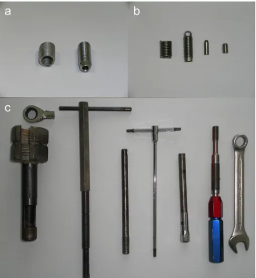
The implant was composed of two stainless steel helicoils (AISI 304), M8 × 1.25 × 16 mm, two modified stainless-steel screws (AISI 316), M 8 × 1.25 × 20 mm, an artificial ligament made in braided polyester fiber with a diameter of 3 mm. The special instrumentation was composed of a threaded mandrel with hook, a pre-winder, an extractor, a guided tapper, a socket wrench, a hex key, an open-end wrench, and drill bits of several sizes (6, 7 and 8 mm) (Fig. 1). The implant and the instrumentation were prototypes.
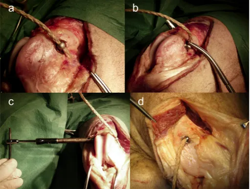
A cranio-lateral approach to the stifle joint was performed. The patella was medially luxated and the joint was observed to confirm the absence of visible pathologies; two Gelpi retractors allowed the exposition of the tibial insertion site of the CCL. The CCL was entirely removed with a scalpel blade and the joint was rinsed with saline solution to eliminate debris. A bone hole was made in the tibia, by placing the drill bit in the center of the insertion area of the CCL, parallel to the tibial crest. Starting with the 6 mm drill bit, the hole was progressively reamed to a diameter of 8 mm. The hole was tapped with a guided tapper; the helicoil was inserted with a modified pre-winder and screwed until the last spire reached a depth of 2–3 mm under the cartilage surface of the tibial condyle (Fig. 2). The artificial ligament was passed through the tibial screw, fixed at the distal end with a knot, and the complex was screwed inside the tibial helicoil (Fig. 3). A tunnel was made along the latero-medial direction in the distal end of the femur, few millimeters over the proximal end of the femoral trochlear ridges, perpendicularly to the longitudinal axis of the femur, starting with a 6 mm drill bit and progressively reaming to 8 mm. After tapping, the helicoil and the femoral screw were inserted; in this case, the last spire of the helicoil was seated at the same level of the femoral lateral cortex (Fig. 4).
The cancellous bone obtained from the drilled holes, in an eventual in-vivo execution, could be placed in a sponge moistened with saline solution and used as autograft for the medial portion of the femoral tunnel to fill the space not occupied by the screw at the end of surgery. The artificial ligament was passed through the joint along the cranio-caudal direction and through the intercondyloid area using a Klemmer forceps, as described in the technique “over-the-top” [26]. The fiber was then inserted in the eyelet of the femoral screw. The patella was reduced inside the trochlea and the limb was stretched out and aligned. The artificial ligament was tensioned to neutralize the cranial drawer motion and was maintained in place temporarily by the Klemmer forceps. The fiber was anchored in the femoral screw eyelet, by the screw-fiber fixing system, tightening the push screw inside the femoral bolt with the hex key, and entering from the medial hole of the femoral tunnel (Fig. 5). In the last passage, the hex key was used in synergy with the socket wrench and the open-end wrench as antirotational tool. The stifle joint was rinsed with saline solution, subcutaneous tissues and skin reapposed with an absorbable braided suture 2/0 (Polyglactin 910) and a non-absorbable suture 3/0 (Nylon), respectively. Cranial drawer sign, cranial tibial thrust, and the articular range of motion were evaluated. A medio-lateral and a caudo-cranial radiograph were performed to verify the position of the implant.
RESULTS
The cranio-lateral approach to the stifle joint is a well-known technique; the application of two Gelpi retractors allowed an excellent exposure of the tibial insertion of the CCL. Special surgical instruments, though still prototypes, were adequate to accomplish surgery in canine cadavers (Fig. 1). The tibial hole as a result was always well placed. The pilot nose of the tap aided in maintaining it well aligned during the tapping. The insertion of the helicoil was simple. In the last screwing phase, the pre-winder was extracted for a better evaluation of the helicoil depth. The correct helicoil depth in the tibia was reached using the sole threaded mandrel (Fig. 2). In 4 cases out of 24 (16.7%), at the tibial point during the insertion of the helicoil with the sole threaded
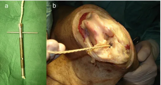
mandrel, there was a certain difficulty to link the hook of the mandrel with the opposite groove placed at the end of the helicoil spire. The problem was resolved extracting the helicoil of few millimeters with the extractor and rinsing the hole to eliminate debris. No difficulties were found during the insertion of the screw-ligament complex into the tibial helicoil (Fig. 3). At the femoral
Fig. 1. From left to right: (a) helicoil and tibial screw; (b) helicoil, femoral screw, piston, and push screw; (c) modified pre-winder with ratchet, threaded mandrel, guided tape, hex key, socket wrench, extractor, open end wrench.
Fig. 2. Tibial phase of the surgery: (a) execution of the tibial bone hole: the drill bit is placed in the center of the insertion area of the CCL, in parallel to the tibial crest; (b) after tapping the tibial hole with a guided tapper, the helicoil is inserted with a modified pre-winder and screwed until the last spire reached a depth of 2–3 mm under the cartilage surface of the tibial condyle (arrow).
L. OMINI ET AL.
tunnel, there was no problem faced during the insertion of the helicoil or the screw (Fig. 4).
In the first 5 cases (20.8%), there were some difficulties during the insertion of the socket wrench on the screw head. The difficulties were due to an excessive ventral inclination of the tunnel that prevented an easy access to the medial entrance of the femoral tunnel, and to the presence of a certain amount of debris around the screw head. In these cases, the problem was resolved by copious lavages with sodium chloride solution to eliminate debris and giving more attention during the alignment of the femoral
Fig. 3. Tibial phase of the surgery: (a) the artificial ligament is passed through the tibial screw, fixed at the distal end with a knot; (b) the complex is screwed inside the tibial helicoil.
Fig. 4. Femoral phase of the surgery: (a) execution of the femoral bone tunnel in lateromedial direction, few millimeters over the proximal end of the trochlear ridges and perpendicularly to the longitudinal axis of the femur; (b) tapping of the femoral hole with the guided tape; (c) insertion of the helicoil with the pre-winder mounted on the threaded mandrel; (d) the artificial ligament passed “over the top”.
tunnel. The screw-fiber fixing system allowed obtaining a firm fixation of the fiber into the eyelet of the femoral screw in all cases (Fig. 5). In all the cases, the post-operative evaluations showed negative cranial drawer test and negative cranial tibial thrust with a normal articular range of motion and correct alignment of the limb. All the post-operative radiographs showed an acceptable insertion of the implants (Fig. 6).
Fig. 5. Femoral phase of the surgery: (a) insertion of the artificial ligament in the eyelet of the femoral screw; (b) temporary fixation of the artificial ligament after tensioning; (c) definitive fixation of the artificial ligament in the eyelet by the internal screw-fiber fixing system, using the hex key and the socked wrench from the medial hole of the femoral tunnel; (d) surgical appearance before closing.
L. OMINI ET AL.
DISCUSSION
In this study, we chose an intra-articular technique because our aim was to restore the biomechanics of the knee similar to the normal CCL [16, 18]. The use of a prosthetic ligament would allow an earlier mechanical load as compared to the biological autograft or allograft. However, to obtain this type of stabilization it was necessary to solve some mechanical problems, well known in the human surgery, the most important being the method of fixation of the artificial ligament, particularly at the tibial site, that is considered the weakest link of the entire implant [8, 13–15, 22, 28, 29].
The helicoil is widely used in the mechanical industry. Its elasticity allows a good adaptation of the filet spires to the bone, outside, and to the screw, inside; it leads to a better force distribution on the entire length of the filet and not only on the first 2–3 spires, as confirmed in a previous work performed by the same research team [23]. In particular, Rossi and Omini analyzed the forces acting on the cranial cruciate ligament during the stance phase of the trot and the mechanical properties of canine tibial cancellous bone. Afterwards, they built a three-dimensional finite element model of the tibial implant, using software ABAQUS standard, evaluating the mechanical behavior with and without the helicoil. They finally tested the implant inserted into the tibial bone under a cyclic load, demonstrating its high mechanical strength [23].
In the project phase of the implant, one of the principal doubts was the mechanical strength of the bone (in particular, the femur) during drilling with drill bits of large diameter; during the study, fracture did not occur in either the femur or the tibia. The use of a starting drill bit of small diameter and the subsequent reaming to larger drill bits is a very effective technique that permits to limit the risk of iatrogenic fracture, to obtain an excellent alignment, and to limit errors during drilling. In an in vivo application, the bones are subjected to load; so, the hazard of fracture remains, but, we have to consider that the hole is filled by the body of the screw, limiting the dead space, and raising the points of least resistance. The option to fill with cancellous bone the portion of the femoral tunnel not occupied by the screw could contribute to a faster calcification and a higher resistance of the bone during the in
vivo application.
In this cadaveric study, due to just one implant size, dogs of medium and large size were solely included. It would not be difficult to produce implants in various sizes, potentially allowing the application in smaller size dogs.
The purpose of this study was to show a new surgical procedure to fix an artificial ligament to the bone. By the results of this cadaveric study, we can assert that the surgical procedure is sufficiently easy to be applied in clinical cases of rupture of the CCL in dogs. Moreover, no major complications occurred during the surgery. The authors can speculate that the fundamentals of this idea, the materials and the technique, could also be applied in other orthopedic pathologies in veterinary and human surgeries.
Although first preclinical studies were encouraging, further tests are necessary before applying in vivo this surgical procedure and clinically comparing its efficacy with the other techniques. Further studies need to be performed to define the exact
mechanical strength of the whole implant, the effects of the forces involved on the bone tissue surrounding the helicoil, and the osteointegration ability of the implant.
REFERENCES
1. Arnoczky, S. P. 1993. Pathomechanics of cruciate ligament and meniscal injuries. pp. 764–771. In: Disease Mechanisms in Small Animal Surgery, 2nd ed. (Bojrab, M. J. ed.), Lea & Febiger, Philadelphia.
2. Batty, L. M., Norsworthy, C. J., Lash, N. J., Wasiak, J., Richmond, A. K. and Feller, J. A. 2015. Synthetic devices for reconstructive surgery of the cruciate ligaments: a systematic review. Arthroscopy 31: 957–968. [Medline] [CrossRef]
3. Boudrieau, R. J. 2009. Tibial plateau leveling osteotomy or tibial tuberosity advancement? Vet. Surg. 38: 1–22. [Medline] [CrossRef]
4. Burgess, R., Elder, S., McLaughlin, R. and Constable, P. 2010. In vitro biomechanical evaluation and comparison of FiberWire, FiberTape, OrthoFiber, and nylon leader line for potential use during extraarticular stabilization of canine cruciate deficient stifles. Vet. Surg. 39: 208–215.
[Medline] [CrossRef]
5. Chen, J., Gu, A., Jiang, H., Zhang, W. and Yu, X. 2015. A comparison of acute and chronic anterior cruciate ligament reconstruction using LARS artificial ligaments: a randomized prospective study with a 5-year follow-up. Arch. Orthop. Trauma Surg. 135: 95–102. [Medline] [CrossRef]
6. Comerford, E. J., Smith, K. and Hayashi, K. 2011. Update on the aetiopathogenesis of canine cranial cruciate ligament disease. Vet. Comp. Orthop. Traumatol. 24: 91–98. [Medline] [CrossRef]
7. Conzemius, M. G., Evans, R. B., Besancon, M. F., Gordon, W. J., Horstman, C. L., Hoefle, W. D., Nieves, M. A. and Wagner, S. D. 2005. Effect of surgical technique on limb function after surgery for rupture of the cranial cruciate ligament in dogs. J. Am. Vet. Med. Assoc. 226: 232–236.
[Medline] [CrossRef]
8. Cook, J. L., Luther, J. K., Beetem, J., Karnes, J. and Cook, C. R. 2010. Clinical comparison of a novel extracapsular stabilization procedure and tibial plateau leveling osteotomy for treatment of cranial cruciate ligament deficiency in dogs. Vet. Surg. 39: 315–323. [Medline] [CrossRef]
9. Doom, M., de Bruin, T., de Rooster, H., van Bree, H. and Cox, E. 2008. Immunopathological mechanisms in dogs with rupture of the cranial cruciate ligament. Vet. Immunol. Immunopathol. 125: 143–161. [Medline] [CrossRef]
10. Freeman, J. W. and Kwansa, A. L. 2008. Recent advancements in ligament tissue engineering: the use of various techniques and materials for ACL repair. Recent Pat. Biomed. Eng. 1: 18–23. [CrossRef]
11. Harper, T. A. M., Martin, R. A., Ward, D. L. and Grant, J. W. 2004. An in vitro study to determine the effectiveness of a patellar ligament/fascia lata graft and new tibial suture anchor points for extracapsular stabilization of the cranial cruciate ligament-deficient stifle in the dog. Vet. Surg. 33: 531–541. [Medline] [CrossRef]
12. Iliadis, D. P., Bourlos, D. N., Mastrokalos, D. S., Chronopoulos, E. and Babis, G. C. 2016. LARS artificial ligament versus ABC purely polyester ligament for anterior cruciate ligament reconstruction. Orthop. J. Sports Med. 4: 2325967116653359. [Medline] [CrossRef]
13. Kowaleski, M. P., Boudrieau, R. J. and Pozzi, A. 2012. Stifle joint. pp. 906–998. In: Veterinary Surgery: Small Animal (Tobias, K. M. and Johnston, S. A. eds.), Elsevier Saunders, St. Louis.
14. Leduc, S., Yahia, L., Boudreault, F., Fernandes, J. C. and Duval, N. 1999. [Mechanical evaluation of a ligament fixation system for ACL reconstruction at the tibia in a canine cadaver model]. Ann. Chir. 53: 735–741 (in French). [Medline]
15. Matsumoto, A. and Howell, S. M. 2005. The WasherLoc and bone dowel: a rigid, slippage-resistant tibial fixation device for a soft tissue anterior cruciate ligament graft. Tech. Orthop. 20: 278–282. [CrossRef]
16. Mitton, G. R., Ireland, W. P. and Runyon, C. L. 1991. Evaluation of the instantaneous centers of rotation of the stifle before and after repair of torn cruciate ligament by use of the over-the-top technique in dogs. Am. J. Vet. Res. 52: 1731–1737. [Medline]
17. Montavon, P. M., Damur, D. M. and Tepic, S. 2004. Tibial tuberosity advancement (TTA) for the treatment of cranial cruciate disease in dogs: evidences, technique and initial clinical results. pp. 254–255. In: Proceedings of ESVOT Congress, 12th ed. (Vezzoni, A. and Shramme, M.), European Society of Veterinary Orthopaedics and Traumatology, Munich.
18. Montgomery, R. D., Milton, J. L., Terry, G. C., McLeod, W. D. and Madsen, N. 1988. Comparison of over-the-top and tunnel techniques for anterior cruciate ligament replacement. Clin. Orthop. Relat. Res. 144–153. [Medline]
19. Morris, E. and Lipowitz, A. J. 2001. Comparison of tibial plateau angles in dogs with and without cranial cruciate ligament injuries. J. Am. Vet. Med. Assoc. 218: 363–366. [Medline] [CrossRef]
20. Newman, S. D. S., Atkinson, H. D. E. and Willis-Owen, C. A. 2013. Anterior cruciate ligament reconstruction with the ligament augmentation and reconstruction system: a systematic review. Int. Orthop. 37: 321–326. [Medline] [CrossRef]
21. Robbe, R. and Johnson, D. L. 2002. Graft fixation alternatives in anterior cruciate ligament reconstruction. The University of Pennsylvania Orthopaedic Journal. 15: 21–27.
22. Rodeo, S. A., Arnoczky, S. P., Torzilli, P. A., Hidaka, C. and Warren, R. F. 1993. Tendon-healing in a bone tunnel. A biomechanical and histological study in the dog. J. Bone Joint Surg. Am. 75: 1795–1803. [Medline] [CrossRef]
23. Rossi, M. and Omini, L. 2008. Studio del comportamento a fatica di un nuovo sistema di fissaggio del legamento crociato. n. 451. In: Atti del Convegno Nazionale AIAS, XXXVII (edn.). (Associazione Italiana per le Analisi delle Sollecitazioni), AIAS, Roma.
24. Shaerf, D. A., Pastides, P. S., Sarraf, K. M. and Willis-Owen, C. A. 2014. Anterior cruciate ligament reconstruction best practice: A review of graft choice. World J. Orthop. 5: 23–29. [Medline] [CrossRef]
25. Slocum, B. and Slocum, T. D. 1993. Tibial plateau leveling osteotomy for repair of cranial cruciate ligament rupture in the canine. Vet. Clin. North Am. Small Anim. Pract. 23: 777–795. [Medline] [CrossRef]
26. Vasseur, P. B. 2003. Stifle joint. pp. 2090–2133. In: Textbook of Small Animal Surgery, 3rd ed. (Slatter, D. ed.), Saunders, Philadelphia. 27. Vezzoni, A., Baroni, E., Demaria, M., Olivieri, M. and Magni, G. 2003. Trattamento chirurgico della rottura del legamento crociato anteriore del
cane mediante osteotomia livellante del piatto tibiale (TPLO): presupposti teorici ed esperienza clinica in 293 casi. Veterinaria. 17: 19–31. 28. Weiler, A., Peine, R., Pashmineh-Azar, A., Abel, C., Südkamp, N. P. and Hoffmann, R. F. 2002. Tendon healing in a bone tunnel. Part I:
Biomechanical results after biodegradable interference fit fixation in a model of anterior cruciate ligament reconstruction in sheep. Arthroscopy 18: 113–123. [Medline] [CrossRef]
29. Weiss, J. A. and Paulos, L. E. 1999. Mechanical testing of ligament fixation devices. Tech. Orthop. 14: 14–21. [CrossRef]
30. Wilke, V. L., Conzemius, M. G., Besancon, M. F., Evans, R. B. and Ritter, M. 2002. Comparison of tibial plateau angle between clinically normal Greyhounds and Labrador Retrievers with and without rupture of the cranial cruciate ligament. J. Am. Vet. Med. Assoc. 221: 1426–1429. [Medline]


