UNIVERSITÀ DEGLI STUDI DI GENOVA
SCUOLA DI SCIENZE MEDICHE E FARMACEUTICHE
DIPARTIMENTO DI MEDICINA SPERIMENTALE (DIMES)
Corso di laurea in MEDICAL-PHARMACEUTICAL BIOTECHNOLOGY
Tesi sperimentale per la Prova Finale
EVALUATION OF T LYMPHOCYTES
INFILTRATION INTO THE BRAIN PARENCHYMA
OF PATIENTS AFFECTED FROM BIPOLAR
DISORDER TYPE I
Relatori
Prof. Gilberto Filaci
Dott.ssa Daniela Fenoglio
Candidato
Dott.ssa Chiara Rosa Maria Uras
Index
1. Abstract ... 1
2. Introduction ... 2
2.1 Bipolar Disorder... 2
2.1.1 Epidemiology ... 2
2.1.2 Clinical signs of the pathology ... 2
2.1.3 Pathogenesis ... 3
2.1.4 Current treatments ... 4
2.2 The relationship between BD and inflammation ... 5
2.3 Main features of CD45RA and CD161... 12
2.4 CD161 and tissue homing ... 17
2.5 Identification of T lymphocyte infiltrates in brain biopsies of people affected from BD I ... 19
3. Objective ... 22
4. Materials and methods ... 22
4.1 Selection of patients and control samples ... 22
4.2 Selection of brain areas ... 23
4.3 Histological analysis ... 23
4.4 Total RNA extraction and quantification... 24
4.5 mRNA retro-transcription ... 26
4.6 Real-Time PCR ... 26
4.7 Data collection and analysis ... 28
4.8 Statistical analysis ... 28
5. Results ... 30
5.1 Expression of CD45RA and CD161 in the inflamed brain areas of BD I patients ... 30
5.2 Quantification of T lymphocytes in the inflamed brain areas of BD I patients ... 32
6. Conclusions ... 35
6.1 Limitations ... 38
6.2 Future perspectives ... 38
1
1. Abstract
Bipolar Disorder (BD) is a serious brain disorder, included between the 50 most dangerous pathologies worldwide, whose etiology is still not clear. The type I (BD I) of this pathology is particularly important because it can be characterized by manic episodes that can lead to suicide attempts. Recent observations have revealed that there could be a correlation between the incidence of the disease and the generation of chronic inflammation in the brain. Particularly, different microstructural abnormalities have been detected in the brain white matter of bipolar patients, which could be related to the uncontrolled activity of microglia. Different studies have revealed a large increase in CD4+ and CD8+ T lymphocytes in the Central Nervous System, due to increased permeability of the Blood-Brain Barrier. This has been observed mainly for the subpopulations of effector memory, terminally differentiated effector memory (CD8+ CD28- CD45RA+) and CD8+ INFγ+ T cells, which have shown significantly different concentrations between the peripheral blood and the inflamed areas of the brain in patients with BD I.
The following study has been performed to evaluate the infiltration of T lymphocytes in the Cingulate Cortex (CC) of patients with BD I.
An histological analysis of brain specimens taken from the superior Anterior Cingulate Cortex (sACC) and ventral Anterior Cingulate Cortex (vACC) areas has been performed. Then, it has been measured the expression of two inflammatory markers, CD161 and CD45RA, typical of the terminally differentiated effector memory (TEMRA) and other infiltrating T lymphocytes. Finally, the expression of CD45RA and CD161 genes have been compared between patients and healthy controls using statistical analysis.
The results of this work show an important increase of TEMRA in the ACC of individuals with BD I with respect to healthy controls. This could corroborate the hypothesis that inflammation can contribute to the development of the disease. However, further investigations are needed to clarify the relation between T lymphocytes infiltration and brain structural modifications that characterize patients with BD I.
2
2. Introduction
2.1 Bipolar Disorder
2.1.1 EpidemiologyThe Bipolar Disorder (BD) is a mood disorder that has been estimated as the 46th greatest cause of disability and mortality among 291 diseases and causes of injuries worldwide1. The pathology has its onset usually in early adulthood and is characterized by intermittent episodes of mania, hypomania, and major depression2.
Bipolar Disorder can be classified in type I (BD I), that is characterized from mania, hypomania and major depressive events, and type II (BD II), that is marked by at least one hypomanic event and at least one major depressive event2. The prevalence of BD in adults worldwide is 1 to 3 percent3, but the ratio of affected females to affected males is 1:14. The age of onset is approximately 18 years old for BD I and 20 years old for BD II5.
2.1.2 Clinical signs of the pathology
The BD can manifest with manic, hypomanic or depressive events, including mixed features (i.e. symptoms of mood episodes of opposite polarity)2.
A series of symptoms, like irritability, anxiety, mood lability, agitation, aggressiveness, sleep disturbance, and hyperactivity could be prodromal for the pathology6, 7, 8.
Euphoric mania is a typical syndrome of BD I and can evolve in different forms thanks to substance abuse, head injury, and persistence of illness9. Patients can manifest high mood coupled with disinhibition, disobedience to the social rules, tendency to talk with strangers, and disregards of risks derived from some actions10. Moreover, they have an excessive sense of self-confidence and wellbeing and little sleepiness perception2.
The BD affects the cognitive functions, inducing in manic phase higher distractibility, increased mental activity and difficulty to distinguish more and less important thoughts2. In addition, some
3
patients can also become very aggressive and offensive to other people, because they don’t understand their needs10.
In the case of hypomania, the symptoms are less intense than the ones of manic events2. The affected individuals retain a certain capacity to control the feeling of self-confidence and can organize better their thoughts, making them even more productive. They have reduced aggressive behaviour and don’t develop psychotic symptoms11.
Patients with BD can also experience episodes of major depression (MD). They are characterized by a decreased capacity to experience pleasure, memory and concentration impairment, coupled with a strong reduction of whole body energy9. Reduced sleep ability is also typical of MD, with feelings of anxiety, agitation, worthlessness, and excessive guilt9, 12.
Some patients have also shown physical sufferance and decreased psychosocial functioning9. It becomes clear that the ill patients are affected from MD when they start to don't take care of themselves, manifest self-harm, don't interact with other people, and try to attempt suicide9.
Patients with BD usually manifest comorbid psychiatric disorders, like anxiety disorders, excessive substance use, Attention Deficit Hyperactivity Disorder (ADHD), eating disturbances, personality alterations, and borderline syndrome5, 13, 14, 15, 16.
Eventually, patients with BD have higher susceptibility to chronic pathologies, like diabetes, cardiovascular diseases, HIV infection, and Chronic Obstructive Pulmonary Disease (COPD)17.
2.1.3 Pathogenesis
It is not clear, until now, which is the major causative event of the BD.
The etiology could involve genetic, biological, psychological, and social factors18.
The genetic risk incidence for the first degree relative of a proband is 5 to 10 percent, while for a monozygotic co-twin the probability of developing BD is 40 to 70 percent19, 20.
4
The onset of the pathology and its progression are the results of the interaction between different genes, but until now candidate genes are still unknown21, 22.
Some susceptibility loci have been identified and among them CACNA1C is the most important. It codes for a subunit of a neuronal calcium channel that is involved in channel gating, important for the transmission of signals between cells23. Even other biological pathways are involved in the development of BD, like cardiac β-adrenergic signaling, cardiac hypertrophy signaling, corticotrophin-releasing hormone signaling, endothelin 1 signaling, glutamate signaling, and phospholipase C signaling22.
Instead, the epigenetic contribution, which underlies the pathology, consists of some processes of histone or DNA methylation and acetylation induced by different agents like toxins, chronic, and post-traumatic stress24. For example, some patients have shown methylation changes in the glutamic acid decarboxylase 67 gene of the hippocampus, which codes for an enzyme that is involved in GABA neurotransmission24.
Modifications of the brain structure could represent other etiologic causes of the pathology. Particularly, changes in white matter (WM) organization can lead to disruption and impairment of neural connections between prefrontal cortex and limbic structures25. On the other side, also the grey matter is important in this process of brain structure’s shape modification, since it can decrease with age and its reduction is particularly evident in patients with BD26.
Moreover, stress factors, like childhood maltreatments could be associated with the onset of BD and the disease worsening, but further studies should be made on this27.
2.1.4 Current treatments
Patients usually need a maintenance treatment after remission from an episode of BD. It is important to prevent other manic events and depressive events28. The best combination includes
5
pharmacological treatment and psychotherapy, to reduce possible relapses and increase the adherence, but especially to decrease the risk of suicide29.
The most used drugs are: first, mood stabilizers (e.g. lithium, valproate, carbamazepine), that suppress the shift between mania and depression29, 30, 31, 32, 33, 34, 35; second, antipsychotics (e.g. antidepressants), that manage the incapacity of the mind to distinguish true from false events36.
2.2 The relationship between BD and inflammation
A fundamental aspect of BD is the modification of the morphology of Central Nervous System (CNS)37. This can be the result of the development of an autoinflammatory reaction, which involves both innate and adaptive immune systems. Apart from inducing neurodegeneration, it can cause the onset of dyslipidemia, atherosclerosis, insulin resistance, and premature mortality38.
During inflammation, the tight junctions of the CNS become looser, and inflammatory cells have a higher possibility to penetrate the brain tissue39.
For what concerns naïve T lymphocytes, the extravasation mechanism includes an interaction with the endothelial cells of blood vessel walls. This mechanism is allowed by selectins that act in concert with chemokines, inducing the modification of lymphocyte membrane’s integrins structure, which allow them to link the adhesion molecules of the endothelial cell barrier and exceed them, reaching the perivascular space40. For more detail, it has been observed that under inflammatory conditions the Blood-Brain Barrier (BBB) can expose Ig-superfamily adhesion molecules and selectins, which mediate the interaction with CD4+ T lymphocytes and CD8+ T lymphocytes, respectively, allowing them to cross the barrier and reach the brain parenchyma41, 42. Instead, the mediators of inflammation can cross the areas not covered from the BBB, like the circumventricular organ. They can be transported from the afferent vagal fibers to some specific nuclei, like the nucleus of the solitary tract, or can pass through the BBB thanks to active transporters. Sometimes immune cells, like macrophages, monocytes and T lymphocytes, can pass the BBB (pericytes and/or endothelial cells), after secreting inflammatory mediators43. In the brain of patients affected from BD, a huge quantity
6
of cytokines has been indeed revealed43. T helper lymphocytes constitute a cell population which is found increased in the peripheral blood of BD patients, where they can release cytokines and exert a pro-inflammatory action on the CNS44. This cytokine shooting induces the over-activation of the cells of the microglia that contribute to the inflammatory process45. These cells have the role to protect the CNS during acute phases of infections and injuries. If their over-activation becomes chronic, it can cause serious damages to the neurons46. In particular, the process could cause the reduction of synaptogenesis, dendritic loss, oxidative stress, up to the neurons and oligodendrocytes apoptosis47.
Particularly, Th1 lymphocytes can release IL-1, IL-2, IL-6, TNF, and IFNγ, while Th2lymphocytes can secrete IL-4, IL-5, and IL-10 and Th3 cells can produce TGFβ44. During manic phase of Bipolar Disorder, the secretion of IL-4 can regulate the switch between cell and antibody-mediated immune response, for example driving the naïve T cells to the Th2 polarization48, 49. At the same time, the production of IL-6 can induce B and T lymphocytes secretion of acute inflammatory proteins, like high-sensitivity C reactive proteins (hs-CRPs)50.
The storm of cytokines productions and secretions can have different effects on brain integrity51. TNFα can interact with two neuronal receptors, p55 and p75, activating an apoptotic signaling cascade. In combination with IL-1 it can enhance the expression of serotonin and dopamine transporters, disrupting monoamine signaling52. Moreover, they can also stimulate the production in the presynaptic terminals that present dopamine receptors of Reactive Oxygen Species (ROS) which can lead to further induction of the monoamine signaling52.
The brain of BD patients presents serious modifications with respect to the healthy individuals53, particularly in the astroglia, oligodendrocytes, and microglia (Figure 1). This occurs mainly in the subgenual anterior cingulate gyrus (sACC), in the dorsolateral prefrontal cortex (DPFC), in the orbitofrontal cortex (OFC) and the amygdala54, 55. Suicidal bipolar patients are characterized by more extended microglia in the DPFC, anterior cingulate gyrus and mediodorsal thalamus respect to the healthy controls and non-suicidal mood-disorder patients, probably because there is a higher
7
correlation between inflammation and BD for them56. In any case, a great number of patients with BD of both categories have a reduced cellular density in the oligodendroglia57.
Figure 1. Relationship between the disruption of BBB and BD: the immune cells, like monocytes, T lymphocytes and neutrophils, can interact through specific adhesion molecules with the endothelial cells surface and enter the CNS. Once they are inside, they can stimulate, through the release of cytokines like TNF-α and IFN-γ, the oxidative stress, which is characterized from an increased production of Reactive Oxygen Species and the development of an inflammatory process characterized from microglial activation, myelin damage, neurons and oligodendrocytes disfunction. Abbreviations: BBB =
Blood-Brain Barrier, ROS = Reactive Oxygen Species. Adapted from Patel et al., 201558.
Noteworthy, in manic phase of BD there is a significant increase of inflammatory cytokines and cells in the CNS, relating to an important loss of structural integrity in the WM. This can compromise the structural connectivity of the midline regions of the brain59, 60.
The impaired structural connectivity involves the activation of the immune-inflammatory cell pathway, inducing the secretion of different molecules. Particularly, there is an increase in the number
8
of soluble receptors which are specific for IL-6 (s IL-6R) and IL-2 (s IL-2R). Furthermore, large quantities of IgG1 immunoglobulins and complement factors C3 and C6 are responsible for the inflammation61, 62, 63. In the circumstance of the acute manic phase of BD there is an over-secretion by macrophages of IFNγ, which is involved in the activation of indoleamine-2,3-dioxygenase (IDO)64. IDO is a heme-containing enzyme produced by immune-modulating cells, that by means of the catabolism of L-tryptophan induces angiogenesis and maturation of myeloid-derived suppressor cells (MDSC). MDSCs can regulate the activity of T lymphocytes, dendritic cells, macrophages, and NK cells, inducing the activation of inflammatory cells that can cause the impairment in structural connectivity of the brain65.
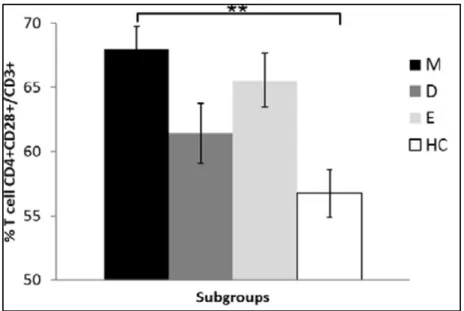
Outside the brain, in peripheral blood of BD I patients with mania there is an important increase of the CD4+ T lymphocytes, particularly the CD4+ CD28+ cell subset (Figure 2). It includes both naïve (CD45+ CCR7+) and central memory (CD45- CCR7+) CD4+ T cells. On the contrary, there is a reduction in the quantity of CD8+ T lymphocytes, regarding mainly the effector memory (CD8+ CD28- CD45RA-) and terminally differentiated effector memory (CD8+ CD28- CD45RA+) T cells37. The effector memory T cells (TEM) are inflammatory cells that exert a cytotoxic action on respect to target cells, which can be direct or mediated from the interaction between TCR and the antigen, causing the release of perforin, granzymes and granulysin or Fas-Fas ligand interaction. The terminally differentiated effector memory T cells (TEMRA) have the same function of the TEM cells but are localized mainly in peripheral tissues66.
Apart from TEM and TEMRA cells, during mania it has been detected in the peripheral blood a lower amount of CD8+ IFNγ+ T lymphocytes with respect to healthy controls, and an increase of early activated (CD4+ CD28+) T cells37, as it is shown in Figure 2 and Figure 3. The CD8+ IFNγ+ T cells are cytotoxic T lymphocytes that include some TEMRA cells and can stimulate the elimination of the target cells through the secretion of IFNγ. In fact, they can stimulate the expression of the MHC-I complex on the surface of the target cells and can cause the polarization of the CD4+ T lymphocytes
9
to the Th1 type. Indeed, they induce the migration of the T cells to the sites of inflammation and the activation of their cytotoxic activity66.
10
Figure 2. Quantification of CD4+ CD28+/CD3+ T cells in the peripheral blood of BD patients during the maniac phase (black), depressive phase (dark grey), euthymic phase (light grey), and in healthy controls (white). Abbreviations: M = mania, D = Depression, E = Euthymia, HC = healthy controls. Adapted
from Magioncalda et al., 201837.
Figure 3. Quantification of (A) CD8+ IFNγ+ T cells, (B) TEM and (C) TEMRA cells in the peripheral blood
of BD patients during manic, depressive, and euthymic phases and in healthy controls. Abbreviations:
M=mania, D=depression, E=euthymia, HC=healthy controls. Adapted from Magioncalda et al., 201837.
A B
11
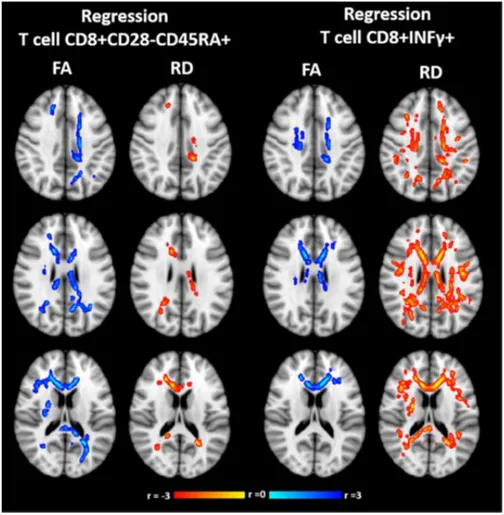
Contemporary with the reduction of TEM, TEMRA and CD8+ IFNγ+ T lymphocytes in the peripheral blood of patients with BD I, there can be the development of some alterations in the WM microstructure. They characterize mainly the oligodendroglia and myelin fractions of Body of the Corpus Callosum (BCC) and Superior Corona Radiata of the left hemisphere (SCR-L), as it is possible to observe by Diffusion Tensor Imaging analysis, or DTI (Figure 4). Between the different phases of BD I, only in mania a frequent coexistence of these two events is detectable37.
The reduction of the TEMRA lymphocytes in the peripheral blood could be related to their infiltration in the brain of patients with BD I. In fact, when they shift from CD45RO to CD45RA expression they become low proliferative and able to secrete high quantities of cytokines. The higher exposure of adhesion molecules and chemokine receptors on their surface, might be the reason for them to leave the bloodstream and localize in the peripheral tissues, like the brain. This event could be responsible for the auto-inflammatory processes that can induce the alteration of WM microstructure in manic patients37.
12
Figure 4. Correlation between WM alterations and T cell regression in all phases of BD, measured with DTI. The direct correlations are shown in blue – light blue, while the inverse correlations are shown in yellow-red. Abbreviations: FA = Fractional Anisotropy, RD = Radial Density. Adapted from
Magioncalda et al., 201837.
2.3 Main features of CD45RA and CD161
CD45 is a 180-220 kDa integral membrane protein of type I, coded from the Protein Tyrosine Phosphatase gene (PTP), that belongs to the group of tyrosine phosphatases67. It is also called Leukocyte Common Antigen (LCA), because it is found in the surface of various leukocytes and it also characterizes the other hematopoietic cells68, 69. In particular, it represents 5-10% of all glycoproteins of B and T cells surface70, 71.
The protein CD45 is made of four parts: an extracellular segment, a transmembrane portion, and two intracellular catalytic domains (Figure 5). The CD45 can be found in different isoforms: CD45RA, CD45RB, CD45RC, CD45RAB, CD45RAC, CD45RO, CD45R(ABC). All the isoforms of CD45 derive from the alternative splicing of a gene with 34 exons, of whom 3 exons (4, 5 and 6) are
13
combined to generate eight different transcripts and proteins72, 73, 74, 75. The exons codify for a sequence of 200 amino acids that is localized near the N-terminus of CD45 and can be modified through O-linked glycosylation. The extracellular domain contains a cysteine-rich region and three fibronectin type III repeats, that are highly N-glycosylated. This modification stabilizes the protein structure and can allow its transport to the cell surface 76, 77. The intracytoplasmatic tail has two tyrosine phosphatase domains, D1 and D2. Only D1 has tyrosine phosphatase activity, while D2 can anchor to the cytoskeleton through the linker protein fodrin, which is spectrin-like, and modulates D1 activity and specificity78. CD45RO is an important marker of memory and effector T cells, while CD45RA characterizes naïve T cells. The expression of CD45RA decreases during T cell aging while the one of CD45RO increases74, 79, 80, 81. Some highly differentiated CD4+ or CD8+ T cells CD45RO+ that accumulate during adulthood in patients with chronic inflammatory diseases can revert to CD45RA expression82.
Figure 5. CD45RO and CD45RABC (CD45RA isoform) structure is characterized by phosphate domains, O-linked glycosylation and N-linked glycosylation that influence their stability, activation and
14
The way through which CD45 interacts with its ligand is determined by the level of glycosylation of its extracellular domain78. The enzymes that are responsible for glycosylation are expressed at different times of T cell differentiation84, 85. They are represented mainly from Core-2 -O-N-acetylgalactosamine transferase (C2GnT), alpha (2,6)-sialyltransferase I (ST6Gal-I), alpha (2,3)-sialyltransferase IV (ST3Gal-IV), and alpha (1,3)-fucosyltransferase VII (FucT-VII). The most important roles of CD45 protein are stimulation of cell growth, differentiation, mitotic cycles and oncogenic transformation. It is recruited in the central supramolecular activation cluster (cSMAC) from the immunological synapse (IS) generating by T cells for the interaction between the antigen of Antigen Presenting Cells (APCs) and the T-cell receptor (TCR). It is then expelled and can localize in the distal supramolecular activation cluster (dSMAC)86, 87, 88, 89, 90. The “kinetic segregation model” establishes that the TCR signaling requires the separation of MHC-bound TCR from tyrosine phosphatases91. CD45 can cause dephosphorylation of the Lymphocyte Specific Protein Tyrosine Kinases (LCKs) of the Src family, at their C-terminal negative regulatory tyrosine Y50592. It can also stimulate the dephosphorylation of the auto/transphosphorylation site Y394 within the kinase domain93, 94. The first process can activate LCKs, while the second one can reduce their functionality93, 95, 96. Both mechanisms are shown in Figure 6. CD45 can also inhibit the activity of Janus Kinases (JAKs), that interacting with Signal Transducer and Activator of Transcription (STAT) transcription factors can promote cytokines and chemokines gene expression97, 98, 99.
15
Figure 6. CD45RA is a receptor that can regulate the activity of LCKs through the phosphorylation of
Y192 and Y505 genes. Adapted from Rheinländer et al., 201882.
CD161 or NKR-PB1 (Killer Cell Lectin-like receptor subfamily B) is an integral membrane protein of type II, coded from the gene KLRB1, that belongs to the superfamily of C-type lectin receptors (CTLRs)100. Particularly, it belongs to the subgroup of Killer cell Lectin-like Receptors (KCLRs). The gene that codifies for CD161 is localized in chromosome 12 and is part of a NK gene complex (NKC), which includes also CD69 and CD94101. The complex is characterized by the alternation between the sequences of NKR-PB1 and CTLRs, codifying for proteins that can interact with CD161 for their structural homology102. CD161 structure is composed by 3 regions, which are a
158-aminoacid extracellular domain with motifs typical of C-type lectins, with several C residues and N-linked glycosylation sites, a 29-aminoacid transmembrane domain and a 38-aminoacid
16
it interacts with the right antigens it can stimulate an increase in the intracellular calcium and the secretion of IL-1β and IL-12103. Moreover, NK cells can expose on their surface CD161 and its interaction with the antigens can induce an upregulation of the T cell receptors (TCRs) expression. Other groups of immune agents, like CD1d - restricted NKT cells, are characterized by the presence of CD161100, 104. For what regards NK cells, the expression of CD161 on their surface can promote the recognition in mice of a specific molecule, which is a C-type lectin related molecule (CLR), in the target cells. The main ligand of CD161 in humans is Lectin-like Transcript 1 (LLT1), which is exposed by monocytes and B cells in the peripheral blood, PMA-stimulated PBMCs and IL-2 activated NK cells or T cells105, 106. The receptor CD161 has charged residues in the transmembrane region that can allow the interaction with LLT1. When CD161 binds to LLT1 there is a reduction in the quantity of receptor in the cell surface, because some of its copies become internalized. The expression of lysosomal-associated membrane protein - 1 (CD107a), which is a marker of degranulation in the NK cells, is slightly reduced after the interaction between CD161 and LLT1107. Indeed, a result of this process is the loss of cytolytic activity of the NK cells. This happens mainly in those cytotoxic T lymphocytes that secrete IL-12, which can up-regulate CD161 exposure103. The interaction between CD161 and LLT1 has also shown to reduce the secretion of IFNγ from the NK cells108.
Between the features of the CD161 receptor there is the ability to induce high expression of α4β1 and αLβ2 integrins and LFA-1 Mg2+-binding site (observed in CD4+ T lymphocytes), promoting the trans-endothelial migration without the need of chemotactic stimuli103. The mechanism could start from the interaction of the CD161 molecule with specific ligands on the blood vessel walls. This recognition process can also be responsible for T lymphocytes acquisition of a specific tissue homing as it has been observed in the intestine, liver109, 110. The T cells can also infiltrate in the CNS, accumulating there and leading to an inflammatory process. This is valid mainly for CD8+ T lymphocytes111.
17
Furthermore, CD161 is also expressed in CD4+ T cells precursors of the Th17 lineage and it differentiates the cell lineage from Th1 and Th2 lineages112.
It can be detected also in plastic Th17 lymphocytes that secrete IFN-γ, typical of autoinflammatory diseases like Multiple Sclerosis (MS)113 and Chron’s Disease114, 115. These cells are non-classical Th1, ex-Th17 lymphocytes that can produce IL-17 and IFNγ, but also new transcription factors and chemokine receptors (i.e. CXCR3), with respect to the classical Th17 cells116, 117. The expression of
IL-17 is made possible from the interaction between CD161 and CD3 in the cell surface. The ex-Th17 cells have important pathogenic features like longevity, multiple cytokines production, and
resistance to Treg - mediated immunosuppression. The cells have an increased capacity to produce GM-CSF, TNF, IL-2, and IFNγ on respect to Th1 and Th17 cells118. Besides, the action of Treg cells on ex-Th17 cells doesn’t reduce the secretion of TNF. At the end, ex-Th17 cells, like the peripheral blood Th17 cells, are phenotypically similar to effector memory T cells (TEM). They are more differentiated concerning the Th1 cells, retained the high proliferative capacity of the Th17
precursors. Particularly, they are CCR7- like the TEM cells, but they produce higher quantities of Bcl-2, which is an antiapoptotic factor118.
2.4 CD161 and tissue homing
CD161 has been proposed to play an important role in trans-endothelial migration119. Supporting this hypothesis, it has been demonstrated that the CD161+ CD4+ T cells can migrate better through the
endothelium than the CD161- CD4+ T cells120. In fact, CD161 can induce in these cells the up-regulation of α4β1, αLβ2 integrins and LFA Mg2+- binding site, that allow the extravasation. In
detail, α4β1 integrin can interact with Vascular Cell Adhesion Molecule-1 (VCAM-1) and induce CD4+ and CD8+ T cells infiltration in the brain of patients with MS, a CNS autoinflammatory disease111. α
Lβ2 integrin is important for the interaction with the Intercellular Cell Adhesion Molecule – 1 (ICAM-1), which is exposed on the surface of endothelial cells, allowing a strict
18
from the chemokine gradient and mechanical stimuli. Indeed, it can change its conformation thanks to the intracellular interaction with the Talin/Kindlin system and can acquire high affinity for ICAM-1122, 123, 124, 125. In fact, Talin is a protein with an amino terminal 50-kDa head that in correspondence of the Fo domain, preceding the FERM domain (4.1, ezrin, radixin, and moesin), can activate theα4β1 integrin126. Kindlin is an adaptor protein, member of the integrin adhesion complex, that can activate integrins interacting directly with their cytoplasmic tails. In this way, they can promote integrin-mediated cell–extracellular matrix adhesion and signaling127. Subsequently to the αLβ2/ICAM-1 interaction, LFA Mg2+-binding site is activated and involved in cycles of adhesion and deadhesion125, 128. The α4β1 integrin has shown to be important for the migration of CD4+ Th1 cells and CD8+ T cells in the CNS129. In fact, it is activated from the chemokine gradient of the target tissue and can interact with the VCAM-1 molecule that is exposed in the surface of endothelial cells belonging to the BBB130. Expression of VCAM‐1 is upregulated in inflammatory conditions on endothelial cells of the BBB and of the blood‐leptomeningeal barrier, as well as on choroid plexus epithelium of the blood‐cerebrospinal fluid barrier, further increasing α4β1/VCAM‐1‐mediated T‐cell adhesion to inflamed CNS endothelium56, 131. Instead, in the case of CD8+ T lymphocytes it has been observed that the interaction of αLβ2 integrin with the ICAM-1 doesn’t influence cell migration132, 133. On the contrary, P-selectin glycoprotein ligand 1 can bind to the P-selectin, exposed on the endothelial cells, and contribute to the recruitment of the CD8+ T lymphocytes in the blood vessels134. It has been observed the importance of CD161 for the extravasation in experiments using neutralizing antibodies directed specifically against the marker (Figure 7)129. In fact, CD161+ CD4+ T cells can allow the T lymphocytes homing into specific tissues, like the brain119. Lymphocyte homing to peripheral tissues in vivo is dictated by the interplay of integrin and selectins, like the α4β1 integrin and the P-selectin 129. Once the T lymphocytes overcome the BBB, they can migrate in the CNS parenchyma due to the high concentration of different chemokines. In detail, Tumor Necrosis Factor (TNF) and CXC Chemokine Receptor 3 (CXCR3) can attract CD4+ and CD8+ T lymphocytes respectively, as it has been observed in patients with MS135.
19
Figure 7. Infiltration of T lymphocytes in the brain parenchyma (A) in absence or (B) in presence of
anti-α4β1 antibodies. Adapted from Martin-Blondel et al., 2015129.
2.5 Identification of T lymphocyte infiltrates in brain biopsies of people affected from
BD I
The increase in T lymphocyte tissue infiltrate is a phenomenon that has been detected in different chronic inflammatory diseases. As it is present physiologically in different peripheral tissues109, 110, it can be found also in the brain111. In the brain T cells can preserve the integrity of the CNS during acute phases of infections and injuries. If the activity of cells like T lymphocytes, macrophages, and epithelial cells, becomes permanent, it can cause reduction of synaptogenesis, dendritic loss, and oxidative stress, until neuronal apoptosis65. Recently it has been demonstrated that different neurological disorders can be characterized by structural modifications of the brain areas. Subgenual Anterior Cingulate Cortex (sACC) and ventral Anterior Cingulate Cortex (vACC) have found to be altered in the brain of patients affected from BD I. Due to the structural modifications, vACC has reduced connectivity with the Medial Prefrontal Cortex (mPFC) in bipolar patients with psychosis and increased connectivity with mPFC in bipolar patients without psychosis136. On the other side, the left sACC has increased connectivity with Posterior Cingulate Cortex (PCC) and right sACC137.
Previous studies37 have also demonstrated that there is a significant expansion of CD4+ CD28+ T cell subsets (that include both naïve and central memory CD4+ T cells) in peripheral blood of the
20
manic phase of BD patients than healthy controls. Moreover, the frequency of total CD8+ T cells has shown a significant decrease in the manic phase for healthy controls. Interestingly, this reduction has
shown to be dependent on the contraction of CD8+ CD28- T cell subsets, including both TEM (CD8+ CD28- CD45RA-) T cells and TEMRA (CD8+ CD28-CD45RA+) lymphocytes, and associated
with the significant reduction of CD8+ IFNγ+ T cells. Hence in the manic phase of BD, there is an increase of early activated (CD4+ CD28+) T cells and a decrease of TEM (CD8+ CD28-CD45RA-) and TEMRA (CD8+ CD28-CD45RA+) cells, paralleled by the reduction of CD8+ IFNγ+ T cells (corresponding to functionally activated CD8+ T cells). Interestingly, the loss of the CD28 co-receptor, which is observed in a heterogeneous CD8+ T cell population, occurs after prolonged stimulation37. The expression of the CD45RA molecule in these cells characterizes the shift from T
EM
to TEMRA cells. This represents a progression to a low proliferative/high release of cytokines profile, that is associated with the increased expression of adhesion molecules and chemokine receptors, ultimately dictating the localization of the cells in the peripheral tissues138. CD161 is another molecule linked to the TEMRA cells migration process. It is an integrin that recognizes surface molecules of the endothelial cells and can mediate an extravasation process that is independent of chemotactic stimuli. Blood vessel leakage can allow migrating cells to reach specific tissues, basing on the selective recognition of surface molecules on the endothelial cells109, 110. Hence, TEMRA cells are effector cells prone to migration in the peripheral tissues139. Together, published data from these studies have suggested the occurrence of an acute immune response in manic patients. The study from Magioncalda et al.37 has shown that in mania CD8+CD28-CD45RA+ T cells (which are cells prone to migration in the peripheral tissues) and CD8+ IFNγ+ T cells (which are the activated CD8+ T cells and include activated CD8+ CD28-CD45RA+ T lymphocytes) could leave the bloodstream to migrate into the brain, inducing an immune-related WM damage (Figure 3). Indeed, a hypothetical mechanism of immunological damage in mania has been proposed (Figure 8).
21
Figure 8. The scheme shows that most remarkable DTI and immunological alterations in mania consist in (I) relatively specific WM alterations in the BCC and SCR-L (upper panel); (II) reduction in the frequency of circulating CD8+ CD28-CD45RA+ and CD8+ IFNγ+ T cell subpopulations (left panel); (III) significant partial correlation between the cited WM and immunological alterations (middle panel). The hypothesis is that CD28-CD45RA+ T cells and CD8+ IFNγ+ T cells leave the bloodstream to migrate
into the brain where they induce an immune-related WM damage (right panel). Abbreviations: WM= White Matter, FA =Fractional Anisotropy, M = mania, HC = healthy controls, BCC = Body of
22
3. Objective
The objective of this study was to analyze T cell infiltrates in brain biopsies of suicidal BD patients, in comparison to brain samples from healthy controls dead from non-psychiatric causes, to verify whether the disappearance of TEMRA cells (CD8+CD28-CD45RA+) from the circulation were associated with their accumulation in the brain at sites pathogenically related to BD development.
4. Materials and methods
4.1 Selection of patients and control samples
Brain samples have been obtained from the Department of Forensic and Legal Medicine (University of Genoa) that provided 10 human specimens from patients with BD and 10 control samples from non-psychiatric individuals, stored at -80 °C in 6-well plates. The specimens have been isolated from patients and healthy controls at 30 minutes post-mortem. The study has been approved by the Ethical Committee of San Martino Hospital, in Genoa. The subjects have been diagnosed by expert psychiatrists (Department of Neuroscience, Rehabilitation, Ophthalmology, Genetics, and Maternal and Child Health, Section of Psychiatry, University of Genoa) via retrospective review of all available medical records and anamnestic information.
The inclusion criteria for the patients selection were: - Diagnosis of BD I according to DSM 5;
- Age between 18 and 60 years old;
- Suicidal people have been chosen since their behavior is characteristic of the active and acute phase of the illness.
The exclusion criteria were:
- Diagnosis of Schizophrenia, Mental Retardation, Dementia, and other cognitive disorders; - History of severe somatic and neurological diseases (e.g. traumatic brain injury, stroke, cerebral
vascular malformations, epilepsy);
23 - History of alcohol and substance addiction.
A retrospective analysis of all medical records and anamnestic information has been done for control subjects to exclude the possibility of BD, but they have been selected with the same exclusion criteria.
4.2 Selection of brain areas
Medical examiners from the Department of Forensic and Legal Medicine (University of Genoa) have dissected the brain of patients and healthy controls, isolating portions of the midbrain, brainstem, and cerebellum from the left and right hemispheres. To identify the areas of inflammation, half of the brain has been fixed, by immersion, in 10% buffered formaldehyde, dehydrated and cleared. Then it has been sent for histological and immunohistochemical analysis to the Pathology Unit, Department of Surgical Science, and Integrated Diagnostics (University of Genoa, IRCCS AOU San Martino Hospital). The other half of the brain has been stored at - 80 °C, to be used for transcriptome analysis aimed to verify the presence of infiltrating T lymphocytes.
4.3 Histological analysis
The Cingulate Cortex (CC) has been isolated, following a previous study that has shown that it can be inflamed in some areas during manic phases of BD37, 136. Sections of vACC, vACC/pACC (pregenual Anterior Cingulate Cortex), sACC, PCC (Posterior Cingulate Cortex), MCC (Middle Cingulate Cortex) and BCC areas have been cut through the microtome, referring to the positional coordinates described in a previous study (Table 1)60 and stained with hematoxylin and eosin. This has allowed the characterization of the microglia and of other areas with lower myelin expression. Through a more specific staining, it has been possible to evaluate the presence of perivascular and parenchymal T lymphocytes in the isolated regions of CC and to count them. Particularly, immunohistochemical analysis has been performed to evaluate the presence of T lymphocytes in the analyzed areas, using the CD3-directed 2GV6 Rabbit Monoclonal Primary Antibody (Roche).
24
Table 1. Coordinates of brain regions for the resting-state networks, including vACC, sACC,PCC, MCC and BCC . Abbreviations: vACC=ventral Anterior Cingulate Cortex, sACC= subgenual Anterior Cingulate Cortex, PCC= Posterior Cingulate Cortex, MCC= Medial Cingulate Cortex and BCC=Body
of the Corpus Callosum. Adapted from Martino et al., 201660.
The T lymphocytes detected with the antibodies have been counted with the optic microscope, using a resolution of 400x, in the hot spot area, which is the part in which more immune-labeled cells have been observed. Cells have been subdivided into four ranges of count number (lower than 5, 5-20, 20-50, higher than 50) and the presence of the T lymphocytes in perivascular and parenchymal regions has been evaluated. For each biopsy, only the areas with more significant results of the analysis have been selected for further molecular analyses.
4.4 Total RNA extraction and quantification
Brain samples isolated from the vACC/pACC and sACC regions have been cut using a little bistoury and have been put in a 2.5 ml Eppendorf containing 2 ml of NucleoZOL (Macherey-Nagel) according to the Manufacturer’s instructions. The reagent, made of guanidinium thiocyanate and phenol, can lyse cells, causing the release of the intracellular content, including DNA and RNA. In detail, guanidinium thiocyanate can block DNases and RNases and denature proteins, while phenol is a denaturing agent immiscible with water. A smashing procedure has been performed with a manual homogenizer. The homogenate has been stored at -80 °C. When all samples have been prepared, an aliquot of 200 µl of the homogenized sample has been mixed with 500 µl of NucleoZOL, to allow a
25
better release of the total DNA and RNA. The mixture has been diluted with 280 µl of RNAse - free water. Everything has been shaken and left for 10 minutes at room temperature. It has been performed centrifugation at 12000 x g for 15 minutes. All the lipids, protein components, cell debris and a high percentage of DNA molecules have been precipitated in alcohol, in the bottom of the tube, while RNA, due to its polarity and lower density, has remained in the water phase of the supernatant. 500 µl of supernatant has been transferred in a new Eppendorf and mixed with 500 µl of isopropanol, to precipitate the RNA. Isopropanol is an alcoholic compound with amine groups (NH4+) that has lower dielectric constant than water and can make the interaction between RNA phosphates and cationic functional groups of the salts stronger. This reduces the solubility of ribonucleic acids in water. The mixture has been taken at room temperature for 10 minutes and centrifuged at 12000 x g for 10 minutes. After the removal of the supernatant, the pellet containing RNA has been washed 2 times with 500 µl of 75% Ethanol and resuspended in RNAse - free water.
The quantification of RNA, isolated with the NucleoZOL protocol (Macherey-Nagel), has been performed with Nano-Drop® ND-1000. The analysis has revealed for all the samples a concentration between 100 ng/μl and 350 ng/μl, which is optimal for the retro-transcription and production of cDNA molecules for Real-Time PCR. It has also been evaluated the 260/280 ratio, which is based on the measurement of absorbance of the UV light at a wavelength of 260 nm and 280 nm. The first value refers to nucleic acids since the UV light is absorbed mainly by the aromatic rings. The second one is referred to proteins and phenolic compounds. The ratio is calculated from the instrument to eventually reveal the presence of contamination from phenols or proteins that have been generated during the RNA purification. The values of the ratio that have been detected are all around 1.8, indicating that the DNA purification has been correctly performed. Since NucleoZOL used in our procedure to extract RNA can absorb at 230 nm, we also measured the 260/230 ratio which is calculated starting from the absorbance of UV light at 230 nm and 260 nm of wavelength, in order to check possible NucleoZOL contamination. Only phenols, like NucleoZOL, can absorb at 230 nm and so the ratio is related to their concentration in the sample. The values that have been detected are in
26
most cases between 1,5 and 2,0. This means that phenols have not been removed completely from the samples, but their concentration has not been so high to affect the retrotranscription that has been performed next in the study.
4.5 mRNA retro-transcription
For the priming, 100 ng of RNA molecules have been annealed with random oligo (dT) primers of 20 nucleotides that bind randomly stretches of poly-adenine, or poly(A), that is localized in the 3’ region of mRNA molecules. To allow the pairing between the primers and the RNA molecules, the sample has been kept for 5 minutes at 65 °C and then for 5 minutes in ice to block the reaction.
The reverse transcription has been performed with the QuantiTect Reverse Transcription kit (Qiagen) using 4 µl of Reaction Buffer, 0.5 µl of RNAse inhibitor (RI), 2 µl of dNTPs and 1 µl of Reverse Transcriptase.
4.6 Real-Time PCR
Real Time-PCR analysis has been developed with the Quantifast® SybrGreen PCR kit (Bio-Rad SsoAdvanced Universal SYBR Green Supermix). The reaction has been run with PCR cycles reported in Table 2.
Table 2. Phases of Real Time – PCR cycles for the amplification of CD45RA and CD161 genes
Table 2. Phases of Real-Time PCR cycles
Denaturation 95 °C for 30’’
Annealing 95 °C for 10’’ 50 cycles
27
A 96 well plate has been prepared to put in each well 1 μl of cDNA (containing 25 ng of DNA), and 9 μl of the Sybrgreen mix (composed of 5 µl of Sybrgreen, 1 µl of Primer Forward, 1 µl of Primer Reverse and 2 µl of RNAse-free water). In the analysis, the expression of CD45RA and CD161 genes has been accomplished in triplicate. Since RPLP13 and UBE2D2 are stably expressed in both brain hemispheres during different neurodegenerative diseases140 they have been used as Reference Genes.
The sequences of the primers used in the analysis are reported in Table 3. The expression of CD45RA and CD161 has been evaluated to hypothesize the presence of the infiltrate of T lymphocytes in the brain samples, taken from the vACC/pACC and sACC areas of the brain.
Table 3. Primers used for the Real Time – PCR reaction, specific for target genes (CD45RA and CD161) and reference genes (UBE2D2 and RPLP13)
The run has been acquired on LightCycler®480 Real-Time PCR System (Roche F. Hoffmann-La Roche AG Konzern-Hauptsitz Grenzacherstrasse 124 CH-4070 Basel Schweiz) with Sequence Detection Software according to the Manufacturer’s instruction.
Table 3. Primers for Real-Time PCR
Gene name Primer sequence forward Primer sequence reverse References
CD45RA 5’-ATGGTCCTCTGAATAAAGCCCA-3’ 5’-TCAGCACTATTGGTAGGCTCC-3’
Wu et al.
(2014)141
CD161 5’-AAATGCAGTGTGGACATTCAA-3’ 5’-CTCGGAGTTGCTGCCAATA-3’
Llibre et al.
(2016)142
UBE2D2 5’-TGCCTGAGATTGCTCGGATCT-3’ 5’-TCGCATACTTCTGAGTCCATTCC-3’
Rydbirk et al.
(2016)140
RPLP13 5’-CCTGGAGGAGAAGAGGAAAGAGA-3’ 5’-TTGAGGACCTCTGTGTATTTGTCA-3’
Rydbirk et al.
28
4.7 Data collection and analysis
The Sybr Green I dye is a fluorescent DNA binding dye, that can intercalate in the minor groove of the amplified DNA. The excitation of the dye generates a fluorescent signal whose intensity is directly proportional to the quantity of double-stranded DNA and so to the concentration of DNA in the original samples. The Real-Time PCR amplification has given an amplification plot which has been analyzed with the LightCycler® 480 Real-Time PCR System software.
The software has given the Ct values, which is the PCR cycles number required for the fluorescent signal to cross a fixed threshold in the amplification plot. All measurements have been performed in triplicate for each cDNA to collect only the geometric mean of Ct values for the triplicates with a variance of Ct lower than 20%. The expression of CD45RA and CD161 has been normalized with the geometric mean of Ct of the housekeeping genes (ΔCt) and transformed in fold change respect to the calibrator sample using the ΔΔCt method143.
The melting curves have been analyzed for each sample. Their plot reveals the variation in fluorescence that occurs when the double-stranded DNA dissociates in single strands with previously incorporated molecules of Sybr Green. When the melting point is reached, the fluorescence signal decreases because the dye is released. The melting curves with a single pick correspond to a single amplification product. The values of Ct for the data analysis have been selected also basing on that.
4.8 Statistical analysis
Statistical analysis has been performed with Graph Pad Prism software to compare the expression of CD45RA and CD161 between patients and healthy controls. The distribution of 2-ΔΔCt values has been represented in a histogram with the mean and standard deviation, taking also in consideration the outliers. To evaluate the significance of the difference between the means and variances of the two groups, specific statistical tests have been performed.
29
Firstly, it has been evaluated the normality of the distributions of patients and healthy controls, developing the D’Agostino-Pearson test. It stands on the skewness and kurtosis tests, that are performed to assess the symmetry or asymmetry and the shape of the distributions144.
The D’Agostino-Pearson test has revealed that the distributions of the patients and healthy controls are not normal.
Subsequently, a non-parametric test has been performed, because it allows to compare groups that are not distributed in a normal way, in opposition to the parametric tests.In addition, it can be useful for small samples because it organizes the values in ranks, while the parametric tests consider the single values. This reduces the possibility of statistical errors. The results of the analysis can confirm the null hypothesis (H0), which establishes that the two sampled populations are equal or the alternative hypothesis (H1), which states that they are unequal. However, the non-parametric tests can only be precise about the H0 hypothesis but are not enough potent to confirm the H1 hypothesis.
In detail, it has been performed a Mann-Whitney U test on the distributions of values for patients and healthy controls. It is used to test whether two samples are likely to derive from the same population. It has been evaluated through this analysis the difference between the means and the variances of the two groups. Indeed, the test has given an estimation of the similarity between the compared groups of samples in terms of dimensions and distribution of probability.
30
5. Results
5.1 Expression of CD45RA and CD161 in the inflamed brain areas of BD I patients
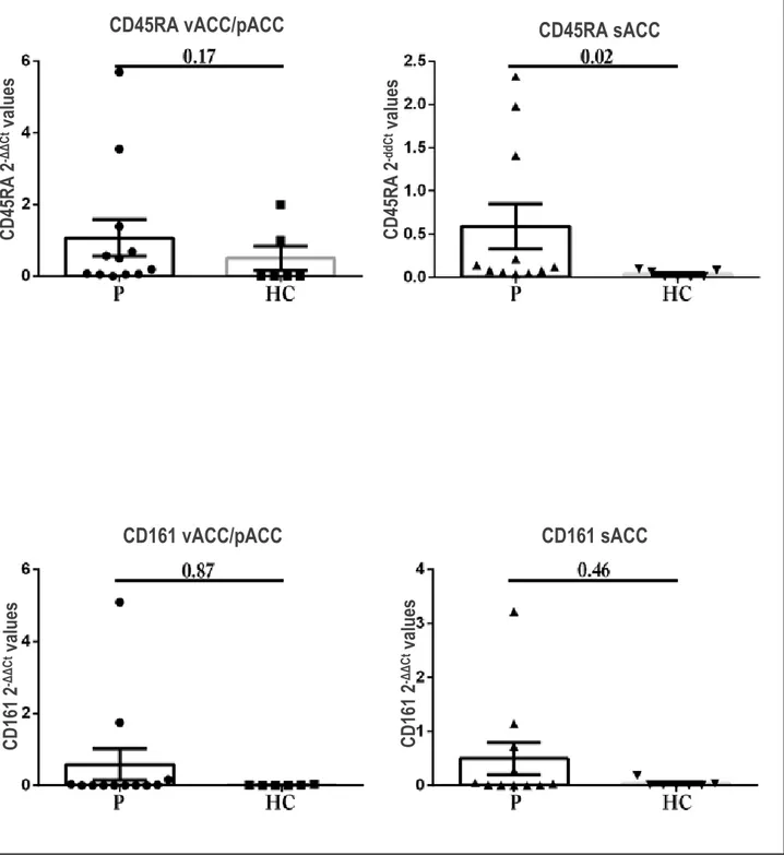
To establish the presence of lymphocyte infiltration of T cells in the brain areas of vACC/pACC and sACC, the expression of lymphocyte markers in brain biopsies has been evaluated, referring to the samples with 2-ΔΔCt higher than 2, that indicates a significant gene expression. The analyses focused on CD45RA and CD161 antigens. This choice was taken since CD45RA is the marker that allows to identify tissue resident TEMRA lymphocytes, while CD161 is important for the migration of those cells through the BBB. A tendency of increased expression for both CD45RA and CD161 antigens in patients with respect to healthy controls has been observed in vACC/pACC and sACC regions. Performing a Mann-Whitney U test for non-parametric measures, the difference between the values of the two groups reached statistical significance only in the case of the CD45RA antigen expressed in sACC (Figure 9).
31
Figure 9. Histogram representation of the values of 2-ddCt in patients and healthy controls relative to
CD45RA and CD161 in the vACC/pACC and sACC areas. Abbreviations: P=patients, HC = healthy controls, vACC=ventral Anterior Cingulate Cortex, pACC=pregenual Anterior Cingulate Cortex, sACC=subgenual Anterior Cingulate Cortex
Although statistical significance hasn’t been achieved in the Mann-Whitney U test, the increased trend in CD161 expression observed in patients could be related to the extravasation of the TEMRA
CD45RA vACC/pACC CD45RA sACC
CD161 vACC/pACC CD161 sACC CD 45 RA 2 -ΔΔ Ct v alu es CD 45 RA 2 -d dC t value s CD 16 1 2 -ΔΔ Ct v alue s C D 16 1 2 -ΔΔ Ct v alue s
32
lymphocytes in the brain parenchyma, which is hypothesized as a mechanism that could trigger the inflammatory process responsible of BD I.
In conclusion, Real-Time PCR analysis has suggested a prevalent expression of CD45RA and CD161 in the inflamed vACC/pACC and sACC areas of both brain hemispheres for the patients on respect to healthy controls. This could corroborate the relation between the infiltration of TEMRA lymphocytes in the CC and the development of BD I.
5.2 Quantification of T lymphocytes in the inflamed brain areas of BD I patients
Histological analysis of brain biopsies taken from patients with BD and healthy controls has been performed. Samples have been taken from the vACC/pACC and sACC areas. In detail, for some individuals, brain specimens have been isolated from both regions, for others only by one of them.
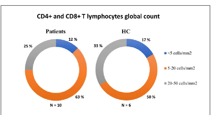
Firstly, the count of total T lymphocytes has been performed in the hotspot areas and it has revealed in most cases a cell number ranged between 5 and 20 per mm2 in patients and controls, for both vACC/pACC and sACC areas (Figure 10). The results of T lymphocytes count in the brain sections of patients and healthy controls haven’t shown any statistically significant difference.
Figure 10. Quantification of CD4+ and CD8+ T lymphocytes in BD I patients and healthy controls. Values of cell count are divided in three ranges: <5 cells/mm2; 5-20 cells/mm2; 20-50 cells/mm2. Abbreviations: HC = healthy controls.
33
In relationship with a potential pathogenic role of the T cell infiltrate, the site of infiltration (if parenchymal or perivascular) has relevance. Importantly, in BD I patients the infiltrating immune cells were detected in both the parenchymal district and the perivascular zone, while in healthy controls they were present only at the perivascular site.
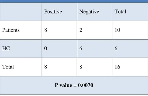
In order to evaluate the significance of the exclusive intraparenchymal detection of T lymphocytes in the patients, Fisher’s exact test has been performed (Table 4). This test offers a valid statistical evaluation for the dichotomous values, that in this case correspond to the presence or absence of the T cells in the CC parenchyma of patients and healthy controls. The calculated P-value is equal to 0.007, which indicates a strong correlation between the presence of the T lymphocytes infiltrate and the development of BD I. This result suggests that the exclusive presence of CD4+ and CD8+ T cells in the CC parenchyma of patients is significant.
Table 4. T lymphocytes detection in the parenchymal district
Positive Negative Total
Patients 8 2 10
HC 0 6 6
Total 8 8 16
P value = 0.0070
Table 4. Fisher’s exact test for the evaluation of T lymphocytes in the brain parenchyma of patients and healthy controls. Abbreviations: HC= healthy controls.
The detection in the vACC/pACC and sACC areas of a remarkable T cell infiltrate, particularly of the CD8+ subtype, at the parenchymal site could support the hypothesis that an immune-mediated
34
mechanism, and the subsequent WM microglia structural alterations, are pathogenically related to onset/development of BD.
35
6. Conclusions
BD is a globally wide-spread pathology that has an important impact on the social and sanitary welfare. In fact, the type I of this pathology can seriously affect the quality of life of patients inducing frequent mood alterations, but also leading to events of aggressiveness, self - harm and in some cases to suicide attempt. For this reason, it is important to identify the pathology and proceed with the treatment in the early phases of BD I. However, the variability of factors that can induce the onset of BD I makes difficult to find an adequate diagnosis and treatment for all patients. There are some valid drugs that can function as mood stabilizers, but they mainly act on the symptoms of the pathology, independently from the cause of it. In fact, their action is not definitive, but requires repetitive administration of the treatment.
Indeed, an interesting field of research is focusing on the formulation of drugs that could act on the determining factors of BD I. Between the determinants of the disease, inflammation has been proposed as responsible of different alterations in the brain structure37, 60.
Supporting this hypothesis, in the peripheral blood of patients the T helper lymphocytes have shown to secrete high quantities of cytokines, that can cross the BBB in different ways. Among them, the TNF can promote neuronal apoptosis and synthesis of ROS in the pre-synaptic terminals, inducing important changes in the brain structure52. Other cytokines produced from T helper lymphocytes can stimulate the secretion of inflammatory mediators, like acute phase proteins, from B and T lymphocytes 50. Also macrophages have shown to be increased in the peripheral blood of patients with BD I, where they produce high quantities of IFNγ that can allow the activation of inflammatory cells from MDSCs and, consequently, the development of structural abnormalities in the brain65.
In line with this, it has been observed that in the case of MS, an autoimmune pathology of the CNS, the T cells can infiltrate the brain tissue thanks to the presence of TNF and CXCR3, that attract CD4+ and CD8+ T lymphocytes respectively135. Similar mechanisms of brain homing could be induced in the BD I.
36
In suicidal patients with BD I it has been observed the presence of different structural abnormalities in the areas of sACC, DPFC, OFC and amygdala, and it has been hypothesized a relation between this aspect and the increased proliferation of microglial cells, which have a pro-inflammatory role53, 54, 55.
Among all the cells that could exert an effector function in the peripheral tissues, the TEM, TEMRA and CD8+ IFNγ+ T lymphocytes have been found reduced in the peripheral blood, in association to the presence of WM lesions, suggesting their trans-endothelial migration and brain homing. All these cell types can have a cytotoxic action during the development of an inflammatory process37, 66.
Particularly, the TEMRA cells secrete IFNγ and are characterized from the surface exposure of CD45RA and CD161145. It is known that IFNγ can induce the activation of macrophages and the Th1 polarization of CD4+ T cells, while CD45RA and CD161 are implicated respectively in the
expression of adhesion molecules and chemokine receptors for tissue homing and the trans-endothelial migration of TEMRA lymphocytes. TEM cells exert their effector function as TEMRA
cells but are mainly concentrated in the peripheral blood66, 109, 110,138.
Taken together, these observations have driven the study toward the research of the presence of CD8+ T lymphocytes, particularly TEMRA lymphocytes, in the CC of patients affected from BD I.
The immunohistochemical analysis of brain specimens obtained from the patients has revealed that there is a migration of CD8+ T lymphocytes in the parenchymal district in patients with BD I, differently from healthy controls. This population could include TEM and TEMRA cells, potentially responsible of the inflammatory state of brain WM.
In our study, evaluation of CD45RA expression in the vACC/pACC and sACC areas has suggested higher presence of T lymphocyte infiltrate in the brain of patients with BD I on respect to healthy controls. For what regards CD161, higher levels of expression have been found on respect to the healthy controls, although the difference didn’t reach statistical significance. These findings support
37
the hypothesis of migration of the TEMRA lymphocytes from the peripheral blood to the brain parenchyma of patients. Since TEMRA cells are activated in inflammatory conditions145, specifically express CD45RA when they exert their effector function and can migrate through the vessel walls thanks to CD161109, 110, 138, the results of the study suggest the involvement of this populations of CD8+ T cells in the inflammatory alterations causing damages, like the loss of connectivity for the neurons, in the CC in the case of BD I.
This is in line with what has been observed in the auto-immune encephalomyelitis, an autoinflammatory manifestation that can occur during MS. Particularly, CD4+ and CD8+ T lymphocytes are recruited in the CNS thanks to the exposure of CD161, that can induce the activation of α4β1 integrin, which interacts with VCAM-1, and αLβ2 integrin, that has structural affinity for ICAM-1111,121. Both VCAM-1 and ICAM-1 are exposed from the endothelial cells, due to the inflammatory state. They are localized in the WM microvasculature of the CNS, allowing the migration of the CD4+ and CD8+ T lymphocytes in the brain parenchyma39.
Also, this study corroborates what has been observed in previous studies, in which the CD45RA has been found expressed from CD8+ memory T lymphocytes (TEMRA) that are highly present during chronic inflammatory diseases146. Particularly, in the early stages of MS, it has been observed an important increase of the TEMRA lymphocytes percentage in Cerebro-Spinal Fluid, derived from blood filtration, from where the cells can migrate in the brain parenchyma during the late phases of disease147.
Basing on what has been observed for MS and in this study, it could be hypothesized that the TEMRA lymphocytes in patients with BD I might develop a specific cytotoxic action against neurons and oligodendrocytes after the stimulation of a self-antigen147, 148. What is the nature of the antigen evoking such immune reaction is unclear. Studies on MS have shown that mature APCs exposing engulfed myelin antigens (like myelin basic protein) or neuronal antigens (as neurofilament light protein), localized in the cervical lymph nodes, can cause the activation of CNS-reactive CD4+ and
38
CD8+ T lymphocytes149. They constitute a cell population that is generated, despite the thymic negative selection, also in healthy individuals150. However, in MS these cells, after antigenic stimulation, can cross the BBB and infiltrate the brain parenchyma, where they are re-activated from the resident APCs, like macrophages, microglial and dendritic cells or B lymphocytes151, 152, 153.
In the case of BD I, the presence of TEMRA lymphocytes in the parenchyma of brain regions involved in the disease suggests that processes resembling those occurring in MS may be promoted. This hypothesis implicates that an immune response is activated in the brain targeting either tissue autoantigens or extrinsic antigens conveyed there by a pathogenic agent (i.e., a virus).
6.1 Limitations
The selection of a low number of individuals from an estimated homogeneous population can have generated a higher biological variability in the sample than expected. Indeed, further evaluations should be done in wider groups of patients and controls to obtain more reliable results. However, this kind of studies has a remarkable technical complexity due to the difficulty in collecting the series of patients (who are suicide patients), related to the forensic issues.
Nevertheless, the study has given significant results, that suggest an inflammatory origin of the BD I.
6.2 Future perspectives
A deeper understanding of the involvement of TEM, TEMRA and CD8+ IFNγ+ T lymphocytes in the structural alterations and loss of connectivity of brain WM in the patients affected from BD I could allow the development of specific therapies aimed at the control of the activity of these cells.
If the infiltration of TEMRA and TEM cells could be limited before the evolution of the pathology, this could represent a pioneer strategy to prevent, at least in part, the neurological dysfunctionalities that could alter brain activity in BD I patients.
39
The formulation of a personalized medicine directed against BD I or even other neurological disorders could improve the quality of life of patients, giving them the possibility to have better social relationships and respect themselves, and decrease the economic and social burden of the diseases.
40
7. Bibliography
1. Murray CJL, Vos T, Lozano R, Naghavi M, Flaxman AD, Michaud C et al., Disability-adjusted life years (DALYs) for 291 diseases and injuries in 21 regions, 1990–2010: a systematic analysis for the Global Burden of Disease Study 2010, The Lancet, 2012; 380, 9859: 2197-2223
2. American Psychiatric Association, Diagnostic and Statistical Manual of Mental Disorders, Fifth Edition (DSM-5), 2013
3. Pedersen CB, Mors O, Bertelsen A, Waltoft BL, Agerbo E, McGrath JJ, Mortensen PB, Eaton WW, A Comprehensive Nationwide Study of the Incidence Rate and Lifetime Risk for Treated Mental Disorders, JAMA Psychiatry, 2014; 71, 5:573-81; doi: 10.1001/jamapsychiatry.2014.16
4. Seedat S, Scott KM, Angermeyer MC, Berglund P, Bromet EJ, Brugha TS, Demyttenaere K, de Girolamo G, Haro JM, Jin R, Karam EG, et al., Cross-national associations between gender and mental disorders in the WHO World Mental Health Surveys, Archives of General Psychiatry, 2010; 66 (7): 785–795; doi: 10.1001/archgenpsychiatry.2009.36
5. Merikangas KR, Jin R, He JP, Prevalence and Correlates of Bipolar Spectrum Disorder in the World Mental Health Survey Initiative, Archives of General Psychiatry, 2011; 68(3):241-251; doi:10.1001/archgenpsychiatry.2011.12
6. Skjelstad DV, Malt UF, Holte A, Symptoms and Signs of the Initial Prodrome of Bipolar Disorder: A Systematic Review, Journal of Affective Disorders, 2010; 126(1-2):1-13; doi: 10.1016/j.jad.2009.10.003
7. Howes OD, Lim S, Theologos G, Yung AR, Goodwin GM, McGuire P, A Comprehensive Review and Model of Putative Prodromal Features of Bipolar Affective Disorder, Psychological Medicine, 2011; 41(8):1567-77; doi: 10.1017/S0033291710001790