1
UNIVERSITA’ POLITECNICA DELLE MARCHE
DOTTORATO DI RICERCA IN
“SCIENZE BIOLOGICHE E CLINICHE SPECIALISTICHE”
Coordinatore: Chiar.mo Prof. Antonio Benedetti
XIV ciclo Trienno Accademico 2012-2015
Effect of Strawberry Antioxidants against
Oxidative and Inflammatory Stress
PhD student:
Relatore
Dott. Massimiliano Gasparrini Prof. Maurizio Battino
Correlatore
2
A chi mi ha sempre sostenuto,
supportato e …sopportato. Grazie!
3
1. ABSTRACT ... 6
2. ITALIAN SUMMARY ... 7
3. INTRODUCTION ... 8
3.1. General overview ... 8
3.2. Oxidative stress in health and disease ... 8
3.3. Oxidative stress and inflammation ... 11
3.4. ROS and RNS ... 14
3.5. Antioxidant defences ... 20
3.6. Lipopolysaccharide ... 26
3.6.1. Structure ... 27
3.6.2. LPS-mediated signaling and the innate immune response ... 28
3.7. Strawberry ... 31
3.7.1. Nutritional aspects ... 32
3.7.1.1. Micronutrients in strawberry... 33
3.7.1.1.1. Folates ... 33
3.7.1.1.2. Vitamin C ... 35
3.7.1.1.3. Other vitamins ... 37
3.7.1.1.4. Essential minerals ... 38
3.7.1.2. Phytochemicals in Strawberry ... 38
3.7.1.2.1. Classification of phenolics ... 38
3.7.2. Strawberry bioactive compounds and human health ... 43
3.7.3. Strawberry and inflammation... 45
3.7.4. The influence of genotype on the nutritional quality of strawberry . 48
4. EXPERIMENTAL DESIGN ... 49
4.1. Part I: Evaluation of the nutritional quality of strawberry fruits ... 49
4.1.1. Objective ... 49
4.1.2. Materials & methods ... 50
4.1.2.1. Strawberry material ... 50
4.1.2.2. Extraction method ... 50
4.1.2.3. Measurement of total antioxidant capacity ... 52
4.1.2.3.1. FRAP ... 53
4.1.2.3.2. TEAC ... 53
4.1.2.3.3. DPPH ... 54
4.1.2.4. Measurement of Total Phenolic Content ... 55
4.1.2.5. Measurement of Total Anthocyanin Content... 56
4.1.2.6. Identification and quantification of anthocyanins in “Alba”
cultivar by HPLC-DAD-MS analysis ... 57
4
4.1.2.8. Measurement of Total Flavonoid Content ... 58
4.1.2.9. Folates identification and quantification ... 59
4.1.2.10. Statistical analysis ... 59
4.1.3. Results ... 59
4.1.3.1. Total antioxidant capacity ... 60
4.1.3.2. Total Phenolic Content ... 61
4.1.3.3. Total Anthocyanin Content ... 61
4.1.3.4. Vitamin C Content ... 61
4.1.3.5. Total Flavonoid Content ... 61
4.1.3.6. HPLC-MS Analysis of anthocyanins in “Alba” cultivar ... 62
4.1.3.7. Folates identification and quantification ... 64
4.1.3.8. Interrelationship among bioactive compounds ... 66
4.1.4. Discussion ... 67
4.2. Part II: : The role of strawberry in the modulation of inflammatory
response induced by LPS ... 71
4.2.1. Objective ... 71
4.2.2. Material and methods ... 71
4.2.2.1. Cell culture ... 71
4.2.2.2. Strawberry for cell treatment ... 72
4.2.2.3. MTT viability assay ... 72
4.2.2.4. TALI
®ROS concentration assay ... 74
4.2.2.5. TALI
®apoptosis assay ... 76
4.2.2.6. Mitochondrial functionality with Seahorse XF24
Analyzer
®: respiratory capacity assay ... 78
4.2.2.7. Determination of nitrite production ... 80
4.2.2.8. Cell RIPA preparation ... 81
4.2.2.9. Cell pellets for Western Blot analysis ... 82
4.2.2.10. Enzymatic activity assays ... 84
4.2.2.10.1. Glutathione Peroxidase (GPx) ... 84
4.2.2.10.2. Glutathione Reductase (GR) ... 84
4.2.2.10.3. Glutathione Transferase (GST) ... 85
4.2.2.10.4. Superoxide Dismutase (SOD) ... 85
4.2.2.10.5. Catalase (CAT) ... 86
4.2.2.11. Lipid and protein oxidation level ... 86
4.2.2.11.1. Lipid peroxidation (TBARS) level ... 86
4.2.2.11.2. Reduced glutathione (GSH) determination ... 87
4.2.2.11.3. Determination of protein carbonyl content ... 87
4.2.2.12. Gene expression analysis with Western Blot ... 88
4.2.2.13. Statistical analysis ... 89
4.2.3. HDF results ... 90
4.2.3.1. MTT viability assay ... 90
4.2.3.2. TALI
®ROS concentration assay ... 92
5
4.2.3.4. XF24 Analyzer
®respiratory capacity assay ... 96
4.2.3.5.Determination of nitrite production ... 98
4.2.3.6. Enzymatic activity assays ... 99
4.2.3.7. Lipid and protein oxidation level ... 102
4.2.3.8. Gene expression analysis ... 103
4.2.4. RAW macrophages results ... 108
4.2.4.1. MTT viability assay ... 108
4.2.4.2. TALI
®ROS concentration assay ... 111
4.2.4.3. XF24 Analyzer
®respiratory capacity assay ... 113
4.2.4.4. Determination of nitrite production ... 115
4.2.4.5. Enzymatic activity assays ... 116
4.2.4.6. Lipid and protein oxidation level ... 118
4.2.4.7. Gene expression analysis ... 120
4.2.5. Discussion ... 125
5. CONCLUSIONS ... 134
6. ACKNOWLEDGEMENTS ... 136
6
1. ABSTRACT
The first aim of this PhD project was to assess and compare the nutritional and phytochemical quality of strawberry fruit extracts of different cultivars and/or varieties, obtained through specific breeding programs: the main purpose of such approach was to evaluate the influence of genetic background on these parameters. The total antioxidant capacity, the radical scavenging activity, the content of total phenolics, flavonoids, anthocyanins, vitamin C and folates were measured in the different strawberry extracts. Among the varieties studied, the cultivar Alba was chosen for its nutritional value and was used in the second part of the Thesis.
The second aim of this project was to evaluate the effects of methanolic purified extracts from Alba cultivar on inflammatory status induced by E.Coli lipopolysaccharide (LPS) on two different cell lines, Human Dermal Fibroblasts (HDF) and RAW 264.7 macrophages. The cell viability, apoptosis rate and Reactive Oxygen Species (ROS) intracellular production were assessed. The protective role of strawberry extracts was estimated by the evaluation of the principal biomarkers related to inflammatory and oxidative stress and the activity of the principal antioxidant enzymes. Moreover, protein expression was evaluated to analyze and clarify the principal molecular pathways involved in strawberry and LPS mechanisms of action. Finally, the oxygen consumption rate related to mitochondria functionality was evaluated.
The results obtained demonstrated that strawberry extracts had an anti-inflammatory effect on LPS-treated cells, through a reduction of ROS and inflammatory and oxidative damages. Strawberry extracts also counteracted the inflammatory response increasing the antioxidant activities, through AMPK-related pathways. An improvement of mitochondria functionality was also demonstrated. The results obtained with this work highlight and confirm the potential health benefit of strawberry against inflammatory and oxidative stress.
7
2. ITALIAN SUMMARY
Il primo obiettivo dello studio è stato quello di valutare e confrontare la qualità fitochimica e nutrizionale di diversi estratti di fragola, prodotti da varietà commerciali ottenue da specifici programmi di incrocio genetico. Nei vari estratti sono state misurate la capacità antiossidante totale, l'attività antiradicalica e il contenuto di polifenoli, flavonoidi, antociani, vitamina C e folati. Sulla base dei risutlati ottenuti la cultivar Alba è stata scelta per condurre le analisi nella seconda parte del progetto.
Il secondo obiettivo è stato quello di valutare gli effetti di estratti metanolici della cultivar Alba sull’infiammazione indotta dal LPS di E.Coli in cellule di fibroblasti di derma umano e macrofagi RAW 264.7. Dopo analisi preliminari di vitalità, apoptosi e produzione di ROS intracellulari, il ruolo protettivo degli estratti è stato stimato attraverso la valutazione dei principali biomarcatori collegati allo stress infiammatorio e ossidativo e in relazione all'attività dei principali enzimi antiossidanti. Analisi di espressione proteica sono state effettuate per identificare le principali vie molecolari coinvolte nell’azione degli estratti di fragola e del LPS E’ stato inoltre misurato il consumo di ossigeno correlato alla funzionalità mitocondriale.
I risultati hanno evidenziato come gli estratti di fragola esercitino un effetto anti-infiammatorio sulle cellule trattate con LPS, riducendo la produzione di ROS e abbassando i livelli dei biomarcatori infiammatori e ossidativi. La risposta infiammatoria è stata altresì contrastata rafforzando l’attività antiossidante e regolando le vie molecolari collegate all’AMPK, registrando inoltre un miglioramento della funzionalità mitocondriale.
I risultati ottenuti sottolineano e confermano il potenziale beneficio per la salute di un consumo di fragola, in particolare nei confronti delle alterazioni indotte da stress infiammatori.
8
3. INTRODUCTION
3.1. General overview
A growing number of epidemiological studies suggest a strong association between a diet rich in fruits and vegetables and a lower incidence of different chronic pathologies, such as obesity (Kovacs et al., 2014; Charlton et al., 2014), infections (Siegel et al., 2010; Crowe et al., 2014), inflammation (Anderson et al., 2012; Urpi-Sarda et al., 2012; Lamprecht et al., 2013; Macready et al., 2014), cardiovascular (Threapleton et al., 2013; Tanaka et al., 2013; Wallace et al., 2013; Eilat-Adar et al., 2013; Wang et al., 2014) and neurodegenerative diseases (Elwood et al., 2013; Marder et al., 2013) and cancer (Grosso et al., 2013; Park et al., 2013a; Leenders et al., 2013; Kruk, 2014; Deschasaux et al., 2014). Taking into consideration the remarkable socio-economic and public health impact of these chronic pathologies, the comprehension of the molecular and biochemical mechanisms that underlie the beneficial effects of a plant-based diet has encouraged different basic, clinical and epidemiological research (Tulipani et al., 2009a). Focusing on fruits, it is quite complex to explain their potential health benefits, given the wide variety of fruits available for consumption and their complex composition. For these reasons, in recent decades, individual subgroups of fruits have been taken into account, to facilitate the observation and promote their specific health benefits. Nowadays, the synergistic and cumulative role in health promotion of the fibers, micronutrients and phytochemicals compounds present in fruit is an established evidence (Giampieri et al., 2012a). A deep sifting of the plant compounds potentially expressing biological activities has been performed, with particular attention to the phytochemical compounds, which are not designated as traditional nutrients. Despite this, the complete identification of the protective substances present in plants and the mechanism by which they can protect against disease still remain a great challenge, that requires a planned and strict collaboration among experts in plant food, nutrition and medical research. In particular, a full understanding of the etiologic pathways related to the different chronic disease could help in the characterization of the health-promoting compounds present in dietary plants.
3.2. Oxidative stress in health and disease
A common denominator in the pathogenesis of most chronic diseases is the involvement of oxidative stress, connected to the production by all aerobic organisms of reactive oxygen species (ROS) and reactive nitrogen species (RNS) including free radicals. These substances are implicated in human physiological and phatological conditions, ranging from rheumatoid arthritis and haemorrhagic shock through cardiomyopathy and cystic fibrosis to gastrointestinal ischaemia, cancer and also aging. Oxidative stress was originally defined by Sies (Sies, 1985) as an extreme
9 situation in terms of the organism oxidation, which occurs with a change in the balance between the generation of oxidants and control by antioxidants. In fact it could result from (i) a reduction in antioxidant defense; (ii) an increase in the production of ROS and RNS or even (iii) a combination of both events. The excessive production of reactive species, no longer adequately controlled by antioxidant defense systems, can lead to a wide range of effects: adaptation by up-regulating the natural defence system, which may completely or in part protect beside damage, tissue injury to different molecular targets and cell death by activating apoptosis and necrosis processes.
At the cellular level the main targets of the attack of ROS are lipids, proteins and nucleic DNA. All cellular membranes are especially vulnerable to oxidation due to their high concentrations of polyunsaturated fatty acids (PUFAs). The damage to lipids, usually called lipid peroxidation, occurs in 3 stages. During the first stage, called initiation, it occurs the attack of a reactive oxygen metabolite, capable of abstracting a hydrogen atom from a methylene group in the lipid. The presence of a double bond near to the methylene group weakens the bond between the hydrogen and carbon atoms, so that it can be easily removed from the molecule. Following hydrogen abstraction, when oxygen is in sufficient concentration in the surroundings, the remaining fatty acid radical reacts with it to form ROO•: this is the second step, called the propagation stage. These radicals themselves are capable of abstracting another hydrogen atom from a close fatty acid molecule, which leads again to the production of fatty acid radicals that undergo the same reactions and interaction with oxygen. The ROO• becomes a lipid hydroperoxide that can further decompose into several reactive species including lipid alkoxyl radicals (LO•), aldehydes (e.g., malonyldialdehyde, 4-hydroxy-2-nonenals), alkanes, lipid epoxides, and alcohols. The propagation stage allows the reaction to continue. The last stage, or chain termination, arises following interaction of one ROO• with another radical or antioxidants (Davies, 2000; Kohen and Nyska, 2002; Zhong and Yin, 2015). Proteins, in particular their SH-groups, can serve as possible targets for attack by ROS, especially OH•, RO•, and nitrogen-reactive radicals. Proteins can undergo direct and indirect damage following interaction with ROS, as well as peroxidation, damage to specific aminoacid residues, changes in their tertiary structure, degradation, and fragmentation. These consequences are considered as a response mechanism to stress and can lead to interference with the creation of membrane potentials, loss of enzymatic activity, altered cellular functions such as energy production and changes in the type and level of cellular proteins. The principal products obtained by protein oxidation are usually aldehydes, keto compounds, and carbonyls (Kohen and Nyska, 2002). Finally, ROS can interact also with DNA causing several types of damage, such as modification of DNA bases, loss of purines, single- and double-DNA breaks, DNA-protein cross-linkage and damage to the deoxyribose sugar and to the DNA repair system. The main responsible of DNA
10 damage is the hydroxyl radicals, that can determine the formation of a wide variety of oxidation product as 8-hydroxydeoxyguanosine, if the guanine at its C-8 position is attacked, or 8 (or 4-, 5-)- hydroxyadenine, if the hydroxyl radicals attack other bases like adenine, in different position. Other less reactive ROS (i.e., O2•- or H2O2), that can direct interact with DNA, are not dangerous at their physiological concentrations, but could serve as sources for other reactive intermediates that can easily attack and cause damage (Kohen and Nyska, 2002)
However, today we know that the damage to various cell components is not the only important effect of these reactive molecules. ROS production, at moderate levels, can be useful to regulate several signaling pathways under both physiological and pathological conditions, affecting different cellular processes, such as proliferation, metabolism, differentiation, and survival, antioxidant and anti-inflammatory response, iron homeostasis and DNA damage response (Trachootham et al., 2008, Ray et al., 2012) (Figure 1).
Figure 1. Cellular signalling pathway regulated by ROS (adapted from Ray et al., 2012).
In other words, the ROS generation, maintained within certain limits, seems to be essential to regulate homeostasis. For these reasons, the importance of a balanced equilibrium between oxidant production and antioxidant defences is essential for preserving health and longevity: in this context dietary antioxidants from fruit may fill an important beneficial role, and clinical and epidemiological data from literature seem to corroborate this hypothesis (Hoelzl et al., 2005; Giacosa et al., 2012; Iqubal et al., 2014; Sadowska-Bartosz and Bartosz, 2014; McCarty et al., 2015).
11
3.3. Oxidative stress and inflammation
A sustained pro-inflammatory state is a major contributing factor to the development, progression, and complication of the most known chronic diseases such as cardiovascular disease, Alzheimer's, and type 2 diabetes. In normal conditions, inflammation is the common, protective, and temporary response of the innate immune system to pathogens and injury stimuli (Joseph et al., 2014).
On the contrary, the interaction of the cellular immune system with endogenous or exogenous antigens results in the generation of ROS and RNS, leading to signaling cascades that can result in hyperactivation of inflammatory responses inducing tissue damage and oxidative stress phenomena (Khansari et al., 2009; Schieber and Chandel, 2014).
Quantifiable inflammatory responses are characterized by the production of cytokines, which are small soluble proteins secreted by different cells that influence the behavior of other cells involved in immunity and inflammation; they also play an important role as hormonal mediators for host defense, growth and repair processes within injured tissues (Liu and Lin, 2012; Dia et al., 2014). Cytokines act as signals between immune cells to coordinate the inflammatory response, and they can take a pro- or anti- inflammatory role (Joseph et al., 2014): interleukin 1β (IL-1 β), interleukin-6 (IL-6) and tumour necrosis factor-alpha (TNF-α) are commonly induced together and act as pro-inflammatory and alarm cytokines. IL-1 β is produced by activated macrophages, while IL-6 is produced by both immune and non-immune cells in response to infection and injury (Dia et al., 2014). Moreover, TNF-α, a pleiotropic cytokine produced by many cell types, including macrophages, monocytes, smooth muscle cells, lymphoid cells and fibroblasts, when released in response to sepsis, can cause shock, disseminated intravascular coagulation and multiorgan failure (Funakoshi-Tago et al., 2011; Dia et al., 2014).
In contrast, interlukin 10 (IL-10) is recognized as an anti-inflammatory cytokine produced by T helper type 2 lymphocytes, T regulatory cells, macrophages and some B cells which inhibit the synthesis of other cytokines and macrophage functions during the late inflammation phase (Liu and Lin, 2012).
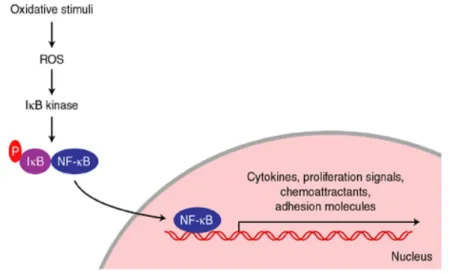
The central orchestrator of the inflammatory response is nuclear factor kappa-light-chain-enhancer of activated B cells (NF-κB), a redox-sensitive transcription factor (Joseph et al., 2014). In unstimulated cells, NF-κB remains inactive in the cytoplasm by the association with inhibitor proteins of the Inhibitor of κB (IκB) family (IκBα and IκBβ). In inflammatory conditions IkB kinase is activated by pro-inflammatory stimuli and phosphorylates IκBs, leading to their ubiquitination and proteasomal degradation (Funakoshi-Tago et al., 2011). These events release free NF-κB dimers in the cytosol, allowing them to translocate into the nucleus where they stimulate the expression of a wide number of genes including those responsible for the production of cytokines
12 and other inflammatory-molecules, as chemokines, cell adhesion molecules, soluble intercellular adhesion molecule-1 and acute phase proteins (Funakoshi-Tago et al., 2011; Joseph et al., 2014). (Figure 2)
Figure 2. Simplified scheme of oxidant regulation of activation of the transcription factor NF-κB. (taken from Cassidy et al., 2003).
Inflammation can be triggered by different stimuli such as endotoxins (i.e., lipopolysaccharide from bacteria), viruses, and changes in levels of ROS, cellular redox status, fatty acids, cytokines, growth factors, and carcinogens (Giampieri et al., 2015a).
In addition to these classic inflammatory stimuli, inflammatory stress can also result from excess body fat and poor diet. Excess body fat and obesity are associated with a concomitant and persistent increase in low-grade inflammation (Giampieri et al., 2015a). In obesity, morphological changes in adipocytes result in altered secretory responses favoring an inflammatory state: elevated circulating inflammatory proteins (TNF-α and IL-6) are in fact observed in this condition and this may explain the critical link between obesity and the development of insulin resistance, type 2 diabetes, and cardiovascular disease (CVD) (Joseph et al., 2014).
Energy intake excess and poor dietary composition typical of the Western diet model may also promote acute (postprandial) and cumulative sustained inflammatory responses in both obese and normal weight individuals. The ROS overproduction, stimulated by an excess of energy intake, results in metabolic oxidative stress and cellular redox imbalance that activate redox-sensitive signaling molecules and ultimately leadto an increased expression of inflammatory genes and oxidative stress markers (Joseph et al., 2014) (Figure 3).
13 Figure 3. Cytokine production by different stimuli (adapted from Forbes-Hernández et al., 2015).
Therefore, during inflammation, cells involved in the inflammatory process are recruited to the damaged site, take up oxygen and release ROS. In addition, inflammatory cells secrete cytokine, which help to further recruit inflammatory cells, generating yet more ROS. Consequently, transcription factors such as NF-κB that encode pro-inflammatory genes, are activated, leading to an increased secretion of cytokines. This vicious cycle supports a protracted environment of oxidative and inflammatory stress, which contributes to several chronic diseases (Joseph et al., 2014) (Figure 4).
Figure 4. Schematic representation of the vicious cycle that supports a protracted environment of oxidative and inflammatory stress (taken from Forbes-Hernández et al., 2015).
14 Therefore, the control of excessive inflammation, through the modulation of pro- and anti-inflammatory cytokine expression in immune cells by potential food components, may represent a strategic tool to avoid immune disorder diseases and maintain health and wellness (Giampieri et al., 2015a).
3.4. ROS and RNS
Free radicals represent reactive chemical species capable of independent existence (Gutteridge, 1994), that possess an unpaired electron in the external orbit (Riley, 1994; Poljsak et al., 2013). An unpaired electron is one that occupies an atomic or molecular orbital by itself; its presence usually causes free radicals to be attracted slightly to a magnetic field and makes them highly reactive, although the chemical reactivity of radicals varies over a wide spectrum.
There are many free radicals in chemistry and biology. ROS is a collective term used to represent both free radical and non-free radical oxygenated molecules such as hydrogen peroxide (H2O2), superoxide (O2•-), singlet oxygen (1/2 O2), and the hydroxyl radical (HO•) (Table 1).
Table 1. Radical and non-radical oxygen species (adapted from Kohen and Nyska, 2002). In the aerobic process, molecular oxygen is reduced to a series of intermediate reactive species through the mechanism indicated (Gutteridge, 1994):
15
HO
2.H
++ O
2.-O
2.-+ 2 H
++ e
-H
2O
2H
2O
2+ e
-HO
-+ HO
.HO
.+ H
++ e
-H
2O
The superoxide radical anion is considered the precursor of all radicals because, in most cases, is the first radical that is produced by cellular oxidase (Ardanaz and Pagano, 2006). It results from a single electron reduction of oxygen by various oxidases, such as dihydro nicotinamide adenine dinucleotide phosphate oxidase, xanthine oxidase, cyclooxygenase, and can act both as reducing agent, yielding in turn an electron to an oxygen molecule, and as an oxidizing agent, with the formation of hydrogen peroxide. This reaction is known as dismutation, which can occur spontaneously but extremely slowly (2 × 10⁵ m/sec) or it can be catalyzed by the superoxide dismutase enzymes, able to accelerate the process up to 104 times.
2 O
2.-+ 2H
+O
2+ H
2O
2The superoxide anion is able to oxidize many biologically important compounds, such as catecholamines, polyphenols, leucoflavine, and to reduce cytochrome c, tetranitromethane and nitroblue tetrazolium (Fridovich, 1986).
Superoxide radical anions may also be formed in the mitochondrial electron transport chain, during the oxidative phosphorylation process (Dröse and Brandt, 2012; Evans et al., 2003).
H2O2, because of its small size and the lack of charge, is much more stable and has a higher diffusion when compared to superoxide anion. H2O2 is able to produce highly reactive radicals as a result of its interaction with metal ions (Gutteridge, 1994). Direct action of H2O2 involves the attack on heme proteins structure with release of iron, enzyme inactivation and oxidation of DNA, lipids, -SH groups, and keto-acids (Kohen and Nyska, 2002). H2O2 can be depleted by catalase, through its conversion into water (Gandhi and Abramov, 2012; Droge, 2002).
It is considered toxic for its ability to convert into hydroxyl radical, considered the most reactive and damaging between the oxygen radicals, through exposure to ultraviolet light or through interaction with metal ions (Fenton reaction).
16 The superoxide anion can cooperate with the hydrogen peroxide in the formation of the hydroxyl radical. This reaction, known as the Haber-Weiss one, takes place both in vitro and in vivo (Liochev andFridovich, 1994).
HO∙ is a highly aggressive radical species, responsible for the oxidative damage of the most biological macromolecules. HO∙ has been reported as the most powerful oxidizing radical: it is characterized by high reactivity and short life-span that allows it to interact indiscriminately at the site of its generation with most organic and inorganic molecules, such as DNA, proteins, lipids, amino acids, sugars, and metals (Kohen and Nyska,2002; Halliwell and Gutteridge, 1999).
Finally, molecular oxygen is not a free radical, but it is considered an oxygenated species endowed with high reactivity (Gutteridge, 1994).
Alongside ROS, an important biological relevance also have RNS. In particular, nitric oxide (NO•), a colourless gas which can diffuse readily between and within cells. NO•, which is toxic in moderate concentrations, possesses different physiological roles: (i) it is an important vasodilator, inhibiting platelet adhesion and aggregation; (ii) it is produced in the brain as a neurotransmitter, (iii) it is involved in immune responses; (iv) it shows a protective effect during the process of ischemia/reperfusion (Wink et al., 1993; Ignarro, 2000). The NO• radical is synthesized by the oxidation of L-arginine by a family of synthases (NOS) of which there are three major isoforms: endothelial (eNOS), neuronal (nNOS) and inducible (iNOS) (Stuehr, 1999). It is considered toxic because it is able to react with O2•- , giving a highly reactive, damaging nitrogen species, namely peroxynitrite, a powerful oxidant versus many biological molecules: it determines depletion of sulfhydryl groups and oxidative damage on most biomolecules, acting similarly to hydroxyl radical, and it is also responsible for DNA damage, protein oxidation and nitration of aromatic amino acid in protein structure (e.g. 3- nitrosotyrosine) (Kohen and Nyska, 2002).
Peroxynitrite can be decomposed to hydroxyl radicals in presence, or not, of transition metals (Gutteridge, 1994; Hou et al., 1999; Stryer, 1995; Green et al., 1990).
ONOO
-+ H
+HO
.+
.NO
2
Other important biochemical reactions involving nitric oxide are: (i) S-nitrosation of thiols and (ii) nitrosylation of transition metal ions. Moreover, the hemoglobin structure may be directly or indirectly altered by NO, respectively through the attachment to heme in the nitrosylation reaction, or by S-nitrosation of the thiol moieties, yielding S-nitrosothiols (van Faassen and Vanin, 2005). ROS can be generated by endogenous or exogenous sources (Figure 5).
17 Figure 5. Exogenous and endogenous sources of ROS (adapted from Kohen and Nyska, 2002). The endogenous production of ROS follows different metabolic pathways (Sauer et al., 2001); the most important site is represented by the electron transport chain (ETC) in the mitochondria (Beal, 2005). Indeed, in animals, about 85% of inspired oxygen is used by mitochondria, the major site of adenosine triphosphate (ATP) production in animals, in non-photosynthetic plant tissues and in leaves in the dark. Mitochondrial ETC consists of a series of redox reactions in which electrons are transferred from donors to acceptor molecules, resulting in a trans-membrane proton translocation which drives the formation of an electrochemical gradient (Forbes-Hernández et al., 2014).
ETC comprises the electron transport complexes I (NADH dehydrogenase), III (cytochrome bc1 complex) and IV (cytochrome c oxidase) that constitute its actual energy–conserving centers and complex II (succinate dehydrogenase), glycerol phosphate dehydrogenase, ubiquinone and cytochrome c (cyt c) which, although incapable of pumping protons across the inner mitochondrial membrane per sé, are essential ETC constituents having critical roles in ETC function and efficiency (Forbes-Hernández et al., 2014). In mitochondrial ETC, electrons released (i) by the oxidation of NADH and (ii) by the oxidation of FADH2 are transferred to the different complexes of the chain, having oxygen as the final acceptor, which is reduced to water (Pieczenik and Neustadt, 2007). The energy released by electrons while they pass through the ETC is used to pump protons from the mitochondrial matrix into the intermembrane space, creating an electrochemical proton
18 gradient across the inner mitochondrial membrane called mitochondrial membrane potential, that is essential to mitochondrial bioenergetics (Brand and Nicholls, 2011).
This electrochemical proton gradient permits ATP synthase, the final complex of the chain, to use the flow of H+ through the enzyme back into the matrix to generate ATP from adenosine diphosphate (ADP) and inorganic phosphate. The complete process is the oxidative phosphorylation, since ADP is phosphorylated to ATP using the energy of hydrogen oxidation in many steps. But in this process a small percentage of electron do not complete the whole series of reactions and directly react with oxygen, resulting in the formation of several ROS as secondary ETC products (Frantz and Wipf, 2010). During normal oxidative phosphorylation 0.4–4.0% of all oxygen consumed in mitochondria is transformed into the O2.- which is transformed to H2O2 by the detoxification enzymes copper/zinc superoxide dismutase or manganese superoxide dismutase (MnSOD) and then to water by glutathione peroxidase (GPx) or peroxidation enzymes (PRx). When these enzymes cannot transform ROS fast enough, oxidative damage occurs and accumulates in mitochondria (Forbes-Hernández et al., 2014) (Figure 6).
Figure 6. Schematic model of mitochondrial oxidative damage. During mitochondrial respiration a small amount of the molecular oxygen consumed by cells is converted into O2.- by reactions occuring in complex I and III. MnSOD enzyme converts O2. to H2O2 which can be converted into
H2O by GPx or PRx. H2O2 can also react with Fe2+ to produce HO.. This radical could attack
mtDNA, decreasing mRNA and altering the expression of proteins essential for ETC (taken from Forbes-Hernández et al., 2014).
19 Additionally, NO is produced inside the mitochondria by mitochondrial nitric oxide synthase and also diffuses into the mitochondria from the cytosol (Forbes-Hernández et al., 2014).
Most of the 10-15% of O2 taken up by aerobic eukaryotes that is not consumed by mitochondria is used by various oxidase and oxygenase enzymes, of which one of the most important is cytochrome P450, contained in the endoplasmatic reticulum of many animal and plant tissues.
The P450 superfamily has been encoded by over 150 genes; these enzymes are involved in the oxidation of a wide range of substrates at the expense of O2. Substrates for cytochromes P450 include insecticides and pesticides, hydrocarbons, and drugs. For these reasons, liver endoplasmatic reticulum is especially rich in P450s. However the product of reaction with P450 is less toxic than the substrates.
Another important multienzymatic complex involved in the generation of ROS, especially during the transduction of intracellular signals, is NADPH oxidase, present in particular in phagocytic cells. This complex contains FAD and a cytochrome b, in form of a dimer, capable of reducing O2 to O2•- . Usually, up to 30% of neutrophil cytochrome is present in the plasma membrane and the rest in the cytoplasm, especially in specific granules, which can supply more b-type cytochrome to the plasma membrane. When the system is activated, the cytoplasmic constituents are phosphorylated and translocate on the membrane where they bind to the cytochrome b, allowing the electron transfer to molecular oxygen, thus producing superoxide. The activation of NADPH oxidase also requires the participation of two proteins of low molecular weight, Rac2, forming part of a cytoplasmic dimer with Rho-GDI, and Rap1A, a member of the Ras family located at the level of the cytoplasmic membrane in intimate contact with the cytochrome (Babior, 1999; Li et al., 2001). This complex is well characterized in phagocytic cells, where it is essential the bactericidal function performed by ROS (O2•-, H2O2 and HOCl) produced by respiratory reactions, which
represent a defense mechanism against invading microorganisms. Finally, in the case of ischaemia, the main source of ROS is the xanthine oxidase, which chairs the metabolism of purines. In healthy animal tissues, this enzyme system is present in endothelial cells of small vessels and in the epithelial cells of the breast tissue. Generally it exists in the form of xanthine dehydrogenase that transfers electrons not to O2 but to NAD+, as it oxidizes xanthine or hypoxanthine into uric acid. However, in certain conditions, as ischaemia, some of the xanthine dehydrogenase can be converted into xanthine oxidase by oxidation of essential –SH groups or by limited proteolysis. Xanthine oxidase produces O2•- and H2O2 when xanthine or hypoxanthine are oxidized.
In addition, ROS can be produced by a host of exogenous processes (Valko et al., 2006). Environmental sources including: x rays and γ rays, ultraviolet light (which produce free radicals when transfer their energy to cellular components or molecules of water (Giampieri et al., 2012b),
20 ionizing radiation, and pollutants such as paraquat and ozone. Even pesticides, tobacco, solvents, anesthetics, and in general all the aromatic hydrocarbons may be responsible for oxidative stress (Pagano, 2002). All of these sources of free radicals, both enzymatic and non-enzymatic, have the potential to cause oxidative damage on a wide range of biological macromolecules (Mateos and Bravo, 2007) (Figure 7).
Figure 7. ROS damage in biological macromolecules (taken from Kohen and Nyska, 2002).
3.5. Antioxidant defences
In all aerobic organisms the production of ROS and RNS is inevitable and, within certain concentrations, essential (Feher, 1985). Exposure to free radicals from a variety of sources has led organisms to develop a series of defence mechanisms (Cadenas, 1997).
Halliwell and Gutteridge proposed, in 1989, a comprehensive definition of an antioxidant as “any substance that, when present at concentrations compared with those of an oxidizable substrate, significantly delays or prevents oxidation of that substrate” (Halliwell and Gutteridge, 1989).
21 Afterwards, the antioxidants were defined by a more general perspective as "all those compounds that protect biological systems against the harmful effects of excessive generation of oxidants," and this definition is currently the most widely used (Krinsky, 1992).
Antioxidant functions lead to a decrease of oxidative stress, DNA mutations, malignant transformations, as well as other parameters of cell damage. Some epidemiological studies highlight the antioxidant ability to contain the effects of ROS activity, reducing the incidence of cancer and other degenerative diseases (Godic et al., 2014).
There is no an universal and best antioxidant: the defenses against oxidative stress are not able to completely avoid damage. In fact, in a biological system, the function of an antioxidant depends on various parameters, such as the type of ROS generated, the manner and the place in which is formed, the extent of damage. The first types of antioxidant defense systems developed against oxidative damage are those that prevent ROS occurrence (preventative mechanisms) and those that block, capture radicals that are formed (repair mechansims) (Cheeseman and Slater, 1993).
These systems can be enzymatic (such as superoxide dismutase, catalase, glutathione peroxidase) and nonenzymatic, such as oxidative enzyme (e.g. cyclooxygenase or lipooxygenase) inhibitors, antioxidant enzyme cofactors (Se, coenzyme Q10), ROS/RNS scavengers (vitamin C, vitamin E), and transition metal chelators (e.g. EDTA) (Huang et al., 2005).
Under physiological conditions, the balance between prooxidant and antioxidant compounds moderately favors prooxidants, thus inducing a slight oxidative stress, requiring the intervention of endogenous antioxidant systems of the organism (Dröge, 2002).
Redox homeostasis of the cell is assured by its complex endogenous antioxidant defense system, which includes endogenous antioxidant enzymes such as superoxide dismutase, catalase, glutathione peroxidase, and non-enzymatic compounds like glutathione, proteins (ferritin, transferrin, ceruloplasmin, and even albumin) and low molecular weight scavengers, like uric acid, coenzyme Q, and lipoic acid (Poljsak et al., 2013).
On the contrary, the main exogenous antioxidants sources are vitamin C and E, carotenoids, and phenolics. Clinical studies proved that, together with omega-3 fatty acids, these antioxidants present in fruits, vegetables, whole grains, legumes, could work as preventative agents regarding disease occurrence, counterpartying the activity of the above-mentioned endogenous antioxidative defense (Willett, 2006).
Another source of exogenous antioxidants is constituted by dietary supplements, as vitamins, minerals, fibres, fatty acids or amino acids, which are either lacking or not found in sufficient amounts in the common diet (Poljsak et al., 2013).
22 The use of exogenous antioxidants may retard the uptake of endogenous antioxidants, for the total “cell antioxidant potential” to remain unaltered (Pisoschi and Pop, 2015). The use of antioxidant supplementation is indeed effective if the initial oxidative stress level is higher than normal or above the individual's set point of regulation (Pisoschi and Pop, 2015).
Thus, the antioxidant supplements may reduce the increased levels of oxidative stress that cannot be inactivated by the endogenous sources (Poljsak et al., 2013).
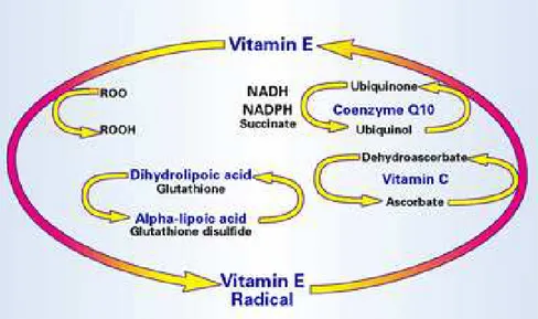
Another antioxidant classification regards their localization in aqueous (water-soluble) or in membrane cell compartments (fat-soluble) (Pisoschi and Pop, 2015). The "scavengers" are water soluble compounds of low molecular weight, as vitamin C, uric acid, glutathione, localized in various cell districts, that interact directly with ROS, neutralizing them. The fat-soluble molecules, such as vitamin E and β-carotene, instead, are located at the level of membranes and are used for the neutralization of lipid peroxidation. Coenzyme Q10 or ubiquinone is an essential component of the inner mitochondrial membrane (Fato et al., 1985; Battino et al., 1990; Lenaz et al., 1992; Rauchova et al., 1992; Samori et al., 1992) and can act neutralizing radical species independently or synergistically with vitamin E. In this sense, it is possible to detect the existence of "networks" of antioxidants, where some antioxidants interact with others to reconstitute their corresponding reduced forms in a coordinated and controlled process, that just now it has been understood in detail (Sen et al., 2000) (Figure 8).
Figure 8. Exemple of antioxidant network. The radical species that are formed in the lipophilic cellular compartment are reduced by Vitamin E, with the formation of the Vitamin E radical. This
can be reduced back to Vitamin E by another fat-soluble antioxidant such as ubiquinol, or by a cytoplasmic antioxidant such as ascorbate.
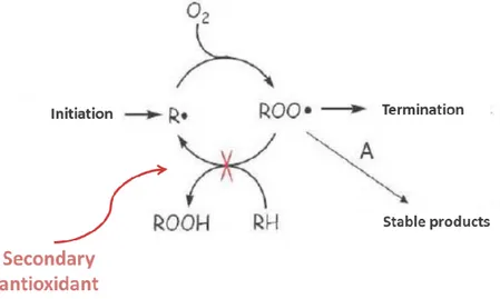
23 Antioxidants can be also classified according to their mode of action: we can identify primary, secondary and tertiary antioxidants. The primary antioxidants are those that convert ROS into less harmful molecules (before they could react with the vital structures), or prevent their production. In this group there are: superoxide dismutase, glutathione peroxidase, glutathione reductase, peroxidase, catalase, the glutaredoxin, thioredoxin, and proteins such as ferritin and ceruloplasmin (Figure 9).
Figure 9. Mechanisms of action of primary antioxidants.
The secondary antioxidants, instead, oxidize and become weak and non-toxic free radicals when they react with ROS/RNS. Examples of these substances are: vitamins C and E, β-carotene, uric acid, bilirubin, albumin, ubiquinone and selenium and, among phenolic compounds, phenolic acids, phenols and flavonoids (Carr and Frei, 1999a; Catani et al., 2001;El-Agamey et al., 2004; Landis and Tower, 2005;Miller et al., 2005;Schrauzer, 2006) (Figure 10).
24 The third system of antioxidants is mainly composed by phospholipase A2 and C, endo- and exonucleases, DNA glycosylase, peroxidase and methionine sulfoxide reductase, whose functions include (i) to repair direct damage to proteins, lipids and carbohydrates, DNA, RNA and (ii) to eliminate irreversibly damaged products (Sahnoun et al., 1997).
Finally, the antioxidants can be also classified taking into account their functions and we can distinguish between:
a) indirect antioxidants: they inhibit the formation of free radicals, by chelation of metals (i.g. Fe2+); b) direct antioxidants: they “capture” ROS and RNS once generated;
c) a large group of compounds that modulate positively not only the cellular capacity to counteract the high levels of ROS and RNS, but also the ability to promote the repair of oxidized biomolecules (Aruoma, 1996).In this sense, the concept of antioxidant capacity is linked not only to the effects of decrease free radicals formation, but also to the prevention of oxidative damage in cellular functions (Aruoma, 1998).
At the end, it has been proposed that antioxidant defenses, in general, act in parallel (several antioxidants may play similar roles) and in serial mode (antioxidant enzymes operate in tandem during their catalytic action): this is the proof of the complexity of this system of molecules (Beckman and Ames, 1998). Secondary antioxidants, for example, may exhibit synergetic effects in combination with primary antioxidants, following several possible mechanisms (Pisoschi and Pop, 2015):
- stabilizing primary antioxidants through the creation of an acidic environment; - regenerating primary antioxidants by hydrogen donation;
- chelating pro-oxidative transition metal cations; - quenching molecular oxygen.
Moreover, it has been revealed that antioxidant enzymes can catalyse the synthesis or the regeneration of non-enzymatic antioxidants (Martysiak-Żurowska and Wenta, 2012).
The synergistic interaction between antioxidants is also favoured by the "protection" that occurs between each enzyme: for example, SOD "protects" GSH-Px and catalase from damage caused by O2.-, while GPx and catalase "protect" in turn SOD from inactivation caused by hydroperoxides and
H2O2 (Chaudieáre & Ferrari-Iliou, 1999).
The principal enzymatic and non-enzymatic antioxidants present in animals and humans are listed below:
25 Enzymatic:
-
Superoxide dismutase (SOD) (E.C. 1.15.1.6). This antioxidant enzyme catalyzes the dismutation of O2•- to O2 and to the less-reactive species H2O2 (Valko et al., 2006). In mammalian tissues, three types of superoxide dismutases can be found: copper-zinc containing superoxide dismutase (SOD1) present in the cytosol, manganese containing superoxide dismutase (SOD2) found in the mitochondrial matrix and extracellular superoxide dismutase (SOD3) (Ghezzi et al., 2005; Sung et al., 2013);-
Catalase (E.C. 1.11.1.6). Catalase is present in the peroxisome of aerobic cells and represents the enzyme involved in the reductive depletion of H2O2 to water. Catalase has one of the highest turnover rates for all enzymes: one molecule of catalase can convert approximately 6 million molecules of hydrogen peroxide to water and oxygen each minute (Rahman, 2007);-
Glutathione peroxidase (GPx) (E.C. 1.11.1.9). Glutathione peroxidase is a selenium-containing enzyme. It catalyses both the reduction of H2O2, and organic hydroperoxides to water or corresponding alcohols. All GPx enzymes are known to add two electrons to reduce peroxides by forming selenoles (Se-OH). Its antioxidant properties enable them to eliminate peroxides as potential substrates for the Fenton reaction (Chaudieáre & Ferrari-Iliou, 1999).Non-enzymatic:
-
Water solubleo Glutathione: One of the main thiol antioxidants is the tripeptide glutathione (GSH), which is an intracellular antioxidant and is considered to be the major thiol-disulphide redox buffer GSH/GSSG of the cell (Masella et al., 2005). The basic protective functions of glutathione against oxidative stress are that it can act as a co-factor for several detoxifying enzymes, involved in amino acid transport through plasma membrane, scavenge hydroxyl radical and singlet oxygen directly, and regenerate vitamins C and E back to their active forms (Masella et al., 2005). The accumulated oxidized glutathione (GSSG) inside the cells and the ratio of GSH/GSSG is a good measure of oxidative stress of an organism (Dröge, 2002b).
o Vitamin C (ascorbic acid): is a very important and powerful water-soluble antioxidant and thus works in aqueous environments of the body. It works synergistically with vitamin E (regenerating α-tocopherol from α-tocopherol
26 radicals in membranes and lipoproteins) and the carotenoids as well as working along with the antioxidant enzymes (Kojo, 2004; Carr and Frei, 1999b). Furthermore, it supports intracellular glutathione levels thus playing an important role in protein thiol group protection against oxidation (Rahman, 2007).
-
Fat-solubleo Vitamin E: This is a fat-soluble vitamin that exists in eight different forms. In humans, α-tocopherol is the most active form, and is the main powerful membrane bound antioxidant hired by the cell (Hensley et al., 2004). The primary function of vitamin E is to protect against lipid peroxidation (Pryor, 2000). During the antioxidant reaction, tocopherol is converted to an α-tocopherol radical by the donation of an hydrogen to a lipid or lipid peroxyl radical. The α-tocopherol radical can subsequently be reduced to the primary α- tocopherol form by ascorbic acid (Kojo, 2004).
o The other non-enzymatic antioxidants that are involved in oxidative stress defense are carotenoids and flavonoids. Carotenoids are pigments that are present in plants and microorganisms. They contain conjugated double bonds and their antioxidant activity result mainly as a consequence of the ability to delocalize unpaired electrons (Mortensen et al., 2001). Flavonoids are a large class of low molecular ubiquitous groups of plant metabolites and are an inseparable part of the human diet (Rice-Evans, 2001). They are benzo-γ-pyrone derivatives containing phenolic and pyran rings (Rahman, 2007). Their antioxidant activity is performed by the phenolic compounds that operate as inhibitors of free radical chain (Valko et al., 2006). Recently, the interest in flavonoids increased, due to their antioxidant capacity, chelating properties and their possible role in the prevention of chronic and age-related diseases.
3.6. Lipopolysaccharide
Lipopolysaccharide (LPS), usually referred to an endotoxin, is an outer membrane structure and an important virulence factor of the cell wall of Gram-negative bacteria (e.g. Escherichia coli, Chlamydia pneumoniae, and periodontopathogens that are common pathogens colonizing the human gastrointestinal tract, including the oral cavity and the gut); it is not found in Gram positive bacteria (Mayer et al., 1985). It may originate from several sources, including infections, diet, and commensal microbiota. The LPS molecule is essential for the viability of most Gram-negative
27 bacteria, exerting a crucial role in outer-membrane integrity as a permeability barrier, protecting bacteria from toxic molecules, bile salts and lipophilic antibiotics (Mayer et al., 1985).
In the circulation system, LPS is able to interact with several cell types, as epithelial cells, macrophages, smooth muscle cells, fibroblasts, B- and T-cells and endothelial cells (Whitfield and Trent, 2014).
3.6.1. Structure
LPS is a complex glycolipid in which the lipid and polysaccharide moieties are joined by a covalent bond. The three structural regions of LPS are: (i) lipid A, a hydrophobic lipid which is responsible for the toxic properties of the molecule, (ii) a hydrophilic core oligosaccharide and (iii) a hydrophilic O-specific side chain (O antigen) (Mayer et al., 1985) (Figure 11).
Figure 11. Chemical structure of LPS (taken from Qiao et al., 2014).
The biological activity of LPS is strictly dependent on the lipid A moiety, which is its most conserved part and connects the molecule to the outer membrane of bacterial. It is a phosphorylated glucosamine disaccharide acylated with hydroxyl saturated fatty acids that further 3-O-acylate the 3-hydroxyl groups of the fatty acids of lipid A (Raetz 1990). In 1986 Munford and Hall demonstrated that the removal of the O-acylated saturated fatty acids or their substitution with
28 unsaturated fatty acids leads to the disappearance of endotoxin activity (Munford and Hall, 1986). The core oligosaccharide is directly attached to lipid A. The O-side chain is a repetitive glycan polymer, which binds to the core oligosaccharide forming the most external part of the LPS (Manco et al., 2010). The repetitive units of the polymer may be linear or branched, producing homo- or heteropolymers (Raetz and Whitfield, 2002). Each repeating unit represents diverse antigen properties, and their sequence and their way of linkage determine the serological O specificity of respective strains (Mayer et al., 1985).
3.6.2. LPS-mediated signaling and the innate immune response
LPS induces the release of a large number of inflammatory cytokines, which play an important role in metabolic processes through the linkage to the pathogen-sensing system. The main elements of LPS-mediated signaling comprise lipoproteins, LPS-binding protein (LBP), cluster of differentiation 14 (CD14) and toll-like receptor 4 TLR4 (Bosshart and Heinzelmann, 2007). These elements act together to initiate a signaling pathway, that ultimately leads to the activation of NFκВ transcription factor. In the circulation, LPS is transported to hepatocytes by LBP, phospholipid transfer protein, and lipoproteins (Munford et al., 1981; Hailman et al., 1996). Approximately 80– 90% of the LPS is bound to the lipoproteins through lipophilic lipid A, including all main lipoprotein classes: (i) HDL, under physiological conditions, (ii) VLDL, when the serum HDL is low, or (iii) TG-rich lipoprotein-LPS complex, that is rapidly eliminated to reduce LPS-induced toxicity or is internalized by macrophages (Brown and Goldstein 1983; Levels et al., 2001; Harris et al., 2002). Therefore, the metabolic fate of LPS may be regulated by the lipoprotein profiles (Berbee et al., 2005).
LBP may transport LPS to lipoproteins or alternatively to soluble or membrane-bound CD14 (Stoll et al., 2004). In this case, the LPS-CD14 complex engages TLR4 via lipid A moiety (Chow et al., 1999). TLRs are necessary for the downstream signaling pathway, since that CD14 misses a transmembrane domain. The TLR4 activation leads to the employment of additional adaptor molecules, which further trigger a cascade that allows NFκВ to diffuse into the nucleus and activate the transcription of proinflammatory cytokines, as TNFα, IL-1β, IL-6, and chemokines (Stoll et al., 2004; Parker et al., 2007; Lu et al., 2008) (Figure 12).
29 Figure 12. Schematic representation of LPS-induced pro-inflammatory cytokines production (taken
from Neves et al., 2013).
Although activation of cytokines is a part of the host defense response to infections, their excessive production and secretion may result in septic shock, systemic inflammatory response syndrome, severe tissue damage and multiple organ dysfunction, including liver, lung, heart, gastrointestinal tract, and kidneys (Haghgoo et al., 1995; Shimazu et al., 1999; Cadenas and Cadenas, 2002; Yokozawa et al., 2003; Chen et al., 2007; Zhang et al., 2009).
3.6.3. LPS and nutrition
In addition to the oral cavity, the other main source of LPS is the gut. Even if LPS has a strong affinity for chylomicrons and is able to cross easily the gastrointestinal mucosa, under physiological conditions, the intestinal epithelium defends itself from LPS translocation (Ghoshal et al., 2009). However, possible mechanisms implicated in LPS translocation from the gut include uptake by intestinal enterocytes and microfold cells, and alterations in the gene expression of host epithelial cells by Gram-negative bacteria (Hathaway and Kraehenbuhl, 2000; Hooper and Gordon, 2001) (Figure 13).
30 Figure 13. Gram-negative-derived LPS pass across the intestinal barriers into the lymphatic
system and then into the bloodstream. Main target tissues are the adipose tissue, the liver, and the endothelium (taken from Manco et al., 2010).
A high-fat diet, obesity, diabetes, and non-alcohol fatty liver disease have been associated with increased permeability of the gastrointestinal mucosa, leading to metabolic endotoxemia (Neves et al., 2013). In mice, a chronic exposure to LPS appears to be related to the development of insulin resistance, weight gain and an insurgent inflammatory status.
The absorption of LPS through the intestinal barrier seems to be enhanched by an high-fat diet, determining a very strong association between this type of diet, the microbiota, and inflammation (Cani et al., 2007).
For this reason, LPS can be considered an important factor directly involved in the onset of obesity induced by a rich-fat diet and type 2 diabetes (Manco, 2009).
In addition, recent studies on mice and pigs fed with a saturated fatty acids-rich diet showed an increase in serum concentrations of LPS and a more active transport of this endotoxin to peripheral tissues through high levels of LBP and soluble CD14 low levels (Laugerette et al., 2012; Mani et al., 2013).
31 The capability of LPS to interact synergistically with this type of fatty acids is related to the strong influence that saturated fatty acids have on the immune system and on TLR4 activation (Fritsche, 2006; Suganami et al., 2007).
Human studies have confirmed the close relationship between high-fat or energy-rich diet and the occurrence of inflammation (Erridge et al., 2007; Amar et al., 2008; Ghanim et al., 2009; Pendyala et al., 2012). Studies on healthy subjects highlighted that an high-fat meal or excessive energy intake, leading to a significative increase in plasma concentration of LPS (Erridge et al., 2007; Amar et al., 2008; Ghanim et al., 2009), expecially in subjects with high metabolic risk, as impaired glucose tolerance and type 2 diabetes (Harte et al., 2012). Dietary fats in fact deeply increase LPS absorption through modification of the gut microbiota, raising the amount of chylomicrons, and increasing the permeability of the gastrointestinal mucosa (Manco et al., 2010).
3.7. Strawberry
Nowadays what we call "food" is not just a tool to satisfy the appetite of the consumer, but it is becoming an essential factor closely related to human health. In this context, the change of lifestyle in modern society has highlighted new crucial aspects from the nutritional point of view. The excessive or inadequate food intake could be one of the causes of the onset of various diseases and illnesses, while the careful choice of food can help to prevent or limit the onset of these diseases (Testoni and Lovati, 2004).
From this point of view, fruits and vegetables represent the foods with the highest amount of bioactive compounds, demonstrating the best beneficial effects on human health. Among commestible fruits, berries of the family Rosaceae (Rubus and Fragaria) are especially rich in antioxidant compounds, as reported by various systematic analysis of a large number of dietary plants (Halvorsen et al., 2002; Proteggente et al., 2002; Sun et al, 2002; Bagchi et al., 2003).
In addition, edible berries may represent a potential important contribution to the intake of fresh fruit for the populations in countries where, as declared by World Health Organization (WHO), there is a limited availability of fruits and vegetables, as in northern latitudes. For example, in a Norwegian diet, berries represent 21.7% of the total plant antioxidants in the diet (Halvorsen et al, 2002), with strawberries, black currant and cranberry being the most significant source among them. One of the most commonly consumed berries both in fresh and processed forms such as jams, yogurts, desserts or juice, is strawberry (Fragaria x ananassa Duch), due to the nutritional quality and the several bioactive compounds that it contains. Strawberries are also economically and commercially important and therefore represent one of the most studied fruit from the agronomic,
32 genomic and nutritional points of view. Strawberry is a relevant source of folate (Olsson et al., 2004; Tulipani et al., 2008a), is rich in vitamin C and contains various phytochemicals, which may greatly influence the nutritional and organoleptic qualities of this fruit (Scalzo et al., 2005a; Proteggente et al., 2002; Deighton et al., 2000; Tulipani et al. 2009a; Giampieri et al., 2012a). Their health effects are mainly attributed to the high levels of antioxidant compounds, mainly phenolic compounds, such as anthocyanins, flavonols, flavanols, condensed tannins (proanthocyanidins, ellagitannins and gallotannins), hydroxybenzoic and hydroxycinnamic acid derivatives, and hydrolyzable tannins (Giampieri et al., 2012a; 2013). These compounds are reported to possess important antioxidant, anti-inflammatory, anticancer, and antineurodegenerative biological properties (Giampieri et al., 2013).
3.7.1. Nutritional aspects
The nutritional quality of the strawberry is related to the quantity and the quality of the phytochemical compounds that it contains (Table 2). This composition determines the nutritional profile and the possible preventive effect of strawberry against many chronic diseases (Tulipani et al., 2011):
• the content of vegetable fiber (1.6 g/100 g fresh weight (FW)) has a satiating effect, determining the reduction of the calorie intake in the diet. Fiber also allows for adjustment of the glycemic index, slowing the rate of digestion;
• strawberries are a natural source of essential fatty acids: in particular, the seeds are rich in unsaturated fatty acids;
• the high content of vitamin C makes the strawberry one of the main dietary sources of this compound and makes it particularly important from the nutritional point of view;
• in addition to traditional nutrients, strawberries are one of the richest dietary sources of phytochemicals known as non-nutritional compounds, represented in particular by the class of polyphenols, main responsible of the fruit color and the characteristic aroma (Mazzoni et al. 2013, 2016).
33 Table 2. Chemical composition of strawberry (Source INRAN). The data are expressed per 100 g
of fresh weight.
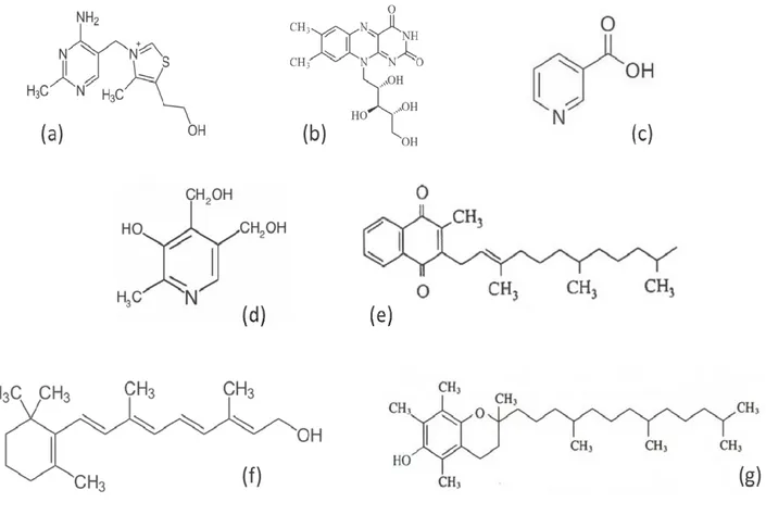
3.7.1.1. Micronutrients in strawberry
3.7.1.1.1. Folates
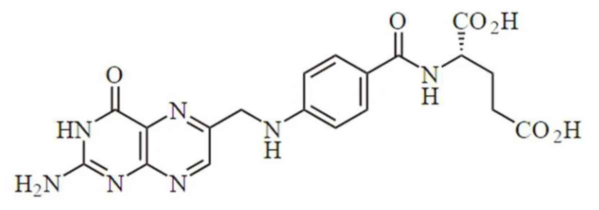
The terms folate and folic acid are often both used to describe this water-soluble B-complex vitamin, also called vitamin M or B9. However, the term folates refers to all derivatives of tetrahydrofolic acid, which are present in food in about 150 different chemical forms, and to the metabolically active forms in the human body (Giampieri et al., 2012a). The elementary structure of these essential dietary compounds consists of a pteridine ring linked to a paraaminobenzoic acid, conjugated with usually five to eight L-glutamic acid residues. These compounds differ one to another in three points inside the molecule: i) the reduction-state of the pteridinic ring, ii) the type of mono carbonaceous unit bound to it, iii) and the number of glutamic acid residues. Commonly,
34 folates are present in conjugated form to one or more residues of glutamic acid (Bates and Heseker, 1994) (Figure 14).
Figure 14. Chemical structure of folic acid.
The essential biological function of folate is to work as a coenzyme in transferring one-carbon units to a variety of target molecules. In particular folates can act as acceptors and/or donors of one-carbon units in the synthesis of nucleic acids (eg. Purine synthesis) or in the re-methylation of homocysteine to methionine. Folate is also very important to prevent the insurgence of diseases related to their deficiency, such as megaloblastic anemia and some types of cancer. Their assumption is strongly recommended in particular for women of childbearing age to prevent the terrible consequences that can be verified in the fetus in the early stages of pregnancy in the case of a deficiency of this compound (Giampieri et al., 2012a).
On the other hand, folic acid is the more stable, fully oxidised monoglutamate form of folate (Pteroyl-L-Glutamic acid) most often used in dietary supplements and fortified foods. Folic acid occurs rarely in foods or in the human body, and requires the reduction to tetrahydrofolic acid for assimilation into the folate body pool.
In association with vitamin C, folate plays a crucial role in emphasizing the nutritional quality of strawberry. Among fruit, strawberry is one of the richest natural sources of this indispensable micronutrient; its content is considered in the range of 20-25 μg/100 g (FW). To date, still few studies are focused on the evaluation of the folate content of fresh strawberries (Muller, 1993; Vahteristo et al., 1997; Konings et al., 2001; Tulipani et al., 2008a), of its stability during storage and of its retention after fruit processing. According to the existing data (Tulipani et al., 2008a) the intake of dietary folate through strawberry consumption (in 250 g of strawberries are present ~60 μg folate on average) can supply 30% of the European folate RDA (Giampieri et al., 2012a). The
35 increasing awareness of the relevance of folate content in strawberry as an interesting nutritional quality parameter makes strongly necessary the optimization of correct, accurate and reliable analytical methods for its determination, in order to confirm and support the available data.
3.7.1.1.2. Vitamin C
Vitamin C (or ascorbic acid) is a water-soluble vitamin with several biological functions in humans (Figure 15).
Figure 15. Chemical structure of vitamin C.
First of all, vitamin C is a highly reactive and effective antioxidant, able to act both directly and indirectly by ROS scavenging through enzymatic and non-enzymatic reactions. Even in small quantities it plays an important role in protecting essential biomolecules in the body (i.e. proteins, carbohydrates, lipids, DNA and RNA) from damages by free radicals and ROS generated (i) during normal metabolism, (ii) in pathological conditions and (iii) through exposure to toxins and polluants (i.e. smoking). Furthermore, due to its one electron reduction potentials, vitamin C plays a crucial role in regenerating the active form of other antioxidant molecules (i.e. vitamin E), and in scavenging the relative antioxidant-derived radicals: for this reason it is defined “the terminal water-soluble small molecule antioxidant” (Carr and Frei, 1999a).
Vitamin C is also an essential cofactor for the synthesis of hormones, collagen, neurotransmitter norepinephrine or small intracellular molecules like carnitine, and seems to be involved in the functionality of the immune system. In Europe the recommended dietary allowance (RDA) for vitamin C was recently revised upward from 60 mg daily. This RDA value continues to be calculated as the daily amount necessary for the prevention of vitamin C deficiency disease, such as scurvy, rather than the prevention of chronic disease and the promotion of optimum health. Nevertheless, the results of different prospective, observational and case-control studies indicate that higher intakes of vitamin C from diet are frequently correlated with a lower incidence of cardio
36 and cerebrovascular diseases (Carr & Frei, 1999b; Yokoyama et al., 2000), most types of cancers (Steinmetz and Potter, 1996), and other health problems such as lead toxicity. Moreover, vitamin C supplementation seems also to be a beneficial adjunt to conventional therapies for individuals with atherosclerosis (Dawson et al., 1999), hypertension and diabetes mellitus (Duffy et al., 1999).
However, discrepancies among different observational studies and randomized controlled trials still persist, and further studies are required to evaluate the potential effects of vitamin supplementation. At the moment, there is no reason to limit high vitamin C intakes by a fruit and vegetables-rich diet since no toxicity and pro-oxidative events have been ever observed. Notwithstanding, it is still difficult to distinct the health-promoting effects of vitamin C from the potential effects of the other nutrient and non-nutrients compounds present in foods.
One of the aspects of major nutritional relevance in strawberry fruit is the extremely high content of vitamin C (even higher than citrus fruit). It is interesting to note that a cup of strawberries (corresponding to two servings) is sufficient to cover the daily RDA for vitamin C, since its content varies between 0.1 and 1 mg g-1 FW (Cordenunsi et al., 2002; Kafkas et al., 2007), with an average of 0.6 mg g-1 FW (Kalt et al., 1999; Proteggente et al., 2002; Keutgen & Pawelzik, 2007) for several strawberry genotypes (Table 3).