UNIVERSITÀ POLITECNICA DELLE MARCHE
FACOLTÀ DI MEDICINA E CHIRURGIA
DOTTORATO DI RICERCA
in
SCIENZE BIOMEDICHE
XXX CICLO
ROLE OF THE Na
+/Ca
+EXCHANGER 1 (NCX1)
IN THE PROTECTIVE RESPONSE ELICITED BY
GLUTAMATE IN CARDIAC CELLS EXPOSED TO
HYPOXIA/REOXYGENATION (H/R)
Coordinatore:
Chiar.mo Prof. Salvatore Amoroso
Tutore:
Candidata:
Dr. Vincenzo Lariccia
Dott.ssa Marta Maiolino
INDEX
1. INTRODUCTION...1
1.1 General features of myocardial I/R ...2
1.2 Pathophysiology of myocardial ischemic injury...3
1.3 Pathophysiology of myocardial reperfusion injury...5
1.4 Myocardial calcium handling ...6
1.4.1 Calcium overload during I/R ...8
1.5 The sodium calcium exchanger (NCX) ...10
1.5.1 Role of NCX in myocardial I/R injury ...13
1.6 Cardioprotective strategies against I/R injury ...16
1.7 Myocardial energy metabolism...19
1.7.1 Energy metabolism impairment during I/R ...20
1.8 Role of amino acids in cardiac metabolism ...22
1.8.1 Glutamate as metabolic substrate...24
1.9 Excitatory amino acid transporters (EAATs) ...26
1.10 NCX and EAATs interplay ...28
1.11 Aim of the thesis ...30
2. MATERIALS AND METHODS ...31
2.1 Cell Culture ...31
2.2 Isolation of rat adult ventricular cardiomyocytes ...31
2.3 In vitro hypoxia/reoxigenation challenge (H/R) ...33
2.4 Determination of cell viability ...34
2.4.1 LDH release assay...34
2.4.2 FDA/PI double staining ...35
2.5 Analysis of ATP production ...35
2.6 Bioenergetic analysis ...36
2.6.2 Extracellular consumption acidification rate (ECAR) ...38
2.7 ROS detection ...39
2.8 Western blotting ...40
2.8.1 Antibodies ...42
2.9 Real-time confocal imaging ...43
2.9.1 Analysis of NCX1 activity ...43
2.10 Drug and chemicals...44
2.11 Data processing and statistic ...44
3. RESULTS ...45
3.1 Effect of glutamate on H/R injury: involvement of NCX1...45
3.2 Effect of glutamate on H/R injury: involvement of EAATs ...49
3.3 Effect of glutamate exposure on ATP production ...50
3.4 Effect of glutamate on mitochondrial function following H/R injury ...54
3.5 Effect of glutamate exposure on ROS production following H/R injury ...57
3.6 Analysis of NCX 1 and EAATs expression following H/R injury ...58
3.7 Effect of glutamate on NCX1 activity alteration following H/R injury. ...60
4. DISCUSSION ...63
4.1 Rationale ...63
4.2 Glutamate counteracts H/R-induced injury in cardiac cells by sustaining ATP production in a NCX1 dependent manner. ...64
4.3 Glutamate counteracts H/R-induced injury in cardiac cells by sustaining ATP production in an EAATs dependent manner ...66
4.4 Glutamate-induced normalization of NCX1 activity during H/R relies on EAATs/NCX1 dependent pathway ...67
4.5 Glutamate-enhanced ATP response during H/R relies on an improved mitochondrial oxidative phosporylation ...68
4.6 Conclusion ...69
1. INTRODUCTION
Disorders characterized by ischemia/reperfusion (I/R), included myocardial infarction, stroke, and peripheral vascular disease, alone account for >12% of all deaths; more than HIV/AIDS, tuberculosis, lung and breast cancer together (Ferrer Gracia, Hernandez-Antolin et al. 2007). The World Health Organization (WHO) continues to emphasize the importance of cardiovascular diseases (CVDs) as a major cause of death in the Western world, with 30 to 50% of patients dying each year within a few hours of the onset of symptoms of acute myocardial infarction (AMI) (Herlitz, Dellborg et al. 2008). The majority of all CVD deaths (greater than 75 percent) are due to heart attacks and strokes. Growing evidence reports that mortality rates from CVD vary considerably between males and females and also between countries. In Europe, more than 4 million people die from CVD across every year, accounting for 45% of all deaths; 49% of deaths among women and 40% among men, with 1.4 million of these deaths before the age of 75 years. The 2016 Heart Disease and Stroke Statistics update of the American Heart Association (AHA) has reported that in the USA 15.5 million persons ≥20 years of age have CVD, whilst the reported prevalence increases with age for both women and men. Further, each year in the Western countries there are approximately 1 million of myocardial infarctions and 700,000 patients undergoing cardioplegic arrest for various cardiac surgery (Roger 2007). Minimizing ischemic time in both of these clinical scenarios has appropriately received a great deal of attention owing to the long-established relationship between duration of ischemia and the extent of myocardial injury. Once coronary flow is restored, however, the myocardium is susceptible to another form of insult stemming from reperfusion of the previously ischemic tissue (Kalogeris, Baines et al. 2012). Given that cardiac ischemia is either unpredictable (due to myocardial infarction) or inevitable (in the operating room), there is great interest in developing strategies to minimize reperfusion-mediated injury.
1.1 General features of myocardial I/R
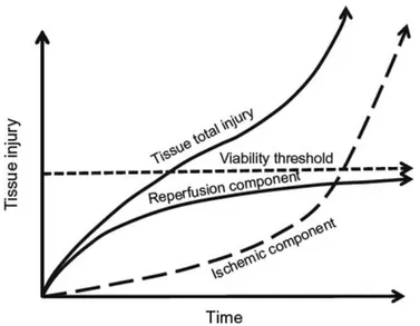
Myocardial ischemia occurs for a mismatch between blood flow and metabolic requirements, when the rate of oxygen and metabolic substrates delivery to the heart is insufficient to meet the myocardial energy requirements. Restoration of oxygenated blood flow, although necessary to re-establish delivery of oxygen and nutrients to support cell metabolism, may induce pathogenic processes that exacerbate the ischemic injury, thus producing a further injury known as I/R injury. Thus, total injury sustained by a tissue represents the sum of damage attributable to ischemia per se plus that invoked by reperfusion, which can be manifested as myocardial necrosis, arrhythmia, myocardial stunning, endothelial and microvascular dysfunction including the no-reflow phenomenon (Kalogeris, Baines et al. 2012). The extent of the ischemic insult is determined primarily by the magnitude and duration of the interruption in the blood supply, whereas the subsequent damage induced by reperfusion is due to mediator release into the bloodstream draining revascularized tissues and subsequent delivery to remote organs. The mechanism of I/R is multifactorial and involves divergent biological pathways. I/R is responsible for endothelial activation leading to microvascular dysfunction and deterioration of coronary flow reserve (Ferguson, Smith et al. 1986, Taqueti and Ridker 2013), vasoconstriction and spasm (de Servi, Poma et al. 1988), myocardial contractile dysfunction (Weisel 1993), reperfusion arrhythmias(Anselmi, Abbate et al. 2004), activation of the inflammation cascade (cytokines, chemokines, complement activation, neutrophil activation) (Levy and Tanaka 2003, Merchant, Nadaraj et al. 2008) and coagulation imbalances (Raivio, Lassila et al. 2009). In summary, the restriction in blood flow to the heart and the following restoration of oxygen and substrates supply leads to cell damage which might result in the development of myocardial infarction, contractile dysfunction and arrhythmias. As a result of intensive investigation over decades, a detailed understanding of
the anatomic and biochemical substrates that contribute to the genesis of I/R injury have been well characterized (Kalogeris, Baines et al. 2012, Lejay, Fang et al. 2015).
1.2 Pathophysiology of myocardial ischemic injury
Myocardial ischemia results in a characteristic pattern of metabolic and ultrastructural changes that may lead to irreversible injury (Fig. 1.1). In absence of oxygen, mitochondrial oxidative phosphorylation rapidly stops leading to mitochondrial membrane depolarization, ATP depletion, and inhibition of myocardial contractile function (Kalogeris, Baines et al. 2012). This process is exacerbated by the breakdown of any available ATP, as the F1-F0 ATPase functions in reverse to maintain the mitochondrial membrane potential. A compensatory increase in anaerobic glycolysis for ATP production leads to the accumulation of hydrogen ions and lactate, resulting in intracellular acidosis (pH to <7.0) and inhibition of mitochondrial fatty acid and residual energy metabolism. Thus, the impaired ATP synthesis rate is the main cause of the imbalances in ionic state across cellular membranes, largely due to the inability of ATP-dependant pumps to function. In detail, at cellular level there is initially an increased K+ efflux related to an increased osmotic load due to the accumulation
of metabolites and inorganic phosphate. An increase in free Mg2+ is followed by a decrease in
total Mg2+. Then, a substantial decline in ATP leads to Na+/K+-ATPase inactivation, resulting
in a further decline of K+ and an increase in Na+. The mechanisms underlying the increased
Na+ early in ischemia are failure to extrude Na+ via the Na+/K+-ATPase and Na+ influx via
Na+/H+ exchange, Na+-HCO3– cotransport (NBC) and the voltage-gated Na+ channel. Na+
efflux via the Na+/K+-ATPase is attenuated during ischemia, therefore activation of other
mechanisms leads to increased intracellular Na+. Both the Na+/H+ exchanger and the
response to the increased intracellular Na+ concentration, the activation of the sodium/calcium
exchanger (NCX), which will be discuss further in detail, in its reverse mode of operation results in intracellular Ca2+ overloading as the cell tries to extrude Na+.
Fig. 1.1: Main events of ischemia injury. Ischemia decreases energy content, acidifies intra- and extracellular environments, and alters ionic homeostasis. Ca2+ accumulation and reactive oxygen species (ROS) production impair cell integrity (Paradis, Charles et al. 2016).
Moreover, in addition to disruptions in ionic homeostasis, cellular systems are overwhelmed by an increase in reactive oxygen species (ROS) production and oxidative stress, which can result in further defective Ca2+ handling (Tompkins, Burwell et al. 2006). More specifically
free radicals can impair Ca2+ stimulated SR and sarcolemmal Ca2+ ATPase activity, thereby
reducing calcium transport into the SR or out of the cell, and exacerbating Ca2+ overload
within the ischemic cell (Netticadan, Temsah et al. 1999). The rise in cytosolic Ca2+ results in
cellular damage by activation of Ca2+ dependent phospholipases, proteases, and calpains,
causing further membrane swelling and rupture, in addition to ischemic contracture. In particular, activation of intracellular proteases (e.g., calpains) damages the myocardial interstitium and microvasculature by producing hypercontracture and contracture band
necrosis. The necrosis of myocytes and no myocytes triggers an inflammatory reaction with subsequent organization and healing (Casey, Arthur et al. 2007). The degree of tissue injury varies in extent with the magnitude of the decrease in the blood supply and with the duration of the ischemic period (Frank, Bonney et al. 2012).
1.3 Pathophysiology of myocardial reperfusion injury
Under experimental circumstances and in clinical situations, ischemia may be followed by reperfusion, that is, the readmission of oxygen and metabolic substrates with washout of ischemic metabolites. Although, reperfusion is essential to salvage ischemic tissue, it has the potential to cause further irreversible cell damage called as reperfusion injury (Fig. 1.2). Restoration of blood flow leads to increasing intracellular pH due to H+ washout and
inadequately restored ATP synthesis, Ca2+ overload and reduced contractility, coronary
endothelial dysfunction, low production of nitric oxide and oxidative stress, migration of inflammatory cells, disruption of membranes and induction of apoptotic and necrotic signals in the myocardium (Garcia-Dorado, Ruiz-Meana et al. 2009). Clearly the recovery of pH, oxidative stress and Ca2+ overload can induce the abrupt opening of the mitochondrial
permeability transition pore (mPTP), a large conductance pore in the inner mitochondrial membrane, which dissipates mitochondrial membrane potential and strongly contributes to myocardial hyper-contracture, apoptosis and necrosis. Opening of the mPTP leads to the release of the enzyme cytochrome c, a potent activator of the apoptotic pathways, and leads to the hydrolysis of ATP rather than synthesis. This causes a rapid decline in the intracellular ATP concentrations, which causes the disruption of ionic and metabolic homeostasis and activation of degradative enzymes and ultimately results in irreversible cell damage and necrotic death (Schriewer, Peek et al. ; Casey, Arthur et al. 2007).
Fig. 1.2: Main events of reperfusion injury. Reperfusion restores pH, produces more ROS, and induces Ca2+ uptake, triggering inflammation, mitochondrial permeability transition pore (mPTP) opening, and cell death. ADP, adenosine diphosphate; ATP, adenosine triphosphate; Pi, inorganic phosphate; ΔΨm, membrane potential (Paradis, Charles et al. 2016).
The consequent Ca2+ increase also enhances ROS accumulation due to the activation of
xanthine oxidase, NADPH oxidases, and mitochondria respiratory chain uncoupling (Yu, Lee et al. 2014), which further exacerbates membrane damage by directly promoting opening of the mPTP, and thus contribute to cell death during reperfusion (Perrelli, Pagliaro et al. 2011). The clinical outcome of reperfusion-mediated injury is also determined by a third phase of ROS production that occurs during post-reperfusion repair that is characterized by tissue remodelling and adaptation (Zorov, Juhaszova et al. 2006). Growing evidence suggests that a defect in myocardial Ca2+ transport system with consequent intracellular Ca2+ overload is one
of the main contributors of myocardial I/R induced injury (Wei, Zhou et al. 2007).
1.4 Myocardial calcium handling
Ca2+ is a very essential intracellular messenger involved in many physiological processes,
myocytes intracellular Ca2+ cycling can be understood as a balance between Ca2+ influx and
efflux pathways governed by a dynamic network of physical and chemical signals. In physiological conditions, resting levels of cytosolic Ca2+ are maintained at submicromolar concentrations by three different mechanisms; (i) Limiting Ca2+ infiltration into the myocytes;
(ii) Sequestering Ca2+ in the intracellular compartments; (iii) Extruding Ca2+ out of the cells against a 10 000-fold ionic gradient. Defined regulation of Ca2+ signals in the heart, which is
crucial for the maintenance of the rhythmic contraction and force development, depend on the free intracellular Ca2+ level and the Ca2+ sensitivity of the cardiomyocytes. In cardiac cells Ca2+ ions are stored inside the SR, which can be utilized for fast contraction and other
physiological activities. Calcium release from intracellular stores plays a central role in cardiac excitation–contraction (E-C) coupling (Fig. 1.3). During depolarization, a small amount of Ca2+ first enters through the L-type Ca2+ channel (LTCC), which are located in
the membrane of T-tubules near the junctional region of the SR. This “calcium trigger” is thought to directly activate the calcium release channels driven in the junctional SR. Opening of these calcium-sensitive Ca2+ release channels known as ryanodine receptors (RyR)
partially empties the internal store of calcium. This mechanism is known as calcium-induced Ca2+ release (Diaz, O'Neill et al. 2004). Binding of calcium to troponin C in the contractile
apparatus initiates muscle contraction (systole), whereas calcium reuptake into the SR primarily by the phospholamban-regulated sarcoplasmic reticulum Ca2+-ATPase (SERCA) allows for cardiac relaxation (diastole). In normal hearts, sympathetic stimulation activates β1-adrenergic receptor, which in turn stimulates the production of cyclic cyclic adenosine monophosphate (cAMP) and thereby activates protein kinase A (PKA). PKA phosphorylates phospholamban and RyR, both of which contribute to an increased intracellular [Ca2+]i
1.4.1 Calcium overload during I/R
The pivotal role of calcium cycling and homeostasis has long been recognized in contractile, metabolic, electrical and ionic alterations associated with myocardial ischemia, as well as in hibernation, stunning and mitochondrial dysfunction associated with reperfusion. During ischemia, the ischemic tissues become dependent on anaerobic glycolysis for their ATP supply. This leads to an accumulation of lactate, protons, and NAD+ which in turn causes a
drop in cytosolic pH. In an attempt to re-establish normal pH, the cell extrudes H+ ions in
exchange for Na+ via the plasmalemmal Na+/H+ exchanger (NHE) (Murphy and Allen 2009).
Prolonged ischemia as well as reperfusion of ischemic heart cause intracellular Ca2+
overloading due to massive Ca2+ entry and dysfunction of Ca2+ sequestration mechanisms.
Ca2+ infiltration during ischemia may predict the severity of Ca2+ influx during reperfusion
leading to reperfusion-induced injury. This increase in cytosolic Ca2+ is greatly exacerbated
upon reperfusion, where removal of extracellular H+ ions further increases the proton gradient
across the plasmalemma, thereby accelerating NHE exchanger function. In addition to detrimental alterations in plasmalemmal Ca2+ handling, the endoplasmic/sarcoplasmic
reticulum (ER/SR) Ca2+ store is also affected during I/R. In particular, Ca2+ reuptake into the
ER/SR by the SERCA ATPase is impaired by I/R, whereas Ca2+ release through the RyRs is
enhanced (Netticadan, Temsah et al. 2000, Sanada, Komuro et al. 2011), both of which further exacerbate the lethal elevations in intracellular Ca2+. Such dysregulation of Ca2+
homeostasis results in the alteration of a cascade of Ca2+-dependent cellular events which are
Fig. 1.4: Ion exchanges during ischemia: excretion of H+ due to pH lowering, deactivation due to loss of ATP, and reduction of Na+/Ca2+ exchange due to lowered extracellular pH and intracellular accumulation of Na+(Sanada, Komuro et al. 2011).
I/R-induced elevations in cytosolic Ca2+ leads to the pathological activation of
Ca2+/calmodulin-dependent protein kinases (CaMKs), which also contribute to cell death and
organ dysfunction following ischemia. In addition, another target for Ca2+ are the calpains
(Verburg, Murphy et al. 2009). This family of cysteine proteases is activated by elevation of Ca2+ and degrades a panoply of intracellular proteins, including cytoskeletal, ER, and
mitochondrial proteins that predispose the myocyte to lethal reperfusion injury (Croall and Ersfeld 2007). One of the ways cells deal with this lethal increase in Ca2+ is to take it up into
the mitochondria via the mitochondrial Ca2+ uniporter (MCU), a protein that uses the negative Δψm to drive uptake of the positively charged Ca2+ ions into the matrix. However, if the
elevations in mitochondrial Ca2+ become excessive, they can trigger the mPTP response
(Tompkins, Burwell et al. 2006). Any major and chronic imbalance in Ca2+ handling,
exceeding the physiological shifts in [Ca2+]i, may result in gradual cellular [Ca2+]i overload,
leading to highly increased arrhythmia propensity, or cell injury.
One of the key regulator in maintaining the balance of the intracellular Ca2+ is NCX, which
contributes to calcium overload during I/R. Since calcium overload is perhaps the most critical factor in determining the biochemical basis of ultimate myocytes cell death (Talukder, Zweier et al. 2009), the coordinated regulation of Ca2+ homeostasis through NCX need to be
clearly understood in order to develop novel therapeutic strategies for patient care and for the prevention of I/R injury.
1.5 The sodium calcium exchanger (NCX)
NCX is a membrane associated protein that catalyses electrogenic exchange of 3 Na+ ions and
1 Ca2+ ion across the plasma membrane in a high capacity, and low Ca2+ affinity fashion.
This transporter can operate in either the Ca2+ -efflux or Ca2+ -influx mode depending on the
prevailing electrochemical driving forces of the substrate ions and the membrane potential. Three mammalian isoforms have been cloned to date (NCX1-3) and their splice variants are
expressed in a tissue-specific manner (Philipson and Nicoll 2000, Lytton 2007) to fulfil physiological demands in excitable and non-excitable tissues (Carafoli 1987, Carafoli 1988, Blaustein and Lederer 1999) NCX2 and NCX3 have been found in the brain and skeletal muscle, whereas NCX1 is widely distributed in mammalian cells. The canine NCX1 subtype, which has been found to be predominantly expressed in the heart, was the first to be purified and cloned. The cardiac NCX consists of 970 amino acids with a molecular mass of 110 kDa. Biochemical analyses over the years have indicated the presence of 9 transmembrane segments (TMSs) (Ren and Philipson 2013). However, major advancement in understanding the structural basis has only recently been achieved by solving the crystal structure of a prokaryotic homologue of the exchanger, which revealed the presence of 10 α-helical TMSs rather than the 9 TMS proposed for NCX1 (Giladi, Lee et al. 2017). This model incorporates an additional TMS8 and an extracellular C-terminus (Fig. 1.5). Interestingly, NCX1 possess a
short motif (about 30 residues) that is similar to the Na+/K+-ATPase with residues 248–252 and 300–304 involved in its regulation by phospholemmon (Blaustein and Lederer 1999). The NH2-terminal portion of the loop has a 20-amino acid domain rich in hydrophobic and basic amino acids and is called the XIP (exchange inhibitor peptide) region. This region is associated with the sodium inactivation process and it can bind several peptide mimetic compounds. In this regard, recent research showed that a new cell penetrating peptide called P1 is capable to blocks the auto inhibitory XIP domain and enhances NCX activity (Molinaro, Pannaccione et al. 2015). Around the central zone of the loop in the NH2-terminal portion, there is a sequence of 135 amino acids containing highly acidic residues (two zones of three aspartyl each) that bind [Ca2+]i with high affinity; this region is responsible for the [Ca2+
]i-dependent or allosteric regulation of the exchanger (Matsuoka, Nicoll et al. 1995).
Fig. 1.5: The schematic molecular structure of NCX. The molecule contains 10 TM segments, a P-loop domain, and the second α repeat (α-2). The central regions of the two α repeats facing opposite in the membrane and contain amino acids that are essential for normal NCX function. The two CBD domains are different (CBD1,CBD2). CBD1 binds four Ca2+ ions with high affinity. CBD2 contains the variable alternative splicing region (Emery, Whelan et al. 2012) .
The properties of Ca2+-binding proteins that participate in Ca2+-dependent regulation of
splicing of closely related genes. The isoform-dependent and alternative splicing-dependent modification of Ca2+-binding regulatory domains are especially important for Ca2+
-transporting proteins operating in excitable tissues, since feedback interactions of Ca2+ with
allosteric regulatory domains dynamically modulate the Ca2+-transport rates in accordance
with dynamic changes in cellular Ca2+ oscillations (Tal, Kozlovsky et al. 2016).
The differential role of NCX subtypes has been extensively investigated in ischemic renal and neuronal injury using various pharmacological tools as well as genetic models. The pathological role of NCX has been studied in I/R-induced renal injury using NCX1 heterozygous mice and it was observed that I/R-induced renal dysfunction, histological damage and Ca2+ accumulation in necrotic tubular epithelium were more markedly in NCX1
wild type than in NCX1 heterozygous mice (Yamashita, Kita et al. 2003). Some reports also supported the deleterious role of NCX revealing the efficacy of pre- and post-ischemic treatments with KB-R7943, an inhibitor of the reverse mode of NCX, which attenuate the I/R-induced renal injury by suppressing the intracellular Ca2+ overload (Yang, Jia et al. 2013,
Yang, Jia et al. 2013). In the brain, unlike other tissues, NCX is present in the three different gene products NCX1, NCX2 and NCX3 with a distinct distribution pattern in different brain regions. Moreover, NCX subtypes may carry variable roles in ischemic injury and the mode of action of each subtype may vary in ischemia and reperfusion states exerting different roles during in vitro and in vivo anoxic conditions leading to a new paradigm in the pathogenesis of ischemic damage (Thurneysen, Nicoll et al. 2002). Thus, NCX subtype-specific strategies, which should be based on appropriate timing of administration, have become an alternative therapeutic approach to limit the severity of ischemic injury (Shenoda 2015). Indeed, the majority of in vivo studies using the focal cerebral ischaemia model indicate that blocking NCX activity is neurodamaging while increasing NCX activity is neuroprotective (Jeffs, Meloni et al. 2007, Sisalli, Secondo et al. 2014). More interestingly, this hypothesis was
supported by the observed increase in NCX1 and NCX3 activity responsible for Ca2+ cycling
from ER and mitochondria leading to neuroprotection (Sisalli, Secondo et al. 2014). In this scenario, a promising selective compounds able to stimulate the activity of NCX1 and to prevent neuronal degeneration in vitro and in vivo models of ischemia has been recently synthesized (Molinaro, Cuomo et al. 2008). Over the last few years, although extensive studies have potentially revealed new molecular targets in cerebral ischemia, a thorough understanding of the role played by NCX still remains a controversial issue.
1.5.1 Role of NCX in myocardial I/R injury
In cardiac cells, NCX1 is one of the essential regulators of Ca2+ homeostasis playing an
important role participating in nodal pace-maker activity (Groenke, Larson et al. 2013), cell metabolism (Magi, Lariccia et al. 2012, Magi, Arcangeli et al. 2013) and E-C coupling (Aronsen, Swift et al. 2013). In E-C coupling, contraction is initiated by influx of Ca2+ through voltage-dependent Ca2+ channels, which then triggers a release of Ca2+ from the
SR by a Ca2+-induced Ca2+ release mechanism. Relaxation is accomplished by the extrusion
of Ca2+ from the cell by both NCX activity and by the reuptake of Ca2+ into the SR. In this
scenario, the role of NCX as a primary Ca2+ extrusion mechanism in the heart is widely
accepted. However, the expression and activities of NCX can be regulated by a variety of factors, among which, membrane potential, as well as Na+ and Ca2+ gradients, are crucial.
NCX can be also a source for Ca2+-influx, which can be enhanced by either decreasing the
Na+ or increasing the Ca2+ gradients across the transarcolemmal membrane, bringing Ca2+
into cardiomyocytes in certain circumstances. Usually, the so called “reverse mode” of NCX becomes predominant in pathological settings, which can significantly alter Ca2+ homeostasis
systemic levels. During I/R, NCX acts reversely to induce Ca2+ influx, which strengthens
Ca2+ overload that is mainly involved in cell structural damage, arrhythmia and systolic
dysfunction (Bers 2008). Although ischemia and reperfusion are often referred to simultaneously, NCX may act differently during the ischemia and reperfusion phase. During ischemia, intracellular changes in Na+ levels, pH and rigor shortening of the heart have been
shown to be mediated by an increase in the Na+/H+ exchanger function. This leads to a rapid
increase in intracellular Na+ that alters the sarcolemmal Na+ gradient, which in turn results in
Ca2+ overload and swamping of the SR. In order to compensate, the NCX is upregulated to
assist the SR in maintaining Ca2+ homoeostasis. However, following long periods of
ischemia/hypoxia, reoxygenation of the cardiomyocytes, in which the cell tries to recover and re-establish the Na+ and Ca2+ gradients, is not able to promote recovery (Chen and Li 2012).
The role of NCX in cardiac cells following I/R has been largely investigated in different in vitro and in vivo models. Recent reports have shown that diabetic cardiomyocytes display an impaired [Ca2+]i homeostasis due to an altered NCX activity (Hattori, Matsuda et al. 2000,
Ma, Zhu et al. 2008) that may precede clinically manifested cardiac dysfunction. Indeed, NCX protein levels as well as NCX activity during Ca2+ efflux have been found diminished in
diabetic cardiac endothelial cells suggesting that the abnormal cardiac function might be due to the compromised ability to maintain Ca2+ homeostasis (Sheikh, Hurley et al. 2012).
Thus, the expression level and activity of NCX1 in the heart is an important factor in understanding cardiac pathophysiology. Other studies by using two different NCX1-KO mouse models revealed that the complete removal of the exchanger is associated with embryonic lethality, whereas its overexpression is linked to severe remodelling and heart failure. In particular, in heterozygous global NCX1-KO mice, with a 50% level of NCX1 expression, there are no significant structural as well as functional alterations of myocytes, whereas ventricular specific NCX1-KO mice, with NCX1 expression in only 10-20% of
myocytes, tolerate better I/R injury (Imahashi, Pott et al. 2005) but display cardiac alterations as animals age (Jordan, Henderson et al. 2010). Since the protein expression levels and/or regulation of NCX isoform/variants are disease related in many pathological states (e.g., heart failure, cardiac arrhythmia, and I/R injury), the selective pharmacological targeting of tissue-specific NCX variants could be beneficial, although this remains still a matter of debate (Giladi, Shor et al. 2016). The majority of studies often refers to a negative role of NCX during I/R, since a reduction of Ca2+ entry through its pharmacological or genetic inhibition
would significantly limit the damage induced by I/R preventing the detrimental effect of Ca2+
overload (Weber, Piacentino et al. 2003, Imahashi, Pott et al. 2005). The lack of Ca2+ entry
via the reverse-mode of NCX would decrease the detrimental effects of Ca2+ overload and
better preserve ATP, which would prolong the activity of the Na+/K+-ATPase, leading to less
Na+ accumulation during ischemia in NCX-KO hearts. On reperfusion, the better-preserved
ATP in the NCX-KO hearts facilitates Na+ extrusion by Na+/K+-ATPase (Imahashi, Pott et al.
2005). Further, NCX mediates the increase in Ca2+- CaMKII activity at the onset of
reperfusion, which also increases the phosphorylation of Thr17 site of PLN, without changes in total protein, consistent with an increase in CaMKII activity. NCX inihibition produces a significant decrease in the phosphorylation of Thr17 of PLN with consequent decrease in infarct size, lactate dehydrogenase (LDH) release, and apoptosis produced by I/R injury. Taken together, these experiments indicate that the reverse NCX mode is a major pathway of Ca2+ influx upon reperfusion able to activate CaMKII which is considered a maladaptive
mediator of cardiac ischemic injury (Salas, Valverde et al. 2010). Moreover, prior stimulation of the reverse-mode NCX not only reduces the infarct size but also attenuates arrhythmias induced by I/R (Zhang, Cheng et al. 2015, Castaldo, Macri et al. 2016). However, recent studies have demonstrated that NCX1 activity can be strategic for cardioprotection against I/R evoked by conditioning programs, whereby short periods of
subcritical ischemic stimuli confer protection. Collectively these findings confirm that NCX1 may have a deleterious role in I/R, but it may be also beneficial in promoting cardioprotection. Thus, up to date a dual role of NCX1 in cardiac ischemia is emerging: on the one hand its inhibition during I/R might lead to cell survival, on the other hand its activity can be strategic as a trigger for the induction of ischemic tolerance.
1.6 Cardioprotective strategies against I/R injury
The pathophysiology of ischemic heart disease is complex, and a therapeutic strategy capable of activating multiple arms of the cardioprotective signalling network and/or simultaneously counteracting multiple mediators of I/R injury would likely have the greatest efficacy in mitigating ischemic death. It is well known that the extent of cell dysfunction, injury, and/or death is influenced by both the magnitude and the duration of ischemia. In recognition of this fact, revascularization and restoration of blood flow as soon as possible remains one of the the mainstay of all current therapeutic approaches to ischemia (Fig. 1.6). Over the years, huge efforts have been made to ensure early reperfusion according to the “time is muscle” principle: the quicker the perfusion is restored, the more heart tissue is saved. In particular, if reperfusion is instituted within 2 to 3 h of the onset of ischemia, the amount of salvage greatly exceeds the amount of myocardium undergoing irreversible reperfusion injury. Prompt restoration of blood flow can lead to the conversion from reversible to irreversible injury of a population of myocytes that have been severely impaired during the prior period of ischemia. For that reason, the treatment of reperfusion injury following ischemia is primarily supportive, even though no specific target-oriented therapy has been validated so far (Garcia-Dorado, Rodriguez-Sinovas et al. 2014). Therefore, there is a great need for the development of novel ischemic heart disease therapies that can make the heart more
resistant to ischemic death, the so called “cardioprotective” interventions. Pharmacological, mechanical, and combined reperfusion techniques have been refined over the years, and their efficacy has been greatly advanced by the development of adjunctive therapies (Gerczuk and Kloner 2012) .
Fig. 1.6: Total injury sustained by a tissue subjected to I/R. Tissue injury and/or death occur as a result of the initial ischemic insult due to the magnitude and duration of the interruption in the blood supply, and then subsequent damage induced by reperfusion. Therapy against I/R injury will be more effective when administered after a period of ischemia that corresponds to the therapeutic window, where the major component of the total injury is due to reperfusion (Bulkley 1987).
Growing evidence has been shown that I/R-induced cell damage can be prevented or limited by triggering intrinsic adaptive response through non-pharmacological strategies such as ischemic pre-conditioning (PreC), ischemic post-conditioning (PostC) and hypothermia. Since 1986, the cardioprotective potential of conditioning programmes, whereby short periods of subcritical ischemic stimuli performed before (PreC) (Murry, Jennings et al. 1986) or after (PostC) (Vinten-Johansen and Shi 2011) the index ischemia, has been demonstrated by several reports. Intensive investigation of the mechanisms underlying PreC and PostC have identified a number of signal transduction pathways conveying the cardioprotective signal from the sarcolemma to the mitochondria, some of
which are overlapping. In fact, these pathways induce activation of signalling elements to preserve mitochondrial function during the early reperfusion following the index ischemia (Perrelli, Pagliaro et al. 2011). Another form of conditioning capable to increase myocardial salvage can be performed in a distant organ and it is known as “remote conditioning” (Healy, Feeley et al. 2015). Among these interventions, PreC has demonstrated the most consistent ability to confer robust cardioprotection in several different mammalian models (Cohen and Downey 1996). However, the clinical value of this approach is limited by the fact that patients suffering from myocardial ischemia do not present any symptoms until after the onset of ischemia. Among the non-pharmacological interventions, therapeutic hypothermia can also have a beneficial effect in limiting infarct size by lowering myocardial temperature during ischemia via reductions in metabolic demand, inflammatory response and platelet aggregation (Kanemoto, Matsubara et al. 2009). The fact that so many different interventions are capable of reducing ischemic death means that there are likely several pathways involved in the robust cardiac signalling network that can ultimately elicit a cardioprotective state. While the effects of myocardial ischemia on electrical propagation and stability have been studied in depth, the effects of I/R on energy metabolism has not been widely appreciated yet. Since changes in cardiac metabolism are understood to be an underlying component in almost all cardiac myopathies, the cardioprotective potential of interventions aimed to rescue the metabolic derangement occurring during I/R remains an active area of intense investigation. Thus, to optimize the heart’s alternative fuel usage and minimize the I/R-induced damage, the molecular machinery involved in metabolic responses both in physiological and pathological conditions needs to be fully elucidated.
1.7 Myocardial energy metabolism
The heart is a highly active organ that consumes 10% of the body’s total oxygen uptake and produces upwards of 35 kg of ATP every day (Taegtmeyer 1994). This high rate of energy flux required oxygen and a wide variety of metabolic substrates to supply the its vast energy needs.
Fig. 1.7: Contribution of various metabolic pathways to energy production in the normal heart. ATP production in the heart originates primarily from the mitochondrial oxidation of fatty acids, pyruvate (originating from glucose and lactate), ketones, and amino acids (Lopaschuk 2017).
Central to the coordinated energy transduction function is the multi-purpose organelle mitochondrion, which occupies one third of the cell volume in cardiac myocytes making them the cell type with the highest mitochondria content. While a constant supply of substrates through the metabolic network is paramount for mitochondrial conversion of ATP, it is increasingly recognized that metabolites generated by both ATP-producing and non-ATP producing pathways can become critical regulators of cell function (Kolwicz, Purohit et al. 2013). In a normal heart, mitochondria are largely fueled by fatty acyl-CoA and pyruvate,
which are the primary metabolites of fatty acids and carbohydrates, respectively. The entry of long-chain acyl-CoA into the mitochondrion is a regulated process; with the rate-limiting step at the muscle form of the carnitine-palmitoyl transferase I (mCPT-1) reaction (Smith, Perry et al. 2012). The oxidation of pyruvate is regulated at the pyruvate dehydrogenase (PDH) reaction whereas other substrates, including lactate, ketone bodies and amino acids, can enter mitochondria directly for oxidation (McCommis, Chen et al. 2015, McCommis and Finck 2015). The contribution of ketone bodies and amino acids to overall cardiac oxidative metabolism, however, is considered to be minor due to the low availability of these substrates under normal physiological conditions. Metabolism of ketone bodies yields acetyl-CoA while amino acid catabolism yields keto-acids which are further metabolized to enter the TCA cycle (Kolwicz, Purohit et al. 2013) (Fig. 1.7). It is widely accepted that fatty acids are the predominant substrate utilized in the adult myocardium. However, the cardiac metabolic network is highly flexible in utilizing other substrates when they become abundantly available. For example, cardiac extraction and oxidation of lactate becomes predominant during exercise as skeletal muscle lactate production increases (Goodwin and Taegtmeyer 2000). The metabolic flexibility of the heart, which results in modulations of myocardial energetic and contractile function, confers the advantage of adequately supplying ATP under a variety of patophysiological conditions (Schaper, Meiser et al. 1985).
1.7.1 Energy metabolism impairment during I/R
In many types of heart diseases and dysfunctions, metabolism is the first area affected, which can then lead to channelopathies, ion imbalance, decreased contractile function, increased free radical production and cardiac death. Fortunately, the heart is also quite resilient, being able to maintain contractile function even under ischemic and anoxic
conditions. In this light, an understanding of metabolism is crucial for any study of the heart, which under conditions of prolonged stress or ischemia is capable of utilizing glucose, lactate, fatty acids, ketone bodies as alternative metabolic substrates (de Windt, Cox et al. 2002). It is well known that the heart’s primary metabolic substrates under normal conditions are fatty acids and lactate, but under conditions unfavorable for oxidation, i.e., ischemia, anoxia and many types of cardiomyopathy, fatty acid oxidation is inhibited and the heart preferentially performs glycolysis and substrate-level phosphorylation. Without oxygen, the heart cannot break down fatty acids and ketone bodies into acetyl-CoA (Stanley 2004). There is also a decrease in fatty acid consumption and an increase in glycolysis in hypertrophied hearts.
Fig. 1.8: Contribution of various metabolic pathways to energy production in the reperfused ischemic heart. During ischemia, ATP production decreases due to a limitation of oxygen supply and glycolysis increases in attempt to compensate for the decrease in mitochondrial oxidative metabolism. During reperfusion, fatty acid oxidation rapidly increases and dominates as a source of ATP production, glycolysis remains high during reperfusion, and pyruvate is shunted toward lactate production because of the decreased mitochondrial oxidation of pyruvate (Lopaschuk 2017).
Thus, glucose and glycolysis become the primary means of producing ATP, but this yields far less energy per molecule and produces two protons per molecule of glucose
consumed. This energy deficit and acidification increase become especially important during acute ischemia, as a lower pH inhibits glycolysis. The heart is unable to produce sufficient ATP through glycolysis, and it will be forced to turn to anaerobic, non-glycolytic fuels, such as amino acids, to utilize alternative metabolic that do not strictly require oxidation and do not require glycolytic conversion, which contributes to increased acidification (Fig. 1.8).
1.8 Role of amino acids in cardiac metabolism
Amino acids play a central role in cardiac metabolism. They can be readily metabolized into Krebs cycle intermediates; even though not all amino acids are metabolized equally in the heart (Lopaschuk and Ussher 2017). There are several different pathways by which amino acids can be used to supply cardiomyocytes with energy.
Glutamate and α-ketoglutarate are readily interconvertible via transamination reaction. This reaction lead to an increase in Krebs cycle intermediates called anaplerosis and contributes to the maintenance of oxidative capacity (Tepavcevic, Milutinovic et al. 2015).
Asparagine can serve as a means of exporting nitrogen from the cell, or it can be converted to aspartate.
Aspartate can be transaminated to oxaloacetate, an other crucial regulator of levels of Krebs cycle (Drake, Sidorov et al. 2012).
Alanine is usually transported out of the myocardium to remove amine groups from the working cell or it can also be transaminated to become pyruvate (Taegtmeyer, Peterson et al. 1977).
Branched Chain Amino Acid (BCAAs) can be converted to acetyl-CoA, pyruvate or succinyl-CoA through different processes (Huang, Zhou et al. 2011).
However, in the fully functional heart, amino acid metabolism makes up a very small percentage of cardiac ATP production, but as the heart becomes oxygen-limited, they become more important as alternative fuel source (Fig. 1.9). In low/no-oxygen conditions, the heart will switch to anaerobic, non-glycolytic fuels, such as amino acids to meet its energy requirements. In this scenario, amino acids are now becoming more widely appreciated as cardioprotective substrates, due to their potential for non-oxidative metabolism and their low contribution to cellular acidification. Since amino acids are synthesized in a wide variety of pathways and reactions, some of them are more readily converted to metabolic intermediates than others.
Fig. 1.9: Metabolite supply and function in the heart. The heart is capable of oxidizing a wide variety of substrates, including glucose, fatty acids and amino acids to supply its vast energy needs (Kolwicz, Purohit et al. 2013).
In particular, glutamate due to the ease with which this particular amino acid can be converted to α-ketoglutarate, may maintain the levels of Krebs cycle intermediates
keeping the metabolic machinery primed to begin oxidative phosphorylation as soon as oxygen returns improving the recovery after an ischemic event (Maus and Peters 2016).
1.8.1 Glutamate as metabolic substrate
Glutamate is classified as a non-essential amino acid, which means that it can be synthesized endogenously through distinct metabolic pathways (Fig. 1.10). In particular, It can be synthesized from glutamine, α-ketoglutarate and 5-oxoproline (Hu et al. 2010; Cho et al. 2001; Chen et al. 1998) and also serves as a precursor for the biosynthesis of amino acids such as L-proline and L-arginine (Young and Ajami 2000). Glutamate is also a neurotransmitter and may interfere with normal function of the nervous system, causing unintended side effects (Zhou and Danbolt 2014). However, also in the brain, glutamate may serve as an ideal metabolic substitute improving neurotoxic glutamate clearance and anaplerotic flux of TCA cycle intermediates in the stroke-affected brain (Khanna, Stewart et al. 2017, Rink, Gnyawali et al. 2017). In the brain, astrocytes, which are essential partners in neurotransmission, can readily shift the metabolic fate of glutamate removed from the synaptic cleft towards increased oxidative energy metabolism relative to direct formation of glutamine (Schousboe, Scafidi et al. 2014). In the heart, glutamate, which constitutes a major proportion of the free intracellular amino acid pool, is taken up in larger quantities than any other amino acid with an increased uptake during ischemia (Nielsen, Stottrup et al. 2011). Ischemia itself significantly reduces the tissue stores of glutamate, ATP and glutathione while increasing lactate levels (Wischmeyer, Jayakar et al. 2003). In response to ischemia, the heart will significantly enhance alanine production and glutamate consumption, though these effects become significant only when the partial pressure of oxygen in the tissue falls to 5% of normal levels (Peuhkurinen, Takala et al. 1983). During early post ischemic reperfusion myocardial glutamate concentrations
decrease in humans and in a rat isolated heart model (Kristiansen, Lofgren et al. 2008), likely due to an increased glutamate utilization. Numerous in vivo and in vitro studies have been conducted to delineate how glutamate can rescue cardiomyocytes from I/R-induced cell damage, in order to evaluate its potential as therapeutic tool. Indeed, isolation of cardiomyocytes in the presence of glutamate leads to an enhancement of their intracellular glutamate concentration. These glutamate-loaded cells exhibit a greater NADH/NAD+ ratio, higher ATP levels and an enhanced capacity to remove intracellular
reactive oxygen species during oxidative stress compared to control cells (Williams, King et al. 2001)
Fig. 1.10: Schematic overview of anaplerotic glutamate utilization as metabolic
substrate. Glutamate derived from glutamine by glutaminase (GLS) is converted to
α-ketoglutarate by glutamate oxaloacetate transaminase (GOT). Aminooxyacetate (AOA) inhibits GOT and therefore suppresses generation of α-ketoglutarate from glutamine-derived glutamate (Mukhopadhyay, Saqcena et al. 2015).
.Furthermore, exogenously administered glutamate given in the postoperative phase or as an adjunct to cardioplegic solutions reduces infarct size in patients with coronary artery disease and improves left ventricular recovery and function in patients undergoing
coronary artery bypass surgery (Svedjeholm, Vanhanen et al. 1996). These effects are likely depend on preserved transamination of glutamate and K-ATP channel activity (Kristiansen, Nielsen-Kudsk et al. 2005, Lofgren, Povlsen et al. 2010). The general idea is that glutamate may work as reservoir in cardiac output by maintaining metabolic intermediates acting as a substrate for complex I of the respiratory chain and/or as an intermediate for replenishment of citric acid cycle metabolites after ischemia and hypoxia (Drake, Sidorov et al. 2012).
Despite clinical and experimental evidence of a glutamate-induced cardioprotection in ischemia and other cardiac disorders, the underlying mechanisms for this effect has yet to be fully elucidated. The cardioprotective role of glutamate may stem from facilitation of cardiac cell function during anaerobic conditions as well as by supporting a rapid return to optimal function upon restoration of aerobic conditions. Both cases require the glutamate entry in to the cells and then into the mitochondria for enhancing activity-triggered metabolism. It is generally accepted that glutamate can get access to the mitochondrial matrix via the aspartate/glutamate carriers, a required component of the malate/aspartate shuttle (Wang, Chen et al. 2014). However, it has been recently proposed that glutamate may be carried in heart tissue through a family of proteins known as excitatory amino acid transporters (EAATs), which represent the major route of glutamate uptake into cells (Ralphe, Segar et al. 2004, King, Lin et al. 2006).
1.9 Excitatory amino acid transporters (EAATs)
The EAATs belong to the SLC1 family of transporters that also includes two mammalian neutral amino acid transporters as well as a large number of prokaryotic neutral and acidic amino acid transporters. There are five subtypes of EAATS, which are named EAAT1–5 in humans, with the rodent versions of EAAT1 and EAAT2 referred to as
GLAST1 and GLT1 respectively, and the rabbit version of EAAT3 referred to as EAAC1 (Vandenberg and Ryan 2013). Three of these transporters, EAAT1, EAAT3 and the variant form of EAAT2, are expressed in heart, with EAAT1 identified in the mitochondrial inner membrane, whilst EAAT2 and EAAT3 localise to the sarcolemmal membrane and T tubules (Shigeri, Seal et al. 2004). All EAATs are Na+-dependent and
display high affinity uptake of l-glutamate and d- or l-aspartate (Fig. 1.11). Limited data are available about the expression of glutamate transporters involved in the uptake of glutamate and in its role in the control of energy metabolism in cardiomyocytes. In this regard, an association of Glutamate–Aspartate Transporter (GLAST) (Karlsson, Tanaka et al. 2009), Glutamate Transporter 1 (GLT-1) (Genda, Jackson et al. 2011) and Excitatory Amino Acid Carrier 1 (EAAC1) (Magi, Lariccia et al. 2012) with glycolitic enzymes and mitochondria has been reported (Genda, Jackson et al. , Shigeri, Seal et al. 2004). Since EAATs transports glutamate using the favorable Na+ gradient (Tzingounis
and Wadiche 2007), their activity is expected to diminish as Na+ accumulates, and
eventually to stop unless specific mechanisms that preserve the Na+ gradient are
activated. The main Na+ efflux system in the mitochondrial matrix in physiological
conditions is the Na+/H+ exchanger (NHE), however its role in glutamate dependent
influx pathway is presumablytrifling, since H+ is cotransported with Na+ while glutamate
is transported by EAATs (Vandenberg and Ryan 2013). Recent studies have suggested that the Na+/K+-ATPase, the antiporter enzyme which maintains the Na+ and K+ ion
gradients across the membrane, may regulate glutamate uptake via EAATs (Wilmsdorff, Blaich et al. 2013). However, a variable but significant component of the Na+ dependent
glutamate transport activity is resistant to Na+/K+-ATPase inhibition, suggesting that the
Na+ gradient across the membrane may be sustained through a different mechanism
Fig. 1.11: Stechiometry of EAAT. Glutamate transport by the EAATs is coupled to the cotransport of 3 Na+ and 1 H+ followed by the countertransport of 1 K+ (Vandenberg and Ryan 2013).
1.10 NCX and EAATs interplay
Another transporter that may support glutamate entry via EAAT is NCX. It has been already proposed that glutamate and Na+ entry via EAAT induces a Ca2+ response due to
the reverse mode of plasma membrane NCX (Kirischuk, Kettenmann et al. 2007). However, our group for the first time showed a selective interaction between a specific EAAT subtype, EAAC1, and a specific NCX subtype, NCX1 at cytosolic and mitochondrial level. Their physical association, disclosed by their co-localization, coimmunoprecipitation and mutual activity dependency, emphasizes the high selectivity of the interaction, which represents a novel and complementary mechanism enhancing energy metabolism to meet the increased demand both in neuronal and cardiac model. The mitochondrial localization of EAAC1 and NCX1 suggests that such complex may represent another way by which glutamate enters into mitochondria to sustain energy production. According to this alternative pathway, EAAC1 and NCX1 exist as a macromolecular complex, which cooperate favoring glutamate entry into the cytoplasm
into the cells through EAAC1 leading to an increase in cytosolic [Ca2+]. The increased
ATP synthesis needed in many cells in which stimuli have increased cytoplasmic concentrations of calcium ions may be brought about, at least in part, by parallel increases in mitochondrial concentrations of calcium ions activating the intramitochondrial calcium-sensitive dehydrogenases (Denton 2009). The activation of these enzymes is important in the stimulation of the respiratory chain and hence ATP supply under conditions of increased ATP demand. This effect, in the presence of glutamate as substrate, may contribute to boost energy metabolism with a concomitant increase in ATP synthesis. Thus, the EAAC1-NCX1 dependent influx pathway, which play a crucial role in the glutamate-dependent metabolic response in physiological conditions (Magi, Lariccia et al. 2012, Magi, Arcangeli et al. 2013), may have important implications in I/R setting, where substantial changes in energy metabolism occur as a consequence of the reduced oxygen availability.
Fig. 1.11: Coordinated activity between NCX and EAATS. The reverse mode of NCX maintains the transmembrane Na+ gradients, hence, supporting effective operation of glutamate uptake through EAATS.
1.11 Aim of the thesis:
On the basis of this previous finding, an experimental model of hypoxia/reoxigenation (H/R) has been employed in cardiac cells with the aim of:
Investigating whether glutamate supplementation during the reoxygenation phase may improve the energy state of the cells and protect against H/R injury;
Studying whether NCX and EAATs may be critically involved in the glutamate-induced metabolic response during H/R.
2. MATERIALS AND METHODS
2.1 Cell Culture
H9C2 rat cardiomyoblast, purchased from the American Type Culture Collection (CRL-1446), were used as in vitro model of cardiac myocites. Although H9C2 cells are no longer able to beat, they still show many similarities to primary cardiomyocytes, including membrane morphology, g-signalling protein expression and electrophysiological properties (Hescheler et al. 1991; Sipido and Marban 1991). For the experiments two different clones were used: H9c2 Wilde Type (WT), devoid of any detectable endogenous NCX1 and H9c2-NCX1 cells stably expressing H9c2-NCX1 as previously described (Magi, Lariccia et al. 2012). Both cell lines were cultured as monolayer to sub-confluence in polystyrene dishes (100 mm diameter) and grown in Dulbecco's Modified Eagle Medium, DMEM (Invitrogen, Carlsbad, CA) supplemented with 10% heat inactivated fetal bovine serum (Invitrogen), 1% L-glutamine (200 mM) (Invitrogen), 1% sodium pyruvate (100 mM) (Invitrogen), 100 IU/ml penicillin (Invitrogen), and 100 μg/ml streptomycin (Invitrogen). Cells were grown in a humidified incubator at 37°C in a 5% CO2 atmosphere. Medium was changed every two days. Cells were plated the day before to conduct each experiment.
2.2 Isolation of rat adult ventricular cardiomyocytes
The animal experiments were conducted following the protocol approved by the Ethic Committee for Animal Experiments of the University Politecnica of Marche (Ref no. 721/2015-PR) and in strict accordance with the guidelines of the Italian Ministry of Health (D.L.116/92 and D.L.111/94- B). All efforts were made to minimize the number of animals used as well as their suffering.
Rat adult ventricular cardiomyocytes from one-month old male Wistar rats (Charles River, Lecco, Italy) were isolated by Collagenase type II-CLS2 (Worthington Biochemical Corporation, Lakewood, NJ) digestion using a modified Langendorff perfusion system. Firstly, rats were anesthetized with 4% isoflurane in 100% O2 and then intraperitoneally
injected with 1 ml of heparin (5000 IU/ml). After 10 min, the chest was opened and 1 ml of heparin (160 UI/ml) was injected into the right atrium. Then the heart was quickly excised, attached to the modified Langendorff perfusion system (Fig. 2.1) and retrogradely perfused with an O2-saturated HEPES buffered solution containing (in mM): NaCl 140, KCl 4, HEPES
10, Na2HPO4 0.5, MgCl2 1, CaCl2 1.5, glucose 15, pH 7.4 adjusted with NaOH
Figure 2.1: Fixation of the heart on Langendorff perfusion system.
After 2 min, the solution was switched to nominally Ca2+-free HEPES buffered solution for 5
min, followed by perfusion with the same solution containing 100 μM EGTA for 2 min. After that, the heart was perfused with enzyme solution (150-200 U/ml) containing 30 µM blebbistatin (Sigma, Milan, Italy) until the heart became swollen and turned lightly pale (usually 10 min). The digestion was stopped by perfusing HEPES buffered solution containing 10% FBS. Perfusion solutions were constantly gassed with O2 and the temperature
petri dish containing the perfusion solution without Ca2+, with 100 μM EGTA and 30 µM blebbistatin. The ventricular tissue was chopped with small scissors, and single cells were isolated by mechanical dispersion. Single cardiomyocytes were harvested after filtration through a nylon mesh. At this point, Ca2+ was gradually reintroduced by incubating the
cardiomyocytes in the perfusion solution with increasing concentrations of calcium, achieving a concentration of 1.8 mM as in the culture medium (O'Connell, Rodrigo et al. 2007). Once the cardiomyocytes were equilibrated (Ca2+ tolerant), they were plated at a
density of 10 000 cells/cm2 on laminin coated dishes, and cultured in M-199 medium containing L-glutamine, NaHCO3 and Earle's salts, supplemented with 0.2% bovine serum albumin, 1X insulin-transferrin-selenium (Gibco, Grand island, NY, USA), 2 mM L-carnitine, 5 mM creatine, 3 mM taurine, 1% penicillin-streptomycin (Invitrogen), at 37°C in 5% CO2 atmosphere 38. The medium was prepared 2-3 hr prior to myocyte isolation in 2% CO2, 37 °C incubator with the caps loose. Incubate for 1-3 hr to allow myocyte attachment at a rate of about 80%. After attachment, the medium containing unattached myocytes and cell debris was gently aspirated off and fresh culture medium was added to the sides of the plates. After 24 hours cells were subjected to H/R and used for the experiments.
2.3 In vitro hypoxia/reoxigenation challenge (H/R)
The day before the H/R experiment, cells were plated in 6 multiwell plates (120,000 cells/well for H9C2 cells or 10,000 cells/cm2 for cardiomyocytes). Hypoxia was induced in an airtight
chamber in which O2 was replaced with N2 in a glucose-free Tyrode’s solution containing (in
mM): NaCl 137, KCl 2.7, MgCl2 1, CaCl2 1.8, NaH2PO4 0.2 and NaHCO3 10, pH 7.4. After
closing all sealable connectors, the chamber was transferred to an incubator and the cells were subjected to hypoxia at 37 °C. Reoxygenation was initiated by opening the chamber and then replacing the glucose-free Tyrode’s solution with fresh Tyrode’s solution containing 5.5 mM
glucose. The cells were then maintained in the incubator under an atmosphere of 5% CO2, at 37°C. The experimental timeline of the H/R protocol in H9C2 (a) and in isolated rat cardiomyocytes (b) is shown in Fig. 2.2.
a b
Fig. 2.2: Timeline of the experimental protocols (H/R). Schematic diagram showing the H/R timeline protocol in H9c2 cells (a) and in isolated rat adult cardiomyocytes (b). Control groups were incubated under normoxic conditions at 37 °C for the entire protocol. Glutamate (1 mM)-alone or in combination with 1 µM SN-6, 300 µM DL-TBOA or 3 μg/ml oligomycin (for ATP experiments conducted in H9c2-NCX1 cells)-was administered during the reoxygenation phase.
2.4 Determination of cell viability
2.4.1 LDH release assay
Cell injury induced by H/R protocol was quantified by measurement of lactate dehydrogenase (LDH) activity released from the cytosol of damaged cells in the experimental media. LDH is a soluble cytoplasmic enzyme that is present in almost all cells and is released into extracellular space when the plasma membrane is damaged. Cells were subjected to H/R protocol as described above (Fig. 2.2). At the end of the protocol, 100 μl of cell culture medium were removed and mixed with 100 μl of the reaction mixture (Diaphorase/NAD+ mixture premixed with iodotetrazolium chloride/sodium lactate) in a 96 well plate. Then, the plate was incubated for 30 min at room temperature protected from light. After incubation,
LDH activity was assessed by reading the absorbance of the sample medium at 490 nm in a Victor Multilabel Counter plate reader (Perkin Elmer, Waltham, MA, USA).
2.4.2 FDA/PI double staining
For FDA/PI staining, H9c2 were plated on glass coverslips with a density of 120,000 cells/well and then subjected to H/R (Fig. 2.2a). At the end of the protocol, cells were treated with 36 μM FDA (Sigma) and 7 µM PI (Calbiochem., San Diego, CA, U.S.A.) for 10 min at 37°C in PBS. FDA once crossed the cell membrane is hydrolyzed by intracellular esterases producing a green-yellow fluorescence. If cells were damaged or dead FDA couldn’t accumulate in cells. By contrast, PI is membrane impermeant and generally excluded from viable cells. Cell damage induced by H/R allows cell permeation by PI, which shifted its excitation maximum to the red by intercalating between the bases of DNA. Stained cells were examined immediately after the H/R protocol with an inverted Zeiss Axiovert 200 microscope (Carl Zeiss, Milan, Italy) and then analyzed. Excitation light was provided by an argon laser at 488 nm and emission wavelength was 510 nm and 625 nm for FDA and PI respectively.
2.5 Analysis of ATP production
The day before the experiment, cells were plated (5,000 cells/well) in 96 multiwell plates. For experiments conducted in normoxia, cells were first washed with Tyrode’s solution containing (in mM): NaCl 137, KCl 2.7, MgCl2 1, CaCl2 1.8, NaH2PO4 0.2, NaHCO3 10,
glucose 5.5 mM, pH 7.4 and then exposed to different glutamate concentrations (0.5 and 1 mM) in the same Tyrode’s solution for 1h at 37°C. For experiments conducted during H/R protocol, glutamate and the specific pharmacological tools were added in the same Tyrode’s solution at the beginning of the reoxygenation phase and maintained for 1h. After the
incubation period, ATP levels were evaluated using a commercially available luciferase-luciferin system (ATPlite, Perkin Elmer, Waltham, MA) and protein content was determined by Bradford method with a luminescence counter (Victor Multilabel Counter, Perkin Elmer). Then, ATP levels were normalized to the respective protein content and expressed as nmol ATP/mg protein.
2.6 Bioenergetic analysis
The oxygen consumption rate (OCR), an indicator of mitochondrial respiration, and the extracellular acidification rate (ECAR), an indicator of glycolysis, were detected using the XF Cell Mito Stress Test and XF Glycolysis Stress Test by a Seahorse Bioscience XFp Extracellular Flux Analyzer (Seahorse Bioscience, USA). H9c2 cells (40,000 cells/well) were seeded on the XFp cell culture mini plates (Seahorse Bioscience, Billerica MA, USA) and subjected to the H/R challenge (Fig. 2.2a). At the end of the first hour of reoxygenation, the Tyrode’s solution was replaced with 500 μl/well of XF24 running media. The plates were pre-incubated at 37 °C for 20 min in the XF Prep Station incubator (Seahorse Bioscience, Billerica MA, USA) in the absence of CO2 and then run on the XF24 analyzer to obtain OCR and ECAR. Several measures of mitochondrial respiration, including basal respiration, ATP-linked respiration, proton leak respiration and reserve capacity, were derived by the sequential addition of pharmacological agents to the respiring cells, as diagramed in Fig. 2.3. OCAR and ECAR were recorded during specified programmed time periods (three readings each) as the average numbers between the injections of inhibitors mentioned above. The final data calculation was performed after the readings and then normalized for total protein/well.
2.6.1 Oxygen consumption rate (OCR)
For the analyses of (OCR), as indicator of oxidative phosporilation, several parameters of mitochondrial respiration were measured before and after the addition of pharmacological inhibitors (Fig. 2.3). Initially, baseline cellular OCR is measured, from which basal respiration can be derived by subtracting non-mitochondrial respiration. Next oligomycin, a complex V inhibitor used to prevent the ATP-consuming reverse activity of ATP synthase, which may lead to cellular metabolic dysfunction and death, is added. The resulting OCR is used to derive ATP-linked respiration (by subtracting the oligomycin rate from baseline cellular OCR) and proton leak respiration (by subtracting non-mitochondrial respiration from the oligomycin rate). Next carbonyl cyanide-p-trifluoromethox- yphenyl-hydrazon (FCCP), a protonophore, is added to collapse the inner membrane gradient, allowing the ETC to function at its maximal rate, and maximal respiratory capacity is derived by subtracting non- mitochondrial respiration from the FCCP rate. Lastly, antimycin A and rotenone, inhibitors of complex III and I respectively, are added to shut down ETC function, revealing the non-mitochondrial respiration.
Figure 2.3: General scheme of mitochondrial stress test. Oligomycin (1.5 μM), FCCP