The Rockefeller University Press $30.00
Billions of anucleated platelets circulate in
mam-malian blood to prevent blood loss in case of
tissue injury. The lifespan of platelets is short
(4–6 d in mice and 5–9 d in humans; Leeksma
and Cohen, 1955; Robinson et al., 2000); as a
consequence, several million platelets have to be
produced every hour to maintain their
physio-logical blood counts and to avoid the risk of
bleeding. In mammals, platelets are generated in
BM from megakaryocytes (MKs), polyploid,
ter-minally differentiated myeloid cells with a
typi-cal morphology and diameters of up to 100 µm.
The production of platelets from MKs
involves several sequential developmental and
maturation steps. MKs develop from
hemato-poietic stem and progenitor cells, which give
rise to an increasingly restricted lineage
culmi-nating in the formation of megakaryocytic
precursors that generate MKs. During their
differentiation and maturation, MKs localize to
CORRESPONDENCE Steffen Massberg: steffen.massberg@ med.uni-muenchen.de Abbreviations used: 3D, three dimensional; DIC, differential interference contrast; DMS, invaginated demarcation mem-brane system; FL, fetal liver; MK, megakaryocyte; MP-IVM, multiphoton intravital micros-copy; PP, proplatelet; PPF, PP formation; S1P, sphingosine 1-phosphate; TPO, thrombopoietin.
A novel role of sphingosine 1-phosphate
receptor S1pr1 in mouse thrombopoiesis
Lin Zhang,
1,3Martin Orban,
1,3Michael Lorenz,
1,3Verena Barocke,
1,3Daniel Braun,
1,3Nicole Urtz,
1,3Christian Schulz,
1,3Marie-Luise von Brühl,
1,3Anca Tirniceriu,
1,3Florian Gaertner,
1,3Richard L. Proia,
4Thomas Graf,
5,6Steffen-Sebastian Bolz,
7Eloi Montanez,
8Marco Prinz,
9,10Alexandra Müller,
9Louisa von Baumgarten,
2Andreas Billich,
11Michael Sixt,
8Reinhard Fässler,
8Ulrich H. von Andrian,
12Tobias Junt,
11and Steffen Massberg
1,3,131Medizinische Klinik und Poliklinik I and 2Neurologische Klinik, Klinikum der Universität, Ludwig-Maximilian-Universität
München, 81337 Munich, Germany
3Deutsches Herzzentrum München, Technische Universität München, 80636 Munich, Germany
4Genetics of Development and Disease Branch, National Institute of Diabetes and Digestive and Kidney Diseases,
National Institutes of Health, Bethesda, MD 20892
5Centre for Genomic Regulation, 08003 Barcelona, Spain
6Catalan Institution for Research and Advanced Studies (ICREA), 08010 Barcelona, Spain 7Department of Physiology, University of Toronto, Toronto, Ontario, Canada M5S 1A8 8Max Planck Institute of Biochemistry, 82152 Martinsried, Germany
9Pathologisches Institut, Abteilung Neuropathologie, Universitäts Klinikum Freiburg, 79106 Freiburg, Germany 10BIOSS Centre for Biological Signalling, University of Freiburg, 79108 Freiburg, Germany
11Novartis Institutes for BioMedical Research, 4002 Basel, Switzerland 12Department of Pathology, Harvard Medical School, Boston, MA 02115
13Munich Heart Alliance, German Cardiovascular Research Centre, 80802 Munich, Germany
Millions of platelets are produced each hour by bone marrow (BM) megakaryocytes
(MKs). MKs extend transendothelial proplatelet (PP) extensions into BM sinusoids and
shed new platelets into the blood. The mechanisms that control platelet generation
remain incompletely understood. Using conditional mutants and intravital multiphoton
microscopy, we show here that the lipid mediator sphingosine 1-phosphate (S1P) serves
as a critical directional cue guiding the elongation of megakaryocytic PP extensions
from the interstitium into BM sinusoids and triggering the subsequent shedding of PPs
into the blood. Correspondingly, mice lacking the S1P receptor S1pr1 develop severe
thrombocytopenia caused by both formation of aberrant extravascular PPs and
defec-tive intravascular PP shedding. In contrast, activation of S1pr1 signaling leads to the
prompt release of new platelets into the circulating blood. Collectively, our findings
uncover a novel function of the S1P–S1pr1 axis as master regulator of efficient
throm-bopoiesis and might raise new therapeutic options for patients with thrombocytopenia.
© 2012 Zhang et al. This article is distributed under the terms of an Attribution– Noncommercial–Share Alike–No Mirror Sites license for the first six months after the publication date (see http://www.rupress.org/terms). After six months it is available under a Creative Commons License (Attribution–Noncommercial–Share Alike 3.0 Un-ported license, as described at http://creativecommons.org/licenses/by-nc-sa/3.0/).
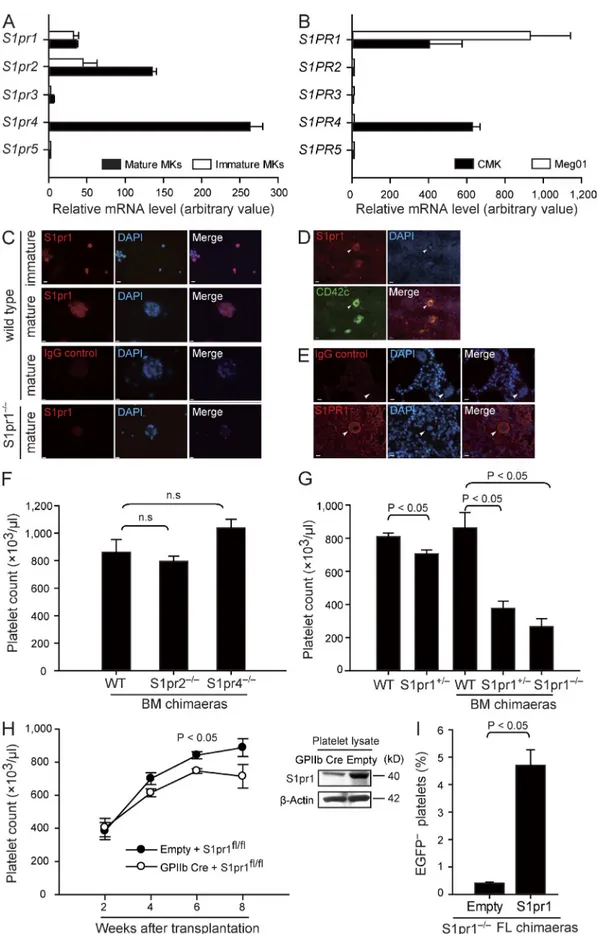
CMK, each express the S1P receptor subtypes 1, 2, and 4
(encoded by S1pr1 and S1PR1, S1pr2 and S1PR2, and S1pr4
and S1PR4 in mice and humans, respectively; Fig. 1, A–E; and
Table S1
). To directly test whether S1P receptors play a role
for megakaryo- or thrombopoiesis, we determined platelet
counts in peripheral blood of WT mice and mice lacking the
S1P receptors expressed by MKs. Loss of S1pr2 or S1pr4 on
hematopoietic cells had no significant effects on peripheral
platelet counts or platelet size (Fig. 1 F,
Table S2
, and not
de-picted). In contrast, ablation of the S1pr1 gene was associated
with dramatically reduced platelet numbers. Loss of one S1pr1
allele (S1pr1
+/mutants) already resulted in a significant
reduction in the number of circulating platelets (Fig. 1 G and
Table S3
). Loss of both alleles (S1pr1
/mice) was
embryoni-cally lethal (Liu et al., 2000); thus, to circumvent embryonic
lethality, we generated chimaeras by transferring fetal liver
(FL) cells from S1pr1
/, S1pr1
+/, or S1pr1
+/+donors into
irradiated WT mice. 6–8 wk after reconstitution, BM cells
from S1pr1
/, S1pr1
+/, or S1pr1
+/+FL chimaeras were
isolated and further transplanted into irradiated secondary
recipient mice. Platelet counts in S1pr1
+/and S1pr1
/chimaeras were reduced by >50% and 70% compared with
S1pr1
+/+chimaeras, respectively (Fig. 1 G and Table S2).
Col-lectively, these results indicate that S1pr1 on hematopoietic
cells controls blood platelet homeostasis, whereas S1pr2 and
S1pr4 are dispensable for this process.
Next we evaluated whether S1pr1 expressed by MKs or
by other hematopoietic lineages regulates the number of
blood platelets. To this end, we reconstituted irradiated mice
with BM cells carrying two floxed S1pr1 alleles (S1pr1
fl/fl)
and transduced with a lentivirus expressing Cre recombinase
under the MK-specific GpIIb promoter (GpIIb-Cre S1pr1
fl/fl)
to delete S1pr1 in the MK/platelet progeny (Fig. 1 H;
Allende et al., 2003). Importantly, platelet counts became
sig-nificantly reduced in GpIIb-Cre S1pr1
fl/flBM recipients as
compared with S1pr1
fl/flcontrol chimaeras (Fig. 1 H).
More-over, lentiviral reexpression of S1pr1 under the MK-specific
GpIb promoter rescued S1pr1
/FL cells to reconstitute
the blood platelet compartment in lethally irradiated mice
(Fig. 1 I). These findings demonstrate that S1pr1 expressed by
the MK lineage intrinsically controls platelet homeostasis.
Normal MK development, platelet life span,
and serum TPO levels in S1pr1-deficient mice
What could be the reason for the severe
thrombocyto-penia in the absence of S1pr1? First we showed that the
life spans of platelets from S1pr1
+/+, S1pr1
+/, or S1pr1
/chimaeras and between WT and S1pr1
+/mutant mice
was similar, excluding a reduced life span as cause for the
reduced platelet counts (Fig. 2 A). We also excluded a defect
in the release of TPO, the principle regulator of
thrombo-poiesis (Kaushansky, 2005a), as cause for the
thrombocy-topenia in S1pr1-null mutants (Fig. 2 B). Finally, we also
could not find evidence for a gross defect in MK
develop-ment, as we found similar numbers of megakaryocytic
progenitor cells in WT and S1pr1
/FL cells populations
the perivascular niche, where they interact with sinusoidal BM
endothelial cells (Avecilla et al., 2004; Patel et al., 2005a). Once
they have settled in the perivascular microenvironment, mature
MKs form dynamic transendothelial pseudopods, which
ex-tend into the lumen of BM sinusoids. These intravascular
pseu-dopodial extensions, termed proplatelets (PPs), continue to
elongate and become tapered into multiple platelet-size beads
connected to each other and with their maternal MKs by thin
cytoplasmic bridges (Italiano et al., 1999; Patel et al., 2005a).
The release of platelets, the final step of platelet formation, then
occurs within the blood, where new platelets are shed as
frag-ments from the tips of intravascular PPs (Stenberg and Levin,
1989; Choi et al., 1995; Italiano et al., 1999; Junt et al., 2007).
MKs are a rare cell population, constituting <0.01% of all
BM cells. This contrasts with the high demand of platelet
pro-duction, implying that the differentiation of MKs (termed
megakaryocytopoiesis) and the subsequent assembly and
re-lease of platelets by MKs (termed thrombopoiesis) are highly
efficient and tightly controlled processes. Among the factors
that modulate megakaryocytopoiesis, thrombopoietin (TPO)
is the major regulator of MK expansion from hematopoietic
stem and progenitor cells, whereas chemokines, including
stromal-derived factor-1 (SDF-1), primarily initiate the
relo-cation of maturing MKs to the perivascular
microenviron-ment (Avecilla et al., 2004). In contrast, the molecular pathways
that control the final steps of thrombopoiesis, particularly the
guidance signals that direct megakaryocytic pseudopodial
ex-tensions into the vascular lumen and trigger the intravascular
release of new platelets, are entirely unknown.
The bioactive sphingolipid sphingosine 1-phosphate (S1P)
and the receptors responsive to this mediator regulate
impor-tant biological functions of various hematopoietic cell types
(Spiegel and Milstien, 2003, 2011; Schwab et al., 2005;
Massberg et al., 2007), including cell migration in the BM
compartment (Ishii et al., 2009; Allende et al., 2010). Here we
report that S1P and the MK S1P receptor S1pr1 receptor are
indispensable for normal BM thrombopoiesis. Using mouse
mutants and by multiphoton intravital microscopy (MP-IVM),
we demonstrate that a transendothelial S1P gradient navigates
megakaryocytic PP extensions into the lumen of BM
sinu-soids. In the blood, PP extensions are exposed to high S1P
concentrations, which initiate the subsequent shedding of
platelets into the circulation. Both processes involve the S1P
receptor S1pr1, triggering activation of the Gi/Rac GTPase
signaling. Correspondingly, lack of S1pr1 on MKs, but not of
other S1P receptors, results in severe thrombocytopenia. Thus,
we have identified the S1P–S1pr1 pathway as a key nodal
point integrating guidance cues that navigate directional PP
elongation and enabling the final step of thrombopoiesis, the
shedding of new platelets into the blood stream.
RESULTS
S1pr1 expression in MKs intrinsically regulates
platelet homeostasis
We observed here that cultured mouse and human MKs,
as well as the human megakaryocytic cell lines Meg01 and
Figure 1. MKs express S1pr1, and S1pr1-deficient mice display severe thrombocytopenia. (A) Relative expression of S1P receptor mRNA by FL-derived mature and immature MKs. (B) Relative expression of S1P receptor mRNA in human megakaryocytic cell lines. (A and B) Data are representative of three independent experiments with triplication. (C) Representative immunostaining of S1pr1 in immature and mature FL-derived MKs. WT MKs stained
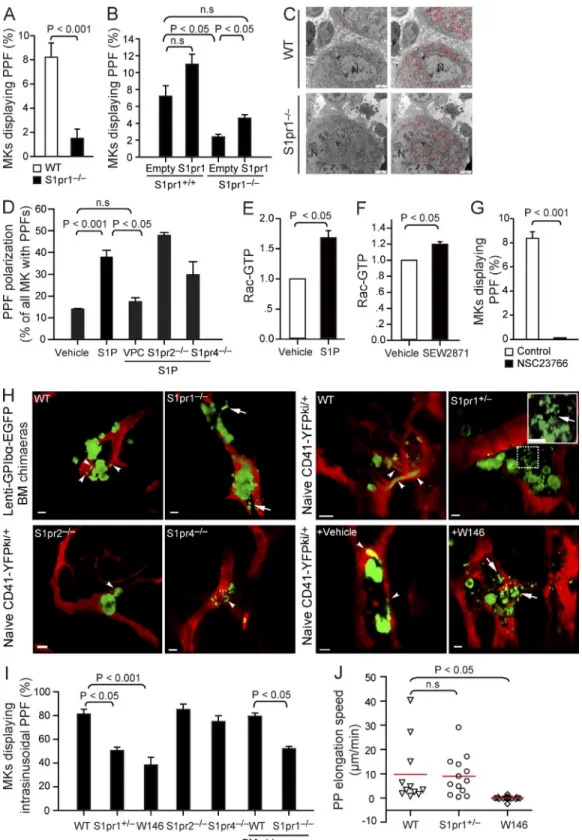
in vitro PPF was reduced by >70% in S1pr1
/MKs, as <2
out of 100 S1pr1
/MKs formed PPs (Fig. 4 A). Importantly,
lentiviral reexpression of GpIb promoter–driven S1pr1 in
S1pr1
/MKs corrected PPF in vitro (Fig. 4 B). These results
clearly indicate that S1pr1 plays a critical and intrinsic role for
PPF by MKs.
When we examined how S1pr1 might control PPF,
we could exclude a primary lack of the invaginated
demarca-tion membrane system (DMS), the predominant reservoir
for PP membranes (Radley and Haller, 1982; Schulze et al.,
2006), in S1pr1
/MKs, as electron microscopy of S1pr1
/BM MKs did not reveal abnormalities of the DMS when
compared with S1pr1
+/+BM MKs (Fig. 4 C). Next we tested
whether S1P serves as a chemoattractant for polarizing MKs
and for inducing the formation of PP protrusions. Within the
normal BM compartment, S1P is rapidly degraded by lyases
and phosphatases expressed by most hematopoietic cells. Thus,
the local S1P concentrations in the BM (with its densely
packed hematopoietic cells) are exceedingly low (unpublished
data), reflecting similar concentrations reported for other
tis-sues such as lymph nodes (Schwab et al., 2005; Pappu et al.,
2007). In contrast, high S1P concentrations exist in the blood
stream (Caligan et al., 2000; Berdyshev et al., 2005; Pappu
et al., 2007). Because of their positioning at the vascular
inter-face, MKs are therefore exposed to a steep transendothelial
S1P gradient. To mimic the situation in the BM, we exposed
cultured MKs to a gradient of S1P in vitro. Notably, PP
extensions developed preferentially toward increasing
con-centrations of S1P but not of vehicle (Fig. 4 D). A similar
result was also obtained with S1pr2
/and S1pr4
/MKs
(Fig. 4 D). VPC23019, a previously described S1pr1 and S1pr3
antagonist (Davis et al., 2005), was used in our study to
selec-tively block megakaryocytic S1pr1 signaling because MKs do
not express S1pr3 (Fig. 1 A). Inhibition of the
megakaryo-cytic S1pr1 using VPC23019 abolished this directionality of
PPF; MKs projected PP extensions into random directions
(Fig. 4 D). These findings suggest that S1P–S1pr1 signaling
is essential for PPF by providing a chemoattractant stimulus
that controls the polarization of PP processes generated by
MKs in culture.
Next we defined the signaling downstream of S1pr1
involved in the regulation of PPF and polarization.
Be-cause Rac GTPase activity controls actin dynamics leading
to membrane protrusion/extensions (Aspenström et al.,
2004), we tested whether activation of Rac GTPases via
(Fig. 2 C) and a normal differentiation of WT and S1pr1
/precursor cells into MKs both in vitro (Fig. 2 D) and in vivo
(Fig. 2, E and F).
Loss of S1pr1 increases MK size but has no effect
on positioning and motility of MKs in vivo
Next we examined whether S1pr1 controls platelet biogenesis
for example by modulating MK motility or their positioning
within the BM compartment. To address this question, we
performed MP-IVM of calvarian BM (Junt et al., 2007) of
two different sets of S1pr1
+/+or S1pr1
/chimaeras, in which
MKs and their progeny were genetically marked: (a) S1pr1
+/+or S1pr1
/CD41-YFP
ki/+FL chimaeras, in which MKs and
platelets express the YFP driven from the endogenous CD41
gene locus (Zhang et al., 2007) and (b) S1pr1
+/+or S1pr1
/lenti-GpIb–enhanced GFP (EGFP) BM chimaeras, in which
MKs and platelets express EGFP under the transcriptional
con-trol of the murine GpIb promoter (Lavenu-Bombled et al.,
2007). The experiments revealed neither differences in MK
size nor in their positioning or motility when we compared
S1pr1
+/+CD41-YFP
ki/+or S1pr1
+/+lenti-GpIb-EGFP
chi-maeras and naive (nontransplanted) S1pr1
+/+CD41-YFP
ki/+or platelet factor 4 (Pf4)–EYFP transgenic mice, in which
EYFP is driven by the MK-specific Pf4 promoter, which
allowed excluding a major influence of irradiation and BM
transplantation (Fig. 3, A–D). As reported previously (Junt et al.,
2007), S1pr1
+/+MKs were large, mostly sessile cells always
lo-cated in close proximity to BM sinusoids (Fig. 3, A and D–G).
In S1pr1
/chimaeras and S1pr1
+/mice, MKs were
signifi-cantly larger compared with S1pr1
+/+chimaeras, whereas the
position and motility of MKs was similar among all genotypes
(Fig. 3, E–G). The aforementioned results suggest that in
con-trast to other cells in the BM (Ishii et al., 2009), neither
posi-tioning nor migration of MKs or their committed progenitors
in marrow spaces is controlled by S1pr1.
S1pr1 is essential for intravascular PP formation (PPF)
During thrombopoiesis, mature MKs extend transendothelial
protrusions, termed PPs, into BM microvessels (Junt et al.,
2007). To test whether S1P/S1P receptor signaling plays a role
during PPF, we cultured MKs in vitro (Lecine et al., 1998)
and found that on average, 9 out of 100 WT MKs
spontane-ously formed PPs as assessed by phase-contrast microscopy.
MKs isolated from S1pr2
/and S1pr4
/mice generated
similar number of PPs (unpublished data). In sharp contrast,
with irrelevant control IgG or anti-S1pr1–stained S1pr1-null MKs served as controls. (D and E) Expression of S1pr1 in murine BM (D) and S1PR1 in human BM sections (E). Arrowheads indicate MKs. CD42c is the marker for MKs. All MKs examined stained positive for S1pr1 or S1PR1. Bars, 10 µm. (F) Platelet counts in peripheral blood. n = 15 for WT BM chimaeras; n = 5 for S1pr2/ BM chimaeras; n = 3 for S1pr4/ BM chimaeras. (G) Platelet counts in
peripheral blood. n = 8 for WT; n = 6 for S1pr1+/; n = 15 for WT BM chimaeras; n = 9 for S1pr1+/ BM chimaeras; n = 14 for S1pr1/ BM chimaeras.
(H) Platelet counts in chimaeras after transferring S1pr1fl/fl BM cells transduced with lenti-GPIIb-cre or empty control vectors into irradiated recipient
mice (left). Expression of S1pr1 in platelets in chimaeras after transferring S1pr1fl/fl BM cells transduced with lenti-GPIIb-cre or empty control vectors.
-Actin served as loading control (right). n = 3 per genotype. (I) Percentage of EGFP platelets in chimaeras after hematopoietic reconstitution of lethally
irradiated mice with EGFP+ S1pr1+/+ BM cells and EGFP S1pr1/ FL cells at a ratio of 20:1. The EGFP S1pr1/ FL cells were transduced with
indicating that activation of Rac GTPases downstream of
S1pr1 is required for PPF.
To evaluate the in vivo relevance of S1P and its receptors
for PPF, we examined CD41-YFP
ki/+mice by MP-IVM. In
CD41-YFP
ki/+mice, 59% of all MKs extended plump or long
PP protrusions into BM sinusoids (
Fig. S1 A
and
Video 1
),
indi-cating active participation in platelet biogenesis. PP protrusions
S1pr1 in MKs controls PPF. As reported previously for
en-dothelial cells (Paik et al., 2004), we found that S1P, and
also the S1pr1-specific agonist SEW2871, enhances Rac
GTPase activity via S1pr1 in megakaryocytic cell lines
(Fig. 4, E and F). Conversely, we observed that
pharmaco-logical inhibition of Rac GTPases by NSC23766
com-pletely abolished PPF in response to S1P in vitro (Fig. 4 G),
Figure 2. Loss of S1pr1 does not change platelet life span, serum TPO levels, and MK development. (A) Platelet life span as-says in the indicated genotypes. n = 3–5 per
genotype. (B) Serum TPO levels. n = 3–5 per
genotype. (C) Quantification of CFU-MK num-bers in FL cells. Data are representative of three independent experiments with triplica-tion. (D) Number of mature MKs in cultured FL cells. Data are representative of four indepen-dent experiments with triplication. (E) Repre-sentative immunostaining of MKs in mouse femoral BM. MKs were detected by the MK-specific marker CD41 (green). DAPI is stained blue. Bars, 10 µm. (F) Quantification of MK numbers per 20× high-power field in femoral
BM. n = 3 mice per genotype. All error bars
Figure 3. Loss of S1pr1 increases the size but has no effect on the positioning and motility of MKs in vivo. (A) Representative MP-IVM images of YFP+ or EGFP+ MKs (green) in BM. BM microvasculature was visualized by intravenous injection of TRITC-dextran (red). Left, naive (nontransplanted)
S1pr1+/+CD41-YFPki/+; middle left, S1pr1+/+;Pf4-EYFP; middle right, S1pr1+/+;lenti-GPIb-EGFP BM chimaeras; right, S1pr1+/+;CD41-YFPki/+ FL chimaeras.
Bars, 20 µm. (B–D) Volumes (B), distances from sinusoids (C), and the instantaneous lateral (x-y) velocity (D) of MKs in the indicated groups. Red lines in C indicate medians; red lines in D indicate means. Error bars represent SEM. n = 13–46 MKs per genotype. Data are pooled from three mice each group.
P-values among the different groups in B–D are >0.05. (E) Surface area of MKs in BM. (F) Distance of MKs from BM sinusoids. (E and F) Red lines indicate medians. (G) Instantaneous lateral (x-y) velocity of MKs. Red lines indicate means. (E–G) Data are pooled from three mice each group.
Figure 4. S1P regulates PPF. (A) The percentage of MKs displaying PPF. PPF is expressed as the percentage of MKs carrying PPs (8,000-10,000 MKs per experiment, five independent experiments with triplications). (B) The percentage of MKs displaying PPF in S1pr1+/+ or S1pr1/ MKs transduced with
lenti-GpIb-S1pr1 or empty control vectors (3,000-8,000 MKs per experiment, two independent experiments with triplications). (C) Representative elec-tron micrographs of WT and S1pr1/ MKs in BM. Arrowheads indicate the DMS. Red color highlights the DMS. N, nucleus. (D) The percentage of MKs
with polarized PPF in the presence or absence of S1P and the S1pr1-specific inhibitor VPC23019 (VPC). n = 127–265 MKs per group. Data are pooled from
three to five independent experiments. (E) Y10/L8057 cells were incubated with 10 µM S1P or vehicle for 2 min. The activities of Rac-GTP were quantified by pull-down assay (n = 5 independent experiments). (F) Y10/L8057 cells were incubated with 1 µM S1pr1 agonist, SEW2871, or vehicle for 5 min. The
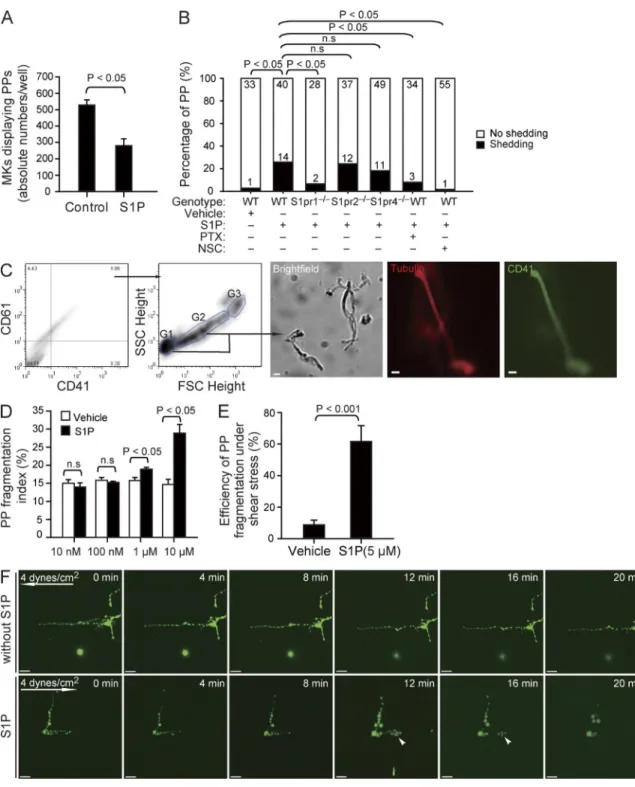
fragmentation at high S1P concentrations, mimicking S1P
plasma levels but not at low concentrations prevailing in the
BM interstitium (Fig. 5, C and D).
In vivo, blood flow–induced shear stress might facilitate
the separation of intravascular cell fragments from MKs (Junt
et al., 2007). We therefore evaluated whether S1P also plays
a role for PP fragmentation under flow conditions. Cultured
MKs exposed to the physiological shear stress of BM
sinu-soids (4 dynes/cm
2; Junt et al., 2007) in the absence of S1P
(serum-free buffer) rarely shed PPs from their MK stems. In
contrast, in the presence of 5 µM S1P, PPs were rapidly
re-leased (Fig. 5, E and F; and
Video 5
), indicating that S1P is
required for PP shedding under static as well as flow
condi-tions. Loss of S1pr2 or S1pr4 did not affect S1P-induced PP
shedding (Fig. 5 B, Fig. S1 B, and
Video 6
). However, lack
of the megakaryocytic S1pr1 receptor completely abolished
S1P-induced release of PPs (Fig. 5 B, Fig. S1 B, and Video 6).
This indicates that S1pr1, but not S1pr2 or S1pr4, plays the
predominant role for S1P-driven PP shedding. To further
clarify the involved signaling pathway, we used pertussis toxin
and NSC23766 to inhibit Gi and Rac GTPase activity,
re-spectively. Both inhibitors blocked S1P-induced
fragmenta-tion of PPs (Fig. 5 B and Fig. S1 B). The observafragmenta-tion that
S1P activates Rac GTPase in MKs via S1pr1 (Fig. 4, E and F)
together with the aforementioned findings suggests that
S1P-induced PP fragmentation depends on S1pr1/Gi/Rac
GTPase signaling.
S1P controls PP shedding into blood via S1pr1 in vivo
To address whether S1P–S1pr1 signaling is also essential for
PP fragmentation in vivo, we examined PP shedding in live
mice by MP-IVM. PP shedding from MKs was a frequent
event in naive (nontransplanted) S1pr1
+/+CD41-YFP
ki/+transgenic mice (Junt et al., 2007) but also in S1pr1
+/+CD41-YFP
ki/+BM chimaeras (
Video 7
). Most MKs shed PP
frag-ments that consist of beaded platelet-like structures (Fig. 6,
A and B; and Video 7), which generate mature platelets by
undergoing consecutive fragmentation steps (Behnke and
Forer, 1998; Junt et al., 2007). More than 60% of the S1pr1
+/+MKs carrying intravascular PP processes showed
fragmenta-tion within 1 h (Fig. 6, A and B; and Video 7). We did not find
any defect in PP fragmentation in S1pr2
/or S1pr4
/mice (Fig. 6 A), whereas this process was severely impaired in
S1pr1 mutants. In S1pr1
/chimaeras, we barely observed
intravascular PP processes because of the aberrant interstitial
PPF reported above (Fig. 4, H and I). However, 70–100% of
the PP processes that had eventually made their way into BM
extended almost exclusively into marrow sinusoids of
CD41-YFP
ki/+mice, whereas we rarely detected extravascular PP
processes (Fig. 4, H and I; and
Video 2
). To determine whether
the S1P receptors expressed by MKs provide the guidance
information necessary to direct PP processes into BM
sinusoids, we examined S1pr1
+/CD41-YFP
ki/+, S1pr2
/CD41-YFP
ki/+, and S1pr4
/CD41-YFP
ki/+mice as well as
lenti-GpIb-EGFP BM chimaeras. Consistent with our in
vitro findings (Fig. 4 D), loss of S1pr2 or S1pr4 did not affect
the formation or polarization of PPs in vivo (Fig. 4, H and I).
In contrast, loss of S1pr1 disrupted PPF and polarization;
cor-respondingly, S1pr1
+/or S1pr1
/MKs projected PP
ex-tensions in random directions (Fig. 4, H and I; and Video 2).
As a consequence, we found aberrant PP processes in the
mar-row interstitial space, whereas intrasinusoidal PPs were rarely
detected in S1pr1-deficient chimaeras (Fig. 4, H and I; and
Video 2). Likewise, when we treated S1pr1
+/+CD41-YFP
ki/+mice with the S1pr1-specific antagonist W146 for 24 h, the
physiological directionality of PPF was entirely disrupted. We
frequently observed long cytoplasmic extensions outside
si-nusoids in mice treated with W146 but not in vehicle-treated
animals (Fig. 4, H and I; and
Video 3
). In addition, inhibition
of S1pr1 also retarded PP growth in vivo, resembling the
re-duced PPF of cultured S1pr1
/MKs in vitro (Fig. 4, A and J).
These results indicate that S1pr1 signaling supports PPF and
elongation along the physiological S1P gradient between BM
interstitium and BM sinusoids and controls the entry of PPs
into the marrow blood stream in vivo.
S1P enhances PP fragmentation via S1pr1 in vitro
Once MK PP processes have entered the blood, they are
ex-posed to significantly higher S1P concentrations compared
with the BM interstitium (unpublished data). To our surprise,
when we mimicked the situation in the blood by incubating
cultured MKs to allow PPF and then adding a high
concen-tration of S1P (instead of exposing MKs to an S1P gradient),
we found a significant reduction in the number of MKs
dis-playing PP extensions (Fig. 5 A). Using differential
interfer-ence contrast (DIC) microscopy of cultured MKs, we observed
that exposure of MKs to a high, homogenous concentration
of S1P results in almost immediate shedding of platelet-like
particles from PPs (Fig. S1 B and
Video 4
). Within 1 h,
platelet-like particles were shed from 26% of PPs in response to
S1P but only from 3% of PPs treated with vehicle (Fig. 5 B).
To further quantify the effect of S1P on PP shedding in vitro,
we determined the number of fragmentation events by
flow cytometry (Fig. 5 C). S1P, but not vehicle, increased PP
absence of 50 µM NSC23766, a Rac GTPase inhibitor (4,000-7,000 MKs per experiment; three independent experiments with triplications). (H) Representa-tive MP-IVM images of MKs with YFP+ or EGFP+ PPs. Green indicates MKs and PPs; red indicates sinusoids. Arrowheads indicate extravascular YFP+ or
EGFP+ PPs; arrows indicate interstitial YFP+ or EGFP+ PPs. The inset shows magnification for the dotted box. The arrow in the inset indicates the
connec-tion between extravascular YFP+ PPs. Bars: (C) 2 µm; (H) 20 µm. (I) MKs displaying intrasinusoidal PPF in vivo presented as a percentage of all MKs
carry-ing PPs (20–30 MKs per group; five independent experiments for WT; three independent experiments for other genotypes). (J) The lateral (x-y) speed of PP elongation was measured in naive (nontransplanted) S1pr1+/+CD41-YFPki/+ mice, S1pr1+/CD41-YFPki/+ mice, and S1pr1+/+CD41-YFPki/+ mice treated with
Figure 5. The effect of S1P on PP fragmentation in vitro. (A) The number of MKs displaying PPF in the absence or presence of 10 µM S1P (230–590 MKs per experiment; three independent experiments with triplications). (B) The number of PPs with or without fragmentation observed by DIC microscopy in vitro over 1 h in the indicated groups. Data are pooled from 4–10 independent experiments for each group (n = 30–60 per group). (C) Representative
dot plots show flow cytometric analyses of PP fragmentation. The first two panels show the gates for PPs. The CD41+CD61+ population was analyzed for
the distribution of PPs according to FSC and SSC. MKs are G3; PPs with higher and lower FSC values are G2 and G1, respectively. The three representative microphotographs in the right show a representative brightfield image, as well as tubulin and CD41 stainings of fragments sorted using the gating strat-egy illustrated in the two plots. (D) Flow cytometric analyses of the PP fragmentation index in the presence or absence of various concentrations of S1P. The PP fragmentation index was calculated as described in Materials and methods. Data are representative of six independent experiments with triplica-tion. (E) PP fragmentation by MKs exposed to shear stress. The efficiency of dynamic PP fragmentation was calculated as described in Materials and methods. Data are pooled from five independent experiments for each group. (F) Representative time-lapse video microscopy of PPs in the presence or absence of 5 µM S1P under shear stress (4 dynes/cm2). Arrows indicate direction of flow; arrowheads indicate PP shedding events. All error bars indicate
Figure 6. The effect of S1P on PP fragmentation in vivo. (A) Percentage of PP fragmentation events observed by MP-IVM over 1 h in the indicated groups. n = 13–33 per group. Data are pooled from three to seven independent experiments. (B) Role of S1pr1 for PP shedding in vivo visualized by MP-IVM.
Representative MP-IVM sequences show that WT MKs frequently shed PPs as shown in the first and the third rows. The inset shows a magnification of a shed PP particle. Asterisks show embedded platelet-like particles. Inhibition or loss of S1pr1 abolishes PP shedding (second and fourth rows). Arrow-heads indicate intrasinusoidal PPs, and arrows show extrasinusoidal PPs in S1pr1/ chimaeras. The dashed lines highlight the sinusoids. Green or yellow
S1pr1 agonists enhance platelet production
Modulation of S1P receptors by FTY720 (fingolimod) has
become a promising strategy for the treatment of patients
with multiple sclerosis (Kappos et al., 2006). Here, we show
that treatment of mice with a single dose of FTY720 leads
to a prompt and transient increase in circulating platelets
(Fig. 7 A). When we used MP-IVM to examine MKs before
and after treatment with a single dose of FTY720, we found
that FTY720 accelerates the shedding of intravascular PP
ex-tensions into the blood stream. As a consequence, the number
of MKs carrying intravascular PPs significantly decreased
immediately after a single dose of FTY720 compared with
vehicle (Fig. 7, B and C). This suggests that FTY720
repre-sents an agonist for megakaryocytic S1pr1 receptors and has
the potential to rapidly mobilize PPs into the blood, most
likely by supporting fragmentation of intravascular PPs (Fig. 7,
B and C). Treatment with the S1pr1-specific agonist SEW2871
also caused an increase in circulating blood platelets (Fig. 7 D),
further supporting that activation of S1P–S1pr1 receptor
sig-naling enhances thrombopoiesis.
DISCUSSION
Our results assign a new role for S1P and its receptor S1pr1
as master regulators of thrombopoiesis. In a dose-dependent
and sequential manner, S1P controls two key steps in the
cascade of thrombopoiesis by BM MKs: (1) the polarized
development of PP extensions into the blood stream and
(2) the subsequent shedding of PPs from their
transendothe-lial stems. As a consequence, loss of S1pr1 is not compatible
with normal thrombopoiesis. Collectively, our findings
uncover the molecular pathway that enables the final steps
of thrombopoiesis.
S1P navigates PP extensions into BM sinusoids
and initiates platelet release
Mature MKs form intravascular PP extensions that grow from
the MK cell body at a mean speed of 10 µm/min under shear
conditions in vivo (Fig. 4 J), with the DMS functioning as the
membrane reservoir for PP elongation (Schulze et al., 2006).
During elongation, PPs are equipped with specific proteins
associated with platelets, including von Willebrand factor
(vWF) and fibrinogen receptors. Microtubules, assembled
from /-tubulin dimers, are the primary structural
compo-nent of the engine that drives the elongation of PPs.
Corre-spondingly, PPs fail to form when cultured MKs are exposed
to agents that inhibit microtubule assembly (Italiano et al., 1999)
or sliding (Patel et al., 2005b), and mice lacking 1-tubulin,
sinusoids remained firmly attached to their MK stems; only in
rare instances did MKs release PP fragments (Fig. 6, A and B;
and Video 7). Together, these data indicate that S1pr1 is
criti-cal for both directional PPF and for proper intravascular PP
fragmentation. Defective PP shedding is likely to explain the
increase in size of S1pr1
/MKs (Fig. 3 E). Interestingly, the
frequency of intravascular PP shedding was only moderately
reduced in CD41-YFP
ki/+S1pr1
+/mice (Fig. 6 A),
suggest-ing that a ssuggest-ingle S1pr1 allele is sufficient to maintain
intra-vascular PP shedding and that the mild thrombocytopenia
observed in S1pr1
+/mice is mostly caused by a defect in
navigating PP processes into BM sinusoids (Fig. 4, H and I).
To examine whether S1pr1 regulates the dynamic process
of PP shedding independently from its effects on PP invasion
into BM sinusoids, we next tested the consequences of
short-term pharmacological inhibition of S1pr1. We treated naive
(nontransplanted) S1pr1
+/+CD41-YFP
ki/+mice with a single
dose of the selective S1pr1 antagonist W146 and visualized PP
shedding immediately thereafter. In contrast to protracted
inhi-bition or genetic ablation of S1pr1 (Fig. 4, H and I), this did not
affect the overall number of MKs with intravascular PP
protru-sions. However, <20% of MKs with established intravascular
protrusions managed to release PP fragments into the blood
stream within 6 h after administration of W146; the vast
ma-jority of the intrasinusoidal processes remained attached to their
MK stems (Fig. 6, A and B; and
Video 8
). Repetitive treatment
of mice with W146 for 24 h resulted in a significant reduction
in circulating young reticulated platelets with an elevated RNA
content (Fig. 6 C), consistent with a central role of the S1P–
S1pr1 pathway for PP fragmentation and release of platelets.
Short-term treatment with W146 also reduced platelet counts
in CD1 mice (Fig. 6 D), suggesting that S1pr1 controls
throm-bopoiesis across different strains of mice. W146 maintains an
adequate in vivo receptor blockade for only 5–6 h (Sanna et al.,
2006), and shedding reoccurred 6 h after a single dose of W146,
suggesting that S1pr1 inhibition does not affect the viability of
MKs (Fig. 6 A and Video 8). In rare instances, where PP
shed-ding occurred in the presence of the S1pr1 inhibitor W146, the
time required until an intravascular fragment dissociated from
its MKs stem was significantly prolonged (Fig. 6 E). The failure
to properly shed PPs resulted in the formation of abnormal,
thick intravascular PP processes (Video 8). In line with this
observation, the few PP fragments that were released despite
the presence of W146 were significantly bigger compared with
those in vehicle-treated control mice (Fig. 6 F), reminiscent of
the large platelets observed in S1pr1-null chimaeras (Fig. 6 G
and Table S2).
indicates MKs and PPs; red indicates sinusoids. Bars, 20 µm. (C) Circulating reticulated (young) platelets in mice treated with W146, an S1pr1-specific antagonist, or vehicle as assessed by flow cytometry. n = 3 mice each group. (D) Circulating platelet counts in CD1 mice treated with W146 or vehicle. n = 4
mice each group. (E) The time point of fragmentation detected by MP-IVM in mice treated with W146 (<6 h) or vehicle. Data are pooled from three independent experiments each group. (F) Volumes of PP fragments in mice treated with W146 (within 6 h) or vehicle. Red lines indicate means. Data are pooled from three independent experiments each group. (G) Mean platelet volume in the indicated genotypes. n = 8 for WT; n = 6 for S1pr1+/; n = 15
fragments break down further in the circulation giving rise to
mature platelets of 2–3-µm diameter within the circulation
(Stenberg and Levin, 1989). Blood shear stress contributes to
the shedding of PPs (Junt et al., 2007; Dunois-Lardé et al.,
2009; Thon et al., 2010); however, whether additional signals
are required for efficient PP shedding was completely
un-known. In this study, we show that hydrodynamic forces alone
are not sufficient to allow the release of new platelets from
MK PP extensions. Instead, we found that high
concentra-tions of the bioactive lipid S1P prevailing in the sinusoidal
blood, but not in the BM interstitium, are mandatory for the
release of new platelets from MKs.
From a teleological point of view, the S1P-dependent
sequential guidance of thrombopoiesis comprising (a)
di-rectional PPF along a transendothelial S1P gradient and
(b) subsequent S1P-dependent intravascular PP shedding
leads to the introduction of naive platelets into the circulating
blood and prevents aberrant platelet production within the
BM interstitium. S1P guidance of intravascular PPF,
elonga-tion, and shedding therefore provides grounds for efficient
thrombopoiesis, which seems instrumental given the relative
paucity of MKs.
S1P controls thrombopoiesis via megakaryocytic
S1pr1 receptors
Our study shows that MKs robustly express three different
S1P receptors, S1pr1, S1pr2, and S1pr4. Loss of S1pr1 on
he-matopoietic cells and also conditional deficiency of S1pr1 in
MKs were associated with severe thrombocytopenia.
More-over, gain of S1pr1 function in S1pr1
/MKs rescued their
the most abundant -tubulin in platelets, develop
thrombo-cytopenia (Schwer et al., 2001).
Cultured MKs form PPs that elongate into random
direc-tions (Italiano et al., 1999; Dunois-Lardé et al., 2009). In
con-trast, we show here by MP-IVM that PPF occurs in a highly
polarized fashion in the BM in vivo. This suggests that PPs
integrate a previously unknown guidance signal, which
navi-gates them into the intravascular compartment and avoids the
interstitial BM compartment. Our study has uncovered this
guidance signal and shows that a transendothelial S1P
gradi-ent in the BM controls the directionality of PPF and
elonga-tion. We observed that the interstitial S1P concentrations in
the BM are low, whereas the S1P blood concentration is
orders of magnitude higher. Residing at the vascular interface,
mature MKs are located in a particularly strategic position for
integrating the guidance cues provided by the
transendothe-lial S1P gradient. Equipped with S1pr1, they sense the steep
vascular S1P gradient and extend dynamic PP protrusions
into microvessels along increasing concentrations of this lipid
mediator. A similar S1P gradient also exists in the lymph node
between lymph node interstitium and the lymph fluid, where
it drives the migration of T cells into efferent lymph vessels
(Matloubian et al., 2004). Recent observations showing that
the S1P pathway controls the egress of lymphocytes from the
BM into the blood stream emphasize the biological relevance
of the transendothelial S1P gradient for BM homeostasis
(Allende et al., 2010).
Once MKs have successfully extended their PPs into the
blood, they release fragments from the tips of their
intravas-cular projections (Video 7; Junt et al., 2007). These new PP
Figure 7. The S1P analogue FTY720 and SEW2871 trigger rapid release of platelets. (A) Mice were treated with a single dose of FTY720 (3 mg/kg i.p.) or DMSO (vehicle). Open bars indicate baseline of platelet counts assessed before drug administration; closed bars indicate platelet counts measured 12 h after drug admin-istration. n = 9 mice each group. (B)
Representa-tive MP-IVM images of MKs with YFP+ PPs from
CD41-YFPki/+ mice before and 8 h after a single
injection of FTY720 (3 mg/kg i.p.) or DMSO (vehicle). Green indicates MKs and PPs; red indicates sinusoids. Arrowheads indicate intravascular PPs. Dashed lines indicate BM sinusoidal vessels. Bars, 20 µm. (C) Fold change of MKs displaying PPF 8 h after a single injection of FTY720 (3 mg/kg i.p.) or DMSO (vehicle) com-pared with numbers recorded before treatment.
n = 120–140 MKs per group. Data are pooled
from three independent experiments. (D) Mice were treated with a single dose of SEW2871 (20 mg/kg i.p.) or dimethyl formamide (DMF; vehicle). Open bars indicate baseline of platelet counts assessed before drug administration; closed bars indicate platelet counts measured 12 h after drug administration. n = 3 mice each
strategy and is currently being used in patients with relapsing
multiple sclerosis (Kappos et al., 2006). After administration,
FTY720 is metabolized to phosphorylated FTY720 (FTY720P),
an agonist for four of the five S1P receptors including S1pr1.
FTY720 limits effector lymphocyte egress from lymph nodes
(Matloubian et al., 2004), contributing to its
immunosuppres-sive actions. However, FTY720 has not been examined for its
potential effects on megakaryo- and thrombopoiesis. In this
study, we show that treatment of mice with a single dose of
FTY720 leads to shedding of intravascular PP extensions into
the blood stream, paralleled by a prompt, but transient
in-crease in circulating platelets. This suggests that FTY720 acts
as an agonist on megakaryocytic S1pr1 receptors and has the
potential to rapidly mobilize PPs into the blood, most likely
by supporting fragmentation of intravascular PPs. Whereas
lymphocyte S1pr1 engagement by phosphorylated FTY720
within secondary lymphoid organs triggers
down-modula-tion of the receptor, resulting in funcdown-modula-tional antagonism of the
S1pr1 pathway, an agonistic effect of FTY720 similar to the
one observed here for MK has recently been reported to
pro-mote the recirculation of BM osteoclast precursor monocytes
from the bone surface (Ishii et al., 2009). This indicates that
FTY720 predominantly exerts agonist effects in cells of the
myeloid lineage. Because activation of S1P–S1pr1 receptor
signaling enhances thrombopoiesis in mice, future studies will
have to evaluate potential clinical implications of S1pr1
ago-nists, in particular in the treatment of thrombocytopenia.
Collectively, the present study reveals that S1P, a signaling
lipid circulating in the blood, regulates dynamic intravascular
PP elaboration and PP shedding without affecting MK
matu-ration and positioning. Tonic S1P–S1pr1 signaling is critical
for normal thrombopoiesis in mice. Although the exact role
of S1P–S1pr1 signaling for human thrombopoiesis still needs
to be defined, our findings could have clinical implications
and provide new approaches to treat thrombocytopenia.
MATERIALS AND METHODS
Mice. C57BL6/J (CD45.2), B6.SJL-PtprcaPep3b/BoyCrl (CD45.1), and CD1
mice were purchased from Charles River. -Actin–EGFP mice were pro-vided by A. Wagers (Harvard Medical School, Boson, MA). S1pr1+/ and
S1pr2/ mice were generated as described previously (Liu et al., 2000; Kono
et al., 2004). S1pr4/ mice were provided by D. Guerini (Novartis Institutes
for BioMedical Research, Basel, Switzerland). CD41-YFPki/+ mice were
generated as described previously (Zhang et al., 2007). Pf4-cre and ROSA26-flox-stop-flox-EYFP mice were obtained from the Jackson Laboratory and crossed to get Pf4-EYFP transgenic mice, in which EYFP is driven by the MK-specific Pf4 promoter. FL chimaeras and BM chimaeras were generated as described previously (Massberg et al., 2007). Cytometric analysis showed that >95% of the blood cells were derived from donors in all the BM chi-maeras. S1pr1fl/fl mice were obtained from R.L. Proia. Age- and
gender-matched mice in a C57BL/6 background were used in all experiments. All experimental procedures performed on animals met the requirements of the German legislation on the protection of animals.
Blood cells and serum TPO measurements. We measured blood cell counts in the mice before and 12 h after a single injection of FTY720 (3 mg/kg i.p.; Cayman) or DMSO (Sigma-Aldrich) as vehicle. Platelet counts were assessed in the mice before and 12 h after a single injection of SEW2871 (20 mg/kg i.p.; Cayman) or dimethyl formamide (Sigma-Aldrich). Serum TPO was measured using the Quantikine murine TPO Immunoassay kit
defect in platelet production. These results clearly
demon-strate that S1pr1 expressed by the MK lineage intrinsically
controls platelet homeostasis. It has been shown previously
that signaling via S1pr1 activates Rac GTPases in multiple
hematopoietic lineages, including T cells (Matsuyuki et al.,
2006; Gérard et al., 2009). Consistent with this, we observed
here that Rac activation is triggered in MKs by S1pr1
ago-nists. Rac GTPases are known to regulate actin dynamics and
induce the formation of membrane extensions (Aspenström
et al., 2004). In MKs, the turnover of actin filaments is known
to control platelet formation (Bender et al., 2010).
Corre-spondingly, we found here that Rac GTPase activation
leading to cytoskeletal reorganization is indispensable for
S1P–S1pr1-driven PPF and fragmentation. Although we
observed that simultaneous pharmacological inhibition of all
Rac GTPases by NSC23766 virtually abolishes S1P-driven
thrombopoiesis in vitro, loss of Rac1 alone does not lead to
thrombocytopenia in vivo (McCarty et al., 2005), suggesting
that other Rac GTPase family members, including Rac2,
Rac3, and RhoG, may have redundant functions in
thrombo-poiesis and its control by S1P. In addition, we cannot rule out
that other small Rho GTPases, including cdc42 and RhoA
(Pleines et al., 2010; Pleines et al., 2012), also contribute to
S1P-driven platelet generation by MKs.
Unlike the loss of S1pr1, genetic disruption of S1pr2
or S1pr4 on hematopoietic cells was not associated with
thrombocytopenia. Moreover, loss of S1pr2 or S1pr4 did not
result in any gross defect of MK development, directional
PPF or PP fragmentation in vitro or in vivo. In line with this
finding, one recent study demonstrated that S1pr4-deficient
mice have normal numbers of BM MKs and normal platelet
counts under physiological conditions (Golfier et al., 2010).
Despite its irrelevance for maintenance of physiological blood
platelet counts, S1pr4 was reported to play a subtle role in the
terminal differentiation of MKs and PPF (Golfier et al., 2010),
an effect which we did not observe here in several sets of
in vitro and in vivo assays, most likely because of the different
experimental conditions. Collectively, our present study and
the previous study (Golfier et al., 2010) suggest that S1pr1,
but not S1pr2 or S1pr4, plays a primary role in the control of
thrombopoiesis under physiological conditions.
In contrast to other cells within the BM compartment,
including osteoclast precursors (Ishii et al., 2009) and
lympho-cytes (Allende et al., 2010), MKs clearly do not require S1pr1
signaling for migration and positioning. This corroborates
previous studies showing that these processes are
predomi-nantly orchestrated by fibroblast growth factor-4 (FGF-4) and
SDF-1 (Majka et al., 2000; Avecilla et al., 2004). Likewise, loss
of S1pr1 does not affect proliferation and maturation of
mega-karyocytic progenitor cells into platelet-producing MKs. This
is consistent with the concept that megakaryopoiesis is
regu-lated predominantly by TPO (Kaushansky, 2005b).
S1P receptor agonist increases blood platelet counts
Recently, modulation of S1P receptor signaling by FTY720
(fingolimod) has emerged as a promising immunosuppressive
Lentiviral infection of FL or BM cells. The different lentiviral con-structs were derived from the pTRIP U3 EF1-EGFP vector (a gift from P. Charneau, Institut Pasteur, Paris, France). The EF1 promoter was replaced by a 541-bp core promoter fragment (254 to 287) of the murine GPIb promoter to obtain the vector pTRIP U3 mGPIb-EGFP. The GFP open reading frame was removed, and the coding region for murine S1pr1 was in-serted to obtain the vector pTRIP U3 mGIb-mS1pr1. A subcloned DNA fragment containing a 921-bp fragment of the human GPII promoter (889 to 32), and the Cre coding sequence was directly cloned into the vector pTRIP U3 EF1-EGFP to obtain the vector pTRIP U3 hGPIIb-Cre.
BM or FL cells were incubated with lentiviral particles in the presence of 8 µg/ml polybrene (Sigma-Aldrich) at 37°C for 12 h in serum-free me-dium supplemented with 1% BSA, 100 ng/ml Flt3-ligand, 100 ng/ml stem cell factor (Sigma-Aldrich), 20 ng/ml TPO (ImmunoTools). After transduc-tion with lentiviral vectors, BM and FL cells were injected into irradiated mice (two doses of 6.5 Gy). For reexpression of S1pr1 in S1pr1/ MKs,
S1pr1/ FL cells were transduced with lenti-GPIb-S1pr1 viral vectors to
express S1pr1 under the control of MK promoter GPIb. 105 S1pr1/ FL
cells transduced with lenti-GPIb-S1pr1 or empty lentiviral vector and 2 × 10⁶ BM cells from -actin–EGFP mice were coinjected into irradiated
CD45.1 mice. The percentage of EGFP-positive platelets was determined by flow cytometry. Because the donor chimaerism in peripheral whole blood of all generated chimaeras was >99% and 100% of platelets from -actin–EGFP mice were EGFP positive, almost all EGFP-negative platelets were from S1pr1/ FL donor cells. For conditional deletion of S1pr1 in MKs, S1pr1fl/fl
BM cells were transduced with lenti-GPIIb-cre viral vectors to express Cre recombinase under the control of MK promoter GPIIb and transferred into irradiated mice. S1pr1 floxed allele was excised in MKs in S1pr1fl/fl
BM chimaeras.
Flow cytometry. We analyzed the mean platelet sizes as described previ-ously (Gramaglia et al., 2005). We performed platelet life span assays as described previously (Robinson et al., 2000). We used Thiazole orange (Molecular Probes) to stain residual RNA in juvenile, reticulated platelets and detected them by flow cytometry as described previously (Matic et al., 1998). For analysis of PP fragmentation, FL-derived MK cultures were treated with various concentrations of S1P or vehicle for 4 h and then stained with CD41-FITC and CD61-PE (BD) antibodies. The PP fragmentation index is determined by the percentage of G1 in the PP population (G1 + G2; Fig. 5 C). We collected the PP fraction according to the gate in Fig. 5 C using a FAC-SAria cell sorter and then observed the sorted PPs using brightfield micro-scopy (Fig. 5 C).
Western blot analyses. Y10/L8057 mouse megakaryocytic cells were cultured in IMDM supplemented with 10% FCS and 25 ng/ml TPO for 1 d and then starved overnight on 100-mm dishes coated with 0.5% fatty acid–free BSA (Sigma-Aldrich). The starved Y10/L8057 cells were simulated with 10 µM S1P or vehicle for 2 min or 1 µM SEW2871 or vehicle for 5 min. Rac-GTP activities were measured using Rac assay kit (Cell Biolabs). Platelet ly-sates were subjected to SDS-PAGE and then immunoblotted with antibodies recognizing murine S1pr1 (Imgenex) or -actin (Abcam) as loading controls. Multiphoton intravital imaging of the BM. We prepared the mouse cal-varian BM as described previously (Junt et al., 2007). We used a BioTech TriMScope system (LaVision BioTec) and Ti: Sa laser (MaiTai) to capture images through a 20× water immersion objective lens (NA = 0.95; Olym-pus). Images were acquired with ImSpectorPro (LaVision BioTec). For three-dimensional (3D) acquisition, the stacks were acquired at a 920-nm wavelength at vertical spacing of 2–3 µm to cover an axial depth of 30–100 µm (for YFP or EGFP). Subsequently, the same stacks were acquired at a wave-length of 800 nm (for TRITC-dextran). The distances between MKs and vasculatures were measured in the reconstructed 3D structure using Volocity software (PerkinElmer). If MKs were outside the vessels, the closest distance from MKs to vessels was measured and represented as negative values. If MKs were in direct physical contact with the vessels, the distance was regarded as (R&D Systems). To block S1pr1 in vivo, we treated C57BL6/J or CD1
mice with W146 (3 mg/kg body weight i.p.) or vehicle every 6 h and mea-sured platelet counts in these mice 24 h later.
Immunostaining. Anti–mouse S1pr1 (Zytomed), anti–human S1PR1 (ABR), anti-S1pr2 (Santa Cruz Biotechnology, Inc.), anti-S1pr4 (Santa Cruz Biotechnology, Inc.), antitubulin (Cell Signaling Technology), anti–mouse CD41 (BD), and anti-CD42c (Emfret Analytics) antibodies were used for immunostaining. The samples were examined using Leica microscopy equipped with 40× objective lens (NA = 0.7) or 20× objective lens (NA = 0.5) and commercial charge-coupled device camera (AxioCam; Carl Zeiss). Images were acquired by AxioVision software (Carl Zeiss). For quantification of MK number and size in BM, we counted the total number of MKs in five randomly selected 20× microscopic fields, and the area of MK cell body was analyzed by ImageJ software (National Institutes of Health).
S1P level measurement. Levels of S1P were determined by high- performance liquid chromatography with mass detection as described previously (Berdyshev et al., 2005).
Transmission electron microscopy. BM samples were fixed in 2.5% glutaraldehyde, embedded in epon, and analyzed using an electron micro-scope (EM 902; Carl Zeiss).
Quantitative RT-PCR. Quantitative real-time PCR was performed using the MaximaTM SYBR Green PCR Master mix (Thermo Fisher Scientific) and an ABI PRISM 7700 Sequence Detection System (Applied Biosystems) according to the manufacturers’ instructions. The data were normalized to the mRNA level of glyceraldehyde 3-phosphate dehydrogenase (Gapdh or GAPDH). Primers are listed in Table S1.
MK culture. Mouse embryonic day (E) 12.5–14.5 FL cells were cultivated in TPO-containing medium for differentiation into MKs (Lecine et al., 1998). In brief, mouse FLs were isolated from E12.5–14.5 embryos and kept in DMEM (Invitrogen) supplemented with 10% charcoal-treated FBS (PAN) in the presence of hTPO (ImmunoTools) in a humidified 5% CO2/95% air incubator at 37°C. For isolation of immature MKs, E12.5–
14.5 FL cells were cultured in medium for 12 h and then incubated with FITC-CD41 (BD) and PE-CD34 (BioLegend) antibodies. Immature MKs were identified as a CD41/CD34 double-positive population and enriched by using a FACSAria cell sorter (BD). Mature MKs were enriched by BSA gradient, as described previously (Pang et al., 2006). In brief, the BSA step gradient was prepared by placing PBS containing 1.5% BSA on top of PBS with 3% BSA (PAA). Cells were loaded on top of the gradient, and MKs were settled to the bottom within 30 min at 1× gravity at room temperature. Mature MKs formed a pellet at the bottom of the tube. We distinguished and scored PPs according to the criteria mentioned previously (Chen et al., 2007). To inhibit the activity of Rac GTPases, we cultured MKs in the pres-ence of 50 µM NSC23766 (Tocris Bioscipres-ence) for 3 d and then scored MKs carrying PPs. To establish a stable gradient during the slow process of PPF, we applied chemotaxis -slides (ibidi), which consist of two large reservoirs with different chemoattractant concentrations and a thin (1 mm) slit be-tween these two reservoirs. A linear and stable chemoattractant gradient is established in the slit by diffusion (Zengel et al., 2011). MKs were resus-pended in serum-free medium in the presence or absence of 1 µM VPC23019 (Avanti Polar Lipids, Inc.) and loaded onto chemotaxis -slides coated with poly-l-lysine between two large reservoirs. 10 µM S1P or vehicle was applied into one of the reservoirs of the -slides. MKs were cultivated for 10 h at 37°C, and polarization of PPF was evaluated based on the images captured using an Axiovert microscope (Carl Zeiss) equipped with 20× objective lens (Nikon) and AxioVision software.
CFU-MK assay. We seeded 10,000 isolated BM or FL mononuclear cells into Megacult-C medium (STEMCELL Technologies) and scored CFU-MKs on day 5 according to the manufacturer’s instructions.
S.-S. Bolz, and A. Billich performed MP-IVM, generated chimaeras, generated lentiviral constructs, performed in vitro MK assays, generated FL-derived MKs, and performed S1P measurements; R.L. Proia and T. Graf generated and provided mutant mice and helped with data interpretation; M. Prinz and A. Müller performed electron microscopy; E. Montanez and M. Sixt performed in vitro shedding assay and examined S1pr1 downstream signaling; L. von Baumbarten, R. Fässler, M. Sixt, U.H. von Andrian, and T. Junt helped with MP-IVM, data interpretation, and discussion. Submitted: 22 May 2012
Accepted: 5 October 2012
REFERENCES
Allende, M.L., T. Yamashita, and R.L. Proia. 2003. G-protein-coupled recep-tor S1P1 acts within endothelial cells to regulate vascular maturation. Blood. 102:3665–3667. http://dx.doi.org/10.1182/blood-2003-02-0460 Allende, M.L., G. Tuymetova, B.G. Lee, E. Bonifacino, Y.P. Wu, and R.L.
Proia. 2010. S1P1 receptor directs the release of immature B cells from bone marrow into blood. J. Exp. Med. 207:1113–1124. http://dx.doi.org/ 10.1084/jem.20092210
Aspenström, P., A. Fransson, and J. Saras. 2004. Rho GTPases have di-verse effects on the organization of the actin filament system. Biochem. J. 377:327–337. http://dx.doi.org/10.1042/BJ20031041
Avecilla, S.T., K. Hattori, B. Heissig, R. Tejada, F. Liao, K. Shido, D.K. Jin, S. Dias, F. Zhang, T.E. Hartman, et al. 2004. Chemokine-mediated interaction of hematopoietic progenitors with the bone marrow vas-cular niche is required for thrombopoiesis. Nat. Med. 10:64–71. http:// dx.doi.org/10.1038/nm973
Behnke, O., and A. Forer. 1998. From megakaryocytes to platelets: platelet morphogenesis takes place in the bloodstream. Eur. J. Haematol. Suppl. 61:3–23.
Bender, M., A. Eckly, J.H. Hartwig, M. Elvers, I. Pleines, S. Gupta, G. Krohne, E. Jeanclos, A. Gohla, C. Gurniak, et al. 2010. ADF/n-cofilin-dependent actin turnover determines platelet formation and sizing. Blood. 116:1767–1775. http://dx.doi.org/10.1182/blood-2010-03-274340 Berdyshev, E.V., I.A. Gorshkova, J.G. Garcia, V. Natarajan, and W.C.
Hubbard. 2005. Quantitative analysis of sphingoid base-1-phosphates as bisacetylated derivatives by liquid chromatography-tandem mass spectrometry. Anal. Biochem. 339:129–136. http://dx.doi.org/10.1016/ j.ab.2004.12.006
Caligan, T.B., K. Peters, J. Ou, E. Wang, J. Saba, and A.H. Merrill Jr. 2000. A high-performance liquid chromatographic method to measure sphin-gosine 1-phosphate and related compounds from sphinsphin-gosine kinase assays and other biological samples. Anal. Biochem. 281:36–44. http:// dx.doi.org/10.1006/abio.2000.4555
Chen, Z., O. Naveiras, A. Balduini, A. Mammoto, M.A. Conti, R.S. Adelstein, D. Ingber, G.Q. Daley, and R.A. Shivdasani. 2007. The May-Hegglin anomaly gene MYH9 is a negative regulator of plate-let biogenesis modulated by the Rho-ROCK pathway. Blood. 110: 171–179. http://dx.doi.org/10.1182/blood-2007-02-071589 Choi, E.S., J.L. Nichol, M.M. Hokom, A.C. Hornkohl, and P. Hunt. 1995.
Platelets generated in vitro from proplatelet-displaying human mega-karyocytes are functional. Blood. 85:402–413.
Davis, M.D., J.J. Clemens, T.L. Macdonald, and K.R. Lynch. 2005. Sphingosine 1-phosphate analogs as receptor antagonists. J. Biol. Chem. 280:9833–9841. http://dx.doi.org/10.1074/jbc.M412356200 Dunois-Lardé, C., C. Capron, S. Fichelson, T. Bauer, E. Cramer-Bordé, and
D. Baruch. 2009. Exposure of human megakaryocytes to high shear rates accelerates platelet production. Blood. 114:1875–1883. http://dx.doi .org/10.1182/blood-2009-03-209205
Gérard, A., R.A. van der Kammen, H. Janssen, S.I. Ellenbroek, and J.G. Collard. 2009. The Rac activator Tiam1 controls efficient T-cell traf-ficking and route of transendothelial migration. Blood. 113:6138–6147. http://dx.doi.org/10.1182/blood-2008-07-167668
Golfier, S., S. Kondo, T. Schulze, T. Takeuchi, G. Vassileva, A.H. Achtman, M.H. Gräler, S.J. Abbondanzo, M. Wiekowski, E. Kremmer, et al. 2010. Shaping of terminal megakaryocyte differentiation and proplate-let development by sphingosine-1-phosphate receptor S1P4. FASEB J. 24:4701–4710. http://dx.doi.org/10.1096/fj.09-141473
zero. For analysis of PP shedding, four-dimensional acquisitions were per-formed at 920 nm by capturing 3D image stacks at an interval of 60 s for 60 min. Videos were generated as maximum intensity projections representing a ‘‘top’’ (x–y) view of the volume using Volocity. The centroid positions (x-y) of MKs or PP tips from a series of top-view (x-y) images were measured using ImageJ, and instantaneous lateral (x-y) velocity, a measure of cell motility was determined by dividing the change in cell displacement between each frame by the time interval between frames and was quantified by the Chemotaxis and Migration Tool plugin (ibidi). All mice were treated with 8 µg/kg/d mTPO (ImmunoTools) for 3 d before imaging as de-scribed previously (Junt et al., 2007). W146 (Avanti Polar Lipids, Inc.) or vehicle was injected (i.p.) 3 mg/kg body weight every 8 h for 24 h before imaging. To evaluate PP shedding, the mice were injected (i.p.) with W146 (3 mg/kg body weight) or vehicle and immediately visualized using MP-IVM. For FTY720 experiments, the same MKs were visualized in mice be-fore and 8 h after a single injection of FTY720 (3 mg/kg i.p.) or DMSO using MP-IVM.
PP shedding under shear stress. MKs from -actin–EGFP mice were seeded in -slides VI coated with 100 µg/ml human fibrinogen (Sigma- Aldrich). The slides were then connected to a pump system (ibidi). A laminar shear stress of 4 dynes/cm2 was applied to the cells in the presence of 5 µM
S1P or vehicle. Image stacks were acquired at 2 µm in z to cover a 20-µm vertical distance at 60-s intervals for 20 min. The efficiency of PP fragmenta-tion was determined by (L0min L20min)/L0min × 100%. L0min and L20min
represent the length of PPs at 0 min and 20 min, respectively.
Live cell imaging. Mature MKs were starved in serum-free medium in custom-made Petri dishes coated with 100 µg/ml of human fibrinogen (Sigma-Aldrich) for 4 h before incubation with S1P or vehicle. We treated MKs with 10 µM S1P together with 25 µM NSC23766 (Tocris Bioscience) to inhibit Rac GTPases. We incubated MKs with 500 ng/ml pertussis toxin (Sigma-Aldrich) to inhibit Gi signaling 1 h before the addition of 10 µM S1P. Live cell imaging was performed as described previously (Lämmermann et al., 2008). In brief, MKs were kept on a heated micro-incubator to keep the temperature at 37°C and monitored using a DIC microscope system (Carl Zeiss), equipped with a 40× oil objective lens with NA = 0.7 (Carl Zeiss). Statistics. We used two-tailed type 2 Student’s t test and Kolmogorov– Smirnov test to calculate p-values. We considered p-values of <0.05 as statistically significant.
Online supplemental material. Fig. S1 shows intravascular PPF and DIC microscopy of PP fragmentation. Videos 1 and 2 show intravital visualization of PPF and shedding in WT or S1pr1 mutants. Video 3 shows intravital vi-sualization of PPs in WT mice treated with S1pr1 inhibitor, W146, or its ve-hicle. Videos 4 and 5 show the effect of S1P on PP shedding in vitro. Video 6 shows the effect of S1P on PP shedding in S1pr2/ or S1pr4/ MKs or
S1pr1/ MKs. Videos 7 and 8 show intravital visualization of PP shedding
in WT or S1pr1 mutants or WT mice treated with W146. Table S1 shows the sequences of primers used for qRT-PCR. Table S2 shows the blood cell counts in BM chimaeras. Table S3 shows the blood cell counts in nontrans-planted mice. Online supplemental material is available at http://www.jem .org/cgi/content/full/jem.20121090/DC1.
We thank Dr. Timothy Hla (Cornell University, Ithaca, NY) for helpful discussions and suggestions.
This study was supported by the Deutsche Forschungsgemeinschaft and by the Intramural Research Program of the National Institutes of Health, National Institute of Diabetes and Digestive and Kidney Diseases.
A. Billich and T. Junt are employees of Novartis. The authors have no further conflicts of interest.
Author contributions: L. Zhang and S. Massberg came up with the conception and study design and wrote the manuscript; L. Zhang, M. Orban, M. Lorenz, V. Barocke, D. Braun, N. Urtz, C. Schulz, M.-L. von Brühl, A. Tirniceriu, F. Gaertner,