Received: 9 March 2016 Revised: 6 July 2016 Accepted: 13 July 2016 http://dx.doi.org/10.1259/bjr.20160217
Cite this article as:
Cacicedo J, Navarro A, Del Hoyo O, Gomez-Iturriaga A, Alongi F, Medina JA, et al. Role of fluorine-18 fluorodeoxyglucose PET/CT in head and neck oncology: the point of view of the radiation oncologist. Br J Radiol 2016; 89: 20160217.
REVIEW ARTICLE
Role of fluorine-18 fluorodeoxyglucose PET/CT in head and
neck oncology: the point of view of the radiation oncologist
1,2JON CACICEDO,MD, PhD,3ARTURO NAVARRO,MD,1OLGA DEL HOYO,MD,1ALFONSO GOMEZ-ITURRIAGA,MD, PhD, 4FILIPPO ALONGI,MD,2,5JOSE A MEDINA,MD,6OLGUN ELICIN,MD,7ANDREA SKANJETI,MD, PhD,
7FRANCESCO GIAMMARILE,MD, PhD,1PEDRO BILBAO,MD, PhD,1FRANCISCO CASQUERO,MD, PhD, 8BERARDINO DE BARI,MDand6ALAN DAL PRA,MD
1Radiation Oncology Department, Cruces University Hospital/Biocruces Health Research Institute, Barakaldo, Spain 2Grupo Español de Oncolog´
ıa Radioter´apica en Cabeza y Cuello (GEORCC) 3
Radiation Oncology Department, Hospital Duran i Reynals (ICO) Avda, Gran Via de L´Hospitalet, Hospitalet de Llobregat, Barcelona, Spain 4Radiation Oncology Department, Sacro Cuore-Don Calabria Hospital, Verona, Italy
5
Radiation Oncology Department, Hospital Universitario Virgen de la Victoria, Malaga, Spain 6Radiation Oncology Department, Inselspital, Bern University Hospital, Bern, Switzerland 7
Nuclear Medicine Department, Hospices Civils de Lyon, Universit´e Claude Bernard Lyon 1, Lyon, France 8fESTRO Radiation Oncology, Centre Hospitalier Universitaire Vaudois (CHUV), Lausanne, Switzerland
Address correspondence to: Dr Jon Cacicedo E-mail:[email protected]
Berardino de Bari and Alan Dal Pra contributed equally as last author to this article.
ABSTRACT
Squamous cell carcinoma is the most common malignant tumour of the head and neck. The initial TNM staging, the evaluation of the tumour response during treatment, and the long-term surveillance are crucial moments in the approach to head and neck squamous cell carcinoma (HNSCC). Thus, at each of these moments, the choice of the best diagnostic tool providing the more precise and larger information is crucial. Positron emission tomography with fluorine-18 fludeoxyglucose integrated with CT (18F-FDG-PET/CT) rapidly gained clinical acceptance, and it has become an important imaging tool in routine clinical oncology. However, controversial data are currently available, for example, on the role of18F-FDG-PET/CT imaging during radiotherapy planning, the prognostic value or its real clinical impact on treatment decisions. In this article, the role of 18F-FDG-PET/CT imaging in HNSCC during pre-treatment staging, radiotherapy planning, treatment response assessment, prognosis and follow-up is reviewed focusing on current evidence and controversial issues. A proposal on how to integrate 18F-FDG-PET/CT in daily clinical practice is also described.
INTRODUCTION
Head and neck squamous cell carcinoma (HNSCC) is the most common malignant tumour of the head and neck (HN).1Radiotherapy has a well-established role both in the exclusive and in the adjuvant setting.2,3 Initial diagnosis and staging of HNSCC is based on physical examination, chest imaging, HN endoscopy, and HN CT or MRI. Clin-ical guidelines for HNSCC recommend different imaging approaches for each phase of disease.2–4Moreover, modern imaging modalities have an essential role in the tumour response after treatment and follow-up.5–7 Each of the currently available imaging techniques present different levels of sensibility and specificity, and it is essential for the radiation oncologist to choose the better one, depending
Positron emission tomography with fluorine-18 fludeox-yglucose integrated with CT (18F-FDG-PET/CT) rapidly gained clinical acceptance and has become an important tool in routine clinical HN oncology. According to the National Comprehensive Cancer Network guidelines, PET or PET/CT is suggested (individualized cases) for Stage III (T3, N0, M0 or T1–3N1M0) and Stage IV (T1–T4, N0–N3, M0–M1) due to the possibility of stage migration.2
The Ontario guidelines suggest that 18F-FDG-PET/CT is also indicated when the primary site is unknown or in the staging of locally advanced disease.4
It is noteworthy that not all HN guidelines agree8upon the usefulness of PET in different potential HN clinical
The aim of this review is to explore the most important available literature dealing with the role of18F-FDG-PET/CT imaging in HNSCC in pre-treatment staging, radiotherapy planning, treatment response assessment, prognosis and follow-up. Evi-dences and controversies have been summarized, with particular attention to the point of view of the radiation oncologist. METHODS AND MATERIALS
We performed a comprehensive literature search of the MED-LINE database without any limits to identify relevant studies (published up to the 31 October 2015) dealing with the topic of this review. We used the keywords“HNSCC” or “head and neck cancer” AND “PET-CT” or “PET” with different combinations: (a) staging, (b) clinical impact, (c) radiotherapy planning, (d) treatment response and (e) prognosis. The titles and abstracts were examined for potentially eligible studies for full-text re-trieval. Results have been presented inTables 1–5. In addition, most significant articles are detailed in the text. Additional sources were identified from references cited in the articles identified by electronic searching. Meta-analyses have been also used as source of articles and briefly described in the text when necessary. The inclusion criteria were: articles comparing diagnostic performance (for staging and treatment response) between PET/PET-CT and conventional imaging (CT, MRI and ultra-sonography); articles evaluating the role of PET vs conven-tional imaging for radiotherapy planning; and articles that evaluated pre-treatment PET/CT metabolic parameters to predict the outcome of patients with HNSCC undergoing radical treatment.
The exclusion criteria were: articles regarding the role of PET limited to nasopharynx carcinoma, thyroid or salivary gland
tumours (considered as specific clinical entities); studies in-cluding,10 patients; and non-English written articles. Thereafter, the articles have been classified depending on their main topic in order to be considered in each of the sections of this article (pre-therapeutic staging, impact on treatment deci-sions, monitoring treatment response, radiotherapy planning and prognosis value).
PRE-THERAPEUTIC STAGING Local tumour extension (T stage)
The correct assessment of the size and extent of a primary lesion at staging is crucial to plan surgery and radiotherapy. Indeed, infiltration of adjacent structures is an important issue in clinical routine. For example, the transgression of the midline on the tongue complicates surgery9 or can modify the clinical target volume in radiotherapy treatment planning (RTP). The initial assessment of the local tumour extension is generally performed with clinical examination and endoscopy.
Even though 18F-FDG-PET/CT detects primary HNSCC with high sensitivity (.95%),10,11
contrast-enhanced (CE) CT and MRI have been considered the primary imaging modalities for evaluating T stage of HNSCC due to their superior anatomical resolution and tissue contrast.
Since it is not possible to exactly define the size and extent of a primary lesion based on 18F-FDG uptake, the PET (alone) images are not suitable to define the T stage of a patient. In addition, the main limitation of hybrid PET/CT, if performed with low-dose unenhanced CT, is its inability to accurately assess the extent of tumour spread and its relationship with adjacent structures.
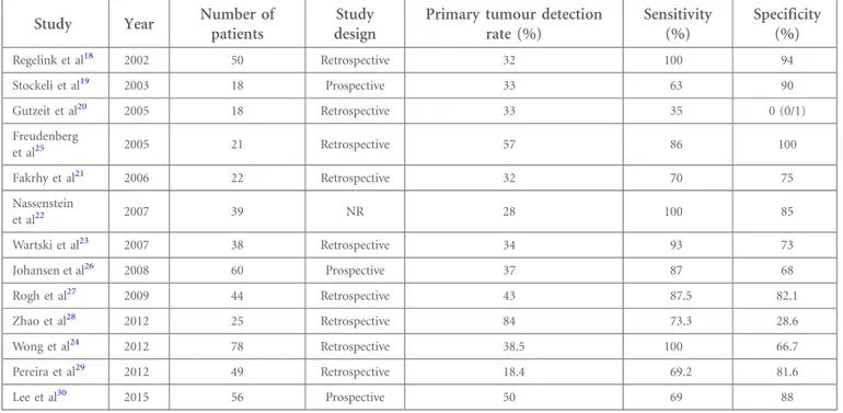
Table 1. Diagnostic performance of positron emission tomography (PET)/CT for unknown primary carcinoma
Study Year Number of
patients
Study design
Primary tumour detection rate (%) Sensitivity (%) Specificity (%) Regelink et al18 2002 50 Retrospective 32 100 94 Stockeli et al19 2003 18 Prospective 33 63 90 Gutzeit et al20 2005 18 Retrospective 33 35 0 (0/1) Freudenberg et al25 2005 21 Retrospective 57 86 100 Fakrhy et al21 2006 22 Retrospective 32 70 75 Nassenstein et al22 2007 39 NR 28 100 85 Wartski et al23 2007 38 Retrospective 34 93 73 Johansen et al26 2008 60 Prospective 37 87 68 Rogh et al27 2009 44 Retrospective 43 87.5 82.1 Zhao et al28 2012 25 Retrospective 84 73.3 28.6 Wong et al24 2012 78 Retrospective 38.5 100 66.7 Pereira et al29 2012 49 Retrospective 18.4 69.2 81.6 Lee et al30 2015 56 Prospective 50 69 88 NR, not reported.
Moreover, although PET/CT performed with contrast-enhanced CT provides both anatomical and metabolic details at the same time, there is no clear recommendation for routine use of PET/ CT in initial T staging.
Some authors showed the high potential value of PET/CT to identify the local tumour extension.11,12 Interestingly, Baek et al11 retrospectively reviewed 69 patients with oral cavity cancer (OCC) who had non-removable dental metallic implants at the time of the pre-treatment imaging work-up and on whom CT or MRI plus PET/CT was performed for the initial staging. The aim was to analyze the clinical impact of PET/CT for pri-mary tumour detection and volume estimation in patients presenting this particular clinical situation. A total of 64 PET/ CT, 64 CT and 27 MRI were analyzed. PET/CT was more ac-curate in detecting primary tumours than CT in patients with OCC and dental artefacts (95.3% vs 75.0%, respectively; p5 0.0016). Among the 27 subjects who had undergone all the three diagnostic modalities, the diagnostic performance for the detection of primary tumours in the oral cavity was 96.3%, 77.8% and 85.2%, respectively (not statistically significant). Rodrigues et al12retrospectively evaluated 44 patients who un-derwent primary tumour resection and neck dissection. They compared the performance of CE-CT, a dedicated HN PET/CT (the latter being a CE-CT) and an optimized whole-body (WB) PET/CT scan. The primary tumour was correctly identified by CE-CT, WB PET/CT and HN PET/CE-CT in 71%, 92% and 95% of cases, respectively. Both (WB and HN) PET protocols demonstrated significantly better performance than did CE-CT in identifying the primary site of the tumour. However, there
was no statistical difference in the detection of the primary le-sion between WB PET/CT and HN PET/CE-CT protocols.12A major limit of this study is the lack of comparison with the MRI performance. Moreover, 66% of the patients participating in the study presented an oropharyngeal carcinoma, making difficult to infer these data in HN neoplasms of different origin. Mandibular invasion
The presence or absence of mandibular invasion is a major determinant in both therapeutic approach and prognosis of HNSCC.13 CT and MRI are commonly used to evaluate the status of the mandible. CT has been reported to be the most accurate method in evaluating discrete cortical bone in-volvement.14 However, MRI is superior to CT for evaluating tumour invasion into medullary cavity of the mandible.15 Gu et al16performed a direct comparison of CT and MRI and PET/CT in the detection of mandibular invasion by OCC. The sensitivity was 47.1%, 58.3% and 58.3% for CT, MRI and PET/ CT, respectively. The specificity was 100%, 97.1% and 97.1% for CT, MRI and PET/CT, respectively. No statistically significant differences in sensitivity and specificity were detected between the three imaging modalities. A recent retrospective study compared the diagnostic performance from PET/CT and MRI for the detection of bone marrow invasion of the mandible in patients with OCC (surgical specimen was used as the standard). PET/CT was found to be more specific than MRI (83% vs 61%, respectively, p 5 0.0015) but less sensitive (78% vs 97%, re-spectively, p5 0.0391). Given the low positive-predictive value (PPV) of MRI, a positive MRI scan should incite to confirm data
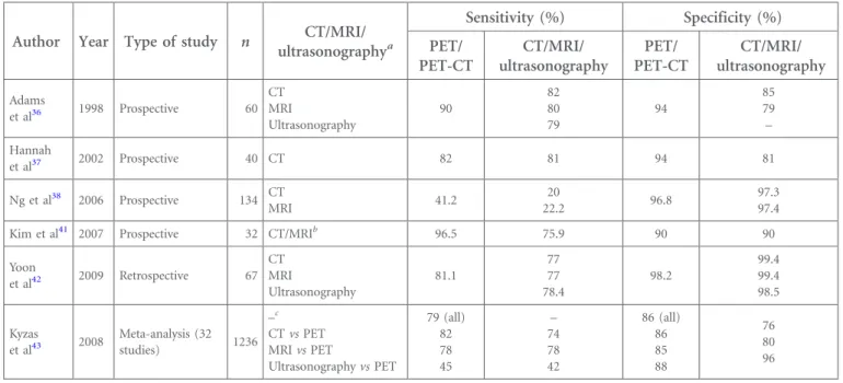
Table 2. Studies comparing positron emission tomography (PET) vs CT, MRI and ultrasonography with histopathology of cervical lymph nodes
Author Year Type of study n CT/MRI/ ultrasonographya Sensitivity (%) Specificity (%) PET/ PET-CT CT/MRI/ ultrasonography PET/ PET-CT CT/MRI/ ultrasonography Adams et al36 1998 Prospective 60 CT MRI Ultrasonography 90 82 80 79 94 85 79 – Hannah et al37 2002 Prospective 40 CT 82 81 94 81 Ng et al38 2006 Prospective 134 CT MRI 41.2 20 22.2 96.8 97.3 97.4 Kim et al41 2007 Prospective 32 CT/MRIb 96.5 75.9 90 90 Yoon et al42 2009 Retrospective 67 CT MRI Ultrasonography 81.1 77 77 78.4 98.2 99.4 99.4 98.5 Kyzas et al43 2008 Meta-analysis (32 studies) 1236 –c CT vs PET MRI vs PET Ultrasonography vs PET 79 (all) 82 78 45 – 74 78 42 86 (all) 86 85 88 76 80 96 a
Type of imaging compared to PET.
bAll patients underwent CT and/or MRI (reported together).
cAlso compared the performance of fluorine-18 fludeoxyglucose PET with that of conventional diagnostic methods (i.e. CT, MRI and ultrasonography
with PET, which shows higher PPV, whereas a negative MRI scan can confidently exclude the presence of bone marrow invasion.15 Cancer from unknown primary
The incidence of cervical metastases from unknown primary cancer (UPC) has been estimated to be around 2–9%. The absence of information about the primary tumour strongly influences the therapeutic approach (i.e. bilateral tonsillecto-mies, additional pharyngeal mucosafield irradiation).17 PET/ CT can identify approximately 30%18–24of tumours in patients presenting cervical lymph node metastases from UPC, in whom the primary was not detected by the comprehensive diagnostic work-up including endoscopy and conventional imaging methods (CT or MRI). Table 1 summarizes the di-agnostic performance of PET/CT in these studies.18–30 It is noteworthy that it should be performed before examination under anaesthesia for targeted panendoscopy and biopsy, avoiding potential false positives due to the inflammation that usually follows these kinds of procedures.26 Thus, a rigorous
physical examination is still essential, considering that small and superficial tumours may not have enough18
F-FDG avidity to be detected by PET/CT, as showed by Thiagarajan5 and Daisne et al.31
Recently, Zhu et al32performed a meta-analysis analyzing a total of 7 studies (246 patients). The primary tumour detection rate, sensitivity and specificity of PET/CT were 44% [95% confidence interval (CI)5 0.31–0.58], 97% (95% CI 5 0.63–0.99) and 68% (95% CI5 0.49–0.83).
The largest prospective study evaluating the diagnostic perfor-mance of PET/CT in UPC has been published by Johansen et al.26 The authors report data about 60 patients presenting a primary tumour detection rate of 30%, and the sensitivity and specificity rates were 86% and 69%, respectively. However, this study had several limits, namely, it used three different PET engines, and among them one was PET/CT, whereas in two cases, it was PET alone.
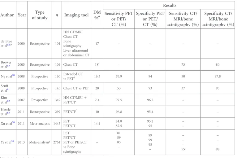
Table 3. Results of positron emission tomography (PET) or PET/CT and other imaging tools in M staging of head and neck (HN) cancer
Author Year Type
of study n Imaging tool DM %a Results Sensitivity PET or PET/ CT (%) Specificity PET or PET/ CT (%) Sensitivity CT/ MRI/bone scintigraphy (%) Specificity CT/ MRI/bone scintigraphy (%) de Bree et al52,b 2000 Retrospective 101 HN CT/MRI Chest CT Bone scintigraphy Liver ultrasound or abdominal CT 17 – – – – Brower et al53 2005 Retrospective 109 Chest CT 18c – – 73 80 Ng et al60 2008 Prospective 160 Extended CT vs PETd 16.3 76.9 94 50 97.8 Senft
et al61 2008 Prospective 145 Chest CT vs PET 28 53 93 37 95
Kim et al62 2007 Prospective 349 HN CT/MRI1 PET/CTe 7.4 97.5 96.2 – – Haerle et al63 2011 Retrospective 299 PET/CTf 10 96.8 95.4 – – Xu et al58 2011 Meta-analysis 1445 PET PET/CT 14.4 84.8 87.5 95.2 95 – – –– Yi et al59 2013 Meta-analysisg 2764 PET PET/CT PET or PET/CT vs Bone scintigraphy – 81 89 85 – 99 99 98 – – – – 55 – – – 98 DM, distant metastasis. aDMs detected by PET. bScreening for DM without PET.
cThis percentage represents metastasis detected by chest CT. There are no comparisons with other imaging. dFrom the skull base to the lower abdomen.
eAll patients underwent contrast-enhanced CT or MRI of the HN. Whole-body fluorine-18 fludeoxyglucose PET/CT was also performed in all patients to
identify second primary or distant metastatic cancers. There was no comparison between PET/CT and other imaging modalities.
fNo comparison was performed between PET/CT and other imaging modalities.
Furthermore, in some cases, PET was acquired for WB, in other cases for half body. Furthermore, the authors did not perform extensive comparison with other imaging modalities.
In summary, physical examination remains essential5 for primary tumour assessment (especially regarding superficial tumour extension in the mucosa) while CE-CT and MRI continue to be the reference imaging modalities, mostly due to the lack of shown superiority of PET over morphological examination; however, PET seems to be a promising staging tool, in particular, when morphological examinations suffer from artefacts due to dental implants. Finally, when com-pared with conventional imaging, PET/CT is recommended to identify primary tumours in patients presenting with cervical lymph node metastases with unknown primary. Nevertheless, it needs opportune integration with other di-agnostic procedure to exclude the relatively high risk of false-positivefindings.
Lymph node involvement (nodal staging)
The information about nodal involvement is crucial in HNSCC, as it strongly influences the treatment and prognosis of the patients.2,3 Current non-invasive staging techniques include clinical examination, ultrasonography, CE-CT and MRI. The criteria adopted in the evaluation of the nodal status are the size, CE and radiological aspect of the nodes (presence of necrosis, analysis of the capsule to identify any sign of extracapsular ex-tension).33 These techniques could define positive nodes with high specificity, but present limitations in the evaluation of small lymph nodes.34–38 The overall diagnostic accuracy (using pa-thology as the reference standard) of CT and MRI for detecting
metastases in the clinical node negative neck (cN0) is relatively low. Sensitivities range from 14% to 80% for CT and from 29% to 85% for MRI, and specificities range from 80% to 100% for both CT and MRI.33,35–37
The sensitivity and specificity of the imaging techniques in-fluence the clinical practice of the radiation oncologist; patients presenting an expected risk of nodal involvement exceeding 20%39undergo a prophylactic treatment of the neck, including a neck dissection or unilateral and/or bilateral neck irradiation.39,40 Considering these rates of microscopic in-volvement, it means that there are at least two thirds of the patients who are treated on the nodal areas without presenting a nodal involvement, only because it is not possible to predict with a better accuracy the“real nodal status of the patients”. Table 2 summarizes studies comparing the performances of different imaging approaches in the study of the nodal status of HNSCC. These studies36–38,41–43 indicate that PET 6 CT is similar or slightly superior to conventional imaging for the di-agnosis of neck metastasis.
Kyzas et al43 performed a meta-analysis on this topic in 2008. Across 32 studies (1236 patients), PET sensitivity was 79% (95% CI5 72–85%) and specificity was 86% (95% CI 5 83–89%). However, for patients with cN0, sensitivity of PET was only 50% (95% CI 5 37–63%), whereas specificity was 87% (95% CI5 76–93%).
Ng et al38evaluated prospectively 134 patients with oral HNSCC with palpably negative neck with 18F-FDG-PET, CT/MRI and
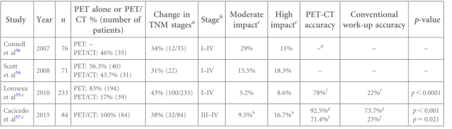
Table 4. Clinical impact of positron emission tomography (PET) or PET/CT
Study Year n
PET alone or PET/ CT % (number of patients) Change in TNM stagesa Stage b Moderate impactc High impactc PET-CT accuracy Conventional
work-up accuracy p-value
Connell et al56 2007 76 PET:– PET/CT: 46% (35) 34% (12/35) I–IV 29% 11% – d – – Scott et al54 2008 71 PET: 56.3% (40) PET/CT: 43.7% (31) 31% (22) I–IV 15.5% 18.3% – – – Lonneux et al55,e 2010 233 PET: 83% (194) PET/CT: 17% (39) 43% (100/233) I–IV 5.2% 8.6% 78% f 22%f p, 0.0001 Cacicedo
et al57,e 2015 84 PET/CT: 100% (84) 38% (32/84) III–IV 9.5%h 16.7%h
92.5%g 71.4%f 73.7%g 25%f p, 0.001 p5 0.021 n, number of patients.
aDiscrepant TNM stages obtained between conventional work-up and the inclusion of PET or PET/CT. bHead and neck squamous cell carcinoma stages included in the study.
cMedium and high impact: the impact on patient management in these studies was classified as follows: low impact (treatment modality and delivery
unchanged); medium impact (change in the treatment within the same therapeutic modality: changes in the type of surgery on primary cancer and/or neck dissection, and changes in the dose or radiotherapy fields); or high impact (change in treatment intent and/or treatment modality: curative to palliation, surgery to chemoradiation or vice versa).
dNot available.
eIn these two studies, treatment decisions were made by a tumour board (pre-PET staging and treatment management plan, and post-PET staging
and treatment plan). Therefore, the pre-PET treatment decision and post-PET treatment decision were both made by the tumour board.
fThis accuracy was calculated for the cases presenting discordant TNMs (n
5 100) between conventional work-up and from adding a PET (not for the whole population of the study).
gThe overall accuracy regarding the whole population of the study.
hMeaning that the clinical original clinical decision (pre-PET) adopted by the tumour board was changed in approximately one out of five (9.5%
1 16.7%5 26.2%) patients participating in the study, due to the PET/CT.
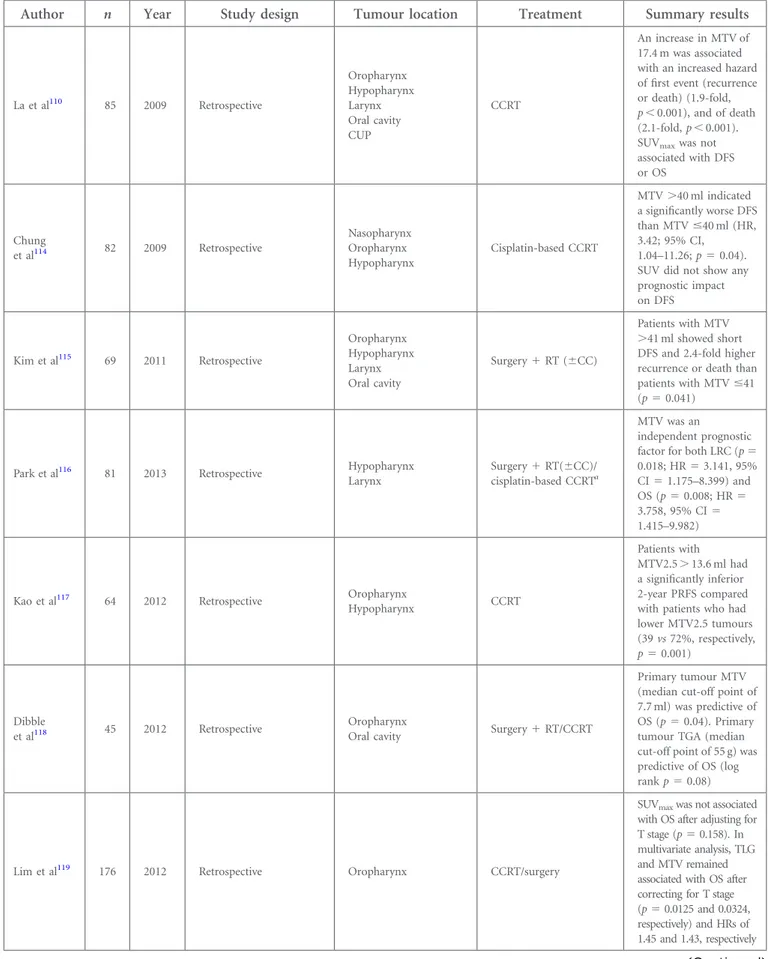
Table 5. Studies evaluating prognostic value of pre-treatment positron emission tomography volumetric parameters
Author n Year Study design Tumour location Treatment Summary results
La et al110 85 2009 Retrospective Oropharynx Hypopharynx Larynx Oral cavity CUP CCRT An increase in MTV of 17.4 m was associated with an increased hazard offirst event (recurrence or death) (1.9-fold, p, 0.001), and of death (2.1-fold, p, 0.001). SUVmaxwas not
associated with DFS or OS Chung et al114 82 2009 Retrospective Nasopharynx Oropharynx Hypopharynx Cisplatin-based CCRT MTV.40 ml indicated a significantly worse DFS than MTV#40 ml (HR, 3.42; 95% CI,
1.04–11.26; p 5 0.04). SUV did not show any prognostic impact on DFS Kim et al115 69 2011 Retrospective Oropharynx Hypopharynx Larynx Oral cavity Surgery1 RT (6CC) Patients with MTV .41 ml showed short DFS and 2.4-fold higher recurrence or death than patients with MTV#41 (p5 0.041)
Park et al116 81 2013 Retrospective Hypopharynx Larynx
Surgery1 RT(6CC)/ cisplatin-based CCRTa
MTV was an
independent prognostic factor for both LRC (p5 0.018; HR5 3.141, 95% CI5 1.175–8.399) and OS (p5 0.008; HR 5 3.758, 95% CI5 1.415–9.982)
Kao et al117 64 2012 Retrospective Oropharynx
Hypopharynx CCRT
Patients with MTV2.5. 13.6 ml had a significantly inferior 2-year PRFS compared with patients who had lower MTV2.5 tumours (39 vs 72%, respectively, p5 0.001) Dibble et al118 45 2012 Retrospective Oropharynx
Oral cavity Surgery1 RT/CCRT
Primary tumour MTV (median cut-off point of 7.7 ml) was predictive of OS (p5 0.04). Primary tumour TGA (median cut-off point of 55 g) was predictive of OS (log rank p5 0.08)
Lim et al119 176 2012 Retrospective Oropharynx CCRT/surgery
SUVmaxwas not associated
with OS after adjusting for T stage (p5 0.158). In multivariate analysis, TLG and MTV remained associated with OS after correcting for T stage (p5 0.0125 and 0.0324, respectively) and HRs of 1.45 and 1.43, respectively
Table 5. (Continued)
Author n Year Study design Tumour location Treatment Summary results
Lee et al120 57 2012 Retrospective Oropharynx Surgery/surgery1
adjuvant therapy
On a univariate analysis, SUVmax, SUVavg, MTV
and TLG of primary tumour were significant predictors of
survival. However, on multivariate analysis, only patients with high MTV ($7.78 cm3) showed significantly worse prognoses (p5 0.037) Tang et al121 83 2012 Retrospective Oropharynx Nasopharynx Hypopharynx Larynx Oral cavity CUP RT/CCRT An increase in total MTV of 17 cm3was associated with a 2.1-fold increase in the risk of disease progression (p5 0.0002) and a 2.0-fold increase in the risk of death (p5 0.0048). SUVmaxwas not
associated with either outcome Moon et al122 83 2013 Retrospective Tonsil RT alone Surgery alone CCRT Surgery CCRT or RT On multivariate analyses, only TLG (HR5 1.020, 95% CI5 1.003–1.037, p5 0.023) was an independent predictive factor associated with decreased OS. MTV and SUVmaxwere not
associated with outcomes
Abd El-Hafez et al123
126 2013 Prospective Oropharynx Surgery/CCRT
TLG and SUVmaxwere
independent prognostic factors for 2-year DSS. Patients with high (T)TLG ($71.4) had a 2-year DFS of 52%, whereas 74% for those with a low (T)TLG (p5 0.007); the 2-year-DSS rates were 53% vs 84%, respectively (p, 0.001). Patients with high (N)SUVmax($7.5)
had a 2-year DFS of 42% vs 70% for patients with a low (N)SUVmax
(p5 0.001); the 2-year-DSS rates were 39% vs 78%, respectively (p, 0.001) Garsa et al124 86 2013 Retrospective Oropharynx CCRT On multivariate analysis, a total MTV.20.5 ml was associated with a 13.0-fold increased risk of death (95% CI5 1.62–100; p 5 0.016) for the p16-positive subgroup compared with
Table 5. (Continued)
Author n Year Study design Tumour location Treatment Summary results
a 4.27-fold increased risk of death (95% CI5 1.28–14.3; p 5 0.018) for the p16-negative subgroup. SUVmax,
SUVmeanfailed to predict
DFS or OS Romesser et al125 100 2014 Retrospective Oropharynx CCRT On multivariate analysis, a larger MTV (,9.7 cm3) retained
a significant correlation with an increased risk for distant metastasis (HR5 2.47; 95% CI5 1.46–4.17; p 5 0.001), disease progression or death (HR5 2.17; 95% CI5 1.40–3.38; p 5 0.001), and death (HR5 2.37; 95% CI5 1.44–3.89; p 5 0.001). SUVmaxfailed to
correlate with any outcome Hanamoto et al126 118 2014 Retrospective Nasopharynx Oropharynx Laryngohypopharyngeal CCRT After multivariate analysis, high MTV (.25.0 ml) and high TLG (.144.8 g) remained as
independent, significant predictors of incomplete response compared with low MTV (OR5 13.4; 95% CI5 2.5–72.9; p 5 0.003) and low TLG (OR 5 12.8; 95% CI 5 2.4–67.9; p 5 0.003), respectively Alluri et al127 70 2014 Retrospective Oropharynx (HPV positive) RT alone/CCRT CCRT1 surgery Surgery1 CCRT
Total MTV and primary tumour MTV remained as independent prognostic markers for EFS. There was no statistically significant association of EFS with SUVmax, SUVmeanand
primary tumour or overall TLG Picchio et al112 19 2014 Retrospective Oropharynx Nasopharynx Larynx RT/CCRT MTV ($32.4 cm3) and TLG ($469.8 g) predicted patients’ outcome with respect to all the considered local and distant disease control end points (LRFS, DMFS and DFS). SUVmeancut-off value
predictive of LRFS and DFS were 10.8
their visual correlation. They reported that 18F-FDG-PET was
twice more sensitive than CT/MRI for detecting cervical nodal metastasis in patients with palpably negative neck (41.2% vs 21.6%, respectively; p5 0.021). Histopathological analysis was used as the gold standard to validate the results obtained with different imaging techniques. The authors concluded that18 F-FDG-PET presented a false negativity rate (of occult neck me-tastasis) of ,15% in T1–3 tumours. However, 18F-FDG-PET, even visually correlated with CT/MRI, was unable to reduce the rate of false negative to,20% in patients with T4 tumour (neck treatment being mandatory regardless of PET results).
Kim et al41evaluated 32 consecutive patients with oropharyngeal HNSCC undergoing18F-FDG-PET and CT/MRI before surgery (all patients underwent curative resection of their primary tumours with also node dissection, with 7 having bilateral dis-sections, for a total of 39 neck sides). Each method was inter-preted separately to assess primary tumour and cervical node status. Histopathology specimen (in 29 of 39 dissected neck sides and in 47 of 163 dissected cervical levels) showed that
18
F-FDG-PET was more accurate than CT/MRI, both in detecting positive neck sides (22/29 vs 28/29, p, 0.05) and on a level-by-level basis (37/47 vs 45/47, p, 0.05). Interestingly,
18F-FDG-PET identified metastatic lesions in approximately two
thirds of the morphologically uninvolved nodes.
Cetin et al44studied 36 patients with HN cancers, clinically and radiographically N0, by means of PET/CT and compared data with neck dissection results. The best threshold of the maximum standardized uptake value (SUVmax) value yielded 84.2%
sen-sitivity and 76.5% specificity for nodal-level staging.
Roh et al45assessed prospectively 91 patients with HNSCC and negative neck palpation. PET/CT was more sensitive on a per-level basis than CT/MRI (69% vs 39%, p, 0.001), as well as on a per-patient basis 71% and 50%, respectively (p5 0.011).
Although PET/CT examination protocols without the use of contrast medium have been utilized, increasing evidence sup-ports the use of CE-CT as a part of routine PET/CT protocols.46,47 Recently, there have been several reports of the possible superiority of PET/CE-CT over standard PET/CT in different clinical settings, including better local48 and nodal analysis.49 Other studies,50,51 confirmed the high accuracy of nodal staging by PET/CT, in particular, if CE-CT is used during PET protocol.
In summary,18F-FDG-PET has high diagnostic performance in the overall nodal staging of patients with HNSCC. When com-pared with conventional imaging, PET/CT is similar or superior for detecting cervical nodal metastases (Table 2). However, this modality is not yet accurate enough to replace the accuracy of neck dissection in the identification of occult cervical metastasis in patients with cN0.36–38,41–43
Detection of distant metastasis
The presence of distant metastases is the most important predictor of patient survival in several cancers. Overall incidence of distant metastasis in HNSCC is relatively low (2–18%).52
Distant metas-tases frequently occur in the lungs and are routinely detected by chest CT (73% sensitivity and 80% specificity).53
It is noteworthy that early detection of metastasis has a major impact on patient management avoiding unnecessary radical treatments.54–57 Xu et al58conducted a meta-analysis evaluating the accuracy of PET and PET-CT in the initial M staging of HNSCC. This meta-analysis suggested that 209 (14.4%) of 1445 patients had distant metastasis or a second primary tumour (SPT). PET-CT pre-sented an overall sensibility of 87.5% (95% CI, 78.7–93.6) and an overall specificity of 95% (95% CI, 93.1–96.4).
Regarding the detection of bone metastases, Yi et al59showed in a recent meta-analysis on .3000 patients, a sensitivity and
Table 5. (Continued)
Author n Year Study design Tumour location Treatment Summary results
Schwartz
et al128 74 2015
Population subanalysis of Phase III trial
(RTOG 0522) Oropharynx Larynx Hypopharynx CCRT (cisplatin and cetuximab) Primary tumour MTV was a strong independent prognostic factor for PFS. SUVmax
was not associated with poor treatment outcomes
Yabuki
et al129 118 2015 Retrospective Larynx RT or CCRT
On multivariate analysis, the 3-year DFS for patients with a high MTV were significantly poorer than those with a low MTV (p, 0.001)
CC, concurrent chemotherapy; CCRT, concurrent chemoradiotherapy; CI, confidence interval; CUP, carcinoma with unknown primary; DFS, disease-free survival; DMFS, distant metastasis-free survival; DSS, disease-specific survival; EFS, event free survival (either recurrence of disease at the primary site, at regional nodes, or at distant metastatic sites or overall patient mortality); HPV, human papillomavirus; HR, hazard ratio; LRC, locoregional control; LRFS, local recurrence-free survival; MTV, metabolic tumour volume; MTV2.5, PET segmentation used applying on isocontour at a SUV of 2.5; n, number of patients; (N)SUVmax, nodal SUVmax; OR, odds ratio; OS, overall survival; PFS: progression-free survival; PRFS, primary
relapse-free survival; RT, radiotherapy; RTOG, Radiation Therapy Oncology Group; SUV, standardized uptake value; SUVavg, average SUV; SUVmax,
maximal SUV; SUVmean, mean SUV; TGA, total glycolytic activity; TLG, total lesion glycolysis; (T)TLG, tumour total lesion glycolysis. aSeveral patients received induction chemotherapy.
a specificity of 81% and 99% for PET, and of 89% and 99% for PET/CT, respectively, which are better than the results obtained by bone scintigraphy. Bone scintigraphy relies on the osteo-blastic response to bone destruction by cancer cells and the accompanying increase in bloodflow. Therefore,18F-FDG-PET is more efficient than bone scintigraphy for bone lesion de-tection considering their frequently lytic character.59
Table 3summarizes available studies comparing the perform-ances of different imaging approaches to detect distant metas-tasis of HNSCC.52,53,58–63Globally, all these studies indicate that PET6 CT is superior to conventional imaging.
18F-FDG-PET shows higher accuracy (90–95%) than CT for the
detection of distant metastasis.60,61,63 Given the very high negative-predictive value (NPV), it suggests that in case of negative PET scan, other imaging techniques are not necessary. Nevertheless, the PPV for detecting SPT or distant metastasis is around 60%, suggesting that additional diagnostic methods are still necessary to exclude false-positive results.62
Senft et al61assessed the added value of18F-FDG-PET (to chest CT) in the screening of distant metastases in patients with HNSCC and high-risk factors (more than or equal to three lymph node metastases, bilateral lymph node metastases, lymph node metastases of$6 cm, low jugular lymph node metastases, regional tumour recurrence and SPT). 145 consecutive patients with HNSCC underwent chest CT and18F-FDG-PET.18 F-FDG-PET improved pre-treatment screening of distant metastasis compared with chest CT, showing higher sensitivity (53% vs 37%) and PPV (80% vs 75%). Moreover, the authors showed that the sensitivity of the combination of CT and18F-FDG-PET was higher (63%) than the sensitivity of each of these techni-ques alone.
Ng et al60prospectively compared18F-FDG-PET and extended-field CE-CT (from the skull base to the lower abdomen). A total of 160 patients with HNSCC of the oropharynx or hypopharynx underwent 18F-FDG-PET and extended-field CT to detect dis-tant metastases or SPT. In the entire study cohort, a total of 26 patients (16.3%) were found to have distant malignant lesions. Diagnostic yields of 18F-FDG-PET and extended-field CE-CT were 12.5% (20 out of 160 patients) and 8.1% (13 of 160 patients), respectively. The patient-based sensitivity of
18
F-FDG-PET for detection of distant malignancies was 1.5-times higher than that of extended-field CE-CT (76.9% vs 50.0%, p 5 0.039), whereas the patient-based specificity of
18F-FDG-PET was not significantly lower than that of
extended-field CE-CT (94.0% vs 97.8%, p 5 0.125).
In summary, when compared with conventional imaging, PET/CT is a valuable tool to rule out the presence of distant metastases in HNSCC, especially in locally advanced tumours.52,53,60–62,64
Second primaries
SPTs are detected in almost 10% of patients with HNSCC,64,65 particularly in patients who smoke and/or in patients who are negative for human papillomavirus.66
The identification of synchronic or metacronic SPT67 could occur both at the HN region (more frequently) and/or elsewhere (lungs, oesophagus, colon etc.), and it can influence the thera-peutic approach61and the prognosis of patients (especially those presenting with HNSCC).68
Strobel et al64 evaluated the role of PET/CT for the initial staging of HNSCC in 589 consecutive patients for the detection of synchronous primaries. They detected 56 secondary cancers in 44 patients. 46 (82%) were found in the aerodigestive tract as follows: lung (26%), HN (15%) and oesophagus (6%). Nine synchronous cancers were detected by endoscopy and lost at PET/CT. The prevalence of synchronous primaries according to the standard of reference (including panendoscopy or bronchoscopy or oesophageal or colon endoscopy when nec-essary) was 9.5%. Of these, synchronous primaries, 47 (84%) were detected in 41 patients (93%) by 18F-FDG-PET/CT. Interestingly, in 32 out of 40 patients (80%) with available follow-up, the treatment was modified because of the detection of a synchronous primary.64 They concluded that
18
F-FDG-PET/CT detects a considerable number of synchro-nous primaries (8.0% prevalence) at the initial staging of patients with HNSCC.
According to Haerle et al65synchronous primary tumours were detected in 4.5% of patients by panendoscopy compared with 6.1% by PET/CT. Indeed 26% of lesions detected on PET/CT were within the coverage of the panendoscopy.6518F-FDG-PET/ CT was superior to panendoscopy. The sensitivity, specificity, PPV and NPV for panendoscopy were 74%, 99.7%, 93% and 98%, respectively. The sensitivity, specificity, PPV and NPV for
18
F-FDG-PET/CT were 100%, 95.7%, 59% and 100%, respectively.
According to these results64,65with a negative18F-FDG-PET/CT,
the extent of endoscopy can be reduced to the area of the pri-mary tumour.
In summary, PET/CT is an accurate method detecting second primaries, with a high NPV.64,65,67–69Nonetheless, it should be stressed that due to a low PPV (approximately 60%65) in this setting, additional diagnostic methods are necessary to exclude false-positive results (inflammation and hyperplasia in the HN region or intestinal polyps can result in false positives). More-over, whenever possible PET/CT should be performed before endoscopy and biopsy to avoid false-positive results.26
POSITRON EMISSION TOMOGRAPHY/CT AND CLINICAL IMPACT ON TREATMENT DECISIONS Although PET/CT imaging is effective for the staging of HNSCC, its impact on patient management is somehow con-troversial. Indeed, to date, the overall impact of PET/CT on treatment decisions in HNSCC has been rarely explored com-pared with the number of studies assessing the impact of PET on staging. However, there are four prospective trials54–57that have specifically analyzed the impact of PET/PET-CT in the treatment approach of HNSCC. These studies followed the same meth-odology and are detailed inTable 4. These studies addressed at the same time the issue of the impact of PET on the initial
staging and management of patients with HNSCC: globally, the PET changed the original treatment plan in approximately 30% of patients.54–57
The largest trial, published by Lonneux et al55 included 233 patients (Stages I–IV) and reported a modification in the original treatment plan in 32 patients (13.7%). In 12 patients (5.2%), the modification was classified as medium (the thera-peutic modality remained the same, but PET altered the treat-ment planning). In 20 patients (8.6%), the impact of PET on patient management was classified as high (change in treatment intent and/or treatment modality, e.g. curative to palliation, surgery to chemoradiation and so on). Interestingly, one of the studies57assessed together the usefulness of PET/CT for staging and its overall impact on management plans specifically in patients with Stages III and IV HNSCC where the treatment plan was altered in 22/84 (26%) patients (Table 4). These results are in line with the current guidelines.2,3
In summary, PET/CT should be included54–57 in the routine diagnosis of patients with Stages III–IV57HNSCC, as it signifi-cantly improves staging accuracy and also has a marked impact on management plans.
RADIOTHERAPY PLANNING
CT is the primary imaging modality in RTP. The CT images are acquired with the patient in the supine position, immobilized with an individual head support and a rigid customized mask to increase positioning accuracy and to prevent movement during image acquisition. All other imaging modalities (such as PET or MRI) are considered as secondary images.70 The secondary images in RTP will have to be registered (fused) to the primary planning CT scan. When the PET and RTP CT images are ac-quired on separate scanners (often in a position that does not correspond to real treatment position), a registration module in the RTP computer system can be used to fuse images. Regardless of the imaging data used (i.e. separate PET or PET/CT) correct co-registration of the PET data with the CT data used for RTP must be verified, since the difference in spatial localization of tumour may lead to false estimation of the gross tumour volume (GTV). Ideally, the fusion process can be executed automatically by a hybrid PET-CT-dedicated RTP scanner performed with the corresponding immobilization devices and reproducing radia-tion delivery condiradia-tions.70
Recognizing the potential of PET/CT-guided treatment plan-ning, some authors recently investigated the role of PET in RTP, specifically for the correct delineation of lymph nodes. Schinagl et al71compared the volume of metastatic lymph nodes between
18
F-FDG-PET/CT segmentation (by ten methods) and CT with the volume as determined by pathological examination. They concluded that beyond the detection of lymph node metastasis (staging), PET has no additional value over CT for the de-lineation of lymph nodes.
Despite the limited role of PET-CT in improving the contouring of nodes,71it seems to have a main role in improving the def-inition of the primary tumour GTV. Indeed, PET/CT in-formation is frequently integrated in RTP. Nevertheless, the use
of 18F-FDG-PET for target volume delineation in RTP for HNSCC has been mainly evaluated in single institution studies.5,31,72–75
Different segmentation methods have been proposed. Visual interpretation of the PET signal, considered the most intuitive method for segmentation, has been commonly applied in many studies. The main limit of this approach is that it is a highly operator-dependent process, and it is influenced by window-level settings.5,74–76This is one of the major weakness in the use of PET-CT in the target volume delineation of HNSCC. This vari-ability could be reduced by using a more objective methodology: isocontouring based on afixed standardized uptake value (SUV) such as a SUV of 2.5–3 g l21
or relative thresholds such as a per-centage of the maximum tumour intensity (40% SUVmax, 50%
SUVmax).75,77 Nevertheless, according to this method, several
structures containing a high physiological18F-FDG uptake, such as the tonsillar area or the vocal cords, can be incorrectly included in the segmented area. Therefore, models using afixed threshold relying on SUV are somehow debatable.73
To overcome this issue, several authors successfully developed advanced adaptive relative threshold segmentation methods based on maximal tumour uptake, background uptake, tumour dimensions and tumour grade.31,75,78,79 Thereafter, other methods including gradient-based73 detections have been in-troduced. In brief, this method relies on the watershed trans-form and hierarchical cluster analysis, to allow a better estimation of the gradient intensity. Interestingly, this method allows automatic delineation and therefore is an operator-independent process. Most studies comparing GTV definitions using18F-FDG-PET against CT or MRI reported a decrease in the GTV, especially when using more sophisticated segmentation methods.5,31,72–74,76,80
However, few groups have validated delineation process using different imaging modalities against surgical resection specimens.31,81–85 In general, all imaging modalities over-estimated the tumour extension compared with surgical speci-men. Nevertheless, none of the image modalities (CT, MRI or PET) completely encompassed the surgical specimen volume because of an underestimation of superficial tumour extension in the mucosa,31as also reported by Ng et al.81
According to Daisne et al,31the GTV delineated from18 F-FDG-PET applying an adaptive signal-to-background method was significantly smaller than GTV delineated by CT or MRI. In addition, GTV-PET was the closest volume to the pathological GTV obtained from surgical specimen. On average, the PET delineated smaller volumes than CT or MRI. Nevertheless, GTV contours at PET were not totally encompassed by those de-lineated with CT or MRI.
Geets et al73validated a gradient-based method in seven patients with laryngeal carcinoma. The calculated volumes for laryngeal tumours according to this methodology73were compared with the macroscopic specimens and, additionally, with the volumes obtained applying the source-to-background ratio developed by Daisne et al.31 Interestingly, the gradient-based method proved
to be more accurate than the source-to-background ratio but neither the threshold-based nor the gradient-based volumes encompassed completely the laryngeal specimens.
Interestingly, in a recent multicentric prospective study by Leclerc et al,86the primary tumour was automatically delineated on the 18F-FDG-PET images using a gradient-based method previously described by this group.73 They confirmed that the use of18F-FDG-PET translated into smaller GTV, clinical target volume and planning target volume for the primary tumour volumes compared with the use of CT, lowering the dose to organs at risk.
On the other hand, there are studies with other tracers such as fluorine-18 fluorothymidine (18
F-FLT) evaluating promising PET-segmentation methods for delineation of the proliferative volume (PV) of tumour. In contrast to18F-FDG,18F-FLT does not accumulate in inflammatory tissue,87
which is frequently found in/near primary tumours of the HN or is induced during the course of chemoradiation. Arends et al88 evaluated 46 patients who underwent18F-FLT PET/CT prior to treatment and in the second and fourth week of therapy. The goal of the study was to compare three semiautomatic PET segmentation methods for derivation of PV in primary HNSCC on sequential
18
F-FLT PET images before and during chemoradiation. The following semiautomatic segmentation methods were applied to sequential PET scans: background-subtracted relative-threshold level, a gradient-based method using the watershed transform algorithm and hierarchical clustering analysis and a fuzzy locally adaptive Bayesian algorithm. The authors88concluded that fuzzy locally adaptive Bayesian algorithm (FLAB) was the best per-forming method for segmentation of the PV on repeat18F-FLT PET/CT scans during chemoradiation. FLAB is less sensitive to image noise than the other segmentation approaches tested in the study. Thisfinding may have other potential implications for radiotherapy indicating that FLAB is a promising candidate for radiation target volume adaptation based on sequential18F-FLT PET scanning.
Currently, there is still no consensus (national/international) between institutions regarding the best method to use for de-lineation. Therefore, data from18F-FDG-PET can complement other diagnostic imaging modalities for management decisions and guidance of RTP, but it cannot replace physical examination or MRI/CT scans to achieve significant details such as assessing invasion of tumour-surrounding tissues.85 Moreover, defining the primary tumour boundaries with PET is a difficult task. In summary, current evidence is based on numerous heteroge-neous small studies with changing methodology for different research questions. PET-based RTP is a promising modality to improve contouring accuracy. PET is the imaging modality that defines the closest volume to the pathological specimen. The main drawback is the lack of standardized method for functional volume segmentation, which highly influences the size and shape of the resulting GTV. Currently, the most accurate seg-mentation method seems to be the gradient-based method validated by Geets et al.73However, it may not completely en-compass the tumour specimen volume.31,73This issue is more
relevant when considering superficial mucosal spread (evaluable by physical examination).5,81 Therefore, even in some contem-porary HNSCC study protocols (European Organisation for Research and Treatment of Cancer-1219; NCT01880359), PET-based target volume delineation is not allowed. Before PET can reliably be incorporated into routine high-precision RTP, operator-independent segmentation tools have to be developed and validated (international consensus), and also, clinical effect on outcomes should be reflected in clinical studies.
MONITORING RESPONSE TO
CHEMORADIOTHERAPY: RESIDUAL DISEASE AND RECURRENCE (FOLLOW-UP)
Early detection of residual or recurrent disease following ra-diotherapy is a diagnostic challenge owing to post-treatment anatomical distortions, mostly related to oedema andfibrosis.89 The key role of a diagnostic tool evaluating treatment efficacy is to correctly identify patients requiring salvage-tailored treat-ments. Moreover, an early detection of the relapse could help in the selection of patients who could be successfully retreated.90In this setting, 18F-FDG-PET/CT is an interesting modality to evaluate response to treatment, as it can assess metabolic activity-rendering malignant process.89
Isles et al91preformed a meta-analysis reporting that18 F-FDG-PET (without CT) is a highly accurate tool for monitoring re-sponse and detecting relapse after chemoradiotherapy (for both the primary site and lymph nodes). Moreover, several studies have demonstrated that18F-FDG-PET/CT also has a higher ac-curacy in the detection of recurrent lesions compared with CT/ MRI.6,92–96These results obtained with PET/CT are not signif-icantly different from those obtained with PET alone.
The timing of PET/CT after the treatment is crucial.6,97–101It is widely accepted that PET has a high NPV (around 90%) if it is performed at least 8 weeks after chemoradiotherapy. Therefore, a negative PET scan after treatment appears to be a consistent predictor of the absence of residual tumour.102 According to other reports, more accurate evaluation is possible when PET/CT is performed 8–12 weeks after treatment.6,97
The meta-analysis of Gupta et al showed a weighted mean (95% CI)-pooled sensi-tivity, specificity, PPV and NPV of post-treatment 18
F-FDG-PET (CT) for the primary site of 79.9% (73.7–85.2%), 87.5% (85.2–89.5%), 58.6% (52.6–64.5%) and 95.1% (93.5–96.5%), respectively. Similar estimates for the neck were 72.7% (66.6–78.2%), 87.6% (85.7–89.3%), 52.1% (46.6–57.6%) and 94.5% (93.1–95.7%), respectively. Moreover, two recent studies showed even further increased accuracy with delayed PET/CT performed approximately 4 months after treatment with NPVs reaching 100%.100,101Intuitively, delaying a response evaluation tool would surely increase its accuracy. However, there is no homogeneous data for optimal window for salvage treatment; probably, it would be wise not to postpone salvage surgery beyond a clinically reasonable point.
There is debate regarding the need for elective neck dissection after radical chemoradiotherapy. There are two prospective studies98,99 addressing the status of neck adenopathy of node-positive HNSCC that had18F-FDG-PET/CT at least 12 weeks
after chemoradiotherapy. Porceddu et al98 prospectively evalu-ated 112 patients presenting with radiological nodal complete response. Residual CT nodal abnormalities were present in 50 patients (45%): 41 were PET negative and 9 were PET pos-itive. Patients with residual CT nodal abnormalities deemed PET negative were uniformly observed regardless of residual nodal size. Importantly, 41 of the 50 patients with a residual nodal abnormality were spared a neck dissection on the basis of neg-ative posttherapy PET, with no subsequent nodal failures in this group. Wang et al99prospectively evaluated 44 restaging PET/CT between 12 and 17 weeks after radiotherapy completion, and 10 PET/CT performed in the follow-up of 44 patients. Imaging data were compared with clinicopathological outcomes. For cervical lymph nodes, sensitivity was 100%, specificity was 98%, PPV was 92% and NPV was 100%. Therefore, both these prospective studies concluded that PET-guided management of the neck after chemoradiotherapy appropriately spares neck dissections in patients with complete response or presenting with PET-negative residual CT lesions.98,99
Recently, Mehanna et al103published a prospective, randomized controlled trial assessing the non-inferiority of PET/CT-guided surveillance (evaluation was performed 12 weeks after definitive chemoradiation. Neck lymph node dissection was only indicated when PET/CT presented an incomplete or equivocal response) to planned neck dissection in a total of 564 patients with locally advanced HNSCC (Stage N2 or N3 disease), who underwent chemoradiation for primary treatment.
Patients were considered to have incomplete nodal responses when PET/CT performed 12 weeks after treatment showed high
18
F-FDG uptake (with or without enlarged lymph nodes in the neck). In addition, results of PET/CT presenting mild or no18 F-FDG uptake in enlarged lymph nodes or mild18F-FDG uptake in normal-sized nodes were classified as equivocal responses. The rest of the PET/CT scans were classified as complete responses. Patients showing an incomplete or equivocal response in the neck but presenting a complete response in the primary location underwent neck lymph node dissection within 4 weeks after PET/CT.
The survival rate was similar (2-year overall survival rate of 84.9% and 81.5% in the surveillance group and in the planned-neck dissection group, respectively) between patients who underwent PET/CT-guided surveillance policy and patients undergoing a planned surgery. Indeed, the hazard ratio for death (upper boundary of the 95% CI for the hazard ratio,,1.50; p 5 0.004) favoured PET/CT-guided surveillance policy.
Moreover, surveillance resulted in considerably fewer operations (approximately 80% of patients were spared neck dissection compared with planned dissection surgery; 54 vs 221), and it was more cost effective. The per-person cost saving was £1492 (approximately $2190 in US dollars), with an additional 0.08 quality-adjusted life years per person.
However, these authors recommended that patients with an equivocal 18F-FDG uptake should continue to undergo neck dissection. In addition, when extrapolating these results to daily
clinical practice, it should be noted that in this study, only a small number of patients [17/564 (3%)] presented N3 disease. Therefore, a direct extrapolation of a PET/CT-guided surveil-lance policy to patients presenting N3 (Stage IVb) disease should not be indicated owing to the small number of such patients recruited in the study.
18F-FDG-PET/CT could have a potential interesting role in the
follow-up of patients with HNSCC. Despite that, the clinical advantages and economic costs of this issue have not yet been largely addressed. One of the largest studies has been published by a group from Pittsburg.90 They evaluated 388 patients retrospec-tively to assess the recurrence rate after radical chemoradiotherapy among patients who underwent PET/CT surveillance. Tumour recurrence was detected in 110 patients (73 asymptomatic and 37 symptomatic). Indeed, 95% (95% CI, 87–98%) of asymptomatic recurrences were observed within 2 years of follow-up. The authors proposed to evaluate patients for recurrence with PET/CT at 2, 5, 8 and 14 months post-treatment. The reason for this protocol is because their study demonstrated that PET/CT detected almost all HNSCC recurrences within 2 years.
IN SUMMARY
(1) The overall diagnostic performance of18F-FDG-PET/CT for response assessment is good, but its PPV is not optimal. By contrast, the NPV is particularly high and negative post-treatment PET/CT is very suggestive of absence of viable disease that can guide daily clinical management decisions. In this context, the timing of PET/CT after the end of the treatments is a crucial issue. Available evidences suggest waiting a minimum of 8 weeks before restaging with PET/CT, and preferably 12 weeks to increase the NPV.
(2) Available evidences suggest that this strategy is safe to avoid neck dissection in patients presenting negative PET/CT after CRT.98,99,102 The safety of this attitude is also confirmed by the results of the PET-NECK study,103 where PET/CT-guided active surveillance showed similar survival outcomes compared with planned neck dissec-tion, and considerably fewer neck dissections, and it was more cost effective. However, extrapolation of a PET-CT-guided surveillance policy to patients with N3 (Stage IVb) disease cannot currently be justified.
(3) Evidence-based recommendations to guide the utilization of PET/CT in the follow-up of patients with HNSCC do not exist.2,90
PROGNOSTIC VALUE
Treatment outcome of HNSCC cancer remains heterogeneous. Identification of novel pre-treatment factors (other than tumour stage, lymph node involvement, anatomical subsite or human papillomavirus status) that potentially predict long-term out-come is of great interest.
Quantifying the prognostic value of PET is challenging. In general, the results of prognostic value of SUV remain un-determined because of the small sample of most of these studies. Moreover, it should be considered that HNSCC prognosis also depends on the initial tumour site; data regarding the prognostic
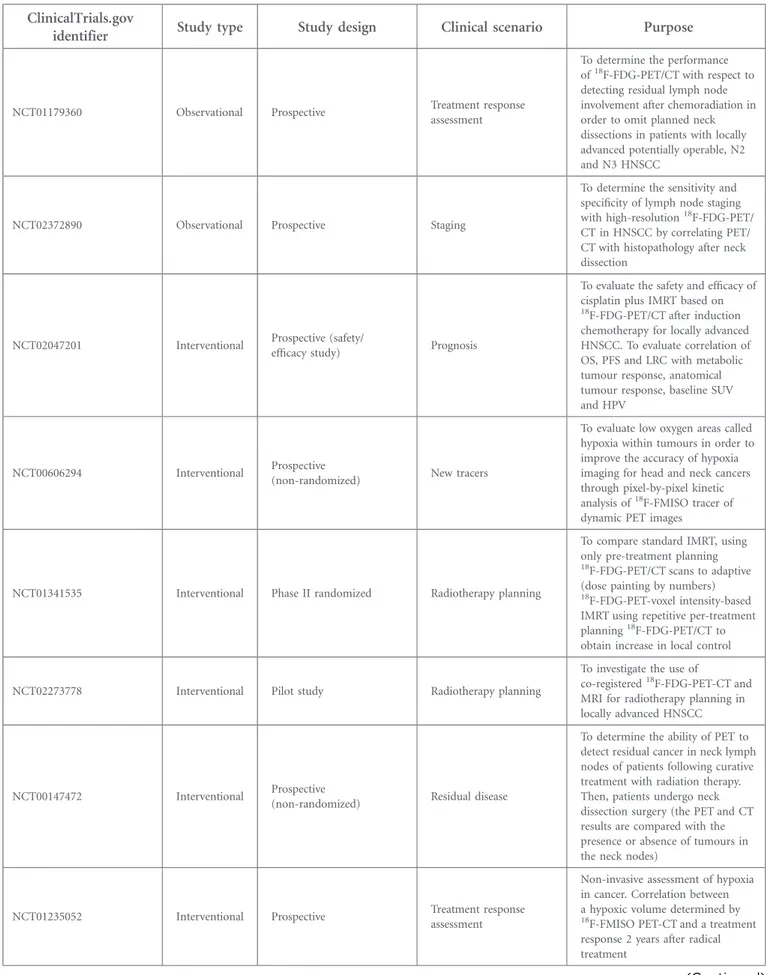
Table 6. Currently ongoing trials evaluating the role of positron emission tomography (PET)/CT in clinical practice
ClinicalTrials.gov
identifier Study type Study design Clinical scenario Purpose
NCT01179360 Observational Prospective Treatment response assessment
To determine the performance of18F-FDG-PET/CT with respect to
detecting residual lymph node involvement after chemoradiation in order to omit planned neck dissections in patients with locally advanced potentially operable, N2 and N3 HNSCC
NCT02372890 Observational Prospective Staging
To determine the sensitivity and specificity of lymph node staging with high-resolution18F-FDG-PET/ CT in HNSCC by correlating PET/ CT with histopathology after neck dissection
NCT02047201 Interventional Prospective (safety/
efficacy study) Prognosis
To evaluate the safety and efficacy of cisplatin plus IMRT based on
18F-FDG-PET/CT after induction
chemotherapy for locally advanced HNSCC. To evaluate correlation of OS, PFS and LRC with metabolic tumour response, anatomical tumour response, baseline SUV and HPV
NCT00606294 Interventional Prospective
(non-randomized) New tracers
To evaluate low oxygen areas called hypoxia within tumours in order to improve the accuracy of hypoxia imaging for head and neck cancers through pixel-by-pixel kinetic analysis of18F-FMISO tracer of
dynamic PET images
NCT01341535 Interventional Phase II randomized Radiotherapy planning
To compare standard IMRT, using only pre-treatment planning
18F-FDG-PET/CT scans to adaptive
(dose painting by numbers)
18F-FDG-PET-voxel intensity-based
IMRT using repetitive per-treatment planning18F-FDG-PET/CT to
obtain increase in local control
NCT02273778 Interventional Pilot study Radiotherapy planning
To investigate the use of
co-registered18F-FDG-PET-CT and
MRI for radiotherapy planning in locally advanced HNSCC
NCT00147472 Interventional Prospective
(non-randomized) Residual disease
To determine the ability of PET to detect residual cancer in neck lymph nodes of patients following curative treatment with radiation therapy. Then, patients undergo neck dissection surgery (the PET and CT results are compared with the presence or absence of tumours in the neck nodes)
NCT01235052 Interventional Prospective Treatment response assessment
Non-invasive assessment of hypoxia in cancer. Correlation between a hypoxic volume determined by
18F-FMISO PET-CT and a treatment
response 2 years after radical treatment
value of PET/CT depending on different tumour locations is scarce in the literature.
Two meta-analyses have been conducted to estimate the effect of SUV on the prognosis of HNSCC. First, Zhang et al104analyzed the potential of SUV [SUVmaxand mean SUV (SUVmean)] as a
prog-nostic marker. These authors concluded that increased SUVmax/ meanof the primary tumour is a poor prognosis factor and has
a potential value in predicting local control, disease-free survival and overall survival. Thereafter, Xie et al105 performed another meta-analysis to evaluate the prognostic value of SUV, confirming that low primary tumour SUV was associated with better survival prognosis. It should be stressed that SUV estimates suffer from poor reproducibility between centres because of the lack of stan-dardization of the acquisition and processing protocols.
Globally, studies evaluating the prognostic utility of PET/CT in HN cancer are quite heterogeneous. Most of them have focused mainly on the SUVmax. Some studies have demonstrated worse
clinical outcomes with higher pre-treatment SUVmax.106–108
Other studies observed the correlation of survival with several PET data such as SUVmean or metabolic tumour volume
(MTV).109,110 Kitajima et al reported111that the pre-treatment SUVmaxof nodal disease (rather than the primary tumour) in
patients with laryngeal cancer was prognostic of recurrence. However, a prospective trial7 conducted at the MD Anderson Cancer Center, Houston, TX, evaluated this particular question and failed to demonstrate any significant clinical correlation
between pre-radiotherapy PET-CT SUV parameters and treat-ment outcomes.
According to other authors, SUVmean109,112 may be a better
prognosis marker than SUVmax, with inferior disease-free
survival in patients presenting higher pre-treatment
SUV-mean.109 These results could be explained considering that
SUVmax reflects the highest intensity of 18F-FDG uptake as
measured in the highest pixels within a concrete region of interest, whereas the SUVmean represents the average of the
intensity of the uptake providing a more global picture of tumour metabolism than SUVmax.109Nevertheless, a potential
pitfall of SUVmean is the lesser degree of reproducibility
rel-ative to SUVmax.109
More recently, there has been an increasing interest in the use of volumetric parameters of metabolism such as the MTV and total lesion glycolysis (TLG), which weights the volumetric burden and volumetric activity of tumours. Pak et al113 conducted a meta-analysis (13 studies including 1180 patients) on volumetric parameters addressing the prognostic value of MTV and TLG in patients with HN cancer. Despite the various methods adopted between studies, these authors concluded that MTV and TLG are accurate prognostic indicators of outcome in patients with HN cancer. Indeed, high MTV and TLG increased the risk of disease progression and death. The studies evaluated in the meta-analysis and more recent contemporary studies110,112,114–129are described inTable 5.
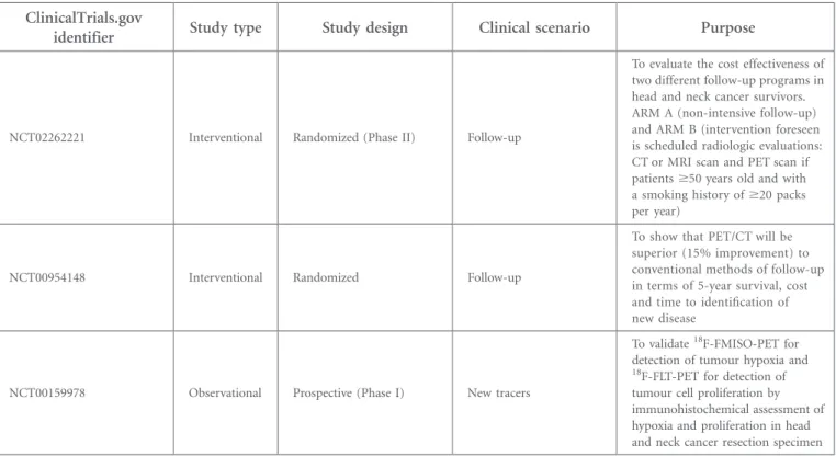
Table 6. (Continued) ClinicalTrials.gov
identifier Study type Study design Clinical scenario Purpose
NCT02262221 Interventional Randomized (Phase II) Follow-up
To evaluate the cost effectiveness of two different follow-up programs in head and neck cancer survivors. ARM A (non-intensive follow-up) and ARM B (intervention foreseen is scheduled radiologic evaluations: CT or MRI scan and PET scan if patients$50 years old and with a smoking history of$20 packs per year)
NCT00954148 Interventional Randomized Follow-up
To show that PET/CT will be superior (15% improvement) to conventional methods of follow-up in terms of 5-year survival, cost and time to identification of new disease
NCT00159978 Observational Prospective (Phase I) New tracers
To validate18F-FMISO-PET for detection of tumour hypoxia and
18F-FLT-PET for detection of
tumour cell proliferation by immunohistochemical assessment of hypoxia and proliferation in head and neck cancer resection specimen
18F-FDG-PET/CT, PET with fluorine-18 fludeoxyglucose integrated with CT;18F-FLT, fluorine-18 fluorothymidine;18F-FMISO, fluorine-18
fluoromiso-nidazole; HNSCC, head and neck squamous cell carcinoma; HPV, human papillomavirus; IMRT, intensity-modulated radiotherapy; LRC, locoregional control; OS, overall survival; PFS, progression-free survival; SUV, standardized uptake value.
In summary, meta-analysis and several studies showed that PET-quantified data such as SUVmax, SUVmean, MTV, TLG are
strongly and negatively correlated with survival. However, given that some uncertainty still exists on which of these parameters are the best predictors, prospective trials are needed to de-finitively settle this issue.
POSITRON EMISSION TOMOGRAPHY/MRI: ADDING NEW POSSIBILITIES
Since information provided by PET/CT and MRI is comple-mentary in many clinical situations, it seems to make sense to combine the two modalities. The high soft-tissue contrast and the different functional imaging techniques of MRI might help to ameliorate the informative value of a hybrid imaging study. Consequently, the discussion about potential applications of this new hybrid technology in oncological imaging and especially in the HN has been generated.130
The feasibility131and diagnostic performance (sensitivity, spec-ificity and accuracy) of clinical PET/MRI have been demon-strated in a significant number of studies,51,85
but technological and logistic challenges, such as errors in attenuation correction and long scan duration continue to be a major focus of the PET/ MRI literature.132
PET/MRI has many potential advantages over PET/CT (including perineural spread of tumours and the infiltration of important anatomical landmarks, such as the pre-vertebral fascia and great vessel walls; lower radiation exposure, higher soft-tissue contrast and several functional techniques).133Realizing this potential in clinics will likely require new radiopharmaceuticals and applica-tions other than WB cancer staging.132
Although PET/MRI is still in the early stages of clinical de-velopment, it is clear that the clinical adoption of PET/MRI is slower than that of PET/CT. This slower evolution is not only due partly to ongoing technological challenges (e.g. accurate attenuation correction of PET images) but also to the complex logistics of combining a WB PET scan with WB or organ-specific MRI. Key applications of PET/MRI that provide information that is clinically relevant and different from that provided by PET/CT still need to be defined.134
Further studies that exploit the functional MRI and molecular PET capabilities may report substantial contributions of PET/ MRI for treatment response predictions, radiotherapy planning, tumour phenotyping and treatment monitoring.131,135,136 Future research involving larger patient series is needed to assess the true impact of this technique in HNSCC and will show whether PET/MRI outperforms PET/CT, MRI, diffusion-weighted MRI or their combination.
FUTURE APPLICATIONS OF POSITRON EMISSION TOMOGRAPHY/CT IN RADIATION ONCOLOGY Table 6 summarizes some of the ongoing clinical trials (www. ClinicalTrials.gov) dealing with the issue of PET/CT in HNSCC treated with radiotherapy.
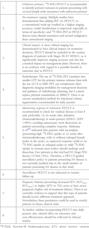
Table 7. Summary: recommendations and key issues
1.
Unknown primary:18F-FDG-PET/CT is recommended
to identify primary tumours in patients presenting with cervical lymph node metastases with unknown primary
2.
Pre-treatment staging: Multiple studies have demonstrated that adding PET (or PET/CT) to a conventional work-up resulted in a higher staging accuracy; nodal classification is improved, especially in terms of specificity; and18F-FDG-PET or PET/CT detects more distant metastases and second malignancy than conventional staging
3.
Clinical impact: A more refined staging has demonstrated to have clinical impact on treatment decisions. PET/CT should be included in the routine diagnosis of patients with Stages III–IV HNSCC, as it significantly improves staging accuracy and also has a marked impact on management plans. However, there is no evidence with regard to a possible benefit in outcomes due to PET/CT
4.
Radiotherapy: The use of18F-FDG-PET translates into smaller GTV for the primary tumour volumes than with the use of CT or MRI. PET can complement other diagnostic imaging modalities for management decisions and guidance of radiotherapy planning, but it cannot replace physical examination or MRI/CT. There is no current standardized method for functional volume segmentation recommended for daily practice
5.
Monitoring response to treatment: PET/CT is recommended to check for residual disease at least 8 and, preferably, 12–16 weeks after definitive chemoradiotherapy in node-positive HNSCC (NPV .90%), avoiding unnecessary neck dissections in patients presenting complete response. Mehanna et al103indicated that patients with incomplete
(presenting high18F-FDG uptake at 12 weeks after
chemoradiotherapy, with or without enlarged lymph nodes in the neck), or equivocal response (mild or no
18F-FDG uptake in enlarged nodes or mild18F-FDG
uptake in normal-sized nodes) should undergo neck dissection. Few patients in this trial had N3 (Stage IVb) disease [17/564 (3%)]. Therefore, a PET-CT-guided surveillance policy to patients presenting N3 disease is not currently justified due to the small number of patients presenting N3 disease in this study 6. Surveillance: PET/CT is not indicated in routine
follow-up
7.
Prognosis: Patients presenting increased SUV (SUVmax,
SUVmean) or higher MTV or TLG seem to have worse
prognosis (higher risk of treatment failure). There is no currently evidence to support that this type of patients should receive different treatment approach.
Nevertheless, these parameters could be used to stratify patients in future clinical trials
8.
To further validate incorporating PET/CT into daily practice also, clinical effect on outcomes and cost effectiveness should be reflected in clinical studies
18F-FDG, fluorine-18 fludeoxyglucose; GTV, gross tumour volume;
HNSCC, head and neck squamous cell carcinoma; MTV, metabolic tumour volume; NPV, negative-predictive value; PET, positron emission tomography; SUV, standardized uptake value; SUVmax, maximal SUV;
Dose escalation to18F-FDG-avid subvolumes of the tumour as well as adapting the RTP during treatment regarding the func-tional tumour changes induced during radiation are two im-portant issues that are currently under investigation.137–139 Madani et al140 performed a Phase I trial to establish the maximum tolerated dose, when the dose was escalated in
18
F-FDG-PET GTV within the anatomically (CT/MRI) based GTV. They demonstrated the feasibility of heterogeneous dose delivery with dose escalation up to 77.5 Gy for18F-FDG-avid tumour areas (the so called “dose painting” approach). Another approach would be adaptation of the biological target volume during the course of radiotherapy in order to reduce
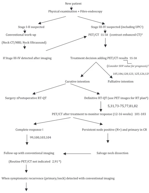
Figure 1. Proposal to incorporate positron emission tomography (PET)/CT (contrast-enhanced CT) into routine clinical practice for head and neck squamous cell carcinoma.†Cervical metastases from unknown primary cancer (UPC). *Evaluate to include MRI to assess soft-tissue invasion or perineural spread if needed depending on primary tumour location.16VPrognosis: uptake parameters and volumetric
parameters can potentially help in identifying patients with worse outcomes (still an area of active investigation).¥Radiotherapy (RT) plan: PET/CT may be helpful, improving contouring accuracy. However, standardized method is lacking.pFollow-up with conventional imaging (CT/MRI) is recommended. When equivocal findings additional PET/CT can be performed.mOnce recurrence is detected, additional PET/CT may be of interest to improve patient counselling; restaging the tumour and planning additional therapy, especially when“aggressive” interventions (extended surgery or reirradiation) may be needed.SHowever, Mehanna et al103indicated that patients with incomplete [patients presenting high fluorine-18 fludeoxyglucose (18F-FDG) uptake at 12 weeks after chemoradiotherapy, with or
without enlarged lymph nodes in the neck] or equivocal response (mild or no18F-FDG uptake in enlarged nodes or mild18F-FDG uptake in
normal-sized nodes) should undergo neck dissection. In addition, it should be noted that few patients in this trial had N3 (Stage IVb) disease [17/564 (3%)]. Therefore, a PET-CT-guided surveillance policy to patients presenting N3 disease is not justified due to the small number of patients presenting N3 disease in this study. CR, complete response; QT, chemotherapy.