Abstract - This study presents the results of
pre-participation musculoskeletal and cardiac screening
using the Lausanne recommendations, which include
a personal and family history, physical examination
and electrocardiography. Cross sectional study using
the Lausanne screenings and the European Society of
Cardiology (ESC) recommendations carried out at
Al-Ahli club in Dubai, United Arab Emirates. 230
male athletes participating in organised sports were
included. Exclusion criteria were those under 14 or
over 35 years old, females and athletes with
established cardiovascular disease. Primary outcome
are the results of Lausanne screening with outline of
the negative, positive and false positive results and
number needed to screen. Secondary outcomes
include
the
results
of
musculoskeletal
and
neurological screening. A total of 174 (76%) athletes
had a negative screening result. Fifty-four athletes
(23%) underwent additional testing. Forty-seven
athletes (20.4%) had false positive screening results.
Seven athletes (3%) had a positive screening result
and four athletes (2%) were restricted from sport.
The number of athletes needed to screen to detect
one lethal cardiovascular condition was 33 athletes.
The Lausanne recommendations are well suited for
the United Arab Emirates. The number needed to
screen
to
detect
one
athlete
with
serious
cardiovascular disease is acceptable at 33.
Keywords: Sudden cardiac death, electrocardiography, physical examination, athlete’s heart, cardiomyopathies
I. INTRODUCTION
Sudden cardiac death (SCD) is the leading cause of mortality in young athletes during exercise, and may result from undiagnosed structural or
electrical cardiovascular disease(1, 2). The incidence of SCD in young athletes varies widely from 0.5-2/100,000/year(3, 4). Sporting activity doubles the relative risk of SCD(5). Pre-participation evaluation in athletes has been implemented in Europe and USA over last decades. In 2005, the European Society of Cardiology (ESC) proposed a common European protocol of cardiovascular pre-participation screening in athletes to prevent risk and occurrence of sudden cardiac death(6). Firstly proposed in Italy(4), this screening consists of taking family and personal history, and undertaking physical examination, and electrocardiographic assessment. Interestingly, the incidence of sudden cardiovascular death in young competitive athletes has been significantly reduced(7). The success of this screening protocol can well be related to the inclusion of electrocardiography in the screening protocol. Specifically, electrocardiography, which is very sensitive to diagnose hypertrophic cardiomyopathy, allows to identify those athletes at risk of sudden cardiac death, suffering from underlying cardiac abnormalities(8-11). Based on the Lausanne recommendations, the International Olympic Committee has approved the European Society of Cardiology recommendations or pre-participation cardiovascular screening in athletes (12).
The current recommendations of the American Heart Association (PPE-4) for athletes include a detailed history and physical examination. In athletes, 75% of medical and orthopedic conditions can be assessed administering a detailed questionnaire(13), but the evidence highlights that the American pre-participation approach is not effective to prevent or detect the risk of sudden cardiac death(14).
Screening of the conditions predisposing to sudden cardiac death is still debated. Specifically, the cost benefit of using electrocardiography is debatable, especially if the screening is used to prevent the sudden death associated with rare hereditary disease(15).
Since musculoskeletal and neurological injuries are frequent in athletes, with specific patterns for different sports (16), it is important that athletes undergo periodic pre-participation assessment as guarantee of safety.
Pre-Participation Musculoskeletal and Cardiac Screening of Male Athletes in
the United Arab Emirates
Alattar A
1, Ghani S
2, Mahdy N
3, Hussain H
3,
Maffulli N
41
Rashid Hospital, P.O. Box: 4545, Dubai, UAE
2
St Georges University London
3
Public Health Affairs, Dubai Health AuthorityP.O. Box: 4545, Dubai, UAE
4
Queen Mary University of London, Barts and The London School of Medicine and Dentistry, Centre for Sports and Exercise Medicine, Mile End Hospital, Department of Musculoskeletal Medicine and Surgery, University of Salerno,
Faculty of Medicine and Surgery, Salerno, Italy (email corresponding author: [email protected])
Following the Lausanne recommendations, we undertook pre-participation evaluation as screening protocol for United Arab Emirates athletes. The aim of this study is to ascertain the effectiveness of this protocol to detect hidden cardiac disorders, and prevent the occurrence of sudden cardiac death.
II. METHODOLOGY
The Dubai Health Authority approved all procedures described in the present investigation, and subjects gave their consent to participate in the study. Both parents signed the consent if the subjects were younger than 16 years of age.
Participants: Two hundred thirty males competitive
athletes, from a sport club in the United Arab Emirates, were examined from December 2011 to February 2012. Exclusion criteria were age under 14 and over 35, female gender, and diagnosis or history of cardiovascular disease. Once a station-based evaluation offered a time-efficient and cost-effective examination, all participants were assessed on a day off training. Recruitment was through general announcement at the club.
Medical questionnaire: A standard questionnaire was
administered to all participants. It was set up according the Lausanne recommendations; the Arabic translation was approved by the Dubai Health Authority. If athletes did acknowledge of family history of sudden death or cardiac disease, parents and siblings were also investigated.
Physical examination: A single fellowship trained sport
physician performed all examinations according to the 4th Edition of the American Preparticipation Physical Examination (PPE-4) criteria. Cardiac examination was based on the ESC Sport's Cardiology Section Consensus statement(6). Measurement of height (cm) and body mass (kg), brachial artery blood pressure in the seated position (mm Hg), precordial auscultation in both supine and standing positions, and examination of Marfan syndrome characteristics was undertaken by a cardiologist experienced in athlete’s heart syndrome.
Resting 12-lead electrocardiography:
Electrocardiography was conducted using regularly calibrated and maintained machines (Philips PageWriter TC50; Philips Healthcare, The Netherlands). The electrodes were placed to ensure consistency of the precordial lead locations. The electrocardiograph traces were printed as hard copy for later analysis, and assessed independently by the cardiologist. PR interval, QRS duration, QT interval, QRS axis, Q, R, S and T wave voltage, and ST segments were measured at each lead. P wave voltage was measured at the V1 lead alone. Left axis deviation was defined as a QRS axis more negative than -30°, and right axis deviation as a QRS axis more positive than +120°. The QT intervals were corrected for heart rate (QTc) using Bazett’s formula. A QTc interval was considered abnormally prolonged if >450 ms in males. Right atrial enlargement was defined as a P wave voltage ≥0.25 mV. Left atrial enlargement was defined as a
biphasic P wave in V1 where the terminal portion was more negative than -0.1 mV and ≥0.04 seconds long. Left and right ventricular hypertrophy was determined by the Sokolow–Lyon voltage criteria. Left ventricular hypertrophy (LVH) was defined by the sum of the S wave in V1 and the R wave in either V5 or V6 being >3.5 mV. Right ventricular hypertrophy was defined by the sum of the R in V1 and the S in V6 being >1.05 mV. In addition, the presence of LVH was assessed by the Romhilt and Estes point-score system with a score of ≥5 being used to define LVH. A Q wave was considered abnormal or pathological if >0.04 seconds long and/or if the depth of the Q wave was >5% of the height of the R wave.
Stages of testing: The Lausanne recommendations were
followed. In step 1, all athletes had a 12-lead electrocardi-ogram. A positive personal history, a family history that indicated the possibility of inherited cardiac disease, posi-tive physical examination or electrocardiographic findings from step 1 prompted further evaluation by an expert car-diologist at a tertiary referral hospital, which included any one or a combination of echocardiography, 24-hour Holter electrocardiography, stress test, electrophysiology test, and even MRI. Symptoms considered to be suggestive of a possible underlying cardiovascular disorder included repetitive syncope during exercise, prolonged periods of palpitations, sustained chest pain and unexplained sudden death in a first degree relative aged <35 years. Athletes with soft murmurs (grade I) not radiated, or who had no electrocardiographic changes, were deemed to have flow murmurs requiring no further action. A flow diagram illustrates the screening protocol for (Figure 1).
Statistical analysis: The statistical analysis was
conducted using computer program SPSS version 20. The qualitative questionnaire results were expressed in absolute numbers form and centile values. Descriptive statistics included mean and standard deviation. All results are presented descriptively.
Tab. 1. Population characteristics.
of sprain, and 36 athletes (15.7%) had a history of muscle strain.
Physical examination
Table 3 shows the distribution of athletes with positive finding on physical examination. Ten athletes (4.3%) had scoliosis, four athletes (1.7%) chronic shoulder instability, ten athletes (4.3%) chronic ankle instability, and 60 athletes (26.1%) pes planus. Three athletes (3.1%) had undergone anterior cruciate ligament reconstruction, and three athletes (3.1%) had undergone meniscectomy. One athlete had a palpable spleen.
Cardiac history and examination
Two athletes had a positive family history of sudden cardiac death before the age of 35 years. Six athletes (2.6%) had a positive history of fainting, nine athletes (3.9%) complained of dizziness, two athletes (2.2%) had shortness of breath, three athletes (1.3%) complained of chest pain, and three athletes (1.3%) complained of palpitation during or after exertion (table 2). Following our examination, two athletes (0.9%) were diagnosed with hypertension, and seven (3%) had a benign systolic ejection murmur (table 3).
Electrocardiography
Table 4 shows all ECG findings. Forty-eight athletes had an abnormal ECG according to the 2010 European Society of Cardiology recommendations. The most common abnormalities were abnormal T wave inversion (10.4%). Two athletes had patterns of Wolff-Parkinson-White syndrome, one athlete prolonged QT, and one athlete atrial fibrillation.
Additional Tests (stage 2)
Table 5 shows the results of stage 2 testing. All positive cases form stage 1 underwent further testing with no loss to follow up. Two athletes had a systolic brachial blood pressure above 140 mm Hg on more than one reading and were referred to their family physician for treatment. A total of 54 (23.5%) had abnormalities at the stage 1 of screening, the reason for the additional tests (stage 2). Diagnosis was confirmed in seven athletes (3%). Figure 2 shows the overview of screening stages, and table 6 gives an overview of the athletes diagnosed with cardiovascular conditions.
Cardiac history subjects
Six athletes had a positive cardiac history, all with normal ECG; they underwent echocardiography, and only one athlete was confirmed to have minor insufficient mitral valve.
Abnormal electrocardiography subjects
Forty eight athletes had ECG abnormalities necessitating further investigations. Two athletes (0.8%) were diagnosed with Wolff-Parkinson-White syndrome following stress ECG, 24-hour Holter and electrophysiology studies showing accessory pathway. Both athletes had no symptoms, opted for ablation and returned to play. One athlete (0.4%) had atrial fibrillation on ECG with history of shortness of breath on exertion. His ECG showed an enlarged left atrium, both exercise test and 24-hour Holter confirmed the diagnosis of atrial fibrillation, and he underwent ablation with temporary restriction from sport. One athlete (0.4%) had positive history of palpitation and positive ECG for T-wave inversions across all leads; his echocardiography showed right ventricular dilatation and hypokinesia; stress test, 24-hour Holter and MRI confirmed the diagnosis of arrhythmogenic right ventricular cardiomyopathy. One athlete (0.4%) was diagnosed with myocardial ischemia following a positive history of chest pain and positive ECG of ST depression. He had further tests, including echocardiography, which showed minor tricuspid and mitral regurgitation, and stress ECG and MRI testing confirmed the diagnosis. Finally, one athlete (0.4%) was diagnosed with prolonged QT syndrome; he had positive history of syncope with exertion, positive family history of sudden cardiac death with ECG of borderline QT syndrome. His 24-hour Holter showed a corrected QT of 490ms.
Total (n = 230)
Mean age (SD), y 20.68 (±5.22)
Mean height (SD), cm 169.13(±10.82)
Mean weight (SD), kg 62.68(±14.61)
Mean body mass index (SD), kg/m2 21.80(±4.17)
Mean body surface area (SD), m2 1.71(±0.24)
Mean pulse rate (SD), BPM 61.54(±12.70)
Mean systolic BP (SD), mm Hg 123.47(±13.47)
Mean diastolic BP (SD), mmHg 68.28(±7.80)
Mean total training time (SD) hours/week 10.06(±0.99)
Teams n (%) Football-First team 36(15.7) Football-Reserve 20(8.7) Sports – U19 13(5.7) Football-U17 29(12.6) Football-U15 27(11.7) Football-U14 20(8.7) Swimming-Elite 13(5.7) Cycling-Elite 12(5.2) Handball-First team 33(14.3) Basketball-First team 27(11.7)
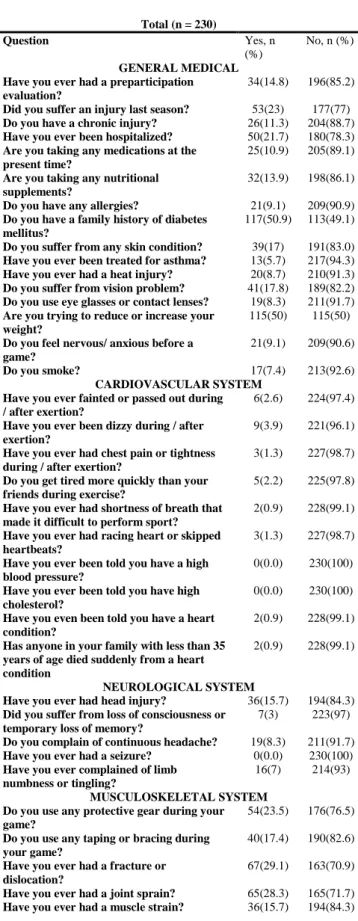
Total (n = 230) Question Yes, n (%) No, n (%) GENERAL MEDICAL Have you ever had a preparticipation evaluation?
34(14.8) 196(85.2)
Did you suffer an injury last season? 53(23) 177(77) Do you have a chronic injury? 26(11.3) 204(88.7) Have you ever been hospitalized? 50(21.7) 180(78.3) Are you taking any medications at the
present time?
25(10.9) 205(89.1)
Are you taking any nutritional supplements?
32(13.9) 198(86.1)
Do you have any allergies? 21(9.1) 209(90.9) Do you have a family history of diabetes
mellitus?
117(50.9) 113(49.1)
Do you suffer from any skin condition? 39(17) 191(83.0) Have you ever been treated for asthma? 13(5.7) 217(94.3) Have you ever had a heat injury? 20(8.7) 210(91.3) Do you suffer from vision problem? 41(17.8) 189(82.2) Do you use eye glasses or contact lenses? 19(8.3) 211(91.7) Are you trying to reduce or increase your
weight?
115(50) 115(50)
Do you feel nervous/ anxious before a game?
21(9.1) 209(90.6)
Do you smoke? 17(7.4) 213(92.6)
CARDIOVASCULAR SYSTEM Have you ever fainted or passed out during / after exertion?
6(2.6) 224(97.4)
Have you ever been dizzy during / after exertion?
9(3.9) 221(96.1)
Have you ever had chest pain or tightness during / after exertion?
3(1.3) 227(98.7)
Do you get tired more quickly than your friends during exercise?
5(2.2) 225(97.8)
Have you ever had shortness of breath that made it difficult to perform sport?
2(0.9) 228(99.1)
Have you ever had racing heart or skipped heartbeats?
3(1.3) 227(98.7)
Have you ever been told you have a high blood pressure?
0(0.0) 230(100)
Have you ever been told you have high cholesterol?
0(0.0) 230(100)
Have you even been told you have a heart condition?
2(0.9) 228(99.1)
Has anyone in your family with less than 35 years of age died suddenly from a heart condition
2(0.9) 228(99.1)
NEUROLOGICAL SYSTEM
Have you ever had head injury? 36(15.7) 194(84.3) Did you suffer from loss of consciousness or
temporary loss of memory?
7(3) 223(97)
Do you complain of continuous headache? 19(8.3) 211(91.7) Have you ever had a seizure? 0(0.0) 230(100) Have you ever complained of limb
numbness or tingling?
16(7) 214(93)
MUSCULOSKELETAL SYSTEM Do you use any protective gear during your game?
54(23.5) 176(76.5)
Do you use any taping or bracing during your game?
40(17.4) 190(82.6)
Have you ever had a fracture or dislocation?
67(29.1) 163(70.9)
Have you ever had a joint sprain? 65(28.3) 165(71.7) Have you ever had a muscle strain? 36(15.7) 194(84.3)
Tab. 2. Distribution of athletes according to medical questionnaire
IV. DISCUSSION
The Lausanne recommendations have been widely debated since their endorsement by the International Olympic Committee in 2006(12). According to Corrado and colleagues(6), we included resting electrocardiography to detect cardiac abnormalities such as cardiac channelopathies, coronary heart disease and cardiomyopathy in young athletes. Of the 230 athletes who were screened between December 2011 and February 2012, a total of 54 (23%) were referred for additional testing because of the presence of abnormalities at the stage 1 of the screening. This rate is comparable to that reported by Baggish et(17), in which 20% of competitive athletes were eligible for additional testing, and higher than in other studies(4) (18). 48 athletes (20%) underwent additional investigations because of abnormal electrocardiographic screening, 6 athletes (2.6%) because of abnormal history and examination. The false positive rate of electrocardiography was 18.3% and, specifically, for stage one screening (history, examination, and ECG), it was 20.4%. This rate is comparable with the rate of 16.9% identified by Baggish et al (17). The spectrum of cardiovascular conditions we have found was comparable with that by Corrado et al(4, 6, 19) and Wilson et al(20). In the present study, 1 athlete had arrhythmogenic right ventricular cardiomyopathy, 2 Wolff-Parkinson-White syndrome, 1 a long QT syndrome, and 1 atrial fibrillation. In the study by Corrado et al(4, 6, 19), the arrhythmogenic right ventricular cardiomyopathy and conduction disorders were the leading causes of sudden cardiac deaths, whereas Maron et al(21) found that, in USA, most sudden cardiac deaths arose from hypertrophic cardiomyopathy. Interestingly, Wilson et al(22) reported that the prevalence of electrocardiographic abnormalities in West Asian and Caucasian athletes was comparable (7.9% vs. 5.8%, p>0.05). Concerning the practicalities of electrocardiography as a screening tool in athletes, Bessem et al(23) showed that only 6.3% of patients who had undergone electrocardiography required further assessment, and the false-positive rate after this screening was 11%. This higher false-positive rate may be partly related to the population screened, as 7% of athletes had been referred for screening because of cardiovascular symptoms. Interestingly, Hevia et al(18) showed that the rate of abnormal electrocardiography is 6.14%, and only 3.27% of subjects needed additional tests. Well-trained sports physician and cardiologist were involved in this study. This improves the quality of the assessment and, reasonably, minimised the false positive rate of electrocardiography. Wilson et al(20), in junior athletes and physically active schoolchildren, reported that further investigations were recommended in 4% (17). This relatively low percentage is supposed to be related to the high expertise level of cardiologists involved in the study. In schoolchildren, the false-positive rate was higher for medical questionnaires than for electrocardiography, and the prevalence of junior athletes diagnosed with a cardiac disease was over twice (0.5%) that of schoolchildren
(0.2%). Therefore, selected groups should be screened to minimise expenditure, and improve testing accuracy. None of the diagnosed athletes were symptomatic, confirming that history and examination alone are inadequate. Comparing a screening protocol with or without electrocardiography, it has been shown that electrocardiography improves sensitivity for detection of cardiac disorders from 45.5% to 90.9%, and the negative predictive value of screening changes from 98.7% to 99.8%, with a false positive rate of 16.9% (17). Marek et al(24) examined the feasibility of a large-scale high school electrocardiography screening program (Young Hearts for Life [YH4L]). Of 32.561 high school students examined between 2006 and 2009, only 2.5% had abnormal electrocardiography and required further evaluation. Therefore, there is need to implement screening and prevent sudden cardiac death in USA. History and physical examination alone are inadequate; electrocardiography has an independent added value for diagnosing cardiac disease, which can lead to sudden cardiac death. Most of the studies do not provide definitive conclusions about the effect of the different screening strategies on the incidence of sudden death in athletes. Given the observational design of the study, we cannot draw definitive conclusions about the role of cardiac screening to reduce the risk of mortality, and incidence of cardiac accidents in athletes. However, previous studies have proved the effectiveness of cardiovascular screening on the incidence of sudden cardiac death in athletes(4, 25, 26). Finally our sample represented a selected population mainly represented by male Arabs (West Asian), and therefore the results should not be extrapolated to females or other ethnic groups..
V. CONCLUSION
Screening with electrocardiography represents a valid clinical strategy to prevent or reduce the risk of sudden cardiac death in young athletes. Implementing the Lausanne recommendations in the United Arab Emirates, screening results will favor the use of electrocardiography. On the other hand, the associated increase in overall false-positive results (20.4%) suggests that some improvements are needed, and future large, multicentre prospective trials could demonstrate how different screening options affect the incidence of sudden death.
Total (n = 230) Finding, n (%) Appearance
Pale 16(7)
Heart
Systolic ejection murmur 7(3) Abdomen
Palpable spleen 1(0.4) Inguinal herniotomy scar 1(0.4) Genitalia Hydrocele 2(0.9) Skin Acne 8(3.5) Chronic urticarial 1(0.4) Eczema 3(1.3) Vitiligo 1(0.4)
Chicken pox scar 1(0.4) Back
Scoliosis 10(4.3)
Kyphosis 1(0.4)
Shoulder/Arm
Chronic shoulder instability 4(1.7) Elbow/Forearm
Reduced elbow extension 2(0.9) Cubitus valgus 1(0.4) Wrist/Hand Hand fracture 1(0.4) Knee Genu varus 5(2.2) Genu valgus 7(3.0)
Anterior cruciate ligament reconstruction
3(1.3)
Meniscectomy 3(1.3)
Leg/Ankle
Chronic ankle instability 10(4.3)
Tibial varum 6(2.6) Foot Pes planus 60(26.1) Pes cavus 3(1.3) Pronated foot 7(3.0) Supinated foot 1(0.4) Halux valgus 2(0.9) Metatarsal abduction 1(0.4) Metatarsal adduction 1(0.4) Heel valgus 1(0.4)
Tab. 3. Distribution of athletes with positive finding on examination
ELECTROCARDIOGRAPHIC
FINDINGS IN ATHLETES (n=230) Numbers Percentage
GROUP 1 (training-related) ECG findings
Sinus Bradycardia (HR < 60) 124 53.9%
1st Degree AV Block (PR > 120ms) 35 15.2%
Partial Right Bundle Branch Block (pRBBB)
28 12.1%
Voltage criteria for Left Ventricular Hypertrophy (LVH)
81 35.2%
Early Repolarization (ER) – overall prevalence
84 36.5%
ER in anterior leads (in isolation or combination)
72 31.3%
ER isolated to inferior and/or lateral leads
47 20.4%
GROUP 2 (training-unrelated) ECG findings
Right Bundle Branch Block (RBBB) 0 -
Left Bundle Branch Block (LBBB) 1 0.4%
Right Atrial Enlargement (RAE) 2 0.8%
Left Atrial Enlargement (LAE) 7 3.0%
Right Axis Deviation (axis ≥ 120°) 1 0.4%
Left Axis Deviation (axis ≤ 30°) 10 4.3%
Right Ventricular Hypertrophy (RVH)
3 1.3%
Prolonged QT interval (QTc>470ms) 1 0.4%
Abnormal T-wave inversions (TWI) – overall prevalence
24 10.4%
TWI in anterior leads 7 3.0%
TWI in inferior leads 18 7.8%
TWI in lateral leads 7 3.0%
Ventricular Ectopics (≥2 per ECG strip) 2 0.8% Pathological Q waves 0 - ST segment depression 1 0.4% Atrial Fibrillation 1 0.4% Wolff-Parkinson-White (WPW) pattern 2 0.8%
Tab.4. Electrocardiographic findings in athletes
ACKNOWLEDGMENT
Dr. Abdulhameed Alattar is an employee of Dubai Health Authority and was the Chief Medical Officer of Al-Ahli football club from 2007 to 2010. This research was funded by Dubai Health Authority.
REFERENCES
1. Maron BJ, Doerer JJ, Haas TS, et al. Sudden deaths in young competitive athletes: analysis of 1866 deaths in the United States, 1980-2006. Circulation. 2009;119(8):1085-92.
2. Harmon KG, Asif IM, Klossner D, et al. Incidence of sudden cardiac death in national collegiate athletic association athletes. Circulation. 2011;123(15):1594-600.
3. Maron BJ, Gohman TE, Aeppli D. Prevalence of sudden cardiac death during competitive sports activities in Minnesota high school athletes. J Am Coll Cardiol. 1998;32(7):1881-4.
4. Corrado D, Basso C, Pavei A, et al. Trends in sudden cardiovascular death in young competitive athletes after implementation of a preparticipation screening program. JAMA. 2006;296(13):1593-601.
5. Corrado D, Basso C, Schiavon M, et al. Does sports activity enhance the risk of sudden cardiac death? J Cardiovasc Med (Hagerstown). 2006;7(4):228-33.
6. Corrado D, Pelliccia A, Bjørnstad HH, et al. Cardiovascular pre-participation screening of young competitive athletes for prevention of sudden death: proposal for a common European protocol. Consensus Statement of the Study Group of Sport Cardiology of the Working Group of Cardiac Rehabilitation and Exercise Physiology and the Working Group of Myocardial and Pericardial Diseases of the European Society of Cardiology. Eur Heart J. 2005;26(5):516-24.
7. Corrado D, McKenna WJ. Appropriate interpretation of the athlete's electrocardiogram saves lives as well as money. Eur Heart J. 2007;28(16):1920-2. 8. Seto CK, Pendleton ME. Preparticipation cardiovascular screening in young athletes: current guidelines and dilemmas. Current Sports Medicine Reports. 2009;8(2):59-64.
9. Magalski A, McCoy M, Zabel M, et al. Cardiovascular screening with electrocardiography and echocardiography in collegiate athletes. Am J Med. 2011;124(6):511-8.
10. Sofi F, Capalbo A, Pucci N, et al. Cardiovascular evaluation, including resting and exercise electrocardiography, before participation in competitive sports: cross sectional study. BMJ. 2008;337:a346. 11. Pigozzi F, Spataro A, Fagnani F, et al. Preparticipation screening for the detection of cardiovascular abnormalities that may cause sudden death in competitive athletes. British Journal Of Sports Medicine. 2003;37(1):4-5.
12. Bille K, Figueiras D, Schamasch P, et al. Sudden cardiac death in athletes: the Lausanne Recommendations. Eur J Cardiovasc Prev Rehabil. 2006;13(6):859-75. 13. Seto CK. The preparticipation physical examination: an update. Clin Sports Med. 2011;30(3):491-501.
14. Rao AL, Standaert CJ, Drezner JA, et al. Expert opinion and controversies in musculoskeletal and sports medicine: preventing sudden cardiac death in young athletes. Archives Of Physical Medicine And Rehabilitation. 2010;91(6):958-62.
15. Behera SK, Pattnaik T, Luke A. Practical recommendations and perspectives on cardiac screening for healthy pediatric athletes. Curr Sports Med Rep. 2011;10(2):90-8.
16. Fuller CW, Ojelade EO, Taylor A. Preparticipation medical evaluation in professional sport in the UK: theory or practice? Br J Sports Med. 2007;41(12):890-6; discussion 6.
17. Baggish AL, Hutter AM, Wang F, et al. Cardiovascular screening in college athletes with and without electrocardiography: A cross-sectional study. Ann Intern Med. 2010;152(5):269-75.
18. Hevia AC, Fernández MM, Palacio JMA, et al. ECG as a part of the preparticipation screening programme: an old and still present international dilemma. British Journal Of Sports Medicine. 2011;45(10):776-9. 19. Corrado D, Basso C, Schiavon M, et al. Screening for hypertrophic cardiomyopathy in young athletes. N Engl J Med. 1998;339(6):364-9.
20. Wilson MG, Basavarajaiah S, Whyte GP, et al. Efficacy of personal symptom and family history questionnaires when screening for inherited cardiac pathologies: the role of electrocardiography. British Journal Of Sports Medicine. 2008;42(3):207-11.
21. Maron BJ, Thompson PD, Ackerman MJ, et al. Recommendations and considerations related to preparticipation screening for cardiovascular abnormalities in competitive athletes: 2007 update: a scientific statement from the American Heart Association Council on Nutrition, Physical Activity, and Metabolism: endorsed by the American College of Cardiology Foundation. Circulation. 2007;115(12):1643-455.
22. Wilson MG, Hamilton B, Sandridge AL, et al. Differences in markers of cardiovascular disease between professional football players of West-Asian and Black African descent. J Sci Med Sport. 2012;15(3):266-71. 23. Bessem B, Groot FP, Nieuwland W. The Lausanne recommendations: a Dutch experience. British Journal Of Sports Medicine. 2009;43(9):708-15.
24. Marek J, Bufalino V, Davis J, et al. Feasibility and findings of large-scale electrocardiographic screening in young adults: data from 32,561 subjects. Heart Rhythm: The Official Journal Of The Heart Rhythm Society. 2011;8(10):1555-9.
25. Pelliccia A, Di Paolo FM, Corrado D, et al. Evidence for efficacy of the Italian national pre-participation screening programme for identification of
hypertrophic cardiomyopathy in competitive athletes. European Heart Journal. 2006;27(18):2196-200.
26. Pelliccia A, Di Paolo FM, Quattrini FM, et al. Outcomes in athletes with marked ECG repolarization abnormalities. N Engl J Med. 2008;358(2):152-61.