E BIOMEDICHE
Dottorato di ricerca in SCIENZE FARMAUCETICHE X Ciclo – Nuova Serie
Design, virtual screening and structural studies of new
molecules with potential antitumor and antiinflammatory
activity
Tutor Dottoranda
Ch.mo Prof. Giuseppe Bifulco Maria Giovanna Chini
Coordinatore Ch.
ma
Preface
My PhD three years course in Pharmaceutical Sciences at the Department of Pharmaceutical and Biomedical Sciences of Salerno University was started in November 2008 under the supervision of Prof. Giuseppe Bifulco.
My research activity was mainly focused onto study of ligand-receptor interactions and structural characterization by computational techniques and NMR spectroscopy in order to identify new antitumor and/or antiinflammatory molecules potentially utilizable in therapy.
These approaches were successfully applied to the characterization of novel inhibitors of Histone deacetilase (HDAC) Nicotinamide Phosphoribosyltransferase (NMPRTase or Nampt), microsomal prostaglandin E2 synthase (mPGES-1), human synovial Phospholipases A2, (hsPLA2), human Farnesoid-X-Receptor (FXR), and agonist of human Pregnane-X-Receptor (PXR) and Bile Acid Pregnane-X-Receptor GPBAR-1 (TGR5).
The entire work was carried out under the direct supervision of Prof. Giuseppe Bifulco.
Furthermore, to improve my knowledge on methodologies for the stereostructural assignment, I moved to the Department of Chemistry of the Bristol University in 2011 (mid-July until mid-November 2011) under the supervision of the Dr. Craig Butts.
During this period in his research laboratory, my research work has included learning and conducting advanced NMR spectroscopic investigations of a number of natural products and synthetic compounds.
In addition to PhD course activities, I was involved in different side projects, mainly regarding the characterization of ligand-targets interactions of ligands on targets involved in other pathologies as e.g. Alzheimer and obesity.
List of publications related to the scientific activity performed during the three years PhD course in Pharmaceutical Sciences:
1. Terracciano, S.; Chini, M. G.; Riccio, R.; Bruno, I.; Bifulco, G. Design, Synthesis, and Biological Activity of Hydroxamic Tertiary Amines as Histone Deacetylase Inhibitors. ChemMedChem 2012, DOI: 10.1002/cmdc.201100531.
2. Chini, M. G.; Jones, C. R.; Zampella, A.; D'Auria, M. V.; Renga, B.; Fiorucci, S.; Butts C. P.; and Bifulco, G. Quantitative NMR-Derived Interproton Distances Combined with Quantum Mechanical Calculations of 13C Chemical Shifts in the Stereochemical Determination of Conicasterol F, a Nuclear Receptor Ligand from Theonella swinhoei. J.
Org. Chem. 2012 dx.doi.org/10.1021/jo2023763.
3. De Marino, S.; Sepe, V.; D’Auria, M. V.; Chini, M. G.; Bifulco, G.; D’Amore, C.; Renga, B.; Petek, S.; Fiorucci, S.; Zampella, A. 4-methylenesterols from Theonella swinhoei sponge are natural PXR agonists and FXR antagonist that modulate liver detoxification genes and innate immunity. Steroids 2012, doi:10.1016/j.steroids.2012.01.006
4. Renga, B; Mencarelli, A; D’Amore, C.; Cipriani, S.; D’Auria, M. V. Sepe, V.; Chini, M. G.; Bifulco, G.; Zampella, A.; Fiorucci. S.; Discovery of Theonellasterol as a highly selective FXR antagonis that protects against liver injury in cholestasis. PLoS ONE 2012, 7, e30443, 1-12.
5. Sepe, V.; Ummarino, R.; D’Auria, M. V.; Chini, M. G.; Bifulco, G.; Renga, B.; D’Amore, C.; Debitus, C.; Fiorucci, S.; Zampella, A. Conicasterol E, a small heterodimer partner sparing farnesoid-X-receptor modulator endowed with a pregnane-X-receptor agonistic activity, from the marine sponge Theonella swinhoei. J Med Chem. 2012, 55, 84-93.
6. Cipriani, S.; Mencarelli, A.; Chini, M. G.; Distrutti, E.; Renga, B.; Bifulco, G.; Baldelli, F.; Donidi, A.; Fiorucci, S. The Bile Acid Receptor GPBAR-1 (TGR5) Modulates Integrity of Intestinal Barrier and Immune Response to Experimental Colitis. PloS One 2011, 6, e25637, 1-11.
7. Monti, M. C.; Chini, M. G.; Margarucci, L.; Riccio, R.; Bifulco, G.; Casapullo, A. The Binding Mode of Cladocoran A to the Human Group IIA Phospholipase A2. Chembiochem 2011, 12, 2686-2691.
8. De Marino, S.; Ummarino, R.; D'Auria, M. V.; Chini, M. G.; Bifulco, G.; Renga, B.; D'Amore, C.; Fiorucci, S.; Debitus, C.; Zampella, A. Theonellasterols and Conicasterols from Theonella swinhoei. Novel Marine Natural Ligands for Human Nuclear Receptors. J. Med. Chem.
2011, 54, 3065-3075.
9. De Simone, R.; Chini, M. G.; Bruno, I.; Riccio, R.; Mueller, D.; Werz, O.; Bifulco, G. Structure-Based Discovery of Inhibitors of Microsomal Prostaglandin E2 Synthase-1, 5-Lipoxygenase and 5-Lipoxygenase-Activating Protein: Promising Hits for the Development of New Anti-inflammatory Agents. J. Med. Chem. 2011, 54, 1565-1575.
10. Chini, M. G.; Terracciano, S.; Riccio, R.; Bifulco, G.; Ciao, R.; Gaeta, C.; Troisi, F.; Neri, P. Conformationally Locked Calixarene-Based Histone Deacetylase Inhibitors. Organic Letters 2010, 12, 5382-5385.
11. Terracciano, S.; Chini, M. G.; Bifulco, G.; D'Amico, E.; Marzocco, S.; Riccio, R.; Bruno, I. Synthesis of new mono and bis amides projected as potential histone deacetylase (HDAC) inhibitors. Tetrahedron 2010, 66, 2520-2528.
12. Di Micco, S; Chini, M. G.; Riccio, R.; Bifulco, G. Quantum Mechanical Calculation of NMR Parameters in the Stereostructural Determination of Natural Products. Eur. J. Org. Chem. 2010, 8, 1411-1434.
13. Colombano, G.; Travelli, C.; Galli, U.; Caldarelli, A.; Chini, M. G.; Canonico, P. L.; Sorba, G.; Bifulco, G.; Tron, G. C.; Genazzani, A. A. Novel Potent Nicotinamide Phosphoribosyltransferase Inhibitor Synthesized via Click Chemistry. J. Med. Chem. 2010, 53, 616-623.
14. Monti, M. C.; Chini, M. G.; Margarucci, L.; Tosco, A.; Riccio, R.; Bifulco, G.; Casapullo, A. The molecular mechanism of human group IIA phospholipase A2 inactivation by bolinaquinone. J. Mol. Recognit. 2009 22, 530-537.
15. Grolla, A. A.; Podesta, V.; Chini, M. G.; Di Micco, S.; Vallario, A.; Genazzani, A. A.; Canonico, P. L.; Bifulco, G.; Tron, G. C.; Sorba, G.; Pirali, T. Synthesis, biological evaluation, and molecular docking of Ugi products containing a zinc-chelating moiety as novel inhibitors of histone deacetylases. J. Med. Chem. 2009, 52, 2776-2785.
16. Chini, M. G.; Riccio, R.; Bifulco, G. DFT/NMR integrated approach: a valid support to the total synthesis of chiral molecules. Magn. Reson.
Table of Contents
Abstract ……… I-III
Page
Introduction ………. 1-56
Chapter 1 Cancer-related inflammation 2
1.1 Inflammation and cancer 3
1.1.1 Inflammation: From Acute to Chronic 4-5 1.1.2 Cancer Development: An Overview 5-6 1.1.3 Connecting inflammation and cancer 6-12 1.1.3.1 Mutagenic Potential of Inflammation 12-13 1.1.3.2 Role of Inflammatory Cells in Tumor
Development
13
1.1.3.3 Key Molecular Players in Linking Inflammation to Cancer
14-18
1.2 Scope and outline of this thesis 18-20
1.3 Methodologies employed 21-56
1.3.1 Molecular docking 21-25
1.3.1.1 Autodock: an Overview 25-34
1.3.2 Quantum Mechanical calculation of NMR Parameters in the Stereostructural Determination of Natural Products
35-40
1.3.3 Quantitative Interproton Distances from Nuclear Overhauser Effect (NOE) Data
40-41
1.3.3.1 The Initial Rate Approximation 42-50
1.3.3.2 Method for Quantitative Interproton Distance Determinations
Page
Result and Discussion
57-332Chapter 2 Design, virtual screening and rationalization of potential HDAC Inhibitors
57-149
2.1 HDAC as drug target 58-68
2.2 Synthesis of new mono and bis amides projected as potential histone Deacetylase (HDAC) inhibitors
69-75
2.2.1 Computational details 76-77
2.3 Design, synthesis, and biological activity of new hydroxamic tertiary amines as histone deacetylase (HDAC) inhibitors.
78-85
2.3.1 Computational details 85-86
2.4 Conformationally Locked Calixarene-Based HDAC Inhibitors
87-98
2.4.1 Computational details 99
2.5 Synthesis, Biological Evaluation, and Molecular Docking of Ugi Products Containing a Zinc-Chelating Moiety as Novel Inhibitors of Histone Deacetylases
100-110
2.5.1 Computational details 110-111
2.6 Structural Basis for the design and synthesis of selective HDAC inhibitors
112-114
2.6.1 Structural analysis 115
2.6.1.1 Common structural features of all isoforms 115-116 2.6.1.2 General features and differences of Class I and II 117-120
2.6.1.3 HDAC1 120-122
Page 2.6.1.5 HDAC3 125-126 2.6.1.6 HDAC8 126-127 2.6.1.7 HDAC4 128-131 2.6.1.8 HDAC6 131-133 2.6.1.9 HDAC7 133-134
2.6.2 Proof of concept: Design, synthesis and biological evaluation of selective HDAC2 inhibitors
135
2.6.2.1 Design of 36-38 135-138
2.6.3 Computational Details 138
2.6.3.1 Homology modeling 138-148
2.6.3.2 Docking calculations 148-149
Chapter 3 A Novel Potent Nicotinamide Phosphoribosyl-transferase Inhibitor
150-164
3.1 A Novel Potent Nicotinamide Phosphoribosyltransferase Inhibitor Synthesized via Click Chemistry
151-163
3.1.1 Computational Details 163-164
Chapter 4 Microsomal Prostaglandin E2 Synthase-1: drug target in inflammation
165-197
4.1 Structure-based discovery of mPGES-1 inhibitors 165-168 4.2 Structure-Based Discovery of Inhibitors of
Microsomal Prostaglandin E2 Synthase-1, 5-Lipoxygenase and 5-Lipoxygenase-Activating
Protein: Promising Hits for the Development of New Anti-inflammatory Agents
4.2.1 Computational Details 182-183
4.3 Design and Synthesis of a Second series of Triazole-based compounds as potent dual mPGES-1 and 5-Lipoxygenase inhibitors.
184-196
4.3.1 Computational Details 196-197
Chapter 5 Marine natural products as hsPLA2 inhibitors 198-223 5.1 Human Group IIA Phospholipase A2: an Overview 199-202 5.2 The molecular mechanism of human group IIA
phospholipase A2 inactivation by bolinaquinone
203-214
5.2.1 Computational details 214-216
5.3 The Binding Mode of Cladocoran A to the Human Group IIA Phospholipase A2
217-222
5.3.1 Computational details 223
Chapter 6 4-Methylensterols as ligands of Human Farnesoid-X-Receptor (FXR) and Human Pregnane-X-Receptor (PXR)
224-275
6.1 The Farnesoid X Receptor (FXR) and Human Pregnane-X-Receptor (PXR) as Modulator of Bile Acid Metabolism
225-237
6.2 Theonellasterols and Conicasterols from Theonella
swinhoei. Novel Marine Natural Ligands for
Human Nuclear Receptors
Page
6.2.1 Computational Details 246-247
6.3 Conicasterol E, a Small Heterodimer Partner Sparing Farnesoid X Receptor Modulator Endowed with a Pregnane X Receptor Agonistic Activity, from the Marine Sponge Theonella
swinhoei
248-255
6.3.1 Computational Details 255-256
6.4 4-Methylenesterols from Theonella swinhoei Sponge are Natural Pregnane-X-Receptor Agonists and Farnesoid-X Receptor Antagonists that Modulate Innate Immunity
257-270
6.4.1 Computational Details. 270
6.5 Discovery of theonellasterol a marine sponge sterol as a highly selective FXR antagonist that protects against liver injury in cholestasis
271-274
6.5.1 Computational Details 274-275
Chapter 7 The Bile Acid Receptor GPBAR-1 (TGR5) Agonists
276-287
7.1 The Bile Acid Receptor GPBAR-1 (TGR5) Modulates Integrity of Intestinal Barrier and Immune Response to Experimental Colitis
277-284
7.1.1 Computational Details 284-285
Page Chapter 8 Structural studies of Natural Products 288-332
8.1 DFT/NMR integrated approach: a valid support to the total synthesis of chiral molecules
289-302
8.1.1 Computational details 302-304
8.2 Quantitative NMR-Derived Interproton Distances Combined with Quantum Mechanical Calculations of 13C Chemical Shifts in the Stereochemical Determination of Conicasterol F, a Nuclear Receptor Ligand from Theonella swinhoei
305-314
8.2.1 Assignment of the relative configuration of 8-14 epoxy ring in conicasterol F
315-331
8.2.2 Computational details 331-332
Conclusions 332-340
I
Abstract
Computational methodologies in combination with experimental techniques as Nuclear Magnetic Resonance (NMR) have become a crucial component in drug discovery process, from hit identification to lead optimization.
The study of ligand-macromolecule interactions, in fact, has a crucial role for the design and the development of new and more powerful drugs. In this project, different aspects of interaction and recognition processes between ligand and macromolecule, and streostructure assignment has been studied through this kind of combined approach with the aim to identify novel potential antitumor and/or antiinflammatory molecules.
In particular, because the strong interconnection between the tumoral and inflammatory pathology has led to the identification of new target utilizable for the therapy, in this project will be described proteins (Histone deacetilase, HDAC; Nicotinamide Phosphoribosyltransferase, NMPRTase or Nampt; microsomal prostaglandin E2 synthase, mPGES-1; human synovial Phospholipases A2, hsPLA2; human Farnesoid-X-Receptor, FXR; human Pregnane-X-Receptor, PXR; Bile Acid Receptor GPBAR-1, TGR5) involved in essential cellular processes and acting at diverse levels and phases of the tumor and inflammation diseases.
The results obtained can be summarized in three main areas of activity, whose relative weight was varied according to the development of the overall project:
a) Support in the design of original scaffolds for the generation of libraries potentially utilizable in therapy. This work was exclusively
conducted in silico by a molecular docking technique in order to direct the design of the new molecules basing on the analysis of ligand-target
II
interactions and the synthetic possibilities. This kind of approach was successfully applied leading to the identification of new potential inhibitors for HDAC enzymes with ciclic (mono and bis amides, paragraph 2.2; conformationally locked calixarenes, paragraph 2.4), and linear (hydroxamic tertiary amines, paragraph 2.3) structures, and isoform selective (paragraph 2.6), and of ligands for microsomal prostaglandin E2 synthase (mPGES)-1 (two series of triazole-based compounds; paragraphs 4.2 and 4.3).
For each of this described studied, the good qualitative accordance between the calculated and experimental data has made possible the identifications of new lead compounds, rationalizing in this way the key features to the target inhibition.
b) Rationalization of the biological activity of compounds by the study of the drug-receptor interactions. Molecular docking was used for the
detailed study of antiinflammatory and antitumoral compounds whose activities are known a priori. In fact, thanks to this procedure, in this thesis several rationalizations of binding modes were reported related to Ugi products derivatives of CHAP 1 (HDAC inhibitors, paragraph 2.5), new and potent inhibitor of NMPRTAse analogs of FK866 and CHS 828(chapter 3), marine natural products as inhibitors of hsPLA2 (BLQ and CLDA, chapter 5), 4-methylen sterols extracted from Theonella swinhoei as ligands of FXR and PXR (chapter 6), and known compounds as taurolitholic acid and ciprofloxacin (chapter 7), agonists of TGR5.
Through the in silico methodology the putative binding modes for the reported molecules was described offering a complete rationalization of docking results, evaluating the influence of the ligand target interactions (e.g. hydrophobic, hydrophilic, electrostatic contacts) on the biological activity.
III
c) Determination of relative configuration of natural products.
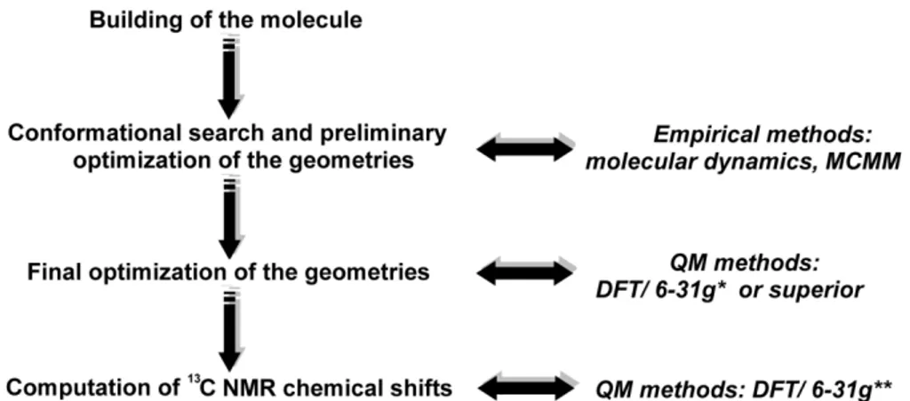
The complete comprehension of the three dimensional structure of synthetic or isolated molecules is fundamental to design and characterize new platform potentially utilizable in therapy. On this basis, the combined approach between the quantum mechanical (QM) calculation of NMR parameters and NMR spectroscopy was revealed a very useful mean to lead the total synthesis of natural product toward the right isomer avoiding waste of time and resources (paragraph 8.1).
Moreover, the stereostructure assignment of marine natural products conicasterol F and its analog thonellasterol I was reported in the paragraph 8.2. by a novel combined approach between the quantitative interproton distance determinations by ROE and quantum mechanical calculations of chemical shifts
1
2
-CHAPTER 1-
3
1.1
Inflammation and cancer
The link between inflammation and cancers, rather than a recent concern, was noticed ~150 years ago. As early as 1863, Virchow indicated that cancers tended to occur at sites of chronic inflammation.1
Although it is now clear that proliferation of cells alone does not cause cancer, sustained cell proliferation in an environment rich in inflammatory cells, growth factors, activated stroma, and DNA-damage-promoting agents, certainly potentiates and/or promotes neoplastic risk.
During tissue injury associated with wounding, cell proliferation is enhanced while the tissue regenerates; proliferation and inflammation subside after the assaulting agent is removed or the repair completed. In contrast, proliferating cells that sustain DNA damage and/or mutagenic assault (for example, initiated cells) continue to proliferate in microenvironments rich in inflammatory cells and growth/survival factors that support their growth. In a sense, tumors act as wounds that fail to heal.2
Today, the causal relationship between inflammation, innate immunity and cancer is more widely accepted; however, many of the molecular and cellular mechanisms mediating this relationship remain unresolved. Furthermore, tumor cells may usurp key mechanisms by which inflammation interfaces with cancers, to further their colonization of the host. Moreover, it was clear that the acquired immune response to cancer is intimately related to the inflammatory response.3,4
Here, the critical points and the pathways connections between these two kinds of pathologies will be described.
4
1.1.1
Inflammation: From Acute to Chronic
Inflammation is a physiologic process in response to tissue damage resulting from microbial pathogen infection, chemical irritation, and/or wounding.5 At the very early stage of inflammation, neutrophils are the first cells to migrate to the inflammatory sites under the regulation of molecules produced by rapidly responding macrophages and mast cells prestationed in tissues.6 As the inflammation progresses, various types of leukocytes, lymphocytes, and other inflammatory cells are activated and attracted to the inflamed site by a signaling network involving a great number of growth factors, cytokines, and chemokines.6 All cells recruited to the inflammatory site contribute to tissue breakdown and are beneficial by strengthening and maintaining the defense against infection.6a
There are also mechanisms to prevent inflammation response from lasting too long.7 A shift from antibacterial tissue damage to tissue repair occurs, involving both proinflammatory and anti-inflammatory molecules.7 Prostaglandin E2,8 transforming growth factor-β,9 and reactive oxygen and nitrogen intermediates6d are among those molecules with a dual role in both promoting and suppressing inflammation. The resolution of inflammation also requires a rapid programmed clearance of inflammatory cells: neighboring macrophages, dendritic cells, and backup phagocytes do this job by inducing apoptosis and conducting phagocytosis.10 The phagocytosis of apoptotic cells also promotes an anti-inflammatory response, such as enhancing the production of antiinflammatory mediator transforming growth factor- β.11 However, if inflammation resolution is dysregulated, cellular response changes to the pattern of chronic inflammation. In chronic inflammation, the inflammatory foci are dominated by lymphocytes, plasma cells, and
5
macrophages with varying morphology.5 Macrophages and other inflammatory cells generate a great amount of growth factors, cytokines, and reactive oxygen and nitrogen species that may cause DNA damage.6a If the macrophages are activated persistently; they may lead to continuous tissue damage.12 A microenvironment constituted by all the above elements inhabits the sustained cell proliferation induced by continued tissue damage, thus predisposes chronic inflammation to neoplasia.1
1.1.2
Cancer Development: An Overview
Cancer defines malignant neoplasms characterized by metastatic growth. It may occur in almost every organ and tissue relating to a variety of etiologic factors, such as genomic instability and environmental stress.5 A two-stage carcinogenesis model is first conceptualized in a mouse model of skin cancer.13 In this model, carcinogenesis is initiated by carcinogen-triggered irreversible genetic alteration and then promoted by dysregulated gene expression of initiated cells that resulted from epigenetic mechanisms and host-selective pressure.6a Once the proliferation advantage is obtained, cancer cells enter the progression stage in which their population expands rapidly.6b This model was subjected to criticism because it oversimplifies and failed to apply to all types of cancer.14
However, cancer development is still accepted as a multistep process, during which genetic alterations confer specific types of growth advantage; therefore, it drives the progressive transformation from normal cells to malignant cancer cells.15 Malignant growth is characterized by several key changes: self-sufficiency of growth signals, insensitivity to antigrowth signals, escaping from apoptosis, unregulated proliferation potential, enhanced angiogenesis, and metastasis.15 Each of these shifts is complicated and
6
accomplished by combined efforts of various signaling processes, and moreover it will find out that inflammation may contribute to the formation of these cancer phenotypes.
1.1.3
Connecting inflammation and cancer
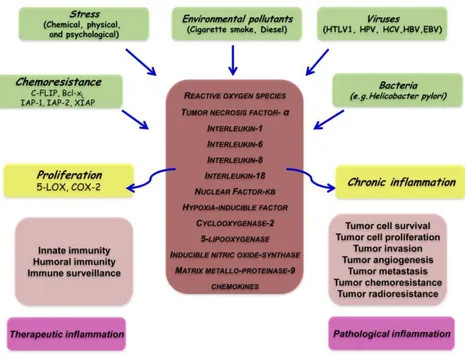
Common wisdom says ‘‘most things in life are a double-edged sword’’. While they are in our favor at one dose or under one condition; they may be disfavor at another dose or under another condition. Inflammation is a part of the host response to either internal or external environmental stimuli. This response serves to counteract the insult incurred by these stimuli to the host. This response can be pyrogenic, as indicated by fever. When acute inflammation or fever is manifested for a short period of time, it has a therapeutic consequence. However, when inflammation becomes chronic or lasts too long, it can prove harmful and may lead to disease. How is inflammation diagnosed and its biomarkers is not fully understood, however, the role of proinflammatory cytokines, chemokines, adhesion molecules and inflammatory enzymes have been linked with chronic inflammation (Figure 1.1). Chronic inflammation has been found to mediate a wide variety of diseases, including cardiovascular diseases, cancer, diabetes, arthritis, Alzheimer’s disease, pulmonary diseases, and autoimmune diseases.16 Chronic inflammation has been linked to various steps involved in tumorigenesis, including cellular transformation, promotion, survival, proliferation, invasion, angiogenesis, and metastasis.17,18 That inflammation is a risk factor formost type of cancers is now well recognized.19
7
Figure 1. 1 Different faces of inflammation and its role in tumorigenesis.
Al already reported, the links between cancer and inflammation were first made in the nineteenth century, on the basis of observations that tumors often arose at sites of chronic inflammation and that inflammatory cells were present in biopsied samples from tumors,1 but there has been a recent resurgence in interest.
Several lines of evidence20 (Table 1.1) — based on a range of findings, from epidemiological studies of patients to molecular studies of genetically modified mice — have led to a general acceptance that inflammation and cancer are linked. Epidemiological studies have shown that chronic inflammation predisposes individuals to various types of cancer. It is estimated that underlying infections and inflammatory responses are linked to 15–20% of all deaths from cancer worldwide.1 There are many triggers of chronic inflammation that increase the risk of developing cancer. Such triggers include
8
microbial infections (for example, infection with Helicobacter pylori is associated with gastric cancer and gastric mucosal lymphoma), autoimmune diseases (for example, inflammatory bowel disease is associated with colon cancer) and inflammatory conditions of unknown origin (for example, prostatitis is associated with prostate cancer). Accordingly, treatment with non-steroidal anti-inflammatory agents decreases the incidence of, and the mortality that results from, several tumor types.21
Table 1. 1 The evidence that links cancer and inflammation 1
Inflammatory diseases increase the risk of developing many types of cancer (including bladder, cervical, gastric, intestinal, oesophageal, ovarian, prostate and thyroid cancer)
2
Non-steroidal anti-inflammatory drugs reduce the risk of developing certain cancers (such as colon and breast cancer) and reduce the mortality caused by these cancers.
3 Signaling pathways involved in inflammation operate downstream of oncogenic
mutations (such as mutations in the genes encoding RAS, MYC and RET).
4
Inflammatory cells, chemokines, and cytokines are present in the microenvironment of all tumors in experimental animal models and humans from the earliest stages of development.
5
The targeting of inflammatory mediators (chemokines and cytokines, such as TNF-α and IL-1β), key transcription factors involved in inflammation (such as NF-κB and STAT3) or inflammatory cells decreases the incidence and spread of cancer.
6 Adoptive transfer of inflammatory cells or overexpression of inflammatory
cytokines promotes the development of tumors.
The hallmarks of cancer-related inflammation include the presence of inflammatory cells and inflammatory mediators (for example, chemokines, cytokines and prostaglandins) in tumor tissues, tissue remodeling and
9
angiogenesis similar to that seen in chronic inflammatory responses, and tissue repair. These signs of ‘smouldering’ inflammation
20a
are also present in tumors for which a firm causal relationship to inflammation has not been established (for example, breast tumors). Indeed, inflammatory cells and mediators are present in the microenvironment of most, if not all, tumors, irrespective of the trigger for development.
In the tumor microenvironment, inflammatory cells and molecules influence almost every aspect of cancer progress, including the tumor cells’ability to metastasize.
22
Thus, whereas there were previously six recognized hallmarks of cancer — unlimited replicative potential, self-sufficiency in growth signals, insensitivity to growth inhibitors, evasion of programmed cell death, ability to develop blood vessels, and tissue invasion and metastasis23 — cancer related inflammation now emerges as number seven (Figure 1.2). In 2000, Hanahan and Weinberg23 proposed a model to define the six properties that a tumor acquires.
10
These are unlimited replicative potential, ability to develop blood vessels (angiogenesis), evasion of programmed cell death (apoptosis), self-sufficiency in growth signals, insensitivity to inhibitors of growth, and tissue invasion and metastasis. Kim and colleagues’ findings,24 together with those of other studies,22,18 indicate that this model should be revised to include cancer-related inflammation as an additional hallmark.23
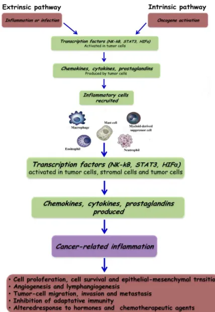
The connection between inflammation and cancer can be viewed as consisting of two pathways: an extrinsic pathway, driven by inflammatory conditions that increase cancer risk (such as inflammatory bowel disease); and an intrinsic pathway, driven by genetic alterations that cause inflammation and neoplasia (such as oncogenes) (Figure 1.3).
The intrinsic pathway was uncovered when addressing why inflammatory cells and mediators are present in the microenvironment of most, if not all, tumors and therefore are present in cases for which there is no epidemiological basis for inflammation. This finding raised the question of whether the genetic events that cause neoplasia in these cases are responsible for generating an inflammatory environment. This question has been addressed only recently, by using preclinical and clinical settings in which various oncogenetic mechanisms can be assessed.
The intrinsic pathway is activated by genetic events that cause neoplasia. These events include the activation of various types of oncogene by mutation, chromosomal rearrangement or amplification, and the inactivation of tumor-suppressor genes. Cells that are transformed in this manner produce inflammatory mediators, thereby generating an inflammatory microenvironment in tumors for which there is no underlying inflammatory condition (for example, breast tumors). By contrast, in the extrinsic pathway,
11
inflammatory or infectious conditions augment the risk of developing cancer at certain anatomical sites (for example, the colon, prostate and pancreas).
Figure 1. 3 Pathways that connect inflammation and cancer. Cancer and inflammation are
12
The two pathways converge, resulting in the activation of transcription factors, mainly nuclear factor-kB (NF-kB), signal transducer and activator of transcription 3 (STAT3) and hypoxia-inducible factor 1 (HIF1α), in tumor cells. These transcription factors coordinate the production of inflammatory mediators, including cytokines and chemokines, as well as the production of cyclooxygenase 2 (COX2) (which, in turn, results in the production of prostaglandins). These factors recruit and activate various leukocytes, most notably cells of the myelomonocytic lineage. The cytokines activate the same key transcription factors in inflammatory cells, stromal cells and tumor cells, resulting in more inflammatory mediators being produced and a cancer-related inflammatory microenvironment being generated. Smouldering cancer-related inflammation has many tumor-promoting effects.
1.1.3.1
Mutagenic Potential of Inflammation
The chronic inflammation microenvironment is predominated by macrophages.6 Those macrophages, together with other leukocytes, generate high levels of reactive oxygen and nitrogen species to fight infection.25 However, in a setting of continuous tissue damage and cellular proliferation, the persistence of these infection-fighting agents is deleterious.6b They may produce mutagenic agents, such as peroxynitrite, which react with DNA and cause mutations in proliferating epithelial and stroma cells.25,26 Macrophages and T lymphocytes may release tumor necrosis factor-α (TNF-α) and macrophage migration inhibitory factor to exacerbate DNA damage.27 Migration inhibitory factor impairs p53-dependent protective responses, thus causing the accumulation of oncogenic mutations.28 Migration inhibitory factor also contributes to tumorigenesis by interfering Rb-E2F pathway.29
13
Within an ileocolitis-associated mouse cancer model, the high susceptibility to inflammation and cancer in hydroperoxide-reducing enzyme-deficient mice suggested that intracellular hydroperoxides might also contribute to tumor initiation.30
1.1.3.2
Role of Inflammatory Cells in Tumor Development
Other than a single mutation, more genetic and epigenetic events are required to drive from initiated cells to malignant tumors.23 Some of these events are also found to be related to chronic inflammation. For instance, angiogenesis, a critical process in tumor progression,31 associates with chronic inflammation, such as psoriasis, rheumatoid arthritis, and fibrosis.23 In addition, the tumor inflammatory microenvironment can facilitate the breakage of the basement membrane, a process required for the invasion and migration of tumor cells.6a A wide population of leukocytes and other types of immune cells infiltrate to the developing tumor site and establish the tumor inflammatory microenvironment.6c Macrophages, neutrophils, eosinophils, dendritic cells, mast cells, and lymphocytes are also found to be key components in the epithelial-originated tumors.6c,12,32 The infiltration of immune cells to tumors may repress tumor growth.33 However, the increasing concern is that inflammatory cells act as tumor promoters in inflammation-associated cancers.6a,34,35 Accumulated mutations in epithelial cells lead to dysregulation of their growth and migration. These dysregulated epithelial cells may also signal to recruit leukocytes.31 In addition, tumor cells may also produce cytokines and chemokines to attract immune cells to facilitate cancer development.6a,c,31
14
1.1.3.3
Key Molecular Players in Linking Inflammation to
Cancer
To address the details of transition from inflammation to cancers and the further development of inflammation-associated cancers, it is necessary to investigate specific roles of key regulatory molecules involved in this process. In fact, in the panoply of molecules involved in cancer-related inflammation, key endogenous (intrinsic) factors can be identified. These include transcription factors (such as NF-kB and signal transducer and activator of transcription 3 (STAT3)) and major inflammatory cytokines (such as IL-1β, IL-6, IL-23 and TNF-α)36,37,38 (Table 1.2).
Table 1. 2 Key Molecular Players Linking Cancer to Inflammation.
Potential linkers Functions in linking inflammation to cancer
Cytokines
IL-6 Promote tumor growth
TNF-α
Induce DNA damage and inhibit DNA repair Promote tumor growth
Induce angiogenic factors
Chemokines
Promote tumor cell growth
Facilitate invasion and metastasis by directing tumor cell migration and promoting basement membrane degradation
NF-Κβ
Mediate inflammation progress, promoting chronic inflammation
Promote the production of mutagenic reactive oxygen species Protect transformed cells from apoptosis
Promote tumor invasion and metastasis
Feedback loop between proinflammatory cytokines iNOS Downstream of NF-nB and proinflammatory cytokines
15
Induce DNA damage and disrupt DNA damage response Regulate angiogenesis and metastasis
COX-2
Produce inflammation mediator prostaglandins
Promote cell proliferation, antiapoptotic activity, angiogenesis, and metastasis
HIF-1α
Promote chronic inflammation
Induced by proinflammatory cytokines through NF-nB Enhance the glycolytic activity of cancer cells
Contribute to angiogenesis, tumor invasion, and metastasis by transactivating VEGF
STAT3
Activated by proinflammatory cytokines
Promote proliferation, apoptosis resistance, and immune tolerance
Nrf2 Anti-inflammatory activity Protect against DNA damage
NFAT Regulate proinflammatory cytokine expression Required in cell transformation
For sick of simplicity, between the molecular players involved in inflammatory networking cancer, the tumor necrosis factor (TNF-α) and NF-kB will be described. The TNF-α was first isolated as an anticancer cytokine than two decades ago.39 Experience since then has indicated that when expressed locally by the cells of the immune system, TNF-α has a therapeutic role. However, when dysregulated and secreted in the circulation, TNF-a can mediate a wide variety of diseases, including cancer.39 TNF-α has itself been shown to be one of the major mediators of inflammation.40 Induced by a wide range of pathogenic stimuli, TNF-α induces other inflammatory mediators and proteases that orchestrate inflammatory responses. TNF-α is also produced by tumors and can act as an endogenous tumor promoter.40 The role of TNF-α
16
has been linked to all steps involved in tumorigenesis, including cellular transformation, promotion, survival, proliferation, invasion, angiogenesis, and metastasis, as outlined below (Figure 1.4).
Figure 1. 4 Inflammatory networking in cancer.
On the other hand, NF-kB is a key coordinator of innate immunity and inflammation, and has emerged as an important endogenous tumor promoter.36 NF-kB is crucial both in the context of tumor or potential tumor cells and in the context of inflammatory cells. In these cell types, NF-ΚB operates downstream of the sensing of microorganisms or tissue damage by the Toll-like receptor (TLR)–MyD88 signaling pathway, and by signaling pathways mediated by the inflammatory cytokines TNF-α and IL-1β. In addition, NF-kB can be activated as a result of cell-autonomous genetic alterations (amplification, mutations or deletions)41 in tumor cells. In tumor cells and epithelial cells at risk of transformation by carcinogens, as well as in inflammatory cells, NF-kB activates the expression of genes encoding
17
inflammatory cytokines, adhesion molecules, enzymes in the prostaglandin-synthesis pathway (such as COX2), inducible nitric oxide synthase (iNOS; also known as NOS2) and angiogenic factors.
In addition, one of the important functions of NF-ΚB in tumor cells or cells targeted by carcinogenic agents is promoting cell survival, by inducing the expression of anti-apoptotic genes (such as BCL2). There is also accumulating evidence of interconnections and compensatory pathways between the NF-KB and HIF1α systems,42 linking innate immunity to the response to hypoxia. There is unequivocal evidence that NF-ΚB is involved in tumor initiation and progression in tissues in which cancer-related inflammation typically occurs (such as the gastrointestinal tract and the liver).43 The NF-ΚB pathway is tightly controlled by inhibitors that function at various stages of the pathway. An example is TIR8 (also known as SIGIRR), a member of the IL-1-receptor family. TIR8 has a single immuno globulin domain, a long cytoplasmic tail, and a Toll/IL-1 receptor (TIR) domain that differs from that of other members of the IL-1-receptor family. Deficiency in the gene that encodes TIR8 is associated with increased susceptibility to intestinal inflammation and carcinogenesis.44 Thus, the balance of inhibitors and activators tunes the extent to which the NF-ΚB pathway operates as an endogenous tumor promoter. Support for the connection between cancer and inflammation is further strengthened by studies of the role of NF-ΚB in tumor-infiltrating leukocytes. In established, advanced tumors, which typically have a microenvironment of smouldering inflammation,20 tumor-associated macrophages (TAMs) have delayed and defective NF-κB activation.45 Evidence suggests that homodimers of the p50 subunit of NF-κB (a negative regulator of the NF-κB pathway) are responsible for this sluggish activation of NF-κB in TAMs and for the protumor phenotype of these cells.46 Thus, NF-κB seems to function as a
18
‘rheostat’ whose function can be tuned to different levels, a property that enables the extent of inflammation to be regulated. Such regulation allows the vigorous inflammation (for example, in inflammatory bowel disease) that predisposes individuals towards developing cancer to be sustained, and enables TAMs to sustain the smouldering inflammatory microenvironment present in established metastatic neoplasia.
Briefly, the mediators and cellular effectors of inflammation are important constituents of the local environment of tumors.
1.2
Scope and outline of this thesis
The study of ligand-macromolecule interactions has a fundamental role for the design and the development of new and more powerful drugs. In this project, different aspects of interaction and recognition processes between ligand and macromolecule has been studied through a combined approach based on computational chemistry techniques and NMR spectroscopy. In particular, these different aspects regard the employment and elaboration of screening methods, the analysis of structural determinants responsible of drug-macromolecule complex formation and the design of new potential bioactive compounds. Several and different proteins, involved in essential cellular processes, have been investigated as biological targets taking into account their implication in tumor and inflammation initiation and progress with the aim to identify and rationalize new molecules potentially utilizable in therapy. As already reported, in some types of cancer, inflammatory conditions are present before a malignant change occurs. Conversely, in other types of cancer, an oncogenic change induces an inflammatory microenvironment that promotes the development of tumors. Regardless of its origin, inflammation in the tumor microenvironment has many tumor-promoting effects. It aids in the
19
proliferation and survival of malignant cells, promotes angiogenesis and metastasis, subverts adaptive immune responses, and alters responses to hormones and chemotherapeutic agents. The molecular pathways of this cancer-related inflammation are now being unraveled, resulting in the identification of new target molecules that could lead to improved diagnosis and treatment. Between them, in this project, the attention was focused on targets (Histone deacetilase, HDAC; Nicotinamide Phosphoribosyltransferase, NMPRTase or Nampt; microsomal prostaglandin E2 synthase, mPGES-1; human synovial Phospholipases A2, hsPLA2; human Farnesoid-X-Receptor, FXR; human Pregnane-X-Receptor, PXR; Bile Acid Receptor GPBAR-1, TGR5) with different mechanisms of action involved in diverse levels and phases of tumor and inflammation process.
In particular, in the chapter 2 the results obtained for the design, in silico screening, and rationalization of binding modes of pan, selective cyclic and linear HDAC inhibitors are summarized.
In chapter 3 the analysis at atomic level of the interactions between NMPRTase and triazole-based analogs of APO866 and CHS2883 are reported.
Although some HDAC inhibitors are already showing therapeutic utility in animal models of inflammatory diseases (such as arthritis, inflammatory bowel diseases, septic shock, ischemia-reperfusion injury, asthma, ecc.),47 and NMPRTase is able to control both cell viability and the inflammatory response,48 by regulating NAD availability, it is important to underline that for all the compounds described in the chapters 2 and 3 only their antitumor activity was evaluated.
The chapters 4 and 5 are related to the design, in silico screening and rationalization of binding modes of mPGES-1 and hsPLA2 inhibitors, key enzymes of arachidonic acid cascade and eicosanoid metabolism. In particular,
20
in the chapter 4 the design and in silico evaluation of two series of triazole-based compounds is described in detail; while in the chapter 5, the putative binding modes of two marine natural products to human synovial Phospholipases A2 were obtained through molecular docking.
However, even if mPGES-149 is becoming a target for cancer suppression thanks to its inhibitory ability to suppress the PGE2 synthesis offering the potential for therapeutic benefit without the potential toxicity associated with COX-2 inhibition, in the chapter 4, only the potential antiinflammatory activities of designed molecules were evaluated. In the chapter 5, the antiinflammatory activity of reported compounds was analyzed in detail, although the group II Group IIA Phospholipase A2 (PLA2-IIA) also plays a role in tumor progression in vivo, and inhibitors of PLA2-IIA suppress the proliferative activity and invasiveness of prostate carcinoma cell lines.50
The chapters 6 and 7 refer to nuclear receptors FXR, PXR and TGR5 ligands. In particular, in the chapter 6, a detail rationalization of the 4-Methylen sterols antagonist and agonist activity on FXR are PXR respectively was reported. On the other hand, in the chapter 7, agonist activity of two well-known ligands to TGR5 is described. For these compounds will be analyzed and described their activity in the controlling of the bile acids metabolism and their involvement with dysfunctions connected with it; in fact the described nuclear receptors (FXR, PXR and TGR5) are important pharmacological targets for a number of diseases, including cancer and metabolic disorders.51
Finally in the chapter 8, it will be described the use of calculation at quantum mechanical level of the NMR parameters (e.g. δ, chemical shifts, and
J coupling constant), as an efficient tool in the stereostructure assignment of
21
1.3
Methodologies employed
Before starting the discussion concerning the results obtained, it is appropriate briefly introduce the methodologies utilized to realize the project.
1.3.1
Molecular docking
Computational methodologies have become a crucial component in drug discovery, from virtual screening for hit identification to lead compound optimization. One key methodology is the molecular docking that consists in the prediction of ligand conformation and orientation within a targeted binding site. The molecular docking is based on the requirement that the 3D structure of the macromolecule is known. Many different programs have been developed, of which DOCK,52 FlexX,53 GOLD,54120 Autodock,55,56,57 Autodock Vina,58 and Glide12259 are among the most popular. The mentioned tools are based on a range of different concepts, and each comes with its own set of strengths and weaknesses. One feature most docking programs share, however, is that they position a flexible ligand into a rigid binding site. Computational feasibility is the main reason for utilizing a rigid macromolecule in the docking calculations, as the number of freedom degrees that have to be considered grows exponentially with the number of accessible receptor conformations. Most molecular docking software have two key parts: (1) a search algorithm and (2) a scoring function.60 For molecular docking to be useful in drug discovery, these key parts should be both fast and accurate. These two requirements are often in opposition to each other, requiring necessary compromises that commonly end in ambiguous results or failure.61
The search algorithm samples different ligand orientations and conformations fitting the macromolecular binding site. This step is
22
complicated by the number of freedom degrees contained in the small molecule, increasing the conformational space to sample. The search methods can be grouped in three categories: systematic methods, random or stochastic methods, and simulation methods. The systematic search algorithms try to explore all the degrees of freedom in a molecule, but they face the problem of huge number of generated conformations.62 The random methods (often called stochastic methods) operate by making random changes to either a single ligand or a population of ligands. A newly obtained ligand is evaluated on the basis of a pre-defined probability function. Two popular random approaches are Monte Carlo and genetic algorithms. About simulations search methods, molecular dynamics is currently the most popular approach. However, molecular dynamics simulations are often unable to cross high-energy barriers within feasible simulation time periods, and therefore might only accommodate ligands in local minima of the energy surface.63 Therefore, an attempt is often made to simulate different parts of a protein–ligand system at different temperatures.64 Another strategy for addressing the local minima problem is starting molecular dynamics calculations from different ligand positions. In contrast to molecular dynamics, energy minimization methods are rarely used as stand-alone search techniques, as only local energy minima can be reached, but often complement other search methods. The scoring function aims to evaluate the results of the search algorithm predicting the affinity for the biological target. This evaluation is very difficult because the binding process is governed by enthalpic and entropic factors and one or of them can predominate. Other elements can affect the scoring method, such as limited resolution of crystallographic targets, inherent flexibility, induced fit or other conformational changes that occur on binding and the participation of water molecules in macromolecule–ligand interactions. Three classes of scoring
23
functions are currently applied: force field-based, empirical and knowledge-based scoring functions.
Molecular mechanics force fields usually quantify the sum of two energies, the macromolecule–ligand interaction energy and internal ligand energy (such as steric strain induced by binding). Most force field scoring functions only consider a single protein conformation, which makes it possible to omit the calculation of internal protein energy, which greatly simplifies scoring. The enthalpic contribution are essentially given by the electrostatic and Van der Waals terms, and is some software (AutoDock, Gold) take into account the hydrogen bond formation between drug and biological target.
The van der Waals potential energy for the general treatment of non-bonded interactions is often modeled by a Lennard–Jones 12–6 function (Equation 1.1):
( ) = 4 −
Equation 1. 1
where ε is the well depth of the potential and σ is the collision diameter of the respective atoms i and j. The exp(12) term of the equation is responsible for small-distance repulsion, whereas the exp(6) provides an attractive term which approaches zero as the distance between the two atoms increases.
The Lennard–Jones 12–6 function is also used to describe the hydrogen bond in macromolecule-ligand complex, but compared to the Van der Waals function, is less smooth and angle dependent.
Figure 1. 5 Schematic representation of functions used to model pair
contribute to binding. Interactions are calculated as a function of the distance ( two atoms i and j. a) van der Waals interact
the smoother attractive part of the potential compared to hydrogen bond term). B) hydrogen bond potential given by a ‘harder’ 12
for two like (blue) or opposite (black) charges of same magnitude calculated using a distance dependent dielectric constant.
The electrostatic potential energy is represented as a Coulombic interactions, as described in equation
Equation 1. 2
where N is the number of atoms in molecules A and B, respectively, and q the charge on each atom. The functional form o
typically very similar to the includes van der Waals contributions
Empirical scoring functions work on the sum of several parameterized functions to reproduce experimental data. The design of empirical scoring functions is based on
sum of individual uncorrelated terms. The coefficients of the various terms are
24
Schematic representation of functions used to model pair-wise interactions that contribute to binding. Interactions are calculated as a function of the distance (
. a) van der Waals interaction given by a 12–6 Lennard–Jones potential (note the smoother attractive part of the potential compared to hydrogen bond term). B) hydrogen bond potential given by a ‘harder’ 12–10 Lennard–Jones potential. C) electrostatic potential opposite (black) charges of same magnitude calculated using a distance dielectric constant.
The electrostatic potential energy is represented as a summation of Coulombic interactions, as described in equation 1.2:
( )= 4! 0 #$ 1 #& 1
where N is the number of atoms in molecules A and B, respectively, and q on each atom. The functional form of the internal ligand energy is typically very similar to the protein–ligand interaction energy, and also includes van der Waals contributions and/or electrostatic terms.
Empirical scoring functions work on the sum of several parameterized roduce experimental data. The design of empirical scoring functions is based on the idea that binding energies can be approximated by a uncorrelated terms. The coefficients of the various terms are wise interactions that contribute to binding. Interactions are calculated as a function of the distance (rij) between Jones potential (note the smoother attractive part of the potential compared to hydrogen bond term). B) hydrogen Jones potential. C) electrostatic potential opposite (black) charges of same magnitude calculated using a distance
summation of
where N is the number of atoms in molecules A and B, respectively, and q f the internal ligand energy is ligand interaction energy, and also
Empirical scoring functions work on the sum of several parameterized roduce experimental data. The design of empirical scoring the idea that binding energies can be approximated by a uncorrelated terms. The coefficients of the various terms are
25
obtained from regression analysis using experimentally determined binding energies and X-ray structural information.
By using the knowledge-based scoring functions protein–ligand complexes are modeled using relatively simple atomic interaction-pair potentials. A number of atom-type interactions are defined depending on their molecular environment.
1.3.1.2
Autodock: an Overview
There are numerous molecular docking software applications that utilize different searching and scoring algorithms and AutoDock is currently one of the most cited of these applications,65 especially in a virtual screening of a compound libraries.66 For the purposes of this project the software AutoDock 3.0.5,55 4.1,56 4.2,57 and AutodockVina58 have been used, where the differences between them are related to the speed, macromolecule sidechains flexibility, optimization of the free-energy scoring function based on a linear regression analysis, AMBER force field, larger set of diverse protein-ligand complexes with known inhibition constants; moreover the Lamarckian Genetic Algorithm (LGA) is a big improvement on the Genetic Algorithm, and both genetic methods are much more efficient and robust than SA in the new version of the software.
The best model obtained with the latest version AutoDock 4.257 in fact, was cross-validated with a separate set of HIV-1 protease complexes, and confirmed that the standard error is around 2.5 kcal/mol.
In AutoDock there are different available search methods, but the Lamarckian Genetic Algorithm (LGA) has been selected for the aim of this study, because it has demonstrated to give the best results compared to the other algorithms.55
The vast majority of genetic algorithms mimic Darwinian evolution
Figure 1.7 note the oneway phenotype. However, in th
(i.e., one which yields a genotype from a given phenotype finish a local search by replacing the
search; see the left-hand side of Figure 1.
Figure 1. 6 This figure illustrates genotypic and
and Lamarckian search.
line, and the space of the phenotypes is re are mapped to phenotypes by a
The result of applying the genotypic mutation the right-hand side of the diagram, and has the
is shown on the left-hand side. It is normally performed in
information about the fitness landscape. Sufficient iterations of the local search local minimum, and an inverse mapping
26
The vast majority of genetic algorithms mimic the major characteristics of Darwinian evolution and apply Mendelian genetics. This is illustrated
note the oneway transfer of information from the genotype to phenotype. However, in those cases where an inverse mapping function exists
yields a genotype from a given phenotype), it is
finish a local search by replacing the individual with the result of the local hand side of Figure 1.6.
This figure illustrates genotypic and phenotypic search, and contrasts Darwinian Lamarckian search.67 The space of the genotypes is represented by the lower horizontal
of the phenotypes is represented by the upper horizontal
are mapped to phenotypes by a developmental mapping function. The fitness function is The result of applying the genotypic mutation operator to the parent’s genotype is shown on
the diagram, and has the corresponding phenotype shown. Local search hand side. It is normally performed in phenotypic space and employs
fitness landscape. Sufficient iterations of the local search
cal minimum, and an inverse mapping function is used to convert from its phenotype to its the major characteristics of and apply Mendelian genetics. This is illustrated in transfer of information from the genotype to the inverse mapping function exists , it is possible to individual with the result of the local
phenotypic search, and contrasts Darwinian represented by the lower horizontal presented by the upper horizontal line. Genotypes developmental mapping function. The fitness function is f(x). operator to the parent’s genotype is shown on corresponding phenotype shown. Local search phenotypic space and employs fitness landscape. Sufficient iterations of the local search arrive at a function is used to convert from its phenotype to its
27
corresponding genotype. In the case of molecular docking, however, local search is performed by continuously converting from the genotype to the phenotype, so inverse mapping is not required. The genotype of the parent is replaced by the resulting genotype, however, in accordance with Lamarckian principles.
This is called the Lamarckian genetic algorithm (LGA), and is an allusion to Jean Batiste de Lamarck’s (discredited) assertion that phenotypic characteristics acquired during an individual’s lifetime can become heritable traits.68
The most important issues arising in hybrids (LGA) of Genetic Algorithm (GA) and the Local Search (LS) revolve around the developmental mapping, which transforms genotypic representations into phenotypic ones.
The genotypic space is defined in terms of the genetic operators mutation and crossover in our experiments by which parents of one generation are perturbed to form their children. The phenotypic space is defined directly by the problem, namely, the energy function being optimized. The local search operator is a useful extension of GA global optimization when there are local ‘‘smooth-ness’’ characteristics (continuity, correlation, etc.) of the fitness function that local search can exploit. In hybrid GA + LS optimizations, the result of the LS is always used to update the fitness associated with an individual in the GA selection algorithm. If, and only if, the developmental mapping function is invertible, will the Lamarckian option converting the phenotypic result of LS back into its corresponding genotype become possible. The fitness or energy is usually calculated from the ligand’s coordinates, which together form its phenotype. The developmental mapping simply transforms a molecule’s genotypic state variables into the corresponding set of atomic coordinates. A novel feature of this application of hybrid global-local
28
optimization is that the Solis and Wets LS operator searches through the genotypic space rather than the more typical phenotypic space. This means that the developmental mapping does not need to be inverted. Nonetheless, this molecular variation of the genetic algorithm still qualifies as Lamarckian, because any ‘‘environmental adaptations’’ of the ligand acquired during the local search will be inherited by its offspring. At each generation, it is possible to let a user defined fraction of the population undergo such a local search. The local search frequencies of just 0.06 have found improved efficiency of docking, although a frequency of 1.00 is not significantly more efficient.67 Both the canonical and a slightly modified version of the Solis and Wets method have been implemented. In canonical Solis and Wets, the same step size would be used for every gene, but we have improved the local search efficiency by allowing the step size to be different for each type of gene: a change of 1 Å in a translation gene could be much more significant than a change of 1° in a rotational or torsional gene. In the Lamarckian genetic algorithm, genotypic mutation plays a somewhat different role than it does in traditional genetic algorithms. Traditionally, mutation plays the role of a local search operator, allowing small, refining moves that are not efficiently made by crossover and selection alone. With the explicit local search operator, however, this role becomes unnecessary, and is needed only for its role in replacing alleles that might have disappeared through selection. In LGA, mutation can take on a more exploratory role.
The LGA yields a maximum number of 256 potential bioactive conformations: run, whose number can be increased performing more docking calculations. Each conformational solution is the result of a selection. The GA, starting from the input geometry, gives rise to a group of n conformations or individuals (whose number can be set up) defining for them translational,
29
rotational and torsional variables. By the scoring function, each individual is labeled by the total interaction energy: fitness. Random pairs of individuals are mated using a process of crossover, in which new individuals inherit geometrical features from their parents leading to new generation of individuals. In addition, some offspring undergo random mutation, in which the translational, rotational and torsional variables are mutated randomly. Selection of the offspring of the current generation occurs based on the individual’s fitness: thus the better solutions go on into the next generations, whereas conformations with a low fitness are discarded. This cycle of crossover, mutation to lead new generation is repeated until the better bioactive conformation (run) is given.
The LS perform an energy minimization of the current found conformation. In each generation a fraction of conformations population undergoes the geometry optimization, based on the local search frequency. Rapid energy evaluation is achieved by precalculating atomic affinity potentials (grid maps) for each atom type in the substrate molecule by grid method.69
These maps are calculated by AutoGrid. In this procedure the protein is embedded in a three dimensional grid and a probe atom is placed at each grid point (Figure 1.7). The energy of interaction of this single atom with the protein is assigned to the grid point.
An affinity grid is calculated for each type of atom in the substrate, typically carbon, oxygen, nitrogen and hydrogen, as well as a grid of electrostatic potential, either using a point charge of +1 as the probe, or using a Poisson-Boltzmann finite difference method, such as DELPHI.70 The energetic of a particular substrate configuration is then found by tri-linear interpolation of affinity values of the eight grid points surrounding each of the atoms in the substrate.
30
Figure 1. 7 Schematic representation of the grid map.
The electrostatic interaction is evaluated similarly, by interpolating the values of the electrostatic potential and multiplying by the charge on the atom (the electrostatic term is evaluated separately to allow finer control of the substrate atomic charges). The time to perform an energy calculation using the grids is proportional only to the number of atoms in the substrate, and is independent of the number of atoms in the protein. An estimated free energy of binding is used to evaluate the docked ligand conformations. This scoring function, based of force field Amber,71 comprises terms above described (directional hydrogen bonding, electrostatics, Van der Waals, internal energy) and entropic contribution: desolvation and torsional entropy. The latter describes the loss of entropy upon interaction with macromolecule followed by immobilization in the active site. The desolvation belongs the displacement