Necrotizing fasciitis of the hand: a case report
Andrea Minini, Stefano Galli, Andrea Salvi, Guido Zarattini
Clinica Ortopedica dell’ Università degli Studi di Brescia
Summary. Necrotizing Fasciitis is a rare life-threatening infection, usually polymicrobial, that frequently
af-fects the extremities in as many as two thirds of the cases. It typically involves primarily the muscular fascia, and then spreads through muscular and subcutaneous tissues. The early diagnosis may be challenging, and appears to be crucial in the management of this condition. We report a case of a 45-year-old man, former drug abuser, diabetic, HCV+, who developed a necrotizing fasciitis of the hand following a minor trauma. Early diagnosis based on clinical, laboratory (LRINEC score) and radiological findings, together with an accurate debridement of the affected site, allowed us to limitate the amputation to the third ray only. The reconstruc-tion with the capitate osteotomy and the coverage with the posterior interosseous flap helped us in further reduction of the functional impairment of the hand. (www.actabiomedica.it)
Key words: Necrotizing fasciitis, LRINEC score, posterior interosseous flap, capitate osteotomy,
dishwater-like pus
Introduction
The term “Necrotizing Fasciitis” (NF) refers to a life-threatening infection, with a bacterial aetiol-ogy, characterized by a rapid necrosis that primarily involves fascial tissues, and then spreads to muscular and subcutaneous tissues. This condition, also known as “flesh-eating disease” frequently engages limbs, of-ten unilaterally, although some cases of bilateral and multifocal involvement are described in literature (1,2).
The incidence of NF ranges between 0,4 (3) and 1,3 (4) /100 000 according to the country (Canada Vs Florida).
This condition can be classified in 4 clinical forms (5,6) depending on the causative organism: one an-aerobic species with one or more facultative anaero-bic streptococci (other than group A) and members of the Enterobacteriaceae (7), Haemolytic streptococcus group A, members of the Vibrio ssp, (8) and, at last, fungineal Candida infections (9).
Clinically NF usually presents as an erythema of the skin surrounding the affected area, with unregular-ly marginated edges, warm to the touch, very painful especially in the early stages; within 3 to 5 days from the onset blisters start to emerge, evolving then in skin necrosis.
Intense fever is a very common finding; at this stage pain and tenderness to the affected area dissolve; this characteristic can actually help in identifying a NF. Gas formation in subcutaneous tissues is frequent mostly in polimicrobial forms, especially in diabetic patients (10).
The diagnosis is essentially clinical, and can rely on a clinical/anamnestic/laboratory score named LRI-NEC (11).
A score of 6 or more has a positive predictive value of 92% and a negative predictive value of 96%.
Still, surgical exploration remains the gold stand-ard for definitive diagnosis (12-14).
E.V. antibiotic treatment should be started imme-diately, together with surgical debridement of affected
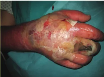
for EV drug abuse until 10 years earlier, was admitted to our E.R. with a swollen and painful hand (Fig. 1, Fig. 2).
He reported a minor crush injury to his right hand between the wings of a gate 3 days before , caus-ing a small wound to the dorsal skin of the proximal phalanx of the third finger.
At first, he started an oral antibiotic prophilaxis with Amoxicilline/clavulanic acid (1 gr x 2/day) and dressed the wound with a topical preparation of gen-tamicine and steroid.
The patient reported the appearance of an erythe-ma spreading to the whole forearm and swelling of his right hand over the following two days; for this reason he headed to our E.R., where he underwent an X-ray scan of his hand (negative for fractures) and was then hospitalized in the Infectious Diseases department with an initial diagnosis of a post-traumatic phlegmon;
thus an E.V. antibiotic therapy has been promptly started (piperacillin/tazobactam and clindamycin).
Blood tests on entry, with regard to the LRINEC score, are reported in (Tab. 1):
The following day, as the symptoms kept wors-ening, the patient underwent a CT scan of his right hand; in view of the CT scan, we stated the need of a timely debridement, and transferred the patient to our ward.
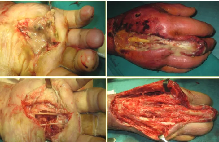
Intraoperatory findings were compatible with NF, with a malodorous “dishwater-like” pus, trombotic sub-cutaneous blood vessels, and little-to none resistance to the digital detachment of subcutis from the extensor ap-paratus to the whole dorsum of the hand (Fig. 3).
The incision for pus drainage was centered dorsally on the third ray, extending to the distal forearm, together with a volar approach for the debridement of all the vis-ible necrotic tissues from the first to the fifth ray. After
Figure 1. Dorsal and volar aspect of the hand at the admission to our E.R.
Figure 2. Dorsal and volar aspect of the hand at the admission to our E.R.
Table 1. Serum parameters of the patient on entry in the ortho-paedic department with regard to the LRINEC score
WBC 8,2x10^3 u/L Glucose 14,37 mmol/L Hb 11.8 g/dL Serum Creatinine 58.3 micromol/L
Na+ 123 mmol/L CRP 181 mg/L
Total LRINEC 8
an abundant wash with hydrogen peroxide and sodium chloride solution, we decided not to close the wound with stitches in order to facilitate the drainage of pus.
Four days later, during the dressing of the wound, we noticed a bulge in the thenar area of the right hand,
compatible with a septic collection (Fig. 4). Therefore we opted for a second debridement with the excision of the whole extensor and flexor apparatus of the third ray (Fig. 5), and we sent a sample of this particulate material to the coltural exam.
Figure 3. Intraoperative findings during the first debridement surgery (dorsal)
Figure 4. Septic collection (volar) at the 4th day after the first
debridement.
coverage of the lesion with a posterior interosseous flap (P.I.F.) we prescribed an angiography of his right upper limb; then we took the patient to the O.R. in
reported a VAS of 1, and a grip strenght of 22,5 Kgs at the Jamar test (65% of the controlateral-non dominant hand) (Fig. 8).
Figure 6. Sequence: isolation and excision of the third ray, capitate wedge ostectomy and closure of the corresponding web space with reabsorbable cerclages. On the lower right corner, post-op X ray
Figure 7. Sequence: skin marks, isolation of the posterior interosseous artery with a perforating vessel, and wound coverage with the posterior interosseous flap
clinical and laboratory parameters, although Burner et al. (16) observed an actual sensitivity of around 77%, lower than that calculated by Wong (11), in particular when used alone.
From a radiological point of view, Mc Gillicuddy et al. (17) developed a scoring system based on CT im-ages which, by assigning scores to certain parameters (as presence of gas within the fascia -5 pts-, muscular and fascial edema -4 pts-, liquid collections – 3 pts-, lymphadenopathy -2 pts-, and subcutaneous edema -1 pt-), it allows to reach a sensitivity of 86% and specific-ity of 92% when the overall total score achieves more than 6 pts.
One of the crucial factors in the management of a patient affected by N.F. is time: survival rate lies ap-proximately around 93% if the time between the admis-sion and the first debridement does not exceed the 24 hours, decreasing dramatically to 75% at 48 hours (18).
In our case the time lapse between the indication to the first debridement and the surgery has been ap-proximately of 26 hours; overall, time elapsed from the admission to the E.R. till the arrival in the O.R. has been around 42 hours.
International literature endorses that it takes on average 3 debridement procedures before reaching the stabilization of the clinical picture (as we noticed even in our case); amputation rate seemed to be variable be-tween the 18% (19), the 21% (8), the 22,5% (18) and the 28% (20), although in this very last case the num-ber of average procedures was reported to be 4.
The promptness of the debridement and his radi-cality have in all likelihood been the main factors that let us avoid the amputation of the patient’s whole hand, limiting it just to his third ray.
References
1. Tocco I, Lancerotto L, Pontini A, Voltan A, Azzena B. “Synchronous” multifocal necrotizing fasciitis. J Emerg Med 2013; 45(6): e187-91.
2. El-Khani U, Nehme J, Darwish A, et al. Multifocal necrotis-ing fasciitis: an overlooked entity? J Plast Reconstr Aesthet Surg 2012; 65(4): 501-12.
3. Kaul R, McGeer A, Low DE, Green K, Schwartz B. Popula-tion-based surveillance for group A streptococcal necrotiz-ing fasciitis: clinical features, prognostic indicators, and mi-crobiologic analysis of seventy-seven cases. Ontario Group A Streptococcal Study. Am J Med 1997; 103(1): 18-24. 4. Mulla ZD, Gibbs SG, Aronoff DM. Correlates of length
of stay, cost of care, and mortality among patients hospital-ized for necrotizing fasciitis. Epidemiol Infect 2007; 135(5): 868-76.
5. van Stigt SF, de Vries J, Bijker JB, et al. Review of 58 pa-tients with necrotizing fasciitis in the Netherlands. World J Emerg Surg 2016; 11-21.
6. Morgan MS. Diagnosis and management of necrotizing fasciitis: a multiparametric approach. J Hosp Infect 2010; 75(4): 249-57.
7. Mandell GL, Bennett JE, Dolin R. Principles and practice of infectious diseases. 7th. Phildelphia: Churchill Living-stone, 2010; 1307-8.
8. Angoules AG, Kontakis G, Drakoulakis E, Vrentzos G, Granick MS, Giannoudis PV. Necrotising fasciitis of upper and lower limb: a systematic review. Injury 2007; 38 Suppl 5: S19-26.
9. Davoudian P, Flint NJ. Necrotizing fasciitis. Contin Educ Anaesth Crit Care Pain 2012; 12: 245-50.
10. Ghafur A, Shareek PS. Skin and soft tissue infections. Med-icine Update 2012; 22: 59-66.
11. Wong CH, Khin LW, Heng KS, Tan KC, Low CO. The LRINEC (Laboratory Risk Indicator for Necrotizing Fasciits) score: a tool for distinguishing necrotizing fascii-tis from other soft fascii-tissue infections. Crit Care Med 2004; 32(7): 1535-41.
fasciitis: current concepts and review of the literature. J Am Coll Surg 2009; 208(2): 279-88.
13. Stevens DL, Bryant AE. Necrotizing soft tissue infections . N Eng J Med 2017; 377(23): 2253-65
14. Stoneback JW, Hak DJ. Diagnosis and management of ne-crotizing fasciitis. Orthopedics 2011; 34(3): 196.
15. Mikić D, Takić-Radovanović T, Luković M, et al. Severe form of streptococcal necrotizing fasciitis of the upper limb--diagnostic and therapeutic challenge: A case report. Vo-jnosanit Pregl 2015; 72(8): 745-9.
16. Burner E, Henderson SO, Burke G, Nakashioya J, Hoffman JR. Inadequate sensitivity of laboratory risk indicator to rule out necrotizing fasciitis in the emergency department. In-fectious Disease 2016; 17(3): 333-6.
17. McGillicuddy EA, Lischuk AW, Schuster KM, et aL. De-velopment of a computed tomography-based scoring system for necrotizing soft-tissue infections. J Trauma 2011; 70(4): 894-9.
18. Wong CH, Chang HC, Pasupathy S, Khin LW, Tan JL, Low CO. Necrotizing fasciitis: clinical presentation, micro-biology and determinants of mortality. J Bone Joint Surg Am 2003; 85-A(8): 1454-60.
19. Mc Henry CR, Piotrowski JJ, Petrinic D, Malangoni MA.
Determinants of mortality for necrotizing soft-tissue infec-tions. Ann Surg 1995; 221(5): 558-65.
20. Elliot DC, Kufera JA, Myers RA. Necrotizing soft-tissue infections: risk factors for mortality and strategies for man-agement. Ann Surg 1996; 224(5): 672-83.
21. O’Brien MS, Singh N. Surgical Tecnique Utilizing Suture-Button device for central Metacarpal Ray Resection. J Hand Surg Am 2016; 41(8): e247-50.
22. Iselin F, Peze W. Ray centralization without bone fixation for amputation of the middle finger. J Hand Surg Br 1988; 13(1): 97-99.
23. Zancolli EA, Angrigiani C. Posterior interosseous island forearm flap. J Hand Surg Br 1988; 13(2): 130-5.
Received: 26 October 2018 Accepted: 10 December 2018 Correspondence:
Andrea Minini
Via M.G. Setti, 10 - Treviglio (BG) Italy Tel. +39 340 82 82 729