Universita di Pavia, I. R. C. C.S. Policlinico San Matteo, Pavia, Italy
Summary: Quantitative assessment of bone resorption inhibition in vivo is not easily accomplished; methods relying on a count of osteoclasts are question- able, and histomorphometric evaluation of the bone mass presents several technical problems as well. The authors developed a simple method to measure the inhibition of bone resorption by study of the proximal tibial metaphysis of growing rats: the height of the perichondrial bone ring was taken as an index of the balance between osteoblastic and osteoclastic activity because any agent that inhibits osteoclasts (without interference with osteoblasts) produces an increase in the height of this anatomical structure. Since the ring is well demarcated by surrounding tissues, its height can be measured with accuracy and used for quantitative assessment of bone resorption inhibition. This model was tested with salmon calcitonin, and it provides evidence in vivo that this hormone inhibits osteoclastic bone resorption.
Drug and hormone inhibitors of osteoclastic activ- ity can be tested in vitro with organ culture tech- niques (5,13,18) or in cultures of isolated osteoclasts (2-4,19), but application of these results to the in vivo environment, especially with regard to the dose nec- essary to produce an inhibitory effect, is problematic. Histomorphometric evaluation of the bone mass can be carried out on cortical bone of experimental ani- mals (14); however, due to the relatively slow rate of cortical bone turnover, inhibition of osteoclastic ac- tivity is neither quickly nor easily revealed. Mor- phometry of the metaphyses, where the rate of bone turnover is higher, presents several technical prob- lems that make this method unreliable. The number of osteoclasts has been used as a parameter of bone
Received April 1,1992; accepted February 22,1993. Address correspondence and reprint requests to Dr. U. E.
Pazzaglia at Clinica Ortopedica e Traumatologica, I1 Facoltl di Medicina e Chirurgia dell’Universitl di Pavia, Ospedale F. Del Ponte, 1-21 100 Varese, Italy.
resorption (1,7); however, when inhibitors are ad- ministered, the number of cells no longer correlates with the real extent of bone matrix resorbed.
The study of the proximal tibial metaphysis of growing rats treated with salmon calcitonin provides a simple method to measure the inhibition of bone resorption.
MATERIALS AND METHODS
Forty Sprague-Dawley rats (Stefan0 Morini, S.
Polo D’Enza, Reggio Emilia, Italy), weighing about 150 g, were used. They were distributed randomly into four groups of 10 animals each. The rats in the first group (A) received 100 U/kg/day of salmon cal- citonin (Sandoz, Milano, Italy) subcutaneously for 21 days; those in the second group (B) received 50 U/kg/day for 21 days; and control group 1 (Cl) re- ceived the same volume of only the vehicle daily for 21 days. Control group 2 (C2) received the vehicle for 21 days and then were killed 60 days after the last
Q U A N T I T A T I V E A S S E S S M E N T OF B O N E R E S O R P T I O N INHIBITION 893
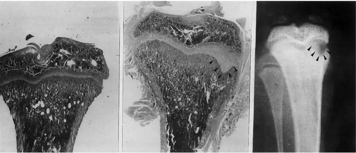
FIG. 1. Low-power view (hematoxylin and eosin, x12.5) of the proximal tibia of control rats (left) and of group A (center). On the medial side, extension of the growth plate inside the metaphysis is present. This is due to persistence of hypertrophic chondro- cytes, which have not been invaded by metaphyseal vessels. The intercolumnar septa in this area are not calcified, as can be seen in the corresponding radiograph. The metaphysis has a cylindrical shape, which is a consequence of metaphyseal modeling arrest.
dose. The mean body weight of the C2 rats at the time of death was 230 g. No significant differences in mean body weight were observed among groups A, B, and C1 at the end of the study. The rats in these groups were killed at 21 days. The left tibia of each animal in each group was carefully dissected from soft tissues without damage to the periosteum and perichondrium.
The specimens were fixed in neutral formalin (10%) and decalcified in EDTA for 2 weeks. The proximal tibia was cut manually with a blade in the median coronal plane. The posterior half of the specimen then was embedded in paraffin, and the plane cut surface was used as a reference for section with a rotatory microtome. Specimens about 10 ym thick were stained with hematoxylin and eosin and mounted in routine fashion.
Assessment of the height of the perichondrial ring
on the medial and lateral sides was performed on the first section with a ocular grid (x100). Its measure was expressed as the mean of the medial and lateral sides. The top end of the ring was identified as the point, just inside the perichondrial groove of Ran- vier, where cells crowd on the outer aspect of the ring and a very thin, bony lamina becomes apparent in the context of cartilage. The bottom end was con- sidered to be the net interruption of the ring, re- sorbed by osteoclasts. A Student t test was applied for comparison among groups.
The numbers of osteoclasts and of nuclei per os- teoclast were assessed in each animal as a mean of the count of five sequential sections: the first median coronal section and the following four sections, each -200 ym deeper than the preceding one. The refer- ence area for the count was composed of the epiph- ysis, the growth plate cartilage, and the metaphysis. The lower limit of the latter was arbitrarily assigned to a line parallel to the growth plate cartilage and at a distance from the proliferative layer equal to twice the thickness of the epiphysis. Only the multinuclear cells on the bone surface were identified as osteo- clasts. The count of osteoclasts was expressed as the mean number of cells for five sections of one speci- men. The nuclei per osteoclast were expressed as the ratio of the number of nuclei to the number of os- teoclasts in the same five sections.
The periosteum and soft tissues were removed from the right tibia. These bones were stored for 7 days in an ethanol-methanol solution (50/50) at 37°C
to extract lipids and water and then dried in an oven at 100°C for 48 h. The length of the tibia was assessed with calipers.
RESULTS
In both groups of rats treated with salmon calcito- nin, an arrest of metaphyseal modeling was present, with an inverted concavity of the metaphysis profile
'
p = 0.002.'p = 0.001.
dp < 0.01. 'Not significant.
(failure of conization) (Fig. 1). The perichondrial ring was thicker and higher than in group C1 (Fig. 2), and the difference was statistically significant. There was
no significant difference in the height of the peri- chondrial ring between control groups C1 and C2 (Table 1). Other features included a persistence of hypertrophic chondrocytes in the outer part of the growth plate, producing a marginal area of thickened
cartilage that was clearly evident on both x-rays and histological sections (Fig. 1). The thickening was more evident on the medial aspect of the tibia1 me- taphysis but was present to a lesser degree on the lateral side as well. The central parts of the growth plate and metaphysis, however, were normal. It al- ready has been documented that such changes in the growth plate are due to arrest of vascular invasion
FIG. 2. Histology of the growth plate and metaphysis of control (left) and group A rats (right) (hematoxylin and eosin, x30). The arrows indicate the top and the bottom end of the perichondrial ring; in treated animals, the metaphysis has a cylindrical shape (arrest of metaphyseal modeling), and the ring extends for the entire height of the unmodeled metaphysis.
Q U A N T I T A T I V E A S S E S S M E N T OF B O N E R E S O R P T I O N INHIBITION 895
FIG. 3. Comparison of osteoclasts in control (left) and group A rats (right) (hematoxylin and eosin, x400). More nuclei are present in the treated group; the nuclei have a dense chromatin and appear pyknotic.
from the metaphysis (15) and that in this model they are associated with failure of mineralization of inter- columnar septa (10).
Many osteoclasts were present at the distal end of the perichondrial ring and on the outer aspect of the metaphysis (Fig. 3). Their number, as well as the nucleikell ratio, was significantly increased in groups
A and B (Table 1). The nuclei had a dense chromatin
and appeared pyknotic. There were no significant differences in the tibial mean length between groups
A and B and group C1 (Table 1).
DISCUSSION
There is a differentiated structure called the peri- chondrial ossification groove of Ranvier (8,12,17) at the periphery of the growth plate of developing tu- bular bones. It consists of an indentation or notch where relatively undifferentiated mesenchymal cells are the progenitors for the osteoblasts forming a thin lamina of bone that encircles the lower portion of the growth plate and is known as the perichondrial ring of Lacroix (llJ6). Because of the modeling of the metaphysis (funnelization) in the proximal tibial metaphysis of rodents, as well as in that of other mammals and humans, the tube of bone is not con- tinuous with the periosteal cortical bone of the di- aphysis but is instead a separate, distinct ring of bone encircling the lower part of the growth plate and the adjacent portion of the metaphysis.
No osteoclasts or evidence of bone resorption are evident along the outer surface of the ring, whereas osteoclasts are concentrated at the distal end and along the inner surface; therefore, the ring grows upward and outward during funnelization as a result of the synthesis of osteoblasts in the proximal and outer portions (ossification groove). Its height is a
function of the rate of osteoblastic activity at its proximal end and of osteoclastic resorption at the distal end. Under healthy conditions, during growth the growth plate and height of the perichondrial ring remain constant. The observation of increased height can be explained only in terms of imbalance of the apposition and resorption rates: either apposition is enhanced or resorption is inhibited. Since there was
no significant difference in the total tibial length be- tween treated animals and controls, it can be as- sumed that the apposition rate was unaffected by calcitonin treatment and that the increased height was due to inhibition of osteoclastic resorption.
A generalized effect on the growth plate was never observed, although it was a constant finding on
the marginal parts of the plate. Since the distribution of osteoclasts in the metaphysis is not homogeneous and they are denser at the periphery, where meta- physeal modeling takes place, if calcitonin inhibits osteoclastic activity then its effects should become apparent at sites where the density of osteoclasts is greater and their metabolic activity is faster.
Both ends of the perichondrial ring are well de- marcated by surrounding tissues, its height can be measured with a satisfactory degree of accuracy, and, if the apposition rate is not affected by the treatment, the height of the ring allows a quantitative evalua- tion of inhibition of bone resorption. This model was tested with a well known in vitro inhibitor of osteo- clastic resorption, calcitonin, and it allowed a his- tometric assessment of bone resorption inhibition, which correlates with the dose of calcitonin.
Paradoxically, the number of multinuclear osteo- clasts was significantly increased, in contrast with the cylindrical shape of the metaphysis and the enlarged perichondrial ring, which indicated an arrest of bone modeling. Stimulation of osteoclast formation may be
are no longer active (9); consequently, no quantitative evaluation of bone resorption may rely on them.
A possible source of error of the assay based on measurement of the perichondrial ring may arise be- cause of the influence of inhibitors not only on osteo- clastic resorption but also on the rate of osteoblast synthesis, since in normal bone turnover these pro- cesses are coupled. However, an inhibition of osteo- blast apposition in growing animals, such as those used in this model, would have resulted in a decrease in the total bone length. An effect on bone length has been observed with other inhibitors of bone resorp- tion, such as EHDP (etidronate) and dichloromethy- lene diphosphonate, when given to rats at doses of 10 and 30 mg/P/kg/day (15). This effect was not ob- served in the present study; however, it is worthwhile to emphasize that comparison with the tibial length of the control group is a necessary step when the assay is performed.
REFERENCES
1. Baron R, Vignery A: Behavior of osteoclasts during a rapid change in their number induced by high doses of parathy- roid hormone or calcitonin in intact rats. Metab Bone Dis
Re1 Res 2:339-346,1981
2. Chambers TJ, Revel1 PA, Fuller K, Athanasou NA: Resorp- tion of bone by isolated rabbit osteoclasts. J Cell Sci 66:383- 399,1984
3. Chambers TJ, McSheehy PM, Thomson BM, Fuller K: The effect of calcium-regulating hormones and prostaglandins
on bone resorption by osteoclasts disaggregated from neo- natal rabbit bones. Endocrinology 116:234-239,1985
hydroxy-1,l-diphosphonate (EHDP) effects on incorpora- tion and accumulation of osteoclast nuclei. Calcif Tissue
Res 22:243-252,1977
10. Pazzaglia U E , Zatti G, Di Nucci A, Coci A: Inhibitory effect of salmon calcitonin on bone resorption: morphological study of the tibial growth plate in rats. Calcif Tissue Int 11. Pratt CW: The significance of the “perichondrial zone” in a
developing long bone of the rat.JAnat 93:llO-122,1959 12. Ranvier LA: Trait6 technique d’histologie, 2nd ed. Paris,
Sary, 1889
13. Reynolds JJ, Dingle JT A sensitive in vitro method for studying the induction and inhibition of bone resorption.
Calcif Tissue Res 4:339-349,1970
14. Russell RG, Kislig AM, Casey PA, Fleisch H, Thornton J, Schenk R, Williams DA: Effect of diphosphonates and cal- citonin on the chemistry and quantitative histology of rat bones. Calcif Tissue Res 11:179-195,1973
15. Schenk R, Merz WA, Muhlbauer R, Russell RG, Fleisch H: Effect of ethane-1-hydroxy-1,l-diphosphonate (EHDP) and dichloromethylene diphosphonate (Clz MDP) on the calcification and resorption of cartilage and bone in the tibial epiphysis and metaphysis of rats. Calcif Tissue Res 16. Shapiro F, Holtrop ME, Glimcher MJ: Organization and cellular biology of the perichondrial ossification groove of Ranvier: a morphological study in rabbits. J Bone Joint Surg
[ Am] 59:703-723,1977
17. Speer DP: Collagenous architecture of the growth plate and perichondrial ossification groove. J Bone Joint Surg [ Am ] 18. Stewart PJ, Stern PH: Vertebral bone resorption in vitro:
effects of parathyroid hormone, calcitonin, 1,25 dihydroxy- vitamin D3, epidermal growth factor, prostaglandin E2 and estrogen. Calcif Tissue Int 40:21-26,1987
19. Yamamoto I, Kitamura N, Aoki J, Shigeno C, Hino M, As- onuma K, Torizuka K, Fuji N, Otaka A, Yajima H: Human calcitonin gene-related peptide possesses weak inhibitory potency of bone resorption in vitro. Calcif Tissue Int 38:339- 341,1986
52:125-129,1993
11:196-214,1973