1
J. Dairy Sci. TBC:1–8
https://doi.org/10.3168/jds.2018-14471
© TBC, THE AUTHORS. Published by FASS Inc. and Elsevier Inc. on behalf of the American Dairy Science Association®. This is an open access article under the CC BY-NC-ND license (http://creativecommons.org/licenses/by-nc-nd/3.0/).
ABSTRACT
The present experiment aimed at understanding the effects of cortisol levels on sheep peripheral blood mono-nuclear cell (PBMC) proliferation and cytokine produc-tion during hyperthermia. To mimic stress related to the exposition of high ambient temperatures, PBMC were cultured at 43°C for 12 h, and subsequently at 39°C for additional 12 h. Cells in normothermia were cultured at 39°C for 24 h. Phytohemagglutinin-stimulated PBMC were cultured with different cortisol levels: 0 ng/mL; 100 ng/mL, representing the physiological cortisol concentration simulating stress condition (Cort100); and 1,000 ng/mL, representing the hyperactivated hypothalamic-pituitary-adrenal axis (Cort1000). Phytohemagglutinin-stimulated PBMC with 0 ng/ mL of cortisol concentration represented the positive control, whereas nonstimulated PBMC without cortisol represented the negative control (NC). The free cell supernatants were collected for the determination of IL-6, IL-1β, and IL-10 by ELISA. Bromodeoxyuridine assay was performed on cells to determine cell prolif-eration. Exposition to hyperthermia negatively affected cell proliferation, IL-6, IL-1β, and IL-10 concentrations in cell supernatants. The interaction of hyperthermia and cortisol level affected both cell proliferation and IL-10 production. Both PBMC proliferation and IL-10 production in positive control, Cort100, and Cort100 decreased at 43°C as compared with 39°C NC. On average, the Cort100 treatment displayed higher con-centrations of IL-6 than NC. The present experiment demonstrated that the action of cortisol concentration simulating stress condition on cell proliferation and cytokine production was a permissive/stimulatory ac-tion during normothermia, whereas it was a suppressive action during hyperthermia. These data confirmed that cortisol concentration simulating stress condition could
have a role in the immune system of sheep via medi-ating cellular homeostasis in the condition of hyper-thermia. The negative effects of hyperthermia on sheep immune responses were apparent when performing an immunological challenge.
Key words: hyperthermia, glucocorticoid, sheep,
noninfectious stressor
INTRODUCTION
Noninfectious stressors are defined disrupters of ho-meostasis of the innate immune response, leading to unbalanced cellular, immunological, and physiological responses (Werling, 2018). Among the different types of noninfectious stressors faced by sheep, heat stress seems to deeply threaten production efficiency of the animals. Further, climate change leads to several vector-borne diseases affecting sheep welfare and health (Gaughan et al., 2009). From this point of view, it becomes crucial to study the effects of thermal stress on the immune efficiency of sheep. Hyperthermia, responsible for heat shock or cellular stress response activation, can alter cells’ metabolism and gene expression (Collier et al., 2006), and the timing and amplitude of the heat shock response can determine cell survival, acclimation, or cell death (Collier et al., 2006).
Glucocorticoids (GC) are produced at high levels during the initial phase of thermal exposure; subse-quently, prolonging the exposure to hyperthermia, a decrement of these hormones is observed (Collier et al., 2008). Furthermore, the increase in circulating hypothalamic-pituitary-adrenal (HPA) axis mediated GC levels is a key factor in the adaptive stress-inflam-matory response (Cooper and Stewart, 2003). The HPA axis activates slowly taking 10 to 60 min from stressor onset for cortisol to reach peak levels, depending on the type of stressor acting, and an hour or more to re-turn to pre-stress levels (Caroprese et al., 2010, 2014). The activation of HPA axis that is caused by stress exposure influences cytokine expression (Uchoa et al., 2014). On the other hand, cytokines, such as IL-1, IL-6, and tumor necrosis factor-α via negative feedback, can
Glucocorticoid effects on sheep peripheral blood mononuclear cell
proliferation and cytokine production under in vitro hyperthermia
M. Caroprese,*1 M. G. Ciliberti,* P. De Palo,† A. Santillo,* A. Sevi,* and M. Albenzio*
*Department of the Sciences of Agriculture, Food and Environment, University of Foggia, Via Napoli, 25, 71122 Foggia, Italy †Department of Veterinary Medicine, University “Aldo Moro” of Bari, S.P. to Casamassima, km 3, 70010, Valenzano (BA), Italy
JDS14471
DRAFT
Received January 19, 2018. Accepted May 15, 2018.
increase the HPA axis sensitivity to later stress chal-lenges (Uchoa et al., 2014). The effects of GC on im-mune system are classified into suppressive, permissive, and anti-inflammatory actions, which were also found during hyperthermia conditions (Sapolsky et al., 2000; Coutinho and Chapman, 2011). It has been proposed that GC might affect the magnitude or direction of the cytokine-induced response by increasing the rate at which the response proceeds (Wiegers et al., 1995). Previous studies demonstrated that as cytokine actions were regulated by GC, also cytokines may have a regu-latory role in GC actions (Arzt et al., 2000; Biola et al., 2001; Heiniger et al., 2001). The precise mechanisms controlling cytokine production and cell proliferation during stress in relation to GC action, have not been fully delineated, but different key mechanisms have been emerging. In a previous experiment in sheep, the possible bi-directional effect of cortisol on the immune system in relation to the magnitude of the stressor and the duration of the stress was discussed (Ciliberti et al., 2017).
Our study focused on the comprehension of the GC effects on sheep immune system when exposed to hy-perthermia; therefore, the aim of the present study was to evaluate the effects of in vitro incremental GC levels on sheep peripheral blood mononuclear cell (PBMC) proliferation and cytokine production in the condition of hyperthermia.
MATERIALS AND METHODS
Animals and Experimental Treatments
Dairy sheep used in the study were allocated at the Segezia research station of the Council for Research and Experimentation in Agriculture. The sheep involved in the experiment were Comisana dairy ewes, with a white coat, aged 3 ± 0.5[AU1: Add units. Identify meaning
of plus/minus value.], reared in external pens of 5 ×
12 m. In the experimental design 8 sheep were chosen with a random design, and an in vitro experiment was performed, with 2 cortisol concentration and 2 different temperature of incubation after mitogen stimulation for a total of 64 samples. Cortisol concentrations were chosen to represent a cortisol concentration simulat-ing stress condition (100 ng/mL, Cort100), and level from an hyperactivated HPA axis status (1,000 ng/mL,
Cort1000), according to results from in vivo studies on
sheep (Caroprese et al., 2010) and also previously re-ported in an in vitro study (Ciliberti et al., 2017). Posi-tive control (PC) was represented by PHA-stimulated PBMC without cortisol (0 ng/mL of cortisol), whereas nonstimulated PBMC without cortisol represented the
negative control (NC). To mimic stress related to the exposition of high ambient temperatures, the experi-ment was conducted at 2 different temperatures: 39°C to simulate normothermia and 43°C to mimic severe hyperthermia, according to Lacetera et al. (2006). To mimic stress related to the exposition of high ambient temperatures, cells (PC, NC, Cort100, and Cort1000) were treated for 12 h at 43°C and subsequently treated for additional 12 h at 39°C. Cells in normothermia (PC, NC, Cort100, and Cort1000) were cultured for 24 h at 39°C. The in vitro study was performed by the split-plot component involving the isolation of sheep PBMC, and the evaluation of their proliferative response and cytokines production after stimulation with the mito-gen phytohemagglutinin-(PHA) and in presence of 2 different concentrations of cortisol under hyperthermia or not. All procedures were conducted according to the guidelines of the EU Directive (2010) on the protection of animals used for experimental and other scientific purposes. The sheep were healthy and their conditions were carefully examined by veterinarians throughout the trial to exclude the presence of any signs of disease. Isolation of PBMC
Blood samples (15 mL) were collected in vacuum tubes from the jugular vein of the sheep. Isolation of PBMC was performed by density gradient centrifuga-tion according to Wattegedera et al. (2004). Peripheral blood mononuclear cells were finally resuspended at a final concentration of 2 × 10⁵ cells/mL in Iscove’s
modified Dulbecco’s medium (Sigma-Aldrich, Milan, Italy) supplemented with 10% FBS and 50 μg/mL of gentamicin.
Lymphocyte Stimulation Assay and Cytokines Measurement
Lymphocyte proliferation assays were performed adding 100 μL of cell suspension into quadruplicate wells of 96-well U-bottom plates. Peripheral blood mononuclear cells were activated with 50 μL of PHA (Sigma-Aldrich) at a final concentration of 10 μg/mL, or not activated with mitogen. Sheep PBMC stimu-lated with PHA were treated with 2 concentrations of cortisol (Sigma-Aldrich; 100, 1,000 ng/mL at final concentration). The plates were incubated at 5% CO2
in a humidified incubator for 24 h. After incubation time, plates were centrifuged at 400 × g for 10 min, and cell-free supernatants from each well were collected and stored at −80°C until ELISA was performed to measure cytokine production. After cell-free supernatant col-lection, cells were incubated with bromodeoxyuridine
(Exalpha Biologicals Inc., Shirley, MA) to test cell pro-liferation. After 18 h of incubation, bromodeoxyuridine incorporation during DNA synthesis was measured by determining optical density with a titer-ELISA spectro-photometer (Power Wave XS, BioTek, Swindon, UK) at 450 nm. During lymphocyte proliferation assay a total number of 256 samples were analyzed (64 samples × 4); for cytokine measurement a total number of 128 samples were analyzed (64 samples × 2).
Measurement of Cytokines in Culture Supernatants by ELISA
The assays were optimized in our laboratory for con-centrations of mouse monoclonal antibodies (mAb), supernatants, polyclonal detecting Ab, and secondary conjugate Ab. The levels of IL-6, IL-1β, and IL-10 in cell-free supernatants were determined by capture ELI-SA performed on 96-well microtiter plates, according to Ciliberti et al. (2017). Both IL-6 and IL-1β incubations were at 37°C for 1 h, and after each step the plates were washed 4 times. All culture supernatants were read in duplicate against a standard curve-fitting 4-parameter logistic curve. The standard curve for IL-6 was obtained using scalar dilution of recombinant ovine IL-6 (Cusa-bio Biotech Co., Wuhan, P.R. China) in a range from 1,750 to 0 ng/mL (R2 = 99.9%). The recombinant ovine
IL-1β (Kingfisher Biotech Inc., St. Paul, MN) for IL-1 β standard curve in a range of 250 to 0 ng/mL was used (R2 = 100%). Interassay and intraassay coefficients of
variation were 2.8 and 3.9% for IL-1β, and 3.1 and 4.2% for IL-6, respectively.
All the incubations for the measurement of IL-10 in plasma were at room temperature, and after each step the plates were washed 4 times. The levels of IL-10 were measured colorimetrically at 450 nm in duplicate (by subtracting absorbance at 540 nm) and quantified by interpolation from standard curve fitted by 4-param-eter logistic curve in a range of 500 to 0 ng/mL (R2 =
99.7%). The IL-10 interassay and intraassay coefficients of variation were 3.7 and 4.1%, respectively.
Statistical Analysis
All variables were tested for normality using the Shapiro-Wilk test (Shapiro and Wilk, 1965). Orthogo-nal contrasts were constructed using the CONTRAST statement procedure of SAS (2013, version 6.1, SAS Institute Inc., Cary, NC). To determine the effect of PHA stimulation, the average of the 3 PHA+ treatment was compared with the NC. To determine the effect of cortisol treatment, the average of the cortisol treatment was compared with the PC. To determine the effect of
cortisol dosage, the average of cortisol treatment at 100 ng/mL was compared with the average of cortisol treat-ment at 1,000 ng/mL. To determine the temperature effect, the average of PHA stimulation, cortisol effect, and cortisol dosage at 39°C were compared with the average of PHA stimulation, cortisol effect, and cortisol dosage at 43°C. To determine the effect of the inter-action among the temperatures tested, and PC, NC, Cort100, and Cort1000, the ANOVA for mixed models using the MIXED procedure of SAS was performed. The interaction between the temperatures tested and the different treatments were used as a fixed effect, and the animal as a random factor nested in the treat-ment. Proliferation, IL-6, IL-1β, and IL-10 were the dependent variables analyzed. When significant effects were found (P < 0.05), the Fisher’s least significant difference test was used to locate significant differences between means for MIXED procedure, whereas the Scheffe test was used to locate the differences among orthogonal contrasts tested.
RESULTS
Cell Proliferation
Contrast analysis of proliferation data showed a significant effect of the temperature on Cort100 (P < 0.0001), Cort1000 (P = 0.015), and PC (P = 0.0004), with a decreasing proliferation at 43°C as compared with 39°C. The PHA stimulation showed a significant effect on cell proliferation both at 39°C (P = 0.018) and 43°C (P = 0.035) as compared with NC. In particular, PHA stimulation resulted in an increase in cell prolif-eration when cells were cultured at 39°C; conversely, PHA stimulation resulted in a decrease in cell prolifera-tion when cells were cultured at 43°C. An interacprolifera-tion between hyperthermia and treatments was detected (P = 0.0008, Figure 1). At 39°C, the treatment Cort100 showed an increase of PBMC proliferation compared with NC (P = 0.002). On the contrary, at 43°C a reduc-tion of PBMC proliferareduc-tion in Cort100 compared with NC was detected (P = 0.006).
Cytokine Production
The results of the effects of the 3 orthogonal contrasts related to PHA stimulation, cortisol effect, and cortisol dosage on cytokine production by PBMC, at both 39 and 43°C, are reported in Table 1. Orthogonal contrasts showed that at 39°C PHA stimulation affected IL-6 production as compared with NC (P = 0.044), whereas cortisol effect and cortisol dosage affected IL-6 produc-tion at 43°C (P = 0.0009 and P = 0.0036, respectively).
The average of PC, Cort100, and Cort1000 treatment displayed a reduction in IL-6 production during hyper-thermia as compared with 39°C (P = 0.027, P = 0.043,
P = 0.009, respectively, Figure 2). Production of IL-6
at 39°C was higher in Cort100 (P = 0.03) and PC (P = 0.03) than in NC (Figure 2).
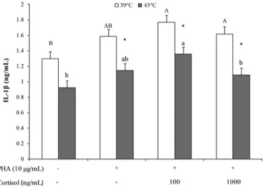
Orthogonal contrasts showed that PHA stimulation increased IL-1β production at 39°C as compared with NC (P = 0.004, Table 1), whereas PHA stimulation decreased IL-1β production at 43°C as compared with NC (P = 0.0004). Furthermore, cortisol treatment and
cortisol dosage affected IL-1β production at 43°C (P = 0.043 and P = 0.044; respectively). The average of PC, Cort100, and Cort1000 treatment displayed a reduction in IL-1β production during hyperthermia as compared with 39°C (P < 0.001, P = 0.025, and P < 0.001, respectively, Figure 3). At 39°C, IL-1β production in-creased in cells cultured with Cort100 (P = 0.005) and Cort1000 (P = 0.01) as compared with NC, whereas at 43°C, IL-1β production was higher in PBMC with Cort100 treatment than NC (P = 0.001) and Cort1000 treatment (P = 0.04, Figure 3).
Orthogonal contrasts showed that PHA stimulation increased IL-10 production at 39°C as compared with NC (P = 0.002, Table 1); cortisol treatment and cortisol
Figure 1. Proliferation of sheep peripheral blood
mononu-clear cells (PBMC; LSM ± SEM) following in vitro stimulation. Phytohemagglutinin (PHA)-stimulated PBMC were treated with 2 concentrations of cortisol [100 ng/mL, as physiological cortisol con-centration simulating stress condition (Cort100), and 1,000 ng/mL, as hyperactivated hypothalamic-pituitary-adrenal axis concentra-tion (Cort1000)], and cultured for 24 h at 39°C (normothermia) or 43°C (hyperthermia). PHA+/cortisol– represented positive control; PHA−/cortisol− represented negative control. A, B indicate differ-ences among experimental treatments at 39°C; a, b indicate differdiffer-ences among experimental treatments at 43°C; * indicates significant differ-ences between 39 and 43°C within each experimental treatment, P < 0.05. OD = optical density.
Table 1. Effects of the orthogonal contrasts [phytohemagglutinin (PHA) stimulation, cortisol effect, and cortisol dosage] on in vitro IL-6,
IL-1β, and IL-10 secretion (mean ± SEM) by sheep peripheral blood mononuclear cells (PBMC) following PHA stimulation at 39 and 43°C temperatures
Item Temperature (°C)
Treatment1
SEM
Effect, P-value
PC Cort100 Cort1000 PHA stimulation Cortisol effect Cortisol dosage
IL-6 39 188.26 191.09 161.68 34.34 0.04 0.81 0.48 43 64.85b 125.08a 57.04b 10.75 0.37 <0.001 0.003 IL-1β 39 1.59 1.77 1.62 0.10 0.004 0.47 0.36 43 1.14ab 1.35a 1.08b 0.08 <0.001 0.04 0.04 IL-10 39 1.57 1.45 1.45 0.06 0.002 0.45 0.93 43 1.08 0.99 0.92 0.10 0.53 0.23 0.32
a,bMeans within a row with different superscripts differ (P < 0.05).
1Treatments: PC = positive control, PHA+/cortisol−; Cort100 = PHA-stimulated PBMC cultured with 100 ng/mL of cortisol; Cort1000 =
PHA-stimulated PBMC cultured with 100 ng/mL of cortisol.
Figure 2. Interleukin-6 secretion by sheep peripheral blood
mono-nuclear cells (PBMC; LSM ± SEM) following in vitro stimulation. Phytohemagglutinin (PHA)-stimulated PBMC were treated with 2 concentrations of cortisol [100 ng/mL, as physiological cortisol con-centration simulating stress condition (Cort100), and 1,000 ng/mL, as hyperactivated hypothalamic-pituitary-adrenal axis concentra-tion (Cort1000)], and cultured for 24 h at 39°C (normothermia) or 43°C (hyperthermia). PHA+/cortisol− represented positive control; PHA−/cortisol− represented negative control. a, b indicate differences among experimental treatments at 39°C; * indicates significant differ-ences between 39 and 43°C for each experimental treatment, P < 0.05.
dosage did not affect IL-10 at 39 or 43°C. The average of PC, Cort100, and Cort1000 treatment displayed a reduction in IL-10 production during hyperthermia as compared with 39°C (P = 0.04, P < 0.001, P < 0.001, respectively, Figure 4). Furthermore, at 39°C PC (P = 0.002) and cells cultured with both cortisol levels (P = 0.04) displayed higher IL-10 concentrations than NC (Figure 4). However, at 43°C PBMC with Cort1000 treatment displayed lower IL-10 production than NC (P = 0.03).
DISCUSSION
In our study, hyperthermia condition deeply influ-enced cell proliferation and cytokine production; fur-thermore, during hyperthermia physiological cortisol concentrations simulating stress condition increased IL-6 and IL-1β production. Moreover, the inhibitory effect of hyperthermia on immune responses was ap-parent in the presence of PHA acting as an immune challenge.
In a previous study, cortisol, when at concentrations simulating stress condition released in vivo after a challenge, exerted a stimulatory action on cell prolif-eration (Ciliberti et al., 2017). In the present study,
during cell stimulation in normothermia conditions, a comparable nonsignificant trend was observed. In particular, the physiological cortisol concentrations simulating stress condition resulted in increasing by 13% of cell proliferation as compared with PC, and by 35% as compared with NC. Such a stimulatory ac-tion of cortisol on cell proliferaac-tion has been found by other authors and attributed to an increased sensitivity to IL-2 action (Wiegers et al., 1995). However, during hyperthermia the physiological cortisol concentrations simulating stress condition showed a decreasing by 50% of cell proliferation as compared with PC, and by 60% as compared with NC. One of the first vital protective strategy the host has to implement when coping with a noninfectious stressor is the delay of growth and rep-lication in order both to reduce energetic costs and to be less vulnerable (Werling, 2018). As a consequence, the cortisol response after a stressor evolved to down-regulate the enhancement of inflammatory responses activated by the prompt activation of the sympathetic nervous system during the first wave of stress responses. From this point of view the cortisol response has the aim to downregulate the inflammatory response and to control cytokine production (Sapolsky et al., 2000). The GC may exert different specific actions, such as permissive, stimulatory, or suppressive actions, in re-sponse to the nature, the magnitude, and the duration of the stressors experienced by most organisms, and to the adaptations needed to survive them by
restor-Figure 3. Interleukin-1β secretion by sheep peripheral blood
mono-nuclear cells (PBMC; LSM ± SEM) following in vitro stimulation. Phytohemagglutinin (PHA)-stimulated PBMC were treated with 2 concentrations of cortisol [100 ng/mL, as physiological cortisol con-centration simulating stress condition (Cort100), and 1,000 ng/mL, as hyperactivated hypothalamic-pituitary-adrenal axis concentra-tion (Cort1000)], and cultured for 24 h at 39°C (normothermia) or 43°C (hyperthermia). PHA+/cortisol− represented positive control; PHA−/cortisol− represented negative control. A, B indicate differ-ences among experimental treatments at 39°C; a, b indicate differdiffer-ences among experimental treatments at 43°C; * indicates significant differ-ences between 39 and 43°C for each experimental treatment, P < 0.05.
Figure 4. Interleukin-10 secretion by sheep peripheral blood
mono-nuclear cells (PBMC; LSM ± SEM) following in vitro stimulation. Phytohemagglutinin (PHA)-stimulated PBMC were treated with 2 concentrations of cortisol [100 ng/mL, as physiological cortisol con-centration simulating stress condition (Cort100), and 1,000 ng/mL, as hyperactivated hypothalamic-pituitary-adrenal axis concentra-tion (Cort1000)], and cultured for 24 h at 39°C (normothermia) or 43°C (hyperthermia). PHA+/cortisol− represented positive control; PHA−/cortisol− represented negative control. A, B indicate differ-ences among experimental treatments at 39°C; a, b indicate differdiffer-ences among experimental treatments at 43°C; * indicates significant differ-ences between 39 and 43°C for each experimental treatment, P < 0.05.
ing homeostasis (Sapolsky et al., 2000). In the present experiment, the action of physiological cortisol concen-trations simulating stress condition on cell proliferation and cytokine production was a permissive/stimulatory action in normothermia, whereas it was a suppressive action during hyperthermia. The present results sug-gested that in the sheep model a further increase of cortisol concentration from 100 to 1,000 ng/mL failed to support an additional effect on cell proliferation, both in normothermia and in hyperthermia. There-fore, the increase of cortisol level did not correspond to the further suppression of immune responses during hyperthermia. The suppressive action of cortisol on cell proliferation could be attributed to the biological mechanisms hypothesized by Van Laethem et al. (2001) in which GC were found able to inhibit T cell responses through early phosphorylating events induced after TCR ligation. The findings of the present experiment further demonstrated that a main role in defining the type of action of cortisol on immune cells is exerted by the nature of the acting stressor, which in this case was hyperthermia. Nevertheless, as suggested by Collier et al. (2008), when the stressor acting on cells is the heat stress, cellular responses involved a controlled network of genes coordinated to minimize the negative effects of the hyperthermia on cellular functions. Bovine mam-mary epithelial cells exposed to hyperthermia (42°C) exhibited morphological changes, reduced cell growth, and downregulation of gene transcripts associated with protein synthesis and cellular metabolism (Collier et al., 2006). It is well known from in vivo studies that during heat stress HPA axis is activated and cortisol concen-tration increased to mediating the ability of sheep to cope with high environmental temperatures (Sejian et al., 2013; Caroprese et al., 2014). The experimental data underlined that in the sheep model the role of cortisol, when at physiological cortisol concentrations simulating stress condition, could be considered as a paramount mediator in restoring cellular homeostasis in the case of heat stress. However, based on the ab-sence of effects, it could be hypothesized that the level of cortisol simulating a hyperstimulation of HPA axis activated a mechanism similar to that of GC resistance, which is found in chronic diseases including asthma, major depression, and cardiovascular conditions (Ro-driguez et al., 2016). However, further research should be done to confirm this also in sheep model.
Hyperthermia is suggested to regulate cell prolifera-tion and inflammatory responses by changing cytokine production. Among proinflammatory cytokines, IL-6 is one of the most powerful cytokines with pleiotropic effects, being involved in hematopoiesis, B cell
differen-tiation, primary antigen receptor-dependent T cell ac-tivation, and their subsequent proliferation (Kishimoto, 2006; Dienz and Rincon, 2009; Tanaka et al., 2014). Interleukin-1β was the first cytokine to be related to HPA activation, as a critical regulator of the adaptive response to stress by the control of stress-related al-terations involving behavior, cognition, and emotions (Goshen and Yirmiya, 2009). In sheep, GC have the ability to influence proinflammatory cytokines as dem-onstrated by both in vivo and in vitro studies (Caroprese et al., 2010; Ciliberti et al., 2017). Accordingly, in the present study, both IL-6 and IL-1β production by cells cultured with cortisol, when at physiological concen-trations simulating stress condition, were altered, and decreased during hyperthermia. The further increase in cortisol concentration to 1,000 ng/mL did not change proinflammatory cytokine production as compared with PC, according to proliferation results. Elevated hyperthermia in mice reduced proinflammatory cyto-kine production as a result of induced expression of activating transcription factor 3, a negative regulator of the IL-6 gene (Hagiwara et al., 2007; Takii et al., 2010). Heat shock protein-72 produced during heat stress has been demonstrated to inhibit the nuclear factor-κB signaling cascade activated by LPS, thus reducing IL-6 production (Chen et al., 2006). The observed alteration in cytokine production induced by GC during hyper-thermia could be considered a further mechanism to restore homeostasis after a noninfectious stressor to re-duce metabolic and health costs connected to cytokine production (Werling, 2018). Interleukin-10 has an im-munosuppressive role in maintenance of the balance in the immune response against invading pathogens (Ma et al., 2015). Interleukin-10 secretion by T cells can be upregulated by glucocorticoids (Elenkov and Chrousos, 1999). In an in vivo study on sheep, an increase in plasma IL-10 production was found when under hy-perthermia (Caroprese et al., 2014). On the contrary, in the present in vitro experiment, hyperthermia lead to decreased IL-10 production by sheep cells when stimulated with PHA, acting as an immune challenge, regardless of cortisol level. Moreover, the lower IL-10 production than NC registered by the highest cortisol level straightened the probably suppressive action of GC also during hyperthermia as a model of a chronic stress. Consequently, the main suppressive effects of hyperthermia on cell proliferation and proinflammatory response could be ascribed to the inhibition of anti-inflammatory cytokines such as IL-10 as well.
The suppression of PBMC proliferation and of cy-tokines observed in the present experiment during in vitro hyperthermia may account for the impairment in
immune responses of sheep under heat stress and help to explain the increased incidence of udder health prob-lems found during summer (Sevi and Caroprese, 2012).
CONCLUSIONS
In the present experiment, the effects of hyperther-mia on cell proliferation and inflammatory responses were evaluated. Hyperthermia, followed by restoring of normothermia, simulated heat waves experienced by heat stressed sheep in livestock production systems. The action of physiological cortisol concentration simu-lating stress condition on cell proliferation and cytokine production was a permissive/stimulatory action during normothermia, whereas it was a suppressive action dur-ing hyperthermia. The finddur-ings of the present in vitro study suggested that the type of action of cortisol on im-mune cells depends on the nature of the stressor acting (i.e., normothermia vs. hyperthermia). The data lead to the hypothesis that cortisol, when at physiological cor-tisol concentration simulating stress condition, can be considered a paramount mediator in restoring cellular homeostasis in the case of hyperthermia by increasing IL-6 and IL-1β production. Results from the present experiment suggested that the reduction in immune competence found in sheep under in vivo hyperthermia could be attributed to the reduction of cell prolifera-tion as a result of alteraprolifera-tion of cytokine producprolifera-tion. The understanding of the mechanisms leading to the reduction of sheep immunological competences under hyperthermia could be crucial to develop new diagnos-tic and therapeudiagnos-tic tools to sustain disorders related to noninfectious stressors in animals[AU2: Please add Acknowledgments with funding sources [required: funder name and location (city and state or city and country); optional: project names and/or grant
numbers] for this research.].
REFERENCES
Arzt, E., D. Kovalovsky, L. M. Igaz, M. Costas, P. Plazas, D. Refojo, M. Páez-Pereda, J. M. Reul, G. Stalla, and F. Holsboer. 2000. Functional cross-talk among cytokines, T-cell receptor, and gluco-corticoid receptor transcriptional activity and action. Ann. N. Y. Acad. Sci. 917:672–677.
Biola, A., P. Lefebvre, M. Perrin-Wolff, M. Sturm, J. Bertoglio, and M. Pallardy. 2001. Interleukin-2 inhibits glucocorticoid receptor transcriptional activity through a mechanism involving STAT5 (signal transducer and activator of transcription 5) but not AP-1. Mol. Endocrinol. 15:1062–1076. https:// doi .org/ 10 .1210/ mend .15 .7 .0657 .
Caroprese, M., M. Albenzio, A. Marzano, L. Schena, G. Annicchi-arico, and A. Sevi. 2010. Relationship between cortisol response to stress and behavior, immune profile, and production performance of dairy ewes. J. Dairy Sci. 93:2395–2403. https:// doi .org/ 10 .3168/ jds .2009 -2604 .
Caroprese, M., M. Ciliberti, G. Annicchiarico, M. Albenzio, A. Muscio, and A. Sevi. 2014. Hypothalamic-pituitary-adrenal axis activation and immune regulation in heat-stressed sheep after supplementa-tion with polyunsaturated fatty acids. J. Dairy Sci. 97:4247–4258. https:// doi .org/ 10 .3168/ jds .2013 -7696 .
Chen, H., Y. Wu, Y. Zhang, L. Jin, L. Luo, B. Xue, C. Lu, X. Zhang, and Z. Yin. 2006. Hsp-70 inhibits lipopolysaccharide-induced NF-kappaB activation by interacting with TRAF6 and inhibiting its ubiquination. FEBS Lett. 580:3145–3152. https:// doi .org/ 10 .1016/ j .febslet .2006 .04 .066.
Ciliberti, M. G., M. Albenzio, C. Inghese, A. Santillo, R. Marino, A. Sevi, and M. Caroprese. 2017. Peripheral blood mononuclear cells proliferation and cytokine production in sheep as affected by corti-sol level and duration of stress. J. Dairy Sci. 100:750–756. https:// doi .org/ 10 .3168/ jds .2016 -11688 .
Collier, R. J., J. Collier, R. Rhoads, and L. Baumgard. 2008. Invited review: Genes involved in the bovine heat stress response. J. Dairy Sci. 91:445–454. https:// doi .org/ 10 .3168/ jds .2007 -0540.
Collier, R. J., C. Stiening, B. Pollard, M. VanBaale, L. Baumgard, P. Gentry, and P. Coussens. 2006. Use of gene expression microarrays for evaluating environmental stress tolerance at the cellular level in cattle. J. Anim. Sci. 84(Suppl):E1–E13.
Cooper, M. S., and P. M. Stewart. 2003. Corticosteroid insufficiency in acutely ill patients. N. Engl. J. Med. 348:727–734. https:// doi .org/ 10 .1056/ NEJMra020529.
Coutinho, A. E., and K. E. Chapman. 2011. The anti-inflammatory and immunosuppressive effects of glucocorticoids, recent develop-ments and mechanistic insights. Mol. Cell. Endocrinol. 335:2–13. https:// doi .org/ 10 .1016/ j .mce .2010 .04 .005 .
Dienz, O., and M. Rincon. 2009. The effects of IL-6 on CD4 T cell re-sponses. Clin. Immunol. 130:27–33. https:// doi .org/ 10 .1016/ j .clim .2008 .08 .018 .
Elenkov, I. J., and G. P. Chrousos. 1999. Stress hormones, Th1/Th2 patterns, pro/anti-inflammatory cytokines and susceptibility to disease. Trends Endocrinol. Metab. 10:359–368.
EU Directive. 2010. EU Directive 2010/63/EU of 22 September 2010 on the protection of animals used for scientific purposes, pp. 33– 79. Official Journal L 276, European Communities Publication, Luxembourg.
Gaughan, J., N. Lacetera, S. E. Valtorta, H. H. Khalifa, L. R. Hahn, and T. Mader. 2009. Response of domestic animals to climate challenges. Pages 131–170 in Biometeorology for Adaptation to Climate Variability and Change. K. L. Ebi et al., ed. Springer Sci-ence + Business Media B.V.[AU3: Add names of all editors. Add location for Springer.]
Goshen, I., and R. Yirmiya. 2009. Interleukin-1(IL-1): A central regu-lator of stress responses. Front. Neuroendocr. 30:30–45. https:// doi .org/ 10 .1016/ j .yfrne .2008 .10 .001.
Hagiwara, S., H. Iwasaka, S. Matsumoto, and T. Noguchi. 2007. Changes in cell culture temperature alter release of inflammatory mediators in murine macrophagic RAW264.7 cells. Inflamm. Res. 56:297–303. https:// doi .org/ 10 .1007/ s00011 -007 -6161 -z .
Heiniger, C. D., M. K. Rochat, F. J. Frey, and B. M. Frey. 2001. TNF-alpha enhances intracellular glucocorticoid availability. FEBS Lett. 507:351–356. https:// doi .org/ 10 .1016/ S0014 -5793(01)03004 -6. Kishimoto, T. 2006. Interleukin-6: Discovery of a pleiotropic cytokine.
Arthritis Res. Ther. 8:S2. https:// doi .org/ 10 .1186/ ar1916. Lacetera, N., U. Bernabucci, D. Scalia, L. Basiricò, P. Morera, and A.
Nardone. 2006. Heat stress elicits different responses in periph-eral blood mononuclear cells from Brown Swiss and Holstein cows. J. Dairy Sci. 89:4606–4612. https:// doi .org/ 10 .3168/ jds .S0022 -0302(06)72510 -3.
Ma, X., W. Yan, H. Zheng, Q. Du, L. Zhang, Y. Ban, N. Li, and F. Wei. 2015. Regulation of IL-10 and IL-12 production and function in macrophages and dendritic cells. F1000Res. 4. https:// doi .org/ 10 .12688/ f1000research .7010 .1.
Rodriguez, J. M., M. Monsalves-Alvarez, S. Henriquez, M. N. Lla-nos, and R. Troncoso. 2016. Glucocorticoid resistance in chronic diseases. Steroids 115:182–192. https:// doi .org/ 10 .1016/ j .steroids .2016 .09 .010.
Sapolsky, R. M., M. Romero, and A. U. Munck. 2000. How do glu-cocorticoids influence stress responses? Integrating permissive, suppressive, stimulatory, and preparative actions. Endocr. Rev. 21:55–89. https:// doi .org/ 10 .1210/ edrv .21 .1 .0389.
Sejian, V., S. Indu, and S. M. K. Naqvi. 2013. Impact of short term exposure to different environmental temperature on the blood bio-chemical and endocrine responses of Malpura ewes under semi-arid tropical environment. Indian J. Anim. Sci. 83:1155–1160.
Sevi, A., and M. Caroprese. 2012. Impact of heat stress on milk pro-duction, immunity and udder health in sheep: A critical review. Small Rumin. Res. 107:1–7. https:// doi .org/ 10 .1016/ j .smallrumres .2012 .07 .012.
Shapiro, S. S., and M. B. Wilk. 1965. An analysis of variance test for normality. Biometrika 52:591–601.
Takii, R., S. Inouye, M. Fujimoto, T. Nakamura, T. Shinkawa, R. Prakasam. K. Tan, N. Hayashida, H. Ichikawa, T. Hai, and A. Nakai. 2010. Heat shock transcription factor 1 inhibits expres-sion of IL-6 through activating transcription factor 3. J. Immunol. 184:1041–1048. https:// doi .org/ 10 .4049/ jimmunol .0902579. Tanaka, T., M. Narazaki, and T. Kishimoto. 2014. IL-6 in
inflamma-tion, immunity, and disease. Cold Spring Harb. Perspect. Biol. 6:a016295. https:// doi .org/ 10 .1101/ cshperspect .a016295.
Uchoa, E. T., G. Aguilera, J. P. Herman, J. L. Fiedler, T. Deak, and M. B. C. de Sousa. 2014. Novel aspects of glucocorticoid actions. J. Neuroendocrinol. 26:557–572. https:// doi .org/ 10 .1111/ jne .12157. Van Laethem, F., E. Baus, L. A. Smyth, F. Andris, F. Bex, J. Urbain,
D. Kioussis, and O. Leo. 2001. Glucocorticoids attenuate T cell receptor signaling. J. Exp. Med. 193:803–814.
Wattegedera, S., K. Sills, C. J. Howard, J. C. Hope, C. J. McInnes, and G. Entrican. 2004. Variability in cytokine production and cell proliferation by mitogen-activate ovine peripheral blood mono-nuclear cells: modulation by interleukin (IL)-10 and IL-12. Vet. Immunol. Immunopathol. 102:67–76. https:// doi .org/ 10 .1016/ j .vetimm .2004 .06 .006
Werling, D. 2018. Non-infectious stressors and innate immune re-sponse. Res. Vet. Sci. https:// doi .org/ 10 .1016/ j .rvsc .2018 .01 .003. In press.[AU4: This paper has been withdrawn. Please remove or replace.]
Wiegers, G. J., M. Labeur, I. Stec, W. E. F. Klinkert, F. Holsboer, and J. M. H. M. Reul. 1995. Glucocorticoids accelerate anti-T cell receptor induced T cell growth. J. Immunol. 155:1893–1902.
![Table 1. Effects of the orthogonal contrasts [phytohemagglutinin (PHA) stimulation, cortisol effect, and cortisol dosage] on in vitro IL-6,](https://thumb-eu.123doks.com/thumbv2/123dokorg/5471725.62123/4.918.460.827.110.361/table-effects-orthogonal-contrasts-phytohemagglutinin-stimulation-cortisol-cortisol.webp)