1
Alma Mater Studiorum – Università di Bologna
DOTTORATO DI RICERCA IN
ONCOLOGIA, EMATOLOGIA E PATOLOGIA
Ciclo XXX°
Settore Concorsuale: SC06
Settore Scientifico Disciplinare: MED/38
Activity of HH/GLI inhibitors in induced pluripotent stem cells model
of pediatric acute myeloid leukemia with CBFA2T3-GLIS2 fusion gene
Presentata da:
Bertuccio Salvatore Nicola
Coordinatore Dottorato
Supervisore
Chiar.mo Prof. Pier Luigi Lollini
Chiar.mo Prof. Andrea Pession
3
TABLE OF CONTENT
Abstracts ... 5
1. Introduction... 7
1.1 Pediatric Acute Myeloid Leukemia ... 7
1.3 Classification and overall outcome ... 7
1.4 Molecular and Genetic Aberrations in Pediatric AML ... 10
1.4.1 Proliferation and/or prosurvival aberrations (Type I mutations)... 10
1.5 Acute megakaryoblastic leukemia ... 16
1.6. CBFA2T3-GLIS2 fusion gene ... 17
1.7 Targeting Hedgehog pathway in AML ... 19
2. Aim ... 21
3. Materials and Methods ... 22
3.1. Cell Lines and reagents ... 22
3.2 Cell viability assay ... 22
3.3 Cell cycle analysis and apoptosis assay ... 22
3.4 Western Blotting... 23
3.5 Quantitative PCR and gene expression. ... 23
3.6 ChIP analysis ... 24
3.7 Cloning of CBFA2T3-GLIS2 fusion gene ... 24
3.8 iPSC Culture ... 24
3.9 iPSCs genome editing using AAVS1 zinc-finger nucleases ... 25
3.10 Characterization of iPSC, knock-in with CBFA2T3-GLIS2 ... 25
3.11 Differentiation of iPSC into hematological lineage ... 27
3.12 Methylcellulose culture ... 28
4
3.14 DNA/RNA extraction and RT-PCR. ... 28
4. Results ... 29
4.1 CBFA2T3-GLIS2-positive cells are sensitive to GLI1/2 inhibitor ... 29
4.3 GANT61 downregulates expression of GLIS2 protein ... 31
4.5 Treatment with GANT61 downregulates expression of HH and BMP signaling target genes 32 4.6 CBFA2T3-GLIS2 chimeric protein regulates DNMT1 and DNMT3B expression ... 33
4.7 iPSC engineered with CBFA2T3-GLIS2 maintain the pluripotent properties ... 34
4.8 Expression of CBFA2T3-GLIS2 in human hematopoietic cells obtained from iPSC recapitulates several molecular features of human pediatric AMKL ... 36
4.9 CBFA2T3-GLIS2 induces aberrant differentiation in megakaryocytic cells ... 37
4.10 CBFA2T3-GLIS2 enhances proliferation and self-renewal of progenitors hematopoietic cells ... 39
4.11 The GANT61 inhibits the proliferation of CBFA2T3-GLIS2 hematopoietic cells derived from iPSC differentiation ... 41
5. Conclusion and Discussion ... 42
5
Abstracts
CBFA2T3-GLIS2 is a fusion gene identified in 7-8% of pediatric acute myeloid leukemia with poor
outcome. This fusion gene drives a peculiar expression pattern and leads to overexpression of some of Hedgehog related genes. GLI similar protein 2 (GLIS2) is closely related to the GLI family, the final effectors of classic Hedgehog pathway. These observations lend compelling weight to the application of GLI inhibitors in the treatment of AML with the aberration CBFA2T3-GLIS2. GANT61 is an inhibitor of GLI proteins. The exposure to the GANT61 resulted in higher sensitivity of cell lines and primary AML cells carrying CBFA2T3-GLIS2 than the AML cells without GLIS2 fusion as well as in promoting apoptosis and downregulation of GLIS2 specific signature genes. Moreover, ChIP analysis revealed direct regulation by GLIS2 chimeric protein of DNMT1 and
DNMT3B, two genes implicated in important epigenetic functions. Furthermore, considering the
limitations of cell lines model we set up a model based on induced pluripotent stem cells (iPSC) derived from healthy donor engineered to express CBFA2T3-GLIS2 fusion gene. We demonstrated that CBFA2T3-GLIS2 and other targets of fusion gene were expressed in hematopoietic cells derived from iPSC differentiation. The CD43+ hematopoietic cells were composed of a higher percentage of CD41+CD42+ megakaryocytic cells. Importantly, we observed strong differentiation alterations, including a lower expression of CD61 on CBFA2T3-GLIS2 megakaryocytes compared to controls and a progressive accumulation of an abnormal CD41lowCD42low population never detected in control conditions. Finally, clonogenic assays in methylcellulose indicated that
CBFA2T3-GLIS2 enhances serial replating activity for several weeks. Moreover treatment with
GANT61 affected proliferation of CBFA2T3-GLIS2 hematopoietic cells derived from iPSC differentiation.
Taken together, our findings indicate that the GLI inhibitor GANT61 may be used to specifically target the CBFA2T3-GLIS2 fusion in pediatric AML.
7
1. Introduction
1.1 Pediatric Acute Myeloid Leukemia
Acute myeloid leukemia (AML) is a group of genetically heterogeneous hematopoietic disorders of the myeloid lineage characterized by the uncontrolled growth and clonal expansion without complete differentiation of a hematopoietic/stem progenitor 1.
Acute myeloid leukemia represents the 15-20% of the entire Acute leukemia in children. The incidence of AML in infants is 1.5 per 100,000 individuals per year, the incidence decreases to 0.9 per 100,000 individuals aged 1–4 and 0.4 per 100,000 individuals aged 5–9 years, after which it gradually increases into adulthood1. The underlying cause of AML is unknown, and childhood AML generally occurs de novo. In adult and elderly patients, AML is often preceded by myelodysplastic syndrome (MDS), but in children, the occurrence of AML preceded by clonal evolution of preleukemic myeloproliferative diseases, such as myelodysplastic syndrome (MDS) or juvenile myelomonocytic leukemia (JMML), is rare. Germline affected individuals, such as those with Fanconi anemia or Bloom syndrome, have an increased risk for developing AML as a secondary malignancy 2, 3. Recently, germ-line mutations in several genes, such as TP53, RUNX1, GATA2 and CEBPA, have been found in families with an unexplained high risk of AML, suggesting a familial predisposition to develop AML 4-9. Children with Down syndrome classically present with a unique megakaryoblastic subtype of AML, classically following a transient myeloproliferative disorder in the neonatal period, which is characterized by somatic mutations in the GATA1 gene. In addition, AML may occur following previous radiotherapy or chemotherapy containing alkylating agents or epipodophyllotoxins, as secondary neoplasm. These are typically characterized by either MLL-rearrangements or by monosomy 7 9, 10.
1.3 Classification and overall outcome
Prognosis of childhood acute myeloid leukemia (AML) has improved significantly over the past decades, the current probability of cure being approximately 60% in most of the developed countries 11. This result has been achieved thanks to not only the use of more effective anti-leukemic agents and significant improvements in supportive care, but also to the progress made in the stratification of patients with a consequent risk-directed therapy 12-14.
8 The first classification of AML was provided by the French-American-British (FAB) classification. The FAB classification was based on the morphological and histological characteristics of the leukemic cells as well as stage of differentiation of the leukemic blasts 15 (Table 1) .
Table 1. French-America-British (FAB) classification for AML
M0 AML with no Romanowsky or cytochemical evidence of differentiation M1 Myeloblastic leukemia with litter maturation
M2 Myeloblastic leukemia with maturation M3 Acute promyelocytic leukemia
M3h Acute promyelocytic leukemia, hypergranular variant M3v Acute promyelocytic leukemia, microgranular variant M4 Actue myelomonocytic leukemia
M4eo Actue myelomonocytic leukemia with dysplastic marrow meosinophils M5 Acute monoblastic leukemia
M5a Acute monoblastic leukemia, poorly differentiated M5b Acute monoblastic leukemia, differentiated M6 Erytroleukemia
M7 Acute megakaryoblastic leukemia
Modified from Bennett JM, Catovsky D, Daniel MT, Flandrin G, Galton DA, Gralnick HR, Sultan C. Proposals for the classification of the acute leukaemias. French-American-British (FAB) co-operative group. Br J Haematol. 1976 Aug;33(4):451-8.
Although the FAB classification remains useful and commonly used is nowadays replaced by the World Health Organization (WHO) classification, which also takes karyotype and molecular aberrations into account (Table 2) 16.
9
Table 2. The World Health Organization (WHO) classification of acute myeloid leukemia Acute myeloid leukemia with recurrent genetic abnormalities
AML with balanced translocations/inversions
Acute myeloid leukemia with t(8;21)(q22;q22); AML/ETO
Acute myeloid leukemia with inv(16)(p13;q22) or t(16;16)(p13;q22); CBFB/MYH11 Acute myeloid leukemia with t(15;17)(q22;q21); PML/RARα
Acute myeloid leukemia with t(9;11)(p22;q23); MLL/AF9
Acute myeloid leukemia (megakaryoblastic) with t(1;22)(p13;q13); OTT/MAL Acute myeloid leukemia with inv(3)(q21;q26.2) or t(3;3)( q21;q26.2); RPN1/EVL1 Acute myeloid leukemia with t(6;9)(p23;q34); DEK/NUP124
AML with gene mutations
Mutation affecting FLT3, NPM1, C/EBPα, KIT, MLL, WT1, NRAS and KRAS
Acute myeloid leukemia with myelodisplasia-related changes
Acute leukemia with 20% or more peripheral blood or bone marrow blasts with morphological features of myelodysplasia or a prior history of a myelodyplastic syndrome (MDS) or myelodyplastyic/myeloproliferative neoplasm (MDS/MPN), or MDS-related cytogenetic abnormalities, and absence of the specific genetic abnormalities of AML.
Therapy-related myeloid neoplasms
Therapy-related acute myeloid leukemia (tAML), myelodysplastic syndrome (tMDS), and myelodyplastyic/myeloproliferative neoplasm (tMDS/MPN) occurring as late complications of cytotoxic chemotherapy and/or radiation therapy administered for a prior neoplastic or non-neoplastic disorder.
Acute myeloid leukemia, not otherwise specified
FAB classification (M0, M1, M2, M3, M3h, M3v, M4, M4eo, M5, M5a, M5b, M6, M7.
Myeloid sarcoma
Tumor mass consisting of myeloid blasts with/without maturation, occurring at an anatomical site other than the bone marrow.
Myeloid proliferations related to Down syndrome
Transient abnormal myelopoiesis
Myeloid leukemia associated with Down syndrome
Blastic plasmacytoid dendritic cell neoplasm
Clinically aggressive tumor derived from the precursors of plasmacytoid dendritic cells, with a high frequency of cutaneous and bone marrow involvement and leukemic dissemination.
Modified from the Vardiman JW et al. The 2008 revision of the World Health Organization (WHO) classification of myeloid neoplasms and acute leukemia: rationale and important changes. Blood. 2009 Jul 30;114(5):937-51.
10 Despite the improvement in overall survival, ~30% of the pediatric patients relapse. The outcome of relapse is poor, and only the ~30%–40% of patients survives 17, 18. Since the high frequency of treatment-related deaths (5%–10%) further intensification of chemotherapy seems no longer feasible 19. Therefore, knowledge on the molecular and genetic background is indispensable to detect novel, leukemia and patient-specific treatment targets 1.
1.4 Molecular and Genetic Aberrations in Pediatric AML
Despite the heterogeneous pattern of genetic lesions, AML is thought to arise from at least two classes of cooperating genetic events 20.
Type I abnormalities result in increased of the proliferation and/or the survival of the leukemic cell. This type of mutations is often activating mutations of genes involved in signal transduction pathways, such as FLT3, KIT, N-RAS, K-RAS and PTPN11 21.
Type II abnormalities impair differentiation and mainly result from genetic aberrations in hematopoietic transcription factors, due to, for instance, translocations t(8;21)(q22;q22)/AML1-ETO and 11q23/MLL rearrangements or from mutations in genes, such as NPM1 and CEBPA 7, 22-26
. Accordingly to the “two-hits” model for leukemogenesis, the mutations conferring prosurvival/proliferative advantage (Type I) need to be associated with the mutations that impair the ability to differentiate of the hematopoietic progenitors for a complete outset of acute myeloid leukemia (Type II) 25.
1.4.1 Proliferation and/or prosurvival aberrations (Type I mutations)
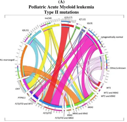
Generally the type I mutations occur in genes encoding for proteins involved in proliferation and survival pathways (Figure 1 A, B).
11
(A)
Pediatric Acute Myeloid leukemia Type II mutations
Type I mutations
(B)
Cytogenetically Normal AML Type II mutations
Type I mutations
Figure 1. Distribution of Type I/II abnormalities in pediatric AML. (A) Cooperating Type I and Type II mutations in
pediatric AML and (B) in cytogenetically normal AML. The circos plot depicts the frequency of the Type II mutations and co-occurrence of Type I mutations in patients with de novo pediatric AML. The length of the arch corresponds to the frequency of the Type II mutation and the width of the ribbon with the percentage of patients with a specific Type I mutation or a combination of Type I mutations. FLT3/ITD denotes FLT3 internal tandem duplication. Modified from de
Rooij JD, Zwaan CM, van den Heuvel-Eibrink M. Pediatric AML: From Biology to Clinical Management. J Clin Med. 2015;4:127-49.
12
FLT3 mutations
The Fms-like tyrosine kinase 3 (FLT3) is a member of the class III subfamily of receptor tyrosine kinases and it is activated by FLT3 ligand 27. FLT3 is expressed in early hematopoietic progenitor cells in the bone marrow 27. High FLT3 levels have been detected in acute myeloid leukemia (AML) 28, 29,27, 30. The most common mutation in AML is the internal tandem duplication (ITD) in the juxtamembrane domain of FLT3 with a 10-15% occurrence in pediatric patients31. FLT3-ITD serves as a prognostic marker since it positively correlates with higher blast counts, increased relapse rate, and worse overall survival 30, 32, 33. Several activating point mutations in the tyrosine kinase domain (TKD) have also been identified.27
Acute myeloid leukemia cells show increased proliferation and survival, as well as impaired hematopoietic differentiation.27 FLT3-ITD or FLT3-TKD activation confers proliferative and survival advantages to cells 27, 34 by activating Src family tyrosine kinases (SFKs), the PI3K/Akt-, mitogen-activated protein kinase (MAPK) pathways, and, in the case of FLT3-ITD, also Stat5.34 Moreover recently it has been demonstrated that FLT3 also blocks differentiation, at least partially, via p27 downregulation. 35,36
KIT activating mutations
Kit, a proto-oncogene in a region on the long arm of chromosome 4 (4q11–4q13), encodes the SCF
receptor (CD117) 37. Ligand independent activation of KIT is driven by mutations that could occur in the extracellular domain of the receptor, in transmembrane/juxtamembrane domain and in the activation loop of the tyrosine kinase domain. Deregulation of KIT, including overexpression and gain of function mutations, has been detected in several human cancers. The proto-oncogene KIT is expressed in approximately 80% of adult AML cases, and its expression is a reliable molecular marker of poor prognosis in AML38, 39. Moreover, KIT mutations are found in 25% to 30% of cases of adult core-binding factor (CBF)-AML, and in 19% of pediatric CBF-AML, which is genetically expressed by the presence of t(8;21)(q22;q22) or inv(16)(p13;q22)4041.
RAS activating mutations
RAS proto-oncogenes, including KRAS, NRAS, and HRAS, encode a membrane-localized G protein
of 21 kDa, they regulate the growth and differentiation of many cell types. RAS proteins are located on the inner surface of the plasma membrane and act as molecular switches that transduce extracellular signals to the nucleus. It is inactive when bound to GDP and active when bound to GTP42, 43. RAS activation caused by its mutation giving rise to an abnormal protein resistant to GTP hydrolysis by GTPase leads to a constitutively active GTP-bound protein that stimulates a critical
13 network of signal transduction pathways that result in cellular proliferation, survival, and differentiation 42-45. RAS mutations at codons 12, 13, and 61 are common events in human cancers45. NRAS or KRAS mutations occur in ∼20% of AML specimens 46, and Ras signaling is activated by somatic mutations in the genes encoding the FLT3 and c-Kit receptor tyrosine kinases in an additional ∼25% to 40% of cases 44
.
PTPN11 mutations
The PTPN11 gene encodes SHP-2, a cytoplasmic protein tyrosine phosphatase. Germline PTPN11 mutations are the causative defect in Noonan syndrome, but somatic mutations have been described in juvenile myelomonocytic leukemia, acute lymphoblastic leukemia, and approximately 4% of children and adults with AML47, 48. Gain of function of SHP-2 contributes to leukemogenesis by upregulating the Ras-Erk and PI3K-Akt pathways leading to aberrant proliferation of myeloid progenitors 49, 50. Moreover, Somatic mutations in PTPN11 are found in about 35% of juvenile myelomonocytic leukemia cases 51. Approximately 4.4% of pediatric AML cases show PTPN11 mutations and the 22% of these carry also mutation in FLT3 and RAS genes 52. The presence of
PTPN11 mutation does not carry prognostic implications in AML, although robust data in
combination with other cytogenetic abnormalities are lacking 47, 53, 54, .
AML with NPM1 mutations
The nucleolar protein nucleophosmin 1 is involved in numerous cellular functions, such as ribosome biogenesis, DNA repair and regulation of apoptosis. Although it is a rather late event in leukaemogenesis, NPM1 mutations emerge as a distinct AML entity. Mutations result in aberrant localization of the protein to the cytosol. An N-terminal nucleolar localisation signal is disrupted, and an export signal is created instead. Mutations in the NPM1 gene are common genetic changes in both pediatric and adult AML, with a incidence of 5% - 10% and 35% respectively 55. In the absence of FLT3-ITD mutations, NPM1 mutations are associated with improved outcomes for patients with CN-AML, even in those older than 60 years. NPM1 mutations are associated with other recurrent genetic abnormalities, such as trisomy of chromosome 8, DNMT3A mutations, FLT3-ITD (40% of the time), FLT3-TKD (10–15%) and IDH mutations (25% of time) 56-58 .
14
Janus kinase 2 (JAK2) activation mutations
The Janus kinase (JAK) family of nonreceptor tyrosine kinases are important mediators of cytokines and Grow Factor signaling, activating signal transducer and activator of transcription (STAT) proteins and other downstream signaling pathways that modulate cell cycling and apoptosis 59
. STATs are constitutively activated in several solid tumors and hematological malignancies 60-62
. Gain-of-function JAK2 V617F mutations are common in myeloproliferative disorders 63, 64 (90% of polycythemia vera cases, 50% of essential thrombocythemia, and in about 70% of patients with AML secondary to other myeloproliferative disorders) but are rare in AML (1.6%) 63,65.
1.4.2. Cytogenetic abnormalities impaired myeloid differentiation (Type II mutations)
The most common cytogenetic abnormalities (Type II) in children are t(8;21)(q22;q22), inv(16)(p13.1q22) (together referred to as core binding factor (CBF)-AML), t(15;17)(q22;q21) and 11q23/MLL-rearranged abnormalities (Figure 1A) 26, 66-68. Together, these account for approximately half of all pediatric AML cases, a much higher frequency than in adults. Translocations involving hematopoietic transcription factors often lead to dysregulated gene expression, either as a result of the fusion partner itself or the recruitment of different co-factors to the transcription complex 1.
CBF (Core Binding Factor leukemia)
CBF-AMLs, which account for approximately 23% of pediatric, contain chromosomal translocations or inversions that target the transcription factors RUNX1 (AML1) or CBFB 69. Normally RUNX1 and CBFB heterodimerize to bind DNA and recruit lineage defining transcription factors to regulate hematopoietic differentiation 70. The fusion products found in CBF-AMLs [RUNX1-RUNX1T1/t(8;21)(q22;q22) or CBFB-MYH11/inv(16)(p13.1q22) or t(16;16)(p13.1;q22)] block myeloid differentiation but are not sufficient to induce leukemia and both fusions requires secondary mutations, including those in the Ras pathway 71-74. Moreover, the AML1 gene is also involved in another translocation t(16;21) in which it results to be in-frame fused with the gene CBFA2T3 localized on chromosome 16. Interestingly, CBFA2T3 gene encode for a protein of the same family of ETO and, functionally, these two chimeric protein could be considered equal 75. Alterations of the CBF are prognostic markers of good prognosis, and a specific subtype of AML, known as core-binding factor AML (CBF-AML), has been define base on that. Nevertheless, the CBF-AML patients are predicted to have a good outcome as long as there are no other negative prognostic markers such as FLT3-ITD or KIT mutations 76.
15
CCAAT enhancer binding protein a (CEBPA) mutations
Mutations in the differentiation-inducing transcription factor CCAAT enhancer binding protein a (CEBPA) are observed in 6–10% of all AML and 15–19% of CN-AML cases and commonly in association with del(9q) and in approximately 4% - 6% of pediatric AML 23, 77, 78. CEBPA is a critical transcription factor that controls gene expression during haematopoiesis 79. Importantly, only bi-allelic, not single, CEBPA mutations (BiCEBPAα) predicts an increased complete remission rate and favourable survival 80, 81. AML with a single CEBPA mutation is associated with survival rates similar to that of AML with wild-type CEBPA 58, 82, 83 Moreover BiCEBPα mutation is found to be stable and highly penetrant. Thus, the stability of biCEBPα supports its role in defining a distinct molecular and clinical subtype of AML 84.
PML/RARα and acute promyelocytic leukemia
PML/RARα traslocation involves the retinoic receptor-alpha gene (RARA) on chromosome 17 and the promyelocytic leukemia gene (PML) on chromosome 15. This aberrations is exclusively associated with the acute promyelocytic leukemia (APL). In APL, PML/RARα fusion protein actively recruits co-repressor complex, thus inducing a sustained transcriptional inhibition and resulting at functional level in a block of myeloid differentiation and increased self-renewal of leukemic progenitors. As a consequence of these biologic properties PML/RARAα acts as a potent transcriptional repressor of retinoic acid signalling. Remarkably, over the 90% of PML/RARα-positive cases could be successfully treated with all-trans retinoic acid (ATRA) that is able to induce the differentiation of this leukemic cells into mature granulocytes 76,85.
Mixed lineage leukemia (MLL) gene alterations
Acute leukemia bearing an MLL-rearrangement (MLL-r) is a distinct leukemia subgroup that needs much attention. While the overall survival of acute leukemias in pediatric patients reaches about 90%, MLL-r leukemia patients still display poor survival rates 86. MLL gene encodes a protein (500 kDa) which serves as a platform for the assembly of a multiprotein complex. The MLL complex influences gene transcription and chromatin by binding to target gene promoters, and reading and writing chromatin signatures.
Based on data published in the literature, MLL-r AML patients display an ectopic genetic program caused by the overexpression of MEIS1/HOXA proteins 87-89. Overexpression of HOXA9 and MEIS1 is essential to drive the development and maintenance of acute myeloid leukemia. In the last years it has been developed a series of new inhibitors that can be potentially used to treat this
16 disease entity 90-95 and many of these is in clinical trial. All these drugs target either a druggable domain that is critical for the function of an MLL fusion or an associated protein 96.
1.5 Acute megakaryoblastic leukemia
Acute megakaryoblastic leukemia [AMKL or AML-M7 in the French–American–British (FAB) classification] is a subtype of AML 15, representing 3–5% of AML and affecting the megakaryocyte lineage. AMKL is higher prevalent in young children (median age approximately 1 year) than in adults (median age approximately 58 years).
Pediatric AMKL is diagnosed as a de-novo disease or in patients harboring constitutive trisomy 21 [Downsyndrome (DS)] 97, who present a 500-fold increased risk to develop AMKL. The 90% of de-novo AMKL harbor both numerical and structural chromosomal alterations, including hyperploids and complex karyotypes 98. Early cytogenetics and fluorescence in situ hybridization analyses identified the recurrent (1;22)(p13;q13) chromosomal translocation 99-101 and led to the identification of the OTT–MAL fusion oncogene 102, 103. OTT–MAL is specifically associated with childhood AMKL and fuses most of OTT and MAL coding regions. The OTT family was proposed to inhibit the RBPJ-dependent transcription of Notch pathway target genes104, which contributes to embryonic hematopoiesis 105 and controls the T-cell development 106 or erythro-megakaryocytic lineage commitment107, 108. MAL is a strong transcriptional coactivator of serum response factor (SRF) 109 through the recruitment of other epigenetic regulators such as ASH2 and SET1110. OTT– MAL alters the transcription of SRF-dependent promoters in transient transactivation in vitro systems and the low frequency of additional mutations found in OTT–MAL AMKL patients suggests that OTT–MAL is sufficient for leukemogenesis in humans111. Other acquired chromosomal abnormalities (e.g., trisomy21, trisomy 19, monosomy 7) are also frequent in AMKL112. In addition both MLL and NUP98 fusions and aberrant expression of homeobox (HOX) are also associated with AMKL 113. Recently, high-throughput sequencing approaches discovered a novel recurrent fusion oncogenes: the CBFA2T3-GLIS2 fusion gene111, 114-116.
17
1.6. CBFA2T3-GLIS2 fusion gene
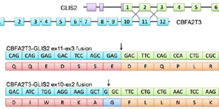
CBFA2T3-GLIS2 also known as ETO2-GLIS2 is a fusion gene resulting from a cryptic inversion of the telomeric region of chromosome 16 leading the the 5’ portion of CBFA2T3 gene fused in-frame to the 3’ region of GLIS2 gene (Figure 2).
Figure 2. Schematic representation of fusion between exon 11 or exon 10 of CBFA2T3 and exon 3 or exon 2 of GLIS2. Modified from Masetti et al. CBFA2T3-GLIS2 fusion transcript is a novel common feature in pediatric,
cytogenetically normal AML, not restricted to FAB M7 subtype. Blood. 2013
The incidence of CBFA2T3-GLIS2 (also known as CBFA2T3-GLIS2) is 17% and 8% in pediatric non-Down syndrome acute megakaryoblastic leukemia (non-DS AMKL, FAB M7) 114, 115, 117 and in pediatric cytogenetically normal AML (CN-AML, in association with several FAB subtypes)116, respectively. The expression of CBFA2T3-GLIS2 is associated with grim prognosis in all the reports published so far 116, 118 (Figure 3).
Figure 3. Probability of 5-year EFS in children with CBFA2T3-GLIS2 fusion transcript in CN-AML. EFS of
CBFA2T3-GLIS2–positive patients (27.4%, SE 10.5) vs CBFA2T3-GLIS2–negative patients (59.6%, SE 3.6; P = .01).
Modified from Masetti at al.CBFA2T3-GLIS2 fusion transcript is a novel common feature in pediatric, cytogenetically normal AML, not restricted to FAB M7 subtype Blood 2013 121:3469-3472.
18 CBFA2T3 is a member of the Eight Twenty One family of transcriptional co-factors that includes ETO and MTGR1. CBFA2T3 expression is essential for HSC maintenance and differentiation, and plays a critical role in the development of megakaryocyte-erythrocyte progenitors in vivo 119, 120. CBFA2T3 participates in high-molecular-weight complexes with several transcription factors (including TAL1, RUNX1, GATA, and ETS) that are able to both activate and repress gene transcription to control HSC commitment toward the erythroid and megakaryocytic lineages, depending on the cofactors found in these transcriptional platforms121-124. In particular, CBFA2T3 represses the expression of erythroid genes 124, 125. During megakaryopoiesis, CBFA2T3 reportedly represses gene expression to inhibit terminal differentiation 126.
The GLIS family zinc-finger protein 2 (GLIS2) is closely related to GLI and zinc-finger protein families. The CBFA2T3-GLIS2 fusion contains most of GLIS2 protein, including its DNA-binding domain, which comprises all five zinc-finger domains. GLIS2 is highly expressed in adult kidney, where it reportedly suppresses the Gli1- activated transcriptome and maintains homeostasis, and is mutated in human nephronophthisis 128.
Although GLIS1/2 factors have been proposed to support the pluripotent stem cell state 129, 130, their contribution to normal HSC biology is unknown.
Different studies show that both CBFA2T3-GLIS2 and GLIS2 expression can increase self-renewal capacity in hematopoietic progenitors. In particular CBFA2T3-GLIS2 enhances self-renewal and inhibits differentiation, whereas the ectopic GLIS2 expression has a variable effect on self-renewal but promotes megakaryocytic differentiation 114.
Importantly, the activity of two transcription factors (GATA1 and ERG) previously involved in DS–AMKL leukemo-genesis is altered by CBFA2T3-GLIS2. GATA1 is strongly down-regulated as a result of both CBFA2T3 and GLIS2 functions, while the ETS-factor/ERG gene, located on chromosome 21, is strongly overexpressed primarily due to the GLIS2 moiety. Definitely, CBFA2T3–GLIS2 AMKL in a single event strongly increases ERG and downregulates GATA1 (Figure 4). It was proposed that the level of deregulation of this balance is in part responsible for the aggressiveness of the disease. According to this hypotesis CBFA2T3–GLIS2 is rarely associated with mutations in coding sequences, suggesting it maybe sufficient to drive leukemogenesis 111.
19
A B
Figure 4. (A) In the normal megakaryocytic differentiation CBFA2T3 (also known as ETO2) complex induces
GATA1 expression and represses ERG expression. (B) ETO2-GLIS2 is a potent inducer of ERG expression and in the same time it is a repressor of GATA1. The deregulation of this balance between ERG and GATA1 is sufficient to block megakaryocytic differentiation and to induce leukemia. (Modified from Thirant et al. 2017. ETO2-GLIS2 Hijacks Transcriptional Complexes to Drive Cellular Identity and Self-Renewal in Pediatric Acute Megakaryoblastic Leukemia)
1.7 Targeting Hedgehog pathway in AML
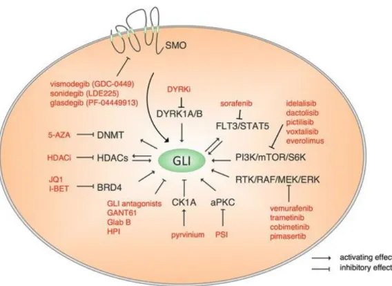
The Hedgehog (HH) signalling pathway has diverse functions in animal development and tissue homeostasis. Dysregulation of this pathway has been implicated in several developmental syndromes and cancers 131. In mammals, the HH signaling cascade is initiated by one of three ligands: Sonic hedgehog (Shh), Indian hedgehog (Ihh), and Desert hedgehog (Dhh) which bind to the transmembrane receptor Patched-1 (PTCH1). Without the presence of ligands, PTCH1 represses activation of the transmembrane protein Smoothened (SMO). Upon HH ligand binding, PTCH1 is internalized, resulting in relief and thus activation of SMO that transduces the signal to the family of Glioma (GLI) zinc finger transcription factors: GLI1, GLI2, and GLI3. GLI1 and GLI2 predominantly represent transcriptional activators while GLI3 acts as transcriptional repressor 132 (Figure 5). HH pathway has recently received much attention for its implication in advanced haematological malignancies and acquired drug resistance in AML with poor prognosis. Interesting it has been demonstrated that the expression of GLI proteins can correlate with poor prognosis in AML and with decrease of DNA methylation in MDS133. Currently there are small molecules that can inhibit HH pathway at different levels (Figure 5) and in particular the SMO inhibitor are in many clinical trials in combination with other conventional drugs in different types of cancer including haematological malignancies (NCT02073838 NCT01842646, NCT01841333, NCT01546038, NCT02367456).
20 It is noteworthy that in a phase 2 trial with untreated AML and high-risk MDS patients, low dose Ara-C chemotherapy in combination with glasdegib improved overall survival when compared to chemotherapy only 134.
Moreover HH pathway can be activated through other pathway including K-Ras, TGF-β, PI3K-AKT, and PKC-α that can be positive regulate GLI protein transcriptional activity 135, 136. Based on these obseravtions, direct inhibition of GLI is a promising option, particularly in settings of SMO-independent GLI activation.
The GANT61 is a small molecule inhibiting DNA binding of GLI family proteins, and it is largely used in preclinical studies 132, 137. It has been reported that GANT61 showed a potent effect on the transcription activity of GLI proteins, binding directly GLI Zing Finger domain and blocking their binding to DNA 138 (Figure 5).
Figure 5. Canonical and non-canonical activation of HH pathway by oncogenic mechanisms. The figure shows the
target drugs that could inhibit HH pathway at different level. From: (Aberger F et al. Acute myeloid leukemia -
21
2. Aim
The overall aim of this research project was to investigate the potential benefit of the GANT61 (a GLI inhibitor) in the subgroup of AML with the CBFA2T3-GLIS2 fusion gene. Indeed, in the last year different studies described the mechanism of CBFA2T3-GLIS2 to induce leukemia transformation.
GLIS2 and GLI proteins family shares a lot of homology of the zinc finger domain (ZFD) indispensable for the DNA binding. Due to the highly homology between the GLIS2 protein and members of the GLI family, the final effectors of classic Hedgehog pathway132, 139, we hypothesized that the GANT61, a small molecule inhibiting the binding of GLI to DNA138, might be used to specifically target the CBFA2T3-GLIS2 fusion in pediatric AML.
Because no model of transformation of normal human cells with CBFA2T3-GLIS2 exist, the second aim of this project was to set-up a human model of induced pluripotent stem cells (iPSC) technology expressing the CBFA2T3-GLIS2 fusion gene. The iPSC technology presents considerable advantages over classical in vitro models, as the immortalized tumor-derived cell lines may contain additional abnormalities due to their derivation, and thus may not properly represent patient’s leukemic cells. Moreover, the iPSCs are capable of self-renewal, and above all can be differentiated in all cell types including hematopoietic lineage.
22
3. Materials and Methods
3.1. Cell Lines and reagents
Human AML cell lines (M07e, HL60, NOMO1, Kasumi-1, MOLM-13, THP-1, OCI-AML3) were obtained from DSMZ; WSU-AML cell line was kindly provided by St. Jude Children's Research Hospital, Memphis, TN, USA. WSU-AML, HL60, NOMO1, MOLM13, THP1 and KASUMI-1 were grown in RPMI 1640 (Lonza), M07cells were grown in Iscove Medium Dulbecco’s modified (IMDM) (Sigma) and OCI-AML3 in Alpha-MEM (Lonza). All culture media were supplemented with 10% Fetal Bovine Serum (EuroClone), 2mM L-glutammine (GIBCO) and 100 U/ml penicillin/streptomycin (GIBCO). Primary cells were obtained from two AML pediatric patients, FAB M7 (AMKL1) positive to fusion gene CBFA2T3-GLIS2 and FAB M4 negative for the same translocation, respectively. Primary cells were grown in Chang Marrow (Technogenetics).
3.2 Cell viability assay
To test the effect of GANT61 (Merck Millipore), cell lines were cultured for 72 hours in the presence of increasing drug concentrations from 0.1 µM to 100 µM, and cell viability was assessed using the WST1 (4-[3-(4-lodophenyl)-2-(4-nitrophenyl)-2H-5-tetrazolio]-1,3-benzene disulfonate), cell proliferation kit (Roche Diagnostic, Basel, Switzerland), according to manufacturer’s instructions. The curve and IC50 were calculated by non-linear dose-response regression with Graphpad Prism software (San Diego, CA, USA). Statistical analyses were performed using Student’s t test at a significance level of p <0.05 (GraphPad Prism Software).
3.3 Cell cycle analysis and apoptosis assay
Cell cycle analysis was performed staining cells with propidium iodide (PI) solution (PI 50 μg/ml; 0.1% sodium citrate; 0,1% Triton X-100) according to standard protocol 140 and after 48 hours of GANT61 treatment.
To assess the extent of induced apoptosis, flow cytometric analysis of Annexin V–FITC/propidium iodide (PI)-stained samples was performed after 24 hours of treatment with GANT61 and in basal condition, following manufacturer's instruction (Annexin-V-FLUOS Staining Kit, Roche Diagnostics, Monza, Italy). The samples were analyzed by a FACS Canto flow cytometer (Beckton Dickinson, Franklin Lakes, NJ). Apoptotic cells were identified as Annexin V-positive, PI-negative cells.
23
3.4 Western Blotting
Western blotting analysis was performed using standard procedures after 48 hours of treatment with GANT61. Cells were lysed using M-PER Mammalian Protein Extraction Reagent, supplemented with the Protease and Phosphatase Inhibitor Cocktail (Thermo Fisher Scientific Inc., Rockford, IL, USA). GLIS2 antibody was purchased from Santa Cruz Biotechnology (Heidelberg, Germany), GLI1 antibody was purchased from Cell Signaling Technology and GLI2 was purchased from Biorad. Secondary antibodies were purchased from Cell Signaling Technology. Densitometry scanning of the bands was performed using a Chemidoc 810 Imager with the appropriate software (UVP, Upland, CA, USA).
3.5 Quantitative PCR and gene expression.
Cell lines (M07e, WSU-AML) and primary cells (AMKL1) positive to CBFA2T3-GLIS2 fusion gene were grown in presence of GANT61 20 µM or vehicle (DMSO) for 48h. Total RNA was extracted by RNeasy spin column method (Qiagen). 250 ug of RNA was reverse transcribed to single-stranded cDNA using the Transcriptor first strand cDNA synthesis kit (Roche Diagnostics) with oligo-dT primers (2.5 μM). qRT-PCR was performed with FastStart Sybr Green (Roche Diagnostics) on the LightCycler 480 apparatus (Roche Diagnostics) using specific primers for BMP2, GLIS2. DDCt method was used to quantify gene expression levels relative to two housekeeping genes, YWHAZ and ATP5B. Quantitative RT-PCR primer sequences were as follows:
Gene expression profile was assessed using GeneChip Human Transcriptome Array 2.0 (Affymetrix, Santa Clara, CA, USA). Microarray target sample processing, target hybridization, washing, staining and scanning steps were completed according to manufacturer's instructions (Affymetrix).
Data normalization and summarization was performed using the RMA (Robust Multi-Array Average) method 141. Before proceeding with differential expression computation, the dimensions of the dataset were reduced by filtering out genes whose IQR is smaller than the 10th percentile of global IQR and whose expression level is below 5 in more than two samples. To score the differences of expression between the two conditions, we used the moderated t-statistics for paired
24 samples (implemented in limma package) with a significance level α =0.05. Differentially expressed genes were then classified as upregulated if logFC>1 and downregulated (logFC< 1).
3.6 ChIP analysis
ChIP analysis was performed using the chromoFlash High sensitivity ChIP kit (Epigentek). In brief, 1 × 10⁶ of WSU-AML and M07e cell lines were fixed with 1% formaldehyde, lysed, and sonicated (Bioruptor Pico sonication device, Diagenode). Sheared chromatin was immune-precipitated for 3h using the following antibodies: anti-CBFA2T3 (Abcam), and normal rabbit non-immune IgG (Epigentek). One tenth of the sheared chromatin (Input) was used as a reference for the quantitative PCR analysis using the primers proximal NCAM1 promoter, DNMT1 and DNMT3B promoter with FastStart Sybr Green (Roche Diagnostics) on the LightCycler 480 apparatus (Roche Diagnostics).
3.7 Cloning of CBFA2T3-GLIS2 fusion gene
CBFA2T3-GLIS2-GFP was cloned through in fusion strategy (In-Fusion® HD Cloning Kit, Takara
Bio, USA) into AAVS1-SA-2A-puro-pA driven by CD43 promoter vector available in Gustave Roussy institute. First, CBFA2T3-GLIS2 tagged with GFP was amplified from a lentiviral vector already described in Thirant et al 2017112, with specific primers containing an overlap region on the vector AAVS1-SA-2A-puro-pA. Second, the PCR insert was cloned following the in fusion kit manifacture’s instruction. The in-fusion reaction was transformed in XL10-Gold Ultracompetent cells (Agilent). The other two vectors encoding the two Zing Finger proteins specific for AAVS1 locus left and right arms were available in Gustave Roussy institute.
3.8 iPSC Culture
iPSC derived from healthy donor were avaliable in iPSC platform, Gustave Roussy institute. iPSC were cultured in Essential 8 medium (ThermoFisher) and in vitronectin (Thermofisher) coated wells. iPSC were passed every 3-4 days in DPBS/0.5mM EDTA Ultrapure (Thermofisher).
25
3.9 iPSCs genome editing using AAVS1 zinc-finger nucleases
The wild-type iPSC were treated with Triple 1x (Thermofisher) to make single cells. 0.8x10⁶ of single cells were electroporeted with CBFA2T3-GLIS2 donor construct in association with the vectors encoding for Zing Finger, using Nucleofector 2B and Stem cell kit (Lonza). Electoroporated iPSC were plated in vitronectin coated plate in presence of 10µM of Y27632 (Miltenyi Biotec). Three days later the electroporated iPSC were selected with puromycin (0.5 μg/mL) for 48h. After 1 week 23 individuals clones were picked, expanded and screened for correct integration at the AAVS1 locus using PCR.
3.10 Characterization of iPSC, knock-in with CBFA2T3-GLIS2
Wild-type iPSC and iPSC from clone 10 with CBFA2T3-GLIS2 knock-in were dissociated in single cells through Triple 1x treatment. 1x10⁶ cells were plated in Nunclon™ Sphera (ThermoFisher) to induce embryoid bodies development. Embryoid bodies (EB) were grown in Essential 6 Medium (ThermoFisher) at least for 24 days replacing medium every 3 days. RNA from embryoid bodies were extracted after 17 days and 24 days and the expression of markers from the three layers upon EB differentiation were tested for iPSC CTRL and iPSC clone 10.
The list of all primers used for PCR and RT-PCR is reported in Table 3.
Table 3
Real-Time PCR
Locus Forward 5’-3’ Reverse 5’-3’
GLIS2 GAGGTTTCAACGCCAGGTACA GCAGACGTAGGGCTTCTCAC
BMP2 GACTGCGGTCTCCTAAAGGTCG CTGTTTCAGGCCGAACATGC
26
YWHAZ AAAAACAGCAGATGGCTCGAGAA GTGAAGCATTGGGGATCAAGAAC
CBFA2T3-GLIS2 CATCAACCAGCAGGAGGACT ACTTGTCCTTGGGAGGGGTA
GAPDH CCAATATGATTCCACCCATGGC CTTGATTTTCGAGGGATCTCGC
NeuroD1 GAGCACGAGGCAGACAAGAAG CCCCCGTTCCTCAGTGAGT
HAND1 AACTCAAGAAGGCGGATGG AGGGCAGGAGGAAAACCTT
GATA 4 GCTATGCGTCTCCCGTCAG GTGACTGTCGGCCAAGACC
NANOG CCATCCTTGCAAATGTCTTCTG CTTTGGGACTGGTGGAAGAATC
Oct4 GCAGCAGATCAGCCACATC CTTGATCGCTTGCCCTTCT
CHIP analysis
NCAM1 GCATCTGCCTCCCTGTCTCT CTCGCAACTCGGAGATCCTT
DNMT1 ACTCAAGGGCTCTCACAAA CGAGGCATTCATTCATTCAT DNMT3B GGGCTACAAGGGGAGT3 GCTCGGAGCGTCCAC PCR screening AAVS1 locus Integration TCTCTTCCGCAT TGGAGTCGCTTTAA ACCGTGGGCTTGTACTCGGTCAT AAVS1 locus
Endogenous CCCCTATGTCCACTTCAGGA CAGCTCAGGTTCTGGGAGAG
Primers for in-fusion cloning reaction
CBFA2T3-GLIS2-GFP AATTCCGCCAACCGGCCACCATGC CGGCTTCAAGACTGAG GAGTGAATTCACGCGTTTACTTGTACAGCTCGTC C
27
3.11 Differentiation of iPSC into hematological lineage
Three clone with CBFA2T3-GLIS2 and CTRL iPSC were induced to differentiate into hematological lineage using 2D co-culture system on matrix adapted from Chou S et al. 142. Briefly 3-4 colonies of each iPSC were picked and put into Geltrex (Thermofisher) coated wells in Essential 8 medium. In the next days we changed medium in StemPro34 enriched with 1X penicillin/streptomycin (PS, GIBCO), glutamine (2 mM), ascorbic acid (50 μg/ml, Sigma) monothioglycerol (MTG, 15 mM, Sigma), BMP4 (5 ng/mL) and VEGF (50 ng/ml) and CHIR (930 ng/mL) for the first 2 days. The medium was replenished and supplemented until day 18 with a sequential cytokine cocktail, as shown in Table 4. Hematopoietic differentiation of iPSC was then assessed at day 13, 15 and 18 using flow cytometry analyses for hematopoietic progenitors (CD34, CD43) and megakaryocytic markers (CD41, CD42 and CD61). All data were analyzed with FACSCantoII or Fortessa with FACS Diva software (BD Biosciences). Antibodies were purchased from BD or eBioscience.
Table 4
BMP4 CHIR VEGF bFGF SCF Flt3L TPO IL6
Day 0 5ng/mL 930 ng/mL 50ng/mL Day 1 5ng/mL 50ng/mL 20ng/mL Day 4 15ng/mL 5ng/mL Day 6 50ng/mL 50ng/mL 50ng/mL 5ng/mL Day 7 50ng/mL 50ng/mL 50ng/mL 5ng/mL 50ng/mL 10ng/mL Day 8 50ng/mL 50ng/mL 50ng/mL 5ng/mL 50ng/mL 10ng/mL Day 11 50ng/mL 50ng/mL 50ng/mL 5ng/mL 50ng/mL 10ng/mL Day 13 50ng/mL 50ng/mL 50ng/mL 5ng/mL 50ng/mL 10ng/mL Day 15 50ng/mL 50ng/mL 50ng/mL 5ng/mL 50ng/mL 10ng/mL Day 18 50ng/mL 50ng/mL 50ng/mL 5ng/mL 50ng/mL 10ng/mL
28
3.12 Methylcellulose culture
To test self-renewal properties CD43+ hematopoietic cells derived from iPSC differentiation were sorted with Fortessa and cultured in methylcellulose (H4230, StemCell Technologies) enriched with Flt3L 10ng/ul, G-CSF 20ng/ul, IL3 10ng/ul, IL6 10ng/ul, SCF 25ng/ul, TPO 10ng/ul, GM-CSF 10ng/ul. All cytokines used were purchased from Peprotech, UK. Cells were replated every 7 days for 4 weeks.
3.13 Culture of hematopoietic progenitors in liquid culture and treatment with GANT61
To test the proliferation properties of hematopoietic progenitors we cultured the CD43+ sorted cells with and without CBFA2T3-GLIS2 in SP34 (Thermofisher) enriched with Flt3L 10ng/ul, G-CSF 20ng/ul, IL3 10ng/ul, IL6 10ng/ul, SCF 25ng/ul, TPO 10ng/ul, GM-CSF 10ng/ul. After 2 days and 10 days we counted the cells number using trypan Blue method. To the test the sensitivity of hematopoietic cells with CBFA2T3-GLIS2 fusion gene we treated the cells with three concentrations of GANT61: 8uM, 4uM and 2uM. After 48h we counted the abosolute number of the cells.
3.14 DNA/RNA extraction and RT-PCR.
DNA and RNA were isolated using the RNeasy Mini and All prep DNA/RNA Micro Kit (Qiagen) and quantified by NanoDrop (ThermoScientific). Reverse transcription was done with SuperScript II from Invitrogen. Q-PCR for CBFA2T3-GLIS2 was performed using SYBR Select master mix (Applied Biosystems) on 7500HT Fast Real-Time PCR System (Applied Biosystems) following manufacturer's recommendations. All Primers used were reported in Table 3 . The GATA3 and ERG expression was tested with TaqMan probe (AppliedByiosystem).GATA3: Hs00231122, ERG: Hs01554629.
29
4. Results
4.1 CBFA2T3-GLIS2-positive cells are sensitive to GLI1/2 inhibitor
To test the effect of GANT61 on cell proliferation, we treated two AML cell lines positive for
CBFA2T3-GLIS2 fusion with increasing concentration of GANT61 for 72h, and, then, we assessed
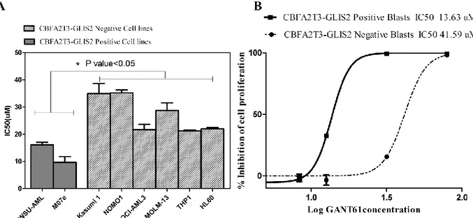
the viability through WST1 assay. Both cell lines showed a marked sensitivity to the inhibitor with a strong reduction of cell proliferation and IC50 ranging between 10 µM and 15 µM (Fig. 6A). In contrast, GANT61 had lower effect on AML cell lines that do not express the CBFA2T3-GLIS2 fusion, suggesting a specific effect of this inhibitor on the chimeric transcription factor. Similar results were obtained on primary leukemia cells isolated from AML patients (Fig.6B), where the IC50 of GLIS2-positive leukemia cells is 13.63 µM and that of other primary cells is 41.59 µM.
Figure 6. A) IC50 of CBFA2T3-GLIS2 and negative cell lines 72h after GANT61 exposure, B Dose-response curves
after 72h of GANT61 treatment of primary cells derived from patients with acute myeloid leukemia either positive or negative for CBFA2T3-GLIS2 fusion gene . *= p value < 0.05
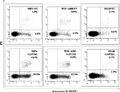
In order to further investigate the mechanism of action of the GANT61, we evaluated the induction of apoptosis in AML cell lines after 24h of treatment. The cytometric analysis indicated that the GANT61 efficiently induced apoptosis in the CBFA2T3-GLIS2-positive cells with an increase of about 30% of cells positive to Annexin V in comparison to the untreated cells (Fig. 7). On the other hand, the HL60 cell line, which lacks CBFA2T3-GLIS2 fusion gene, did not show any significant increase of Annexin V positive cells (Fig. 7).
30
Figure 7. Flow cytometric analysis of Annexin V-FITC/PI stained AML cell lines treated for 24 h with 20 µM of
GANT61. The percentages of early apoptotic cells (Annexin-V FITC+/PI−, lower right quadrant) and late apoptotic/necrotic cells (Annexin-V FITC+/PI+, upper right quadrant) are indicated. NT: sample treated with vehicle alone (DMSO).
Moreover, cell cycle analysis showed an increasing number of CBFA2T3-GLIS2-positive cells (WSU-AML and M07e) in G0/G1 phase, and a decrease number of the cells in S and G2/M phases (Fig. 8).
Figure 8. Cell cycle analysis. Flow cytometric analysis of PI-stained AML cell lines positive to CBFA2T3-GLIS2
31
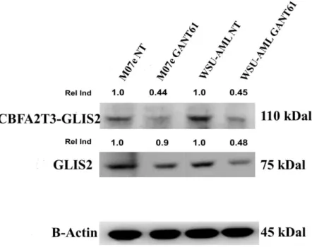
4.3 GANT61 downregulates expression of GLIS2 protein
Since in the classic Hedgehog signaling pathway several target genes involved in feedback mechanisms (HHIP, PTCH1, GLI1) have been described, we hypothesized that CBFA2T3-GLIS2 chimeric protein could regulate wild-type GLIS2 protein with the same feedback mechanism. Thus, we assessed the effect of GANT61 on the expression of GLIS2 protein in two cell lines (M07e, WSU-AML) using GLIS2 antibody. Western blotting analysis confirmed the presence of chimeric protein, but, unexpectedly, showed also the presence of wild-type GLIS2. These results may indicate the role of the fusion protein in regulating wild-type GLIS2. Following treatment, the expression of both proteins were decreased with respect to untreated samples, suggesting that the GANT61 treatment targeted not only CBFA2T3-GLIS2 fusion protein, but also wild-type GLIS2 expression (Fig. 9).
Figure 9. Western blot analysis showing the decrease of GLIS2 protein and CBFA2T3-GLIS chimeric protein in
samples treated with 20 µM Gant61. Thirty micrograms of protein were blotted to each lane. Antibody to β-actin served as a loading control. Molecular weights are indicated at right. The Relative Induction (Rel Ind) is the amount of protein present in treated samples relative to untreated cells after normalizing to β-Actin density. NT, untreated cells. One representative of three independent experiments is shown.
The known targets of GANT61 are GLI1 and GLI2 proteins, the final effectors of Hedgehog classical pathway. Therefore, as shown in figure 10 GLI1/2 proteins were expressed at low level in cell lines with GLIS2 translocation (M07e and WSU-AML) and the treatment with GANT61 did not affect their expression (Fig. 10).
32
Figure 10. Western blotting analysis of GLI1/2 protein in cell lines positive and negative for GLIS2 fusion gene treated
for 48h with 20 µM GANT61.
4.5 Treatment with GANT61 downregulates expression of HH and BMP signaling target genes
Since the expression of CBFA2T3-GLIS2 fusion has been reported to be associated with upregulation of both HH and BMP signaling, as well as with an increase of expression of target genes of these pathways, such as BMP2 and GLIS2, we performed a qPCR analysis to assess the molecular effects of GANT61. As shown in figure 11A, treatment with GANT61 in WSU-AML and M07e cell lines, as well as in primary leukemia cells from the patient harboring the CBFA2T3-GLIS2 fusion (AMKL1), led to a significant reduction of the expression of BMP2 and CBFA2T3-GLIS2. In order to fully characterize the effect of GANT61 treatment on whole transcriptome profile of CBFA2T3-GLIS2-positive cells, gene expression was assessed by microarray analysis. The expression profile confirmed the overexpression of GLIS2 and the activation of major Hedgehog mediators in untreated cell lines and primary leukemia cells expressing CBFA2T3-GLIS2. Moreover, following GANT61 treatment, the expression of target genes of CBFA2T3-GLIS2, such as CRISP3, GATA3, H2AFY or NCAM1 (CD56) was significantly downregulated (p<0.05) (Fig 11B). Besides demonstrating the inhibition of GLIS2 pathway, we were able to identify additional genes that were downregulated by the treatment with GANT61 (p<0.05) (Fig. 11B). Among them, genes involved in cell proliferation (KIF14, MELK, MCM10, NUF2), regulation of cell cycle (CCNA2, CDKN3, CDC7, PRC1), genomic stability (BRCA1, BRCA2), and epigenetic regulators namely DNMT1, DNMT3B were present.
33
Figure 11 A) Quantitative PCR of selected mediators of GLIS2 pathway after 48h treatment of GANT61. *= p value <
0.05 **= p value < 0.01 ***= p value < 0.001. B) Hierarchical clustering of genes differentially expressed between untreated and treated AML leukemia blast cells and cell lines, with a p value<0.05
4.6 CBFA2T3-GLIS2 chimeric protein regulates DNMT1 and DNMT3B expression
Gene expression analysis revealed downregulation of genes involved in epigenetic regulation, such as DNMT1 and DNMT3B. To assess if these genes could be direct transcriptional targets of
CBFA2T3-GLIS2, we performed chromatin immunoprecipitation (ChIP) analysis using a
CBFA2T3-specific antibody on WSU-AML and M07e cell lines. Quantitative PCR was then performed on immune-precipitated DNA using several primer pairs located in the proximal NCAM1, DNMT1 and DNMT3B promoter. Specific around fivefold enrichment was observed at the proximal promoter of these genes in WSU-AML and M07e cell lines expressing the CBFA2T3-GLIS2 fusion gene. These data suggest that the CBFA2T3-CBFA2T3-GLIS2 fusion protein directly binds to the proximal promoter of DNA methyltransferase genes, regulating positively their expression (Fig. 12).
34
Figure 12. ChIP analysis, performed on WSU-AML cell line has shown around five-fold enrichment of chimeric
protein on DNMT1, DNMT3B and NCAM1 promoter. *= p value < 0.05 **= p value < 0.01 ***= p value < 0.001.
4.7 iPSC engineered with CBFA2T3-GLIS2 maintain the pluripotent properties
To study the biological effect of the CBFA2T3-GLIS2 fusion gene in megakaryocytic cells we set up an induced pluripotent stem cells (iPSC) model.
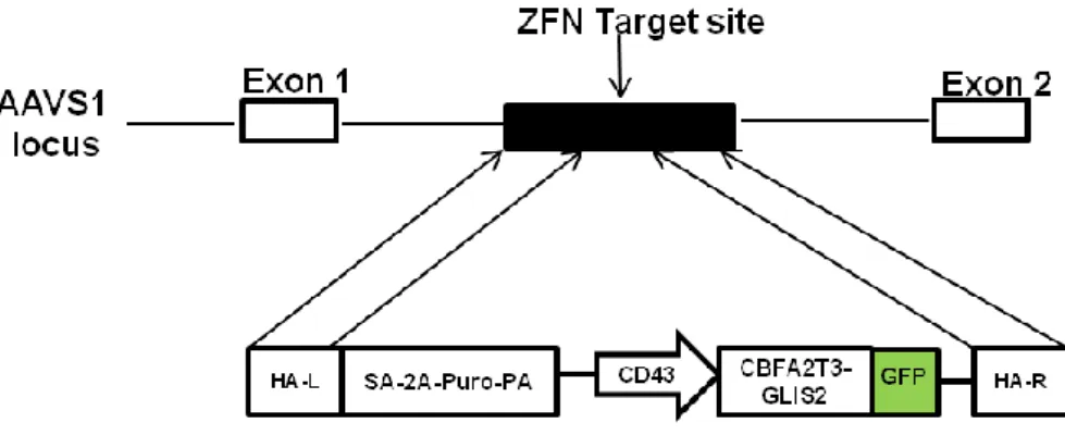
To this aim we used an iPSC derived from healtoy donor engineered with CBFA2T3-GLIS2 fusion gene in AAVS1 locus. First of all CBFA2T3-GLIS2 cDNA tagged with the green fluorescent protein (GFP) driven by human CD43 promoter was cloned in AAVS1 SA-2A-puro-pA donor and the fusion gene was introduced in AAVS1 locus through Zing finger mediated recombination (Fig 13).
35
Figure 13. Gene editing strategy. The constitutively active AAVS1 “safe harbor” locus is shown on the top line and the
targeting construct is shown below. cDNA expression cassettes driving expression of CBFA2T3-GLIS2 cDNA tagged with GFP under the CD43 promoter were inserted by zinc finger-mediated homologous recombination into intron 1 of AAVS1. HA, homologous arms left (L) and right (R); SA-2APuro-PA, puromycin drug resistance cassette.
After Puromycin selection we identified 23 clones with homozygous integration.
From iPSC wild-type and iPSC from clone 10 we induced Embryoid bodies development and spontaneous differentiation into the three embryonic layers. The embryoid bodies were maintained in culture and RNA was extracted after 17 days and 24 days to test expression of the three embryonic layers markers. The three embryonic layers were present in embryoid bodies as shown by expression of NeuroD1 for the neuroectoderm layer, Hand1 for mesoderm and GATA4 for endoderm (Figure 14). Moreover the expression of NANOG and Oct4, two markers of the pluripotent properties, decreased (Figure 14). These results demonstrated that iPSCs engineered with CBFA2T3-GLIS2 mantain pluripotent properties.
A B
Figure 14. Expression of genes specific for the three embryonic layers at day 17 (A) and at day 24 (B) after
36
4.8 Expression of CBFA2T3-GLIS2 in human hematopoietic cells obtained from iPSC recapitulates several molecular features of human pediatric AMKL
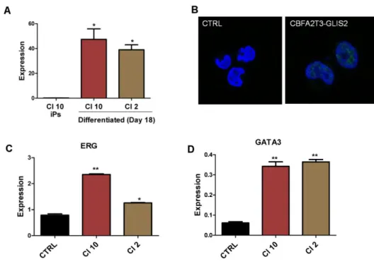
Three independent iPSC clones were amplified and subsequently differentiated into hematopoietic progenitors and then into megakaryocytes using a 2D co-culture system on matrix and compared to control iPSC. At day 18 of differentiation we collected the sovranatant containing hematologic progenitors and we extracted RNA to check in qPCR CBFA2T3-GLIS2 expression and several recently characterized direct targets of the fusion: ERG and GATA3 114.
CBFA2T3-GLIS2 was expressed in hematologic lineage in all clones tested, at the mRNA level
(Figure 15A). The fusion protein was also expressed as shown by expression of GFP in fluorescence microscopy (Figure. 15B). Furthermore the expression of the genes ERG and GATA3 were increased in CBFA2T3-GLIS2 clones (Figure 15C, 15D).
These results demonstrated that iPSC engineered with CBFA2T3-GLIS2 fusion gene reproduce the expression pattern of this leukemia subtype.
Figure 15. Expression pathway of hematopoietic cells derived from iPSC differentiation. (A) CBFA2T3-GLIS2, (B)
expression of CBFA2T3-GLIS2 protein tagged with GFP, (C) ERG, (D) GATA3. Statistical significance is indicated as p values * *p<0.01. *p<0.05.
37
4.9 CBFA2T3-GLIS2 induces aberrant differentiation in megakaryocytic cells
Hematopoietic differentiation of iPSC was then assessed at day 13, 15 and 18 using flow cytometry analysis for progenitors (CD34 and CD43) and megakaryocytic markers (CD41, CD42 and CD61). The CD43+ hematopoietic population in CBFA2T3-GLIS2 cells was composed of a higher percentage of CD41+CD42+ megakaryocytic cells than CTRL cells. Therefore we observed some strong different alterations in different populations of cells (Figure 16H), but in particular a progressive accumulation of an abnormal population expressing low level of CD41 and CD42 never detected in the control conditions. Furthermore the integrin CD61, normally associated with CD41 in a calcium-dependent heterodimeric complex, had lower expression on CBFA2T3-GLIS2 megakaryocytic cells compared to controls at day 15 (Figure 16 D, E) and at day 18 (Figure 16 F, G). The low expression of CD61 was more evident in the abnormal population CD41lowCD42low in comparison with CTRL megakaryocytic cells (Figure 16E, 16G).
Together these data indicated that CBFA2T3-GLIS2 enhances development of aberrant human megakaryocytic deregulating expression of markers CD41, CD42 and CD61.
38
Figure 16. A) Flow cytometry analysis of megakaryocytic markers (CD41,CD42, CD61) markers in CTRL and clone
expressing CBFA2T3-GLIS2 fusion gene at day 13, 15 (B,D,E) and 18 (C,F,G).
(D,E) Expression of CD61 at day 15 gated respectively in CD41+CD42+ and in CD41lowCD42low population. (F,G) Expression of CD61 at day 18 gated respectively in CD41+CD42+ and in CD41lowCD42low population.
(H) Percent number of different populations detected in flow cytometry analysis in hematopoietic cells derived from iPSC differentiation.
39
4.10 CBFA2T3-GLIS2 enhances proliferation and self-renewal of progenitors hematopoietic cells
To test the capacity of self-renewal of progenitor hematopoietic cells with CBFA2T3-GLIS2 we sorted the hematopoietic cells CD43+ at day 15 of differentiation and we cultured them in methylcellulose as well as in liquide culture enriched with specific hematopoietic cytokines.
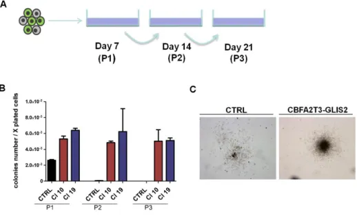
For methylcellulose we counted the colonies and replated the cells every seven days (Figure 17A). As early as at day 7 there were more colonies in CBFA2T3-GLIS2 cells than hematopoietic cells from CTRL (Figure 17B). Moreover the morphology of colonies in CBFA2T3-GLIS2 was more dense in number than CTRL probably due to the high proliferation induced by fusion gene (Figure 17C).
In the second replate, after 7 days the CTRL lost the capacity to make colonies while
CBFA2T3-GLIS2 cells maintained proliferation properties until forth passage (Figure 17B).
Figure 17. (A) Experimental design to test the self-renewal capacity in methylcellulose experiments (B) Ratio of
colonies number upon serial replating of CTRL and CBFA2T3-GLIS2 hematopoietic cells as means ± SEM (duplicate performed in 2 independent experiments). Cells were replated every 7 days for 4 weeks. (C) Morphology of colonies between P1 and P2. P1; passage 1; P2 passage 2; P3 passage 3.
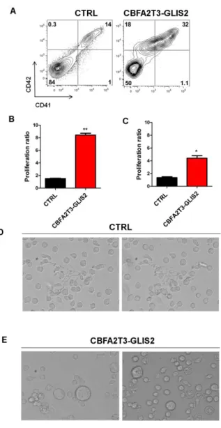
As the methylcellulose culture we tested megakaryocytic markers and proliferation properties also in liquid cultured. To this aim we maintained CD43+ hematopoietic cells in culture for 4 days and
40 10 days. As shown in figure 18 we confirmed the presence of the aberrant megakaryocytic population with low expression of CD41 and CD42 markers (Figure 18A). Moreover the proliferation of CBFA2T3-GLIS2 hematopoietic cells was clearly higher than hematopoietic CTRL cells (Figure 18 B, C). Furthermore preliminary morphology studies demonstrated that in liquid culture only the CTRL cells were induced to acquire pro-platelet morphology as shown in Figure 18D (Figure 18D).
These results indicated that this fusion gene is sufficient to increase cell proliferation, self-renewal properties and block of platelets differentiation in-vitro.
Figure 18. (A) Expression of CD41 and CD42 in hematopoietic cells derived from iPSC differentiation (B) Ratio
number of CD43+ hematopoietic cells after 4 days and (C)10 days of sorting . (D) Morphology analysis of CD43+ hematopoietic cells in the CTRL and in the (E) CBFA2T3-GLIS2 cells . Statistical significance is indicated as p values (Student’s t-test): *p<0.05.
41
4.11 The GANT61 inhibits the proliferation of CBFA2T3-GLIS2 hematopoietic cells derived from iPSC differentiation
To test the capacity of GANT61 to inhibit proliferation we treated hematopoietic cells with or without CBFA2T3-GLIS2 in liquid culture. We exposed the hematopoietic cells at low concentration of GANT61. The number of cells after 48h of treatment revealed that GANT61 inhibited proliferation of hematopoietic cells with CBFA2T3-GLIS2 expression. In particular at 2uM of GANT61 concentration the mortality of cells with fusion gene was around 50% in comparison with any effect in the CTRL (Figure 19).
This result suggests a possible effect of the GANT61 in this particular subgroup of AMKL.
Figure 19. Absolute number of CTRL and CBFA2T3-GLIS2 hematopoietic cells treated with different concentrations of
42
5. Conclusion and Discussion
Despite improvements in patient risk stratification and definition of prognosis achieved through the identification of novel genetic and epigenetic alterations in pediatric AML, the majority of children with this disease are still treated with intensive multi-agent chemotherapy. Treatment resistance, occurrence of relapse, as well as short-term and long-term therapy-related side effects, highlight the need for new targeted therapies. Recently, the identification of the cryptic chromosomal inversion of chromosome 16, leading to the fusion between CBFA2T3 and GLIS2 genes, defined a novel subgroup of childhood AML with an adverse outcome 115, 116, 118. Indeed, while several other groups of AML and subgroups of AMKL require cooperating mutations to induce fully leukemia,
CBFA2T3-GLIS2 imposes leukemic properties by directly regulating ERG, GATA1, in a single
oncogenic hit. This idea is also supported by the lower genetic burden of CBFA2T3-GLIS2 leukemic cells 118, and could also explain the dismal prognosis. More importantly, gene expression profiling of patients harboring this fusion gene demonstrated an upregulation of HH signaling 115, 143
. Emerging evidences are supporting the role of HH pathway deregulation in the pathogenesis and chemo-sensitivity of AML and myelodysplastic syndrome 132, 144. For this reason HH pathway inhibitors could be envisaged as agents able to potentiate the effect of traditional drugs and to restore chemo-sensitivity. GANT61 is a small molecule inhibiting DNA binding of GLI family proteins, and it is largely used in preclinical studies 132, 137. In order to investigate the possible benefit of GANT61 in CBFA2T3-GLIS2-positive leukemia, we approached different in-vitro preclinical models. We demonstrated that CBFA2T3-GLIS2-postive cells were more sensitive to GANT61 than the cell lines without CBFA2T3-GLIS2. Moreover, GANT61 had a marked effect on cell proliferation and in inducing the apoptosis only on CBFA2T3-GLIS2-positive cells, suggesting that the effect of the drug derived from specific inhibition of the chimeric transcriptional factor
CBFA2T3-GLIS2. Interestingly, similar results were obtained also in primary blast cells from a
AMKL patient harboring CBFA2T3-GLIS2fusion gene.
To define the implication of GANT61 treatment on expression of HH signaling target genes, gene expression profiling and qPCR of specific genes have been performed on untreated and treated
CBFA2T3-GLIS2-positive cells. The Expression of CRISP3, GATA3, ERG or H2FAY, known target
genes of CBFA2T3-GLIS2 pathway, after the treatment with GANT61 was lower than in untreated cells, suggesting specific inhibition of GLIS2 transcription factor. Beside the inhibition of HH pathway, we were able to identify additional genes implied in cell proliferation, cell cycle regulation and DNA methylation that were significantly downregulated after treating the cells with GANT61. Of particular relevance, two genes involved in chromatin remodeling and DNA