Open Access
Omic Approach in Non-smoker Female with Lung Squamous Cell
Carcinoma Pinpoints to Germline Susceptibility and Personalized
Medicine
Original Article
Purpose
Lung cancer is strongly associated to tobacco smoking. However, global statistics estimate that in females the proportion of lung cancer cases that is unrelated to tobacco smoking reaches fifty percent, making questionable the etiology of the disease.
Materials and Methods
A never-smoker female with primary EGFR/KRAS/ALK-negative squamous cell carcinoma of the lung and their normal sibs were subjected to a novel integrative “omic” approach using a pedigree-based model for discovering genetic factors leading to cancer in the absence of well-known environmental trigger. A first-step whole-exome sequencing on tumor and normal tissue did not identify mutations in known driver genes. Building on the idea of a germline oligogenic origin of lung cancer, we performed whole-exome sequencing of DNA from patients’ peripheral blood and their unaffected sibs. Finally, RNA-sequencing analysis in tumoral and matched non-tumoral tissues was carried out in order to investigate the clonal profile and the pathogenic role of the identified variants.
Results
Filtering for rare variants with Combined Annotation Dependent Depletion (CADD) > 25 and potentially damaging effect, we identified rare/private germline deleterious variants in 11 cancer-associated genes, none of which, except one, shared with the healthy sib, pinpointing to a “private” oligogenic germline signature. Noteworthy, among these, two mutated genes, namely ACACA and DEPTOR, turned to be potential targets for therapy because related to known drivers, such as BRCA1 and EGFR.
Conclusion
In the era of precision medicine, this report emphasizes the importance of an “omic” approach to uncover oligogenic germline signature underlying cancer development and to identify suitable therapeutic targets as well.
Key words
Exome, Squamous cell carcinoma,
High-throughput nucleotide sequencing, Disease susceptibility, Multifactorial inheritance, Precision medicine
Margherita Baldassarri,
MD1,2Chiara Fallerini,
PhD1Francesco Cetta,
MD3Marco Ghisalberti,
MD4Cristiana Bellan,
MD5Simone Furini,
PhD6Ottavia Spiga,
PhD7Sergio Crispino,
MD8Giuseppe Gotti,
MD4Francesca Ariani,
PhD1,2Piero Paladini,
MD4Alessandra Renieri,
MD, PhD1,2Elisa Frullanti,
PhD1 + + + + + + + + + + + + + + + + + + + + + + + + + + + + + + + + + + + + + + + + + + + + + + + + + + + + + + + + + + + + + + + + + + + + + + + + + + + + + + + + + + + + + + + + + + + + + + + + + + + + + + + + + + + + + + + + + + + + + + + + + + + + + + + + + + + + + + + + + + + + + + + + + + + + + + + + + + + + + + + + + + + + + + + + + + + + + + + + + + + + + + + + + + + + + + + + + + + + + + + + + + + + + + + + + + + + + + + + + + + + + + + + + + + + + + + + + + + + + + + + + + + + + + + + + + + + + + + + + + + + + + + + + + + + + + + + + + + + + + + + + + + + + + + + + + + + + + + + + + + + Correspondence: Alessandra Renieri, MD, PhD Medical Genetics Unit, University of Siena, Azienda Ospedaliera Universitaria Senese, Viale Bracci, 2 - 53100 Siena, Italy Tel: 39-0577-233303Fax: 39-0577-233325
E-mail: [email protected] Received March 9, 2017 Accepted April 25, 2017
*Margherita Baldassarri and Chiara Fallerini contributed equally to this work.
1Medical Genetics Unit, University of Siena,
Siena, 2Genetica Medica, Azienda
Ospedaliera Universitaria Senese, Siena,
3IRCCS MultiMedica, Milan, 4Thoracic Surgery Unit, Azienda
Ospedaliera Universitaria Senese, Siena,
5Section of Pathology, Department of
Medical Biotechnology, Departments of
6Medical Biotechnology and 7Biotechnology,
Chemistry and Pharmacy, University of Siena, Siena, 8Medical Oncology Unit,
Introduction
Lung cancer is an important public health problem and the leading cause of cancer death worldwide [1]. The two major histological types of lung cancer are non-small cell lung can-cers (NSCLC) and small cell lung cancan-cers. NSCLCs consist mostly of lung adenocarcinomas (ADCA) and lung squa-mous cell carcinomas (SQCC). These two NSCLC subtypes have both unique and common clinical and histopathological characteristics. For example, ADCAs are mainly observed in never smokers [2] while SQCC is the most frequent histolog-ical type among smoking patients.
The discovery of common, mutually exclusive, driver genes, epidermal growth factor receptor (EGFR), KRAS proto-oncogene GTPase (KRAS) and anaplastic lymphoma receptor tyrosine kinase (ALK), that represent crucial thera-peutic targets, led to a remarkable improvement in NSCLC therapy. However, only about 50% of NSCLC acquire muta-tions in these genes. Identification of new cancer driver genes currently is a top research priority. Recent studies using next-generation sequencing (NGS) approach characterized NSCLC tissues in order to identify new less common candi-date driver mutations [3,4] highlighting the presence of great variability even within tumor samples belonging to the same histological type.
Up to now, application of NGS technology has been reported in many lung cancer samples leading to the discov-ery of a large set of new somatic mutations in key cancer genes. Nevertheless, no studies are present in literature that performed whole exome sequencing (WES) and RNA-sequencing (RNA-seq) from tumoral and matched non-tumoral lung tissues integrating these with WES of blood DNA from the same individual to identify possible tumor-driver variants which are already present in the germline background and that may predispose to lung cancer. Using WES analysis in a model of cases and controls deriving from the same kindred—discordant sib-pair design—we have recently showed that a set of oligogenic germline mutations, that are detectable in the peripheral blood, may play a role in the development and growth of lung ADCA in never smokers [5]. Noteworthy, our previous results demonstrated that the oligogenic cancer predisposing signature is a “pri-vate signature,” varying from one patient to another.
Herein, we tested further the hypothesis of the oligogenic origin of lung cancer in a never-smoker female who devel-oped a SQCC of the lung. We applied for the first time an integrative “omic” approach of WES and RNA-seq experi-ments in a discordant sib-pair design.
Materials and Methods
1. Samples and DNA/RNA extractionPatient and sib signed the informed consent declaration at the Medical Genetics Unit of the Azienda Ospedaliera Uni-versitaria Senese, Italy, for the use of their biological samples and clinical data for research purposes. The study protocol was approved by the Ethical Committee of the Azienda Ospedaliera Universitaria Senese. Information on histologi-cal diagnosis (by the Pathology Unit) was retrieved from the clinical records. Genomic DNA of patient and healthy sib was isolated from EDTA peripheral blood using QIAamp DNA Blood Kit (Qiagen, Hilden, Germany) according to the manufacturer’s protocol. Formalin-fixed paraffin-embedded (FFPE) samples of patient’s tumoral and non-tumoral lung tissues were obtained from the Pathology Unit of the recruit-ing hospital and analyzed through WES and RNA-seq. DNA was extracted from FFPE lung tumoral and non-tumoral tis-sue samples using MagCore Genomic DNA FFPE One-Step Kit for MagCore System (Diatech Pharmacogenetics s.r.l., Ancona, Italy). RNA was extracted from FFPE lung tumoral and non-tumoral tissue samples of our case using High Pure FFPE-Tissue RNA Isolation Kit (Roche, Basel, Switzerland) following the manufacturer’s instructions. Additional RNAs from three FFPE lung non-tumoral tissues from other lung cancer patients were isolated using the same kit and used as control tissues. RNA samples were processed to remove rRNA using Ribo-Zero rRNA Removal Kit for Human sam-ples (Illumina, Grand Island, NY) following the manufac-turer’s instructions. RNA integrity was verified using the Agilent Eukaryote Total RNA Nano Kit (Agilent Technolo-gies, Palo Alto, CA) on Agilent2100 Bioanalyzer (Agilent Technologies). Both DNA and RNA were quantified by spectrophotometry (ND-2000c, NanoDrop Products, Wilm-ington, DE) and Qubit Fluorometer with Qubit dsDNA HS Assay and Qubit RNA HS Assay Kits (Life Technologies, Carlsbad, CA), respectively.
2. WES and data analysis
WES was performed using the Life Technologies Ion Pro-ton sequencer (Life Technologies) on genomic DNA samples of cases and controls and tumor tissues as previously described [6]. Variant were then annotated against external datasets, including 1000 genomes (http://www.1000geno-mes.org/) and dbSNP. After removing variants with low coverage, in order to identify candidate susceptibility vari-ants, we selected for rare variants with minor allele fre-quency [MAF] ≤ 0.01 or not reported (S1 Fig.). We then filtered excluding variants with clinical significance as
“benign” or “likely benign” and present in an in-house data-base of variants. Additional filtering procedures were thus implemented for retrieving exonic rare variants with a potential detrimental impact on protein function, i.e., vari-ants, insertions and deletions (indels) causing exonic frameshifts and missense variants predicted as deleterious applying the Combined Annotation Dependent Depletion (CADD > 25) and the MetaSVM (Support Vector Machine) bioinformatics tools. Genes affected by mutations were checked in literature in order to identify cancer-related genes. Validation of these variants was carried out using custom NGS panel for Ion PGM sequencer (Life Technologies). 3. RNA-seq and data analysis
RNA-seq was performed using Illumina HiSeq2500 plat-form (Illumina Inc., San Diego, CA), in a 2×100 bp paired-end configuration in High Output mode (V4 chemistry), with a total of at least 250 million reads per lane. After quality check, RNA (50 ng) was used to prepare sample libraries. Sequencing library construction included these steps: library construction using Illumina TruSeq RNA Sample Pre Kit (Illumina), library purification using Beckman AMPure XP beads (Beckman Coulter s.r.l., Milan, Italy), insert fragments test using Agilent High Sensitivity DNA Kit on Agilent 2100 Bioanalyzer (Agilent Technologies), and cBOT automatic cluster (TruSeq PE Cluster Kit v3-cBotHS). Post-library qual-ity controls were performed using the Agilent RNA 6000 Nano kit (Agilent Technologies) on Agilent 2100 Bioanalyzer (Agilent Technologies) and Qubit Fluorometer (Life Tech-nologies). Libraries were then loaded on HiSeq2500 sequenc-ing platform (Illumina) and sequenced ussequenc-ing 2×100 bp pair-end High Output Mode (V4 chemistry) per lane. The reads generated on the HiSeq2500 were provided under FASTQ format.
Sequence reads in FASTQ format were processing using the Fastqc software (http://www.bioinformatics.babraham. ac.uk/projects/fastqc/) for data quality check and removing excess adaptors to get high-quality and clean reads. The high-quality reads were aligned to the GRCh38/hg38 refer-ence human genome (ftp://jgi-psf.org/pub/compgen/phy-to zome/v9.0/Ptrichocarpa/assembly/Ptrichocarpa_210.fa. gz) using the TopHat software ver. 2.0.9 [7]. Transcript assembling and expression quantification were carried out using Cufflinks ver. 2 [8]. Gene expression was expressed as fragments per kilo-base transcript per million mapped reads (FPKM) value. This normalized value was used for visuali-zation on a genome browser (http://genome.ucsc.edu/), as well as to compare read coverage between and throughout different genes. Statistical analysis was performed to com-pute the mean FPKM level with the associated p-value for lung normal tissues together with the mean FPKM level with
the associated p-value for lung cancer tissues. Cuffdiff tool from Cufflinks was used to identify differentially expressed genes [9]. Gene Ontology and pathways analyses were per-formed using both Enrichr (http://amp.pharm.mssm.edu/ Enrichr/) and Database for Annotation, Visualization and Integrated Discovery functional annotation tool (DAVID Bioinformatics Resources 6.7, https://david.ncifcrf.gov).
Results
1. Patient description
A still alive Caucasian female patient, who developed SQCC of the lung in the absence of smoking habit and/or asbestos exposure, was recruited. Her smoker father died at age 63 from lung cancer. She had no known other family history of cancers. At age of 77, after a “screening” chest and abdomen computed tomography scan examination, a 12×7 mm nodule was found in the dorsal segment of the right superior lobe in association with a small “ground glass” area (9.5 mm in diam-eter) in the left superior lobe. The nodule showed a small vol-umetric increase after 3 months and the patient was scheduled for surgery at the Thoracic Surgery Unit of Azienda Ospe-daliera Universitaria Senese, Italy. Intraoperative diagnosis was NSCLC. At gross pathologic examination, a 12×0.5 solid nodule with irregular margin was detected. Histologic exam-ination showed SQCC with a mainly perialveolar and perivas-cular growth pattern. Tumoral cells were p63+, cytokeratin (CK) 5/6+, CK7‒, thyroid transcription factor 1‒, synapto-physin‒, chromogranin‒. Ki67 immunostaining was 20 per cent. There was moderate infiltration of the visceral pleura, but no lymphonodal infiltration. Two years before, patient had undergone to removal of a cutaneous nodule located in the left side of the neck that resulted to be a SQCC of the basal subtype. She subsequently underwent left cervical phadenectomy, that showed infiltration of three cervical lym-phonodes. On the basis of histological analysis, site of tumors and type of lymphonodal involvement, it was stated that the patient had two different primary tumors, both belonging to the squamous subtype. The patient is alive and well 4 years after the first and 2 years after the second operation. 2. “Omic” characterization
We firstly carried out whole-exome sequencing of gDNA from FFPE tumoral and non-tumoral lung tissue. With the aim of identification of somatic driver mutations, we subtracted the germline background to 39,286 variants present in the tumoral lung tissue, leading to 2,543 exonic variants present
only in the tumor. Among them, 1,381 were missense variants and 254 were truncating (S2A Fig.). We then extracted the somatic mutational signature according to base substitutions, as described by Alexandrov et al. (2013) [10]. This analysis showed a predominance of C>T transitions in tumoral lung tissue, not corresponding to the specific cancer signature related to tobacco consumption that are characterized by pre-dominance of C>A transversions (S2B Fig.). Pathway analysis showed that mutations observed in SQCC tissue mostly affected genes involved in extracellular matrix organization (p=0.005), transmembrane transport of small molecules (p=0.010) and collagen formation (p=0.034) pathway, as
pre-viously reported (S2C Fig.) [4]. Overall, no mutations in the known driver genes, such as EGFR, KRAS, AKT, ROS1, nor in other putative driver genes described by Campbell et al. [3] were present in our tumoral sample [3]. EML4-ALK fusion gene was also excluded. The failure of identification of somatic driver mutations rose the hypothesis that germline variants may have a role in etiology of the disease.
We then adopted a novel integrative “omic” approach using a combination of NGS techniques in a pedigree model that compares discordant sibs to test the hypothesis of oligogenic origin of lung cancer (Fig. 1).
We carried out whole-exome sequencing of gDNA from Fig. 1. Flowchart illustrating the study strategy and result. WES, whole exome sequencing; EGFR, epidermal growth factor receptor; CADD, Combined Annotation Dependent Depletion; RNA-seq, RNA sequencing.
WES in tumoral vs. normal tissue
No EGFR, KRAS mutations No mutations in other putative driver genes
(Campbell et al. [3])
WES patient vs. healthy sib in blood
19 cancer genes with CADD > 25 or truncating mutations
RNA-seq in tumoral vs. normal tissue
11 with altered expression
1 BRCA1 pathway 1 EGFR pathway 9 other pathway
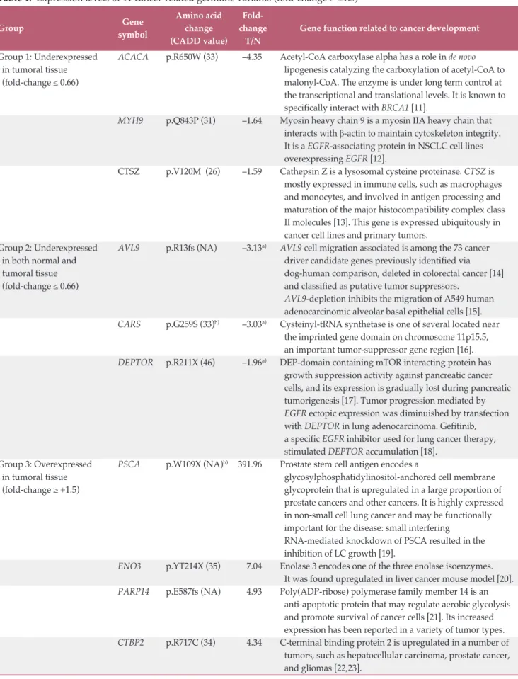
Table 1. Expression levels of 11 cancer-related germline variants (fold-change > ±1.5)
Gene Amino acid Fold-
Group
symbol change change Gene function related to cancer development
(CADD value) T/N
Group 1: Underexpressed ACACA p.R650W (33) –4.35 Acetyl-CoA carboxylase alpha has a role in de novo
in tumoral tissue lipogenesis catalyzing the carboxylation of acetyl-CoA to
(fold-change ≤ 0.66) malonyl-CoA. The enzyme is under long term control at
the transcriptional and translational levels. It is known to specifically interact with BRCA1 [11].
MYH9 p.Q843P (31) –1.64 Myosin heavy chain 9 is a myosin IIA heavy chain that interacts with β-actin to maintain cytoskeleton integrity. It is a EGFR-associating protein in NSCLC cell lines overexpressing EGFR [12].
CTSZ p.V120M (26) –1.59 Cathepsin Z is a lysosomal cysteine proteinase. CTSZ is mostly expressed in immune cells, such as macrophages and monocytes, and involved in antigen processing and maturation of the major histocompatibility complex class II molecules [13]. This gene is expressed ubiquitously in cancer cell lines and primary tumors.
Group 2: Underexpressed AVL9 p.R13fs (NA) –3.13a) AVL9 cell migration associated is among the 73 cancer
in both normal and driver candidate genes previously identified via
tumoral tissue dog-human comparison, deleted in colorectal cancer [14]
(fold-change ≤ 0.66) and classified as putative tumor suppressors.
AVL9-depletion inhibits the migration of A549 human
adenocarcinomic alveolar basal epithelial cells [15].
CARS p.G259S (33)b) –3.03a) Cysteinyl-tRNA synthetase is one of several located near the imprinted gene domain on chromosome 11p15.5, an important tumor-suppressor gene region [16].
DEPTOR p.R211X (46) –1.96a) DEP-domain containing mTOR interacting protein has growth suppression activity against pancreatic cancer cells, and its expression is gradually lost during pancreatic tumorigenesis [17]. Tumor progression mediated by
EGFR ectopic expression was diminuished by transfection
with DEPTOR in lung adenocarcinoma. Gefitinib, a specific EGFR inhibitor used for lung cancer therapy, stimulated DEPTOR accumulation [18].
Group 3: Overexpressed PSCA p.W109X (NA)b) 391.96 Prostate stem cell antigen encodes a
in tumoral tissue glycosylphosphatidylinositol-anchored cell membrane
(fold-change ≥ +1.5) glycoprotein that is upregulated in a large proportion of prostate cancers and other cancers. It is highly expressed in non-small cell lung cancer and may be functionally important for the disease: small interfering
RNA-mediated knockdown of PSCA resulted in the inhibition of LC growth [19].
ENO3 p.YT214X (35) 7.04 Enolase 3 encodes one of the three enolase isoenzymes. It was found upregulated in liver cancer mouse model [20].
PARP14 p.E587fs (NA) 4.93 Poly(ADP-ribose) polymerase family member 14 is an anti-apoptotic protein that may regulate aerobic glycolysis and promote survival of cancer cells [21]. Its increased expression has been reported in a variety of tumor types.
CTBP2 p.R717C (34) 4.34 C-terminal binding protein 2 is upregulated in a number of tumors, such as hepatocellular carcinoma, prostate cancer, and gliomas [22,23].
peripheral blood of the SQCC patient and her unaffected sib as controls (S3 Table). Overall, we identified 37,727 germline variants in the patient and 37,355 germline variants in the healthy sib and 39,286 variants in the tumoral lung tissue.
To identify susceptibility variants, we compared case and control WES data and selected for rare variants (MAF ≤ 0.01) with a potentially damaging effect: truncating variants, i.e., insertions and deletions (indels) causing exonic frameshifts and nonsense mutation, and missense deleterious variants predicted with CADD > 25 and filtering out those with MetaSVM as “tolerated” (S1 Fig.). We obtained 111 potential deleterious variants of which 40 were shared by both siblings. Among the 111 potentially deleterious variants, 19 variants mapped in genes that have been found previously associated with cancer of the lung or other tissues (S4 Table). Validation of these variants led to the confirmation of all variants that were probably responsible for lung cancer susceptibility in our case. All the sequence variants identified were in the heterozy-gous state and were also present in the relative lung tumor tis-sue in heterozygous state as expected.
Finally, we performed RNA-seq analysis of RNA extracted from both FFPE tumoral and non-tumoral lung tissues of our patient in order to detect differences in the expression level that could help the understanding of the functional role of germline variants. RNA-seq generated a mean of 75,134,860 reads per sample (S5 Table).
To assess the potential functional role of the 19 germline cancer-related variants identified by whole-exome sequencing in the patient and predicted as deleterious by bioinformatics tools, we combined results from WES experiments with the respective-gene expression profile from RNA-seq of
non-tumoral and matched non-tumoral tissue. In 19 variants mapping in 19 genes, RNA-seq data showed differential expression level (fold-change > ±1.5) for 11 genes, reinforcing the patho-genic role of the identified variants showing three different effects (Table 1). Three genes, namely ACACA, CTSZ, and
MYH9, showed downregulation in lung cancer compared to
normal tissue. Three genes, namely DEPTOR, AVL9, and
CARS, showed downregulation of both normal and cancer
tis-sue when we compared those tistis-sues with a pool of three nor-mal lung tissues. Lastly, five remaining genes (PSCA, ENO3,
PARP14, CTBP2, FOXM1) showed an upregulation in lung
cancer tissue. All the above mentioned variants were exclusive of the patient with the exception of variant in FOXM1 gene that was present also in the healthy sib.
Among the 11 germline-mutated cancer-related genes, two genes, namely ACACA and DEPTOR, are known to be associ-ated with the known driver genes, BRCA1 and EGFR, respec-tively. In particular, we found a missense variant, that has been predicted as deleterious, c.C1948T, p.Arg650Trp (NM_ 198837.1, exon 16) in ACACA gene (OMIM* 200350) encoding Acetyl-CoA carboxylase alpha, the rate-limiting enzyme for the long-chain fatty acid synthesis [25]. The other mutation is a stopgain variant c.A631T, p.Arg211* in DEPTOR gene (OMIM *612974) encoding DEP domain-containing MTOR-interacting protein.
Interestingly, from RNA-seq data, we found a decreased expression level of ACACA in tumoral rather that in non-tumoral tissue. Sequence information was extracted from RNA-seq to confirm the presence of mutated mRNA and we found that 81.8% of reads carried the mutated allele pinpoint-ing to a loss of heterozygosity (LOH) effect. The same analysis Table 1. Continued
Gene Amino acid Fold-
Group
symbol change change Gene function related to cancer development
(CADD value) T/N
FOXM1 p.P658L (27)b) 3.28 Forkhead box M1 is a transcriptional activator involved in cell proliferation. Consistent with an important role of Foxm1 in cell cycle progression, increased expression of Foxm1 was found in many human tumors. Increased Foxm1 expression in human lung adenocarcinomas and squamous cell carcinomas was associated with increased proliferation of tumor cells [24].
CADD, Combined Annotation Dependent Depletion; T, tumoral lung tissue; N, non-tumoral lung tissue; ACACA, acetyl-CoA carboxylase alpha; EGFR, epidermal growth factor receptor; NSCLC, non-small cell lung cancers; mTOR, mammalian target of rapamycin; NA, not applicable; LC, lung cancer; MAF, minor allele frequency. a)Fold-change calculated by comparing expression levels of normal and tumor tissues with those of a pool of normal lung tissues, b)All listed variants were private except those in CARS (MAF=0.002), PSCA (MAF=0.0076), and FOXM1 (MAF=0.0044) genes.
revealed a decreased expression level of DEPTOR both in nor-mal and tumoral tissue at mRNA level pinpointing to LOH effect already present in the surrounding normal tissue.
Discussion
Among 111 potentially deleterious germline variants, 19 mapped in genes that have previously been associated with cancer of the lung or other tissues (S3 Table). Using the dis-cordant-sib pair design, we confirmed the presence of an oli-gogenic signature present in the patient’s genetic back-ground [5]. In fact, only one out of 11 cancer-related variants was also found in the healthy sib. For all the 11 variants, RNA-seq data reinforced the pathogenic role showing three different effects on the expression levels of harboring gene: three genes (ACACA, CTSZ, MYH9) showed a possible “sec-ond hit” in tumor suppressor genes responsible for gene inactivation; three genes (AVL9, DEPTOR, CARS) showed a possible transcript instability occurring in both tumoral and normal mutation-bearing tissues; five genes (PSCA, ENO3,
PARP14, CTBP2, FOXM1) were upregulated in lung cancer
tissue confirming previous published results and suggesting their role as oncogene. No mutations or gene expression
alteration in genes belonging to the Hedgehog pathway, mainly involved in basal cell carcinoma development, were found in our case.
The oligogenic combination of cancer predisposing genes found in our patient supports the model of “private genetic epidemiology” for lung cancer susceptibility [5]. In fact, we found that 11 cancer-related genes were mutated in the patient and only one in healthy sib (Fig. 2), suggesting that the oligogenic control of lung cancer susceptibility is due to a peculiar signature of multiple germline mutations in can-cer-related genes probably with a dosage effect. This per-sonal signature may play a role in the development and growth of lung cancer and, therefore explain the non-hered-itability of the condition, especially in non-smokers patients.
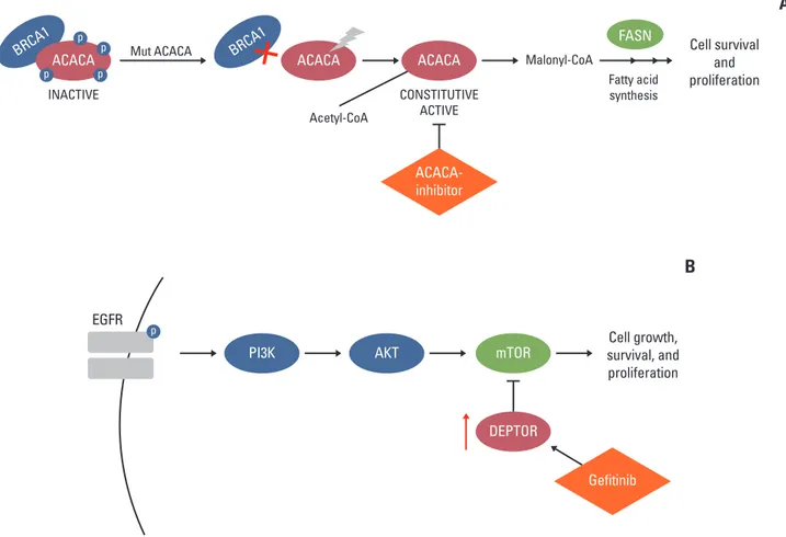
Focusing our analysis on genes that have already been reported in association with known druggable target genes— and likely having a major role in driving tumor development in our case—, we identified two possible suitable targets. Acetyl-CoA carboxylase alpha (ACACA) is a key enzyme in de novo lipogenesis catalyzing the carboxylation of acetyl-CoA to malonyl-acetyl-CoA. ACACA is known to be one of the major BRCA1-interacting protein [11]. Cancer cells strongly need to de novo synthesize fatty acid for membrane and energy production [26] and interaction between ACACA and BRCA1 is likely to be important for BRCA1-mediated tumor suppressor activity [27]. BRCA1 indeed binds the phospho-rylated and inactive form of ACACA and this interaction blocks ACACA activity by preventing its dephosphorylation [14]. The identified variant c.C1948T, p.Arg650Trp is outside the catalytic domain but may have a role in folding of domain conformation determining a different exposure for BRCA1 binding. Based on the evidence that in our tumoral tissue only the mutated transcript is present, we can assume that, even if protein level is decreased, the isoform loses neg-ative control by BRCA1 and remains active. In this case, patient could potentially benefit from a therapy with ACACA inhibitors such as miR-195 [28] or soraphen A [29] (Fig. 3A).
The second druggable variant maps in DEPTOR gene, whose loss determines the activation of mammalian target of rapamycin (mTOR) cascade and promotes cell growth and survival. It is known that DEPTOR mainly acts as a tumor suppressor [17,30]. A recent study in lung cancer demon-strated that activation of EGFR signaling resulted in down-regulation of DEPTOR expression. In the same study, tumor progression mediated by EGFR ectopic expression was dimi-nished by transfection with DEPTOR in lung adenocarci-noma cell lines and that treatment with gefitinib, an EGFR tyrosine kinase inhibitor, stimulated DEPTOR expression leading to a significantly decreased cell proliferation, migra-tion and invasion [18]. In line with these results, we observed an increased expression level of DEPTOR in normal and Fig. 2. Pedigree of the two sib-pairs and representation of
disruptive mutations in cancer-related genes found in the patient and in her unaffected sib.
Patient
Age at diagnosis: 77 yr Age at sampling: 76 yrHealthy sib
ACACA : p.R650W CTSZ : p.V120M MYH9 : p.Q843P AVL9 : p.R13fs DEPTOR : p.R211X CARS : p.G259S PSCA : p.W109X ENO3 : p.YT214X PARP14 : p.E587fs CTBP2 : p.R717C FOXM1 : p.P658L FOXM1 : p.P658L
tumoral tissue, suggesting an enhanced predisposition of activation of mTOR signaling and thus a promotion of tumor progression (Fig. 3B). These results suggested the possibility to use gefitinib in our patient in order to restore DEPTOR level and consequently suppress cell growth and survival.
In summary, we suggest that both “primary” SQCC are triggered by the presence of an oligogenic germline signature consisting of at least 11 mutations, among which two lead to the activation of mTOR and BRCA1. Thus, this finding opens up the possibility of treating one of them with an already commercially available drug and the other with developing molecules.
This study has a number of limitations. First, alterations in non-coding regions and copy number variants would have been missed by our approach. Another limitation is that we focused only on cancer-associated genes, but we cannot exclude that non-cancer genes, in particular those involved in immune system or drug metabolism, could play a role in
cancer development. Finally, additional functional studies will be required to translate our results into effective person-alized therapy.
The present report provides evidence that an “omic” sequencing approach in cancer patient is feasible, and could impact on treatment decisions, namely in patients not suit-able for conventional therapy. In the present era of “precision medicine”, our study demonstrates the utility of applying this comprehensive sequencing protocol in order to effec-tively identify possible therapeutic targets enabling person-alized therapies.
Electronic Supplementary Material
Supplementary materials are available at Cancer Research and Treatment website (http://www.e-crt.org).
Fig. 3. Likely personal germline driver genes in present case and possible therapeutic options. ACACA (A) and DEPTOR (B) pathway involvement. ACACA, acetyl-CoA carboxylase alpha; EGFR, epidermal growth factor receptor; PI3K, phospho-inositide 3-kinase; mTOR, mammalian target of rapamycin.
BRCA1ACACA BRCA1 ACACA Cell survival
and proliferation ACACA DEPTOR EGFR FASN p p p p INACTIVE CONSTITUTIVE ACTIVE Fatty acid synthesis
Mut ACACA Malonyl-CoA
Acetyl-CoA
A
Cell growth, survival, and proliferation mTORB
AKT PI3K ACACA-inhibitor Gefitinib pConflicts of Interest
Conflict of interest relevant to this article was not reported.
Acknowledgments
This work was supported by grant from Regione Toscana–Istituto Toscano Tumori (ITT) and from ASSO (Associazione per lo Svi-luppo della Scienza Oncologica) onlus. The authors thank Fon-dazione Umberto Veronesi for its support to E.F. through the Post-Doctoral Fellowships 2015. The funders had no role in the design or conduct of the study, in the collection, analysis or inter-pretation of the data, or in the preparation, review or approval of the manuscript.
1. Stewart BW, Wild CP. World cancer report 2014. Lyon: Inter-national Agency for Research on Cancer; 2014.
2. Samet JM, Avila-Tang E, Boffetta P, Hannan LM, Olivo-Marston S, Thun MJ, et al. Lung cancer in never smokers: clin-ical epidemiology and environmental risk factors. Clin Cancer Res. 2009;15:5626-45.
3. Campbell JD, Alexandrov A, Kim J, Wala J, Berger AH, Pedamallu CS, et al. Distinct patterns of somatic genome alterations in lung adenocarcinomas and squamous cell carci-nomas. Nat Genet. 2016;48:607-16.
4. Vanni I, Coco S, Bonfiglio S, Cittaro D, Genova C, Biello F, et al. Whole exome sequencing of independent lung adenocarci-noma, lung squamous cell carciadenocarci-noma, and malignant peri-toneal mesothelioma: a case report. Medicine (Baltimore). 2016;95:e5447.
5. Renieri A, Mencarelli MA, Cetta F, Baldassarri M, Mari F, Furini S, et al. Oligogenic germline mutations identified in early non-smokers lung adenocarcinoma patients. Lung Can-cer. 2014;85:168-74.
6. Imperatore V, Mencarelli MA, Fallerini C, Bianciardi L, Ariani F, Furini S, et al. Potentially treatable disorder diagnosed post mortem by exome analysis in a boy with respiratory distress. Int J Mol Sci. 2016;17:306.
7. Trapnell C, Pachter L, Salzberg SL. TopHat: discovering splice junctions with RNA-Seq. Bioinformatics. 2009;25:1105-11. 8. Trapnell C, Roberts A, Goff L, Pertea G, Kim D, Kelley DR, et
al. Differential gene and transcript expression analysis of RNA-seq experiments with TopHat and Cufflinks. Nat Protoc. 2012;7:562-78.
9. Trapnell C, Williams BA, Pertea G, Mortazavi A, Kwan G, van Baren MJ, et al. Transcript assembly and quantification by RNA-Seq reveals unannotated transcripts and isoform switch-ing durswitch-ing cell differentiation. Nat Biotechnol. 2010;28:511-5. 10. Alexandrov LB, Nik-Zainal S, Wedge DC, Aparicio SA, Behjati S, Biankin AV, et al. Signatures of mutational processes in human cancer. Nature. 2013;500:415-21.
11. Magnard C, Bachelier R, Vincent A, Jaquinod M, Kieffer S, Lenoir GM, et al. BRCA1 interacts with acetyl-CoA carboxy-lase through its tandem of BRCT domains. Oncogene. 2002;21:
6729-39.
12. Chiu HC, Chang TY, Huang CT, Chao YS, Hsu JT. EGFR and myosin II inhibitors cooperate to suppress EGFR-T790M-mutant NSCLC cells. Mol Oncol. 2012;6:299-310.
13. Zavasnik-Bergant T, Turk B. Cysteine cathepsins in the immune response. Tissue Antigens. 2006;67:349-55.
14. Tang J, Li Y, Lyon K, Camps J, Dalton S, Ried T, et al. Cancer driver-passenger distinction via sporadic human and dog cer comparison: a proof-of-principle study with colorectal can-cer. Oncogene. 2014;33:814-22.
15. Linford A, Yoshimura S, Nunes Bastos R, Langemeyer L, Gerondopoulos A, Rigden DJ, et al. Rab14 and its exchange factor FAM116 link endocytic recycling and adherens junction stability in migrating cells. Dev Cell. 2012;22:952-66.
16. Hu RJ, Lee MP, Connors TD, Johnson LA, Burn TC, Su K, et al. A 2.5-Mb transcript map of a tumor-suppressing subchro-mosomal transferable fragment from 11p15.5, and isolation and sequence analysis of three novel genes. Genomics. 1997;46:9-17.
17. Li H, Sun GY, Zhao Y, Thomas D, Greenson JK, Zalupski MM, et al. DEPTOR has growth suppression activity against pan-creatic cancer cells. Oncotarget. 2014;5:12811-9.
18. Zhou X, Guo J, Ji Y, Pan G, Liu T, Zhu H, et al. Reciprocal neg-ative regulation between EGFR and DEPTOR plays an impor-tant role in the progression of lung adenocarcinoma. Mol Cancer Res. 2016;14:448-57.
19. Kawaguchi T, Sho M, Tojo T, Yamato I, Nomi T, Hotta K, et al. Clinical significance of prostate stem cell antigen expression in non-small cell lung cancer. Jpn J Clin Oncol. 2010;40:319-26. 20. Li JL, Fei Q, Yu J, Zhang HY, Wang P, Zhu JD. Correlation
between methylation profile of promoter cpg islands of seven metastasis-associated genes and their expression states in six cell lines of liver origin. Ai Zheng. 2004;23:985-91.
21. Barbarulo A, Iansante V, Chaidos A, Naresh K, Rahemtulla A, Franzoso G, et al. Poly(ADP-ribose) polymerase family mem-ber 14 (PARP14) is a novel effector of the JNK2-dependent pro-survival signal in multiple myeloma. Oncogene. 2013;32: 4231-42.
22. Zhang C, Li S, Qiao B, Yang K, Liu R, Ma B, et al. CtBP2
over-References
expression is associated with tumorigenesis and poor clinical outcome of prostate cancer. Arch Med Sci. 2015;11:1318-23. 23. Wang Y, Che S, Cai G, He Y, Chen J, Xu W. Expression and
prognostic significance of CTBP2 in human gliomas. Oncol Lett. 2016;12:2429-34.
24. Kim IM, Ackerson T, Ramakrishna S, Tretiakova M, Wang IC, Kalin TV, et al. The Forkhead Box m1 transcription factor stim-ulates the proliferation of tumor cells during development of lung cancer. Cancer Res. 2006;66:2153-61.
25. Tong L. Acetyl-coenzyme A carboxylase: crucial metabolic enzyme and attractive target for drug discovery. Cell Mol Life Sci. 2005;62:1784-803.
26. Currie E, Schulze A, Zechner R, Walther TC, Farese RV Jr. Cel-lular fatty acid metabolism and cancer. Cell Metab. 2013;18: 153-61.
27. Moreau K, Dizin E, Ray H, Luquain C, Lefai E, Foufelle F, et al. BRCA1 affects lipid synthesis through its interaction with acetyl-CoA carboxylase. J Biol Chem. 2006;281:3172-81. 28. Singh R, Yadav V, Kumar S, Saini N. MicroRNA-195 inhibits
proliferation, invasion and metastasis in breast cancer cells by targeting FASN, HMGCR, ACACA and CYP27B1. Sci Rep. 2015;5:17454.
29. Cordonier EL, Jarecke SK, Hollinger FE, Zempleni J. Inhibition of acetyl-CoA carboxylases by soraphen A prevents lipid accumulation and adipocyte differentiation in 3T3-L1 cells. Eur J Pharmacol. 2016;780:202-8.
30. Peterson TR, Laplante M, Thoreen CC, Sancak Y, Kang SA, Kuehl WM, et al. DEPTOR is an mTOR inhibitor frequently overexpressed in multiple myeloma cells and required for their survival. Cell. 2009;137:873-86.