Vestibulo-Ocular Reèex Modiécation after Virtual Environment Exposure
STEFANO DI GIROLAMO, PASQUALINA PICCIOTTI, BRUNO SERGI, WALTER DI NARDO, GAETANO PALUDETTI and FABRIZIO OTTAVIANI1From the Institute of Otorhinolaryngology , Uni◊ersita` Cattolica del ‘‘Sacro Cuore’’, Rome, and1Institute of Otorhinolaryngology of the II Uni◊ersity of Rome ‘‘Tor Vergata’’, Rome, Italy
Di Girolamo S, Picciotti P, Sergi B, Di Nardo W, Paludetti G, Ottaviani F. Vestibulo-ocular reèex modiécation after◊irtual en◊ironment exposure. Acta Otolaryngol 2000: 121: 211 –215.
Immersion in an illusory world is possible by means of virtual reality (VR), where environmental perception is modiéed by artiécial sensorial stimulation. The application of VR for the assessment and rehabilitation of pathologies affecting the vestibular system, in terms of both diagnosis and care, could represent an interesting new line of research. Our perception of reality is in fact based on static and dynamic spatial information perceived by our senses. During head movements in a virtual environment the images on the display and the labyrinthine information relative to the head angular accelerations differ and therefore a visuo-vestibular conèict is present. It is known that mismatches between visual and labyrinthine information may modify the vestibulo-oculomotor reèex (VOR) gain. We studied the post-immersion modiécations in 20 healthy subjects (mean age 25 years) exposed to a virtual environment for 20 min by wearing a head-mounted display. VOR gain and phase were measured by means of harmonic sinusoidal stimulation in the dark before, at the end of and 30 min after VR exposure. A VOR gain reduction was observed in all subjects at the end of VR exposure which disappeared after 30 min. Our data show that exposure to a virtual environment can induce a temporary modiécation of the VOR gain. This énding can be employed to enable an artiécial, instrumental modiécation of the VOR gain and therefore opens up new perspectives in the assessment and rehabilitation of vestibular diseases. Key words : gain, ◊irtual reality,◊estibular -oculomotor reèex.
INTRODUCTION
Virtual reality (VR) can be deéned as a set of com-puter technologies which provide an interface with a three-dimensional, computer-generated world. Dur-ing VR exposure relationships between the subjects and the real world are modiéed (1). As the subject is immersed in an artiécial world by wearing a head-mounted display (HMD) normal visuo-vestibular in-teraction is altered (2, 3). The resultant visual-vestibular conèict may generate cybersickness, characterized by ataxia, nausea and oscillopsia (4 –7). In recent years concern has been raised about the potential effects of VR (8). Research into the effects of altered or simulated visual environments has shown that subjects adapted to a new visual environ-ment may experience effects of postural instability when they re-enter the natural environment, as shown by post-immersion postural instability after VR exposure (9 –12). In this case the effects are mild and not long-lasting. Kennedy and co-workers (13 – 15) suggest that it is the adaptation to the simulated environment that disrupts balance and coordination on returning to the real environment and therefore the extent of instability will depend on the amount of adaptation that has affected the vestibulo-spinal reèex (VSR).
The vestibulo-ocular reèex (VOR), a low latency reèex (10 –12 ms), is the other output of the vestibu-lar system and allows the eyes to compensate for head rotation in order to maintain a stable gaze during movement. To our knowledge, no data have
been reported in the literature concerning the effect of VR on the VOR. Physiological VOR gain is close to one, thus indicating that ocular movements are able to almost completely compensate for head rota-tion. Different stimuli can create a mismatch between the current gain setting of the VOR and that re-quired to keep an image stabilized on the retina. This mismatch can be generated by changes in the visual stimuli scene in response to head movements, such as would occur when putting on prescription spectacles. Moreover, it can be due to the effects of age, disease or trauma on the vestibular apparatus and eye mus-cles. The VOR is able to make adaptive changes to its gain setting to correct for the difference and re-stabilize the image (plasticity). In fact, Demer et al. (16) demonstrated that visual feedback using tele-scopic spectacles can modify the VOR gain. In par-ticular they observed a signiécant VOR gain increase of 7–46% after a 15 min exposure to combined visuo-vestibular stimuli. Other authors (17) demon-strated plastic reduction of VOR gain in humans induced by mirror reversal of vision during head rotation. Wearing of Dove prism reversing spectacles for many days can reduce human VOR gain by 75% (18).
During VR exposure subjects experience a mis-match between actual VOR gain and the gain de-manded and therefore the sensation of an unstable visual world can arise. The aim of our study was to evaluate the occurrence of VOR gain changes after virtual environment exposure.
MATERIALS AND METHODS
Twenty healthy volunteers (12 males, 8 females) aged between 22 and 34 years (mean 9SD 25.3 92.2) were studied after giving their informed consent. All sub-jects underwent an ophthalmologic examination to verify that they were visually normal and their vestibular reèectivity was tested using the Fitzgerald – Hallpike method.
The subjects were exposed to a PC-generated vir-tual environment (VE) for 20 min by wearing a HMD. The HMD used was the i*glasses!™ manufac-tured by Virtual i*O™. The i*glasses! consist of a head piece with two 7-in. full-color LCDs, each hav-ing a éeld of view of 30°. Each LCD panel has a resolution of 180,000 pixels. A tracker with a sample rate of 250 Hz is mounted on the back of the unit to monitor head position. The update rate is 60 –70 Hz and the entire unit weighs 450 g. The stimulus for the experiment was a computer game called Ascent, pro-duced by Gravity Inc. for Virtual i*O, which was selected because it is relatively easy to learn, non-vio-lent and moderately engaging. Furthermore the game is such that each research participant received essen-tially the same stimulus which could be cycled contin-uously for the required amount of time. This rock jumping game comes bundled with the i*glasses!. The control device was a standard mouse and only the left mouse button was required while playing the game. Players start the game on a ledge overlooking a canyon with rock walls on both sides and hot lava èowing on the ground below. A path of stones is suspended in the air in front of them. The player’s task is to jump from stone to stone and ‘‘ascend’’ to the énal stone, whereupon they automatically pro-ceed to the next level. Players must be careful not to miss any stone or else they will fall into the hot lava. The direction of movement in the game is controlled by a tracker located at the back of the i*glasses!. A cross-hairs device is located in the center of the éeld-of-view. Players must move their heads to look around the virtual world and aim the cross-hairs where they want to go, thus naturally creating eye and head movements. During immersion, participants faced a black screen so that they were forced to look into the HMD rather than at the monitor. Lights in the room were dimmed to reduce glare and reèections within the HMD during immersion.
While engaged in the VR task and at the end of the exposure subjects were seated on a rotatory chair. Horizontal eye movements were recorded by bitem-poral DC-coupled silver –silver chloride electrodes (impedance B20 kV) connected to a Nystagliner Toennies 1996 (Tonnies, Germany) electronystag-mograph. The electronystagmography (ENG) signal
was éltered with a 4-pole, high-pass élter with a bandwidth of 25 Hz. The signal was then digitally sampled at 200 Hz before being stored for subsequent computer analysis. ENG calibration was achieved by measuring the digitized change in ENG potential for saccades to illuminated targets from center to 15° left, and from center to 15° right. ENG recordings were obtained during a torsion test in complete darkness with the subject wearing Frenzel’s glasses. The chair performed pendular sinusoidal movements, with a frequency of 0.1 Hz. Velocity was set at 90°:s. Rise and fall time were both set at 5 s and the plateau was 65 s. The subject’s head was bent by 30°. They were instructed to look straight ahead and alertness was maintained using mental arithmetic and alphabetical listing tasks. The VOR gain, i.e. the ratio between the response amplitude (slow-phase eye velocity) and the stimulus amplitude (head rotation), was automati-cally calculated by Fourier analysis to avoid bias from human intervention.
The subjects underwent ENG recording before (PRE-VR), at the end (POST-VR1) and 30 min after (POST-VR2) VR exposure. One week before the test the subjects were submitted to a double ENG regis-tration (PRE-1 and PRE-2), with a 30 min interval without VR exposure, in order to verify the intra-in-dividual reproducibility and to exclude vestibular adaptation. During the week before the test the sub-jects were asked not to consume drugs, alcohol, caf-feine or nicotine. All tests were carried out at almost the same clock time and under the same environmen-tal conditions.
Statistical evaluations among groups were carried out using a non-parametric paired test (Wilcoxon test) and differences were considered signiécant at pB0.05.
RESULTS
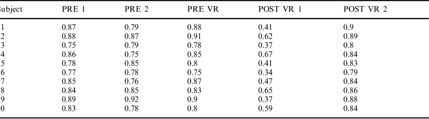
Mean values 91 SD of VOR gain, obtained under the different experimental conditions, are reported in Table I. Data obtained one week before VR exposure (PRE1 and PRE2) showed VOR gain values almost superimposable with those of normative data (19) and data observed immediately before VR exposure (PRE VR), suggesting good intra-individual repro-ducibility and no vestibular adaptation. Immediately after VR exposure (POST VR1), a statistically signié-cant decrease in VOR gain (pB0.005) was observed in all subjects. In two subjects we had to terminate VR exposure due to the onset of severe neurovegeta-tive symptoms. Thirty minutes after VR exposure (POST VR2) VOR gain returned to basal values, not signiécantly different from those observed 1 week before and immediately before VR exposure.
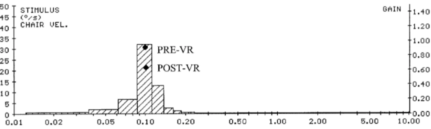
The ENG recordings obtained in one subject are reported in Fig. 1. The upper tracing represents the chair movement, the middle one the angular velocity of the low phase (open and élled symbols) before VR exposure and the lower one the angular velocity of the low phase immediately after VR exposure. The numerical data on the right-hand side of the traces represent the gain as a function of stimulus fre-quency. The amplitude spectra of the stimulus and eye velocity are plotted in Fig. 2.
DISCUSSION
The brain constantly monitors the ocular motor con-trol and shows a remarkable ability to adapt the VOR to new visual circumstances. These physiologi-cal adaptive changes of VOR gain are continuously performed by the vestibulo-cerebellum, in order to correct changes due to age, traumas, diseases of the oculomotor system or modiéed visual conditions (20), and a number of adaptive mechanisms with
Table I. VOR gain before and after VR exposure
PRE 2
PRE 1 POST VR 2
Subject PRE VR POST VR 1
1 0.87 0.79 0.88 0.41 0.9 0.62 0.89 2 0.88 0.87 0.91 3 0.75 0.79 0.78 0.37 0.8 0.84 0.67 0.85 0.75 0.86 4 0.41 0.8 0.85 0.83 0.78 5 6 0.77 0.78 0.75 0.34 0.79 7 0.85 0.76 0.87 0.47 0.84 0.86 8 0.84 0.85 0.83 0.65 0.92 0.89 9 0.9 0.37 0.88 0.83 0.78 0.8 0.59 0.84 10
Fig. 2. Gain calculated as a function of stimulus frequency. The amplitude spectra of the stimulus and eye velocity are plotted.
different time courses and capabilities have been identiéed (21).
When the subject tracks or acquires targets, the head and eyes usually move together and the VOR continuously balances the head movements in order to maintain a stable gaze. During VR exposure the computer, which monitors head movements by means of the tracker positioned on the helmet, continuously shifts the visual scene on the displays according to the recently acquired point of view. Under this condition the physiological strategy of eye–head coordination is no longer available because the new visual scene, usually obtained by complex and integrated move-ments of the head and eyes, is provided by the computer while the eyes are still in the middle of the orbit. Therefore, during VR exposure the function of the semicircular canals is partially substituted by the computer and the VOR, which usually drives the ocular movement in a direction opposite to the head movement, becomes an encumbrance.
This mismatch between the current gain setting of the VOR and that required to keep an image stabi-lized on the retina (visuo-vestibular conèict) might produce an adaptive shift in the oculomotor system, as demonstrated by the VOR gain reduction immedi-ately after exposure to a VE in our subjects (22). This adaptation, which disrupts the eye –head coordina-tion strategy, is the same as the VSR modiécacoordina-tion revealed by the impairment of the postural control (9, 10) observed after a similar simulated environmental exposure. Moreover this vestibular re-adaptation is clinically evident with the onset of ataxia (7), nausea and oscillopsia, all of which disappear when the VOR gain adapts to the new artiécial environment.
The VOR gain modiécation during VR exposure is similar to that observed in patients wearing telescopic spectacles after a 15 min exposure to combined visuo-vestibular stimuli (16 –18). In fact in this situation the current VOR gain also had to be modiéed because
the relationship between the eye movements, con-trolled by the VOR, was no longer able to maintain stable vision during the head movements because of enlarged vision. The visual feedback reveals an incon-gruity between the real and imaginary scenery. This incongruity induces the vestibular system to readapt the VOR. When the subject returns to a natural environment the VOR gain quickly returns to the initial value, as observed in our data obtained 30 min after the end of VR exposure.
It may be concluded that exposure to a VE has the potential to produce not only motion sickness and postural impairment but also adaptive shifts in the central vestibular system. Moreover, an artiécial modiécation of the gain, phase and direction of the eye movements opens new perspectives in the assess-ment and rehabilitation of vestibular diseases. REFERENCES
1. Gobetti E, Scateni R. Virtual reality: past, present and future. In: Riva G, Wiederhold BK, Molinari E, eds. Virtual environments in clinical psychology and neuro-science. Amsterdam; IOS Press, 1998: 3 –20.
2. Di Girolamo S, Di Nardo W, Picciotti P, Paludetti G, Ottaviani F, Chiavola O. Virtual reality in vestibular assessment and rehabilitation. Virtual Reality 1999; 4: 169 –83.
3. Kuhlen T, Kraiss KF, Dohle C, Hefter H, Freund HJ. Virtual holography in diagnosis and therapy of sensori-motor disturbances. In: Sieburg H, Weghorst S, Mor-gan K, eds. Health care in the information age. Amsterdam: IOS Press, 1996: 184 –93.
4. Regan EC, Price KR. The frequency of occurrence and severity of side effects of immersion virtual reality. Aviat Space Environ Med 1994; 65: 527 –30.
5. Lackner JR, Graybiel A. Visual and postural motion aftereffects following parabolic èight. Aviat Space En-viron Med 1980; 51: 230 –3.
6. Howarth PA, Costello PJ. The occurrence of virtual simulation sickness symptoms when an HMD was used as a personal viewing system. Displays 1997; 18: 107 – 16.
7. Kolasinski EM, Gilson RD. Ataxia following exposure to a virtual environment. Aviat Space Environ Med 1999; 70: 264 –9.
8. Dizio P, Lackner JR. Spatial orientation, adaptation and motion sickness in real and virtual environments. Presence 1992; 3: 319 –28.
9. Gray Cobb SV, Nichols SC. Static posture tests for assessment of postural instability after virtual environ-ment use. Brain Res Bull 1998; 47: 459 – 64.
10. Gray Cobb SV. Measurement of postural stability be-fore and after immersion in a virtual environment. Appl Ergon 1999; 30: 47 –57.
11. Kolasinski EM, Jones SA, Kennedy RS, Gilson RD. Postural stability and its relation to simulator sickness. 38th Annual Meeting of the Human Factors and Er-gonomics Society, 1994, Minneapolis (USA): 84. 12. Asai M. Effects of vestibular rehabilitation on postural
control. Acta Otolaryngol Stockh 1997; Suppl 528: 116 – 20.
13. Kennedy RS, Fowlkes JE, Lilienthal MG. Postural and performance changes following exposure to èight simu-lators. Aviat Space Environ Med 1993; 64: 912 –20. 14. Kennedy RS, Stanney JM, Ordy JM, Dunlap WP.
Virtual reality effects produced by head-mounted dis-play (HMD) on human eye-hand coordination, pos-tural equilibrium, and symptoms of cybersickness. Society for Neuroscience, 1997, New Orleans (USA): 7 772 (Abstract).
15. Kennedy RS, Stanney KM. Postural instability induced by virtual reality exposure: development of certiécation protocol. Int J Hum Comput Interact 1996; 8: 2547. 16. Demer JL, Porter FI, Goldberg J, Jenkins HA,
Schmidt K. Adaptation to telescopic spectacles:
vestibulo ocular-reèex plasticity. Invest Ophthalmol Vis Sci 1989; 30: 159 –69.
17. Gohnsor A, Melvill-Jones G. Short-term adaptive changes in the human vestibulo-ocular reèex arc. J Physiol (Lond) 1976; 251: 361.
18. Gonshor A, Melvill-Jones G. Extreme vestibulo-ocular adaptation induced by prolonged optical reversal of vision. J Physiol (Lond) 1976; 4: 256 – 381.
19. Miles FA, Eighmy BB. Long-term adaptive changes in primate vestibuloocular reèex. I. Behavioral observa-tions. J Neurophysiol 1980; 43: 1406.
20. Robinson DA. Adaptive gain control of the vestibulo-ocular reèex by the cerebellum. J Neurophysiol 1976; 39: 954 –69.
21. Melvill Jones G. Adaptive modulation of VOR parameters by vision. In: Berthoz A, Melville Jones G, eds. Adaptive mechanism in gaze control. Amsterdam: Elsevier, 1989: 21– 50.
22. Ebenholtz SM. Motion sickness and oculomotor sys-tem in virtual environments: Teleoperators and virtual environments. Presence 1992; 1: 302 –5.
Address for correspondence: Stefano Di Girolamo Clinica ORL
Policlinico Largo a. Gemelli, 8 IT-00168 Rome
Italy
Tel.: ª39 06 30154858 Fax: ª39 06 35501584