University of Rome “Tor Vergata”
Faculty of Medicine and Surgery
Department of Public Health and Cellular Biology
Doctoral thesis in Pediatria Molecolare e Biotecnologie
XXI Dottorato AA 2007/08
MOLECULAR CHARACTERIZATION AND NOVEL
MUTATIONS IN A LARGE COHORT OF PATIENTS
WITH CHRONIC GRANULOMATOUS DISEASE
Gigliola Di Matteo
Supervisor : Dott. Andrea Finocchi
SUMMARY
Background: Chronic Granulomatous Disease (CGD;
MIM#306400) is a rare primary immunodeficiency affecting the innate immune system. The disease is characterized by a greatly increased susceptibility to severe fungal and bacterial infections. CGD is caused by mutations in any one of four genes encoding subunits of the superoxide-generating phagocyte NADPH-oxidase, resulting in an absence or very low levels of enzyme activity.
Aim: this study represents the first molecular characterization of a
large cohort of italian patients affected by CGD. Its aim was to definetely define the type of molecular lesion in our patients, an indispensable tool for genetic counseling and prenatal diagnosis of CGD. This study focused the great genetic and clinical heterogeneity of the disease and the notion that genetic analysis is a critical step in obtaining a definitive diagnosis of CGD.
Materials and Methods: the molecular study was performed
essentially by DHPLC, to screen the presence of the different mutations and by sequencing of the suspected exons identified by DHPLC. This approch was extremely productive in terms of time and sensitivity. Functional tests were also performed to identify the level of protein expression and activity.
Results: I reported the results of CYBB (encoding the gp91phox
subunit) and NCF-1 (encoding the p47phox subunit) gene sequence analysis in 34 CGD patients from 29 unrelated families and their relatives. This report described nine novel CYBB mutations and their consequences. The carrier analysis was performed in 23 patients’ mothers and 16 female relatives through molecular and FISH studies.
Conclusions: the molecular analysis performed in our cohort of
CGD patients, underlines the breadth and the heterogeneity of the mutations responsible for the disease. It is likely that other factors may affect the clinical severity of CGD such as polymorphisms in oxygen-independent antimicrobial systems or other components of the immune system. Moreover, systematic characterization of molecular defect and polymorphisms in larger cohorts of patients, might help us to better relate clinical form and functional data to the type of the mutation found.
MOLECULAR CHARACTERIZATION AND NOVEL
MUTATIONS IN A LARGE COHORT OF PATIENTS
WITH CHRONIC GRANULOMATOUS DISEASE
Gigliola Di Matteo
Doctoral thesis in Pediatria Molecolare e Biotecnologie University of Rome “Tor Vergata”
PUBLICATIONS
1.Chiriaco M, Di Matteo G, Sinibaldi C., Giardina E, Blancato J, Folgori L, D’Argenio P, Rossi P and Finocchi A. Identification of deletion carriers in X-linked Chronic Granulomatous Disease by Real Time PCR. Submitted to Genetic testings.
2.G Di Matteo, L Giordani, A Finocchi, A Ventura, M Chiriaco, J Blancato, C Sinibaldi, A Plebani, A Soresina, C Pignata, RM Dellepiane, A Trizzino, F Cossu, R Rondelli, P Rossi, D De Mattia, B Martire, with IPINET (Italian Network for Primary Immunodeficiencies) Molecular characterization of a large cohort of patients with Chronic Granulomatous Disease and identification of novel CYBB mutations: An Italian multicenter study Mol Immunol 46(10):1935-41
3.Finocchi, A., Palma, P., Di Matteo, G., Chiriaco, M., Lancella, L., Simonetti, A., Rana, I., Livadiotti, S., Rossi, P., 2008. Visceral leishmaniasis revealing chronic granulomatous disease in one child. Int. J. Immunopathol. Pharmacol. 3, 739–743 4.Graziani S, Di Matteo G, Benini L, Di Cesare S, Chiriaco M,
Chini L, Chianca M, De Iorio F, La Rocca M, Iannini R, Corrente S, Rossi P, Moschese V. 2008. Identification of a Btk mutation in a dysgammaglobulinemic patient with reduced B cells: XLA diagnosis or not? Clin Immunol 128 (3):322-8. 5.F. Angelini, G. Di Matteo, S. Balestrero, F. Brunetti, G.
Mancino, P. Rossi, E. Galli. “Nuclear factor kappaB activity is increased in peripheral blood mononuclear cells of children affected by atopic and non-atopic eczema”. Int J Immunopathol Pharmacol. 2007 Jan-Mar;20/1:59-67.
CONTENTS
SUMMARY ……….…..2
PUBLICATIONS……….…..3
INTRODUCTION……….5
HISTORICAL BACKGROUND ………6
CLINICAL FEATURES, PROPHYLACTIC AND CURATIVE THERAPIES………..7
THE NADPH OXIDASE: MEMBRANE AND CYTOSOLIC COMPONENTS ………..13
CGD DIAGNOSTIC TESTS………..18
DHPLC (DENATURING HIGH PERFORMANCE LIQUID CHROMATOGRAPHY ………21
GENETICS OF CHRONIC GRANULOMATOUS DISEASE…………..22
X-linked CGD……… 24
Autosomal recessive CGD………. 26
AIM OF THE STUDY………30
PATIENTS………...31
MATERIAL AND METHODS………..32
Cell lines………...32 Molecular studies………32 RT-PCR………...35 Immunoblot analysis ………35 FISH ……….36 Statistical analysis………...37
RESULTS AND DISCUSSION………..38
CONCLUSIONS………..51
AKNOWLEDGMENTS ………51
INTRODUCTION
Chronic Granulomatous Disease (CGD) is the most common inherited disorder of phagocytic function caused by defects in the nicotinamide adenine dinucleotide phosphate (NADPH) oxidase, the enzyme that produces microbicidal (and pro-inflammatory) reactive oxygen species (ROS) during the respiratory burst indispensable for killing of phagocyted microorganisms. Consequently, CGD patients are highly susceptible to recurrent life-threatening bacterial and fungal infections, mainly located in the lungs, skin and lymph nodes but they are also affected by inflammatory/granulomatous manifestations ether infectious or non-infectious in multiple organs (1).
NADPH oxidase consist of a heterodimeric membrane-bound flavocytochrome b558 (formed by gp91phox and p22phox) and cytosolic
subunits: p47phox, p67phox, p40phox and two small GTPase, Rac2 and Rap1A. Mutations in any of the genes encoding NADPH oxidase subunits can cause CGD except p40phox and Rap1A. Approximately two thirds of all CGD cases are X-linked and result from mutations in the CYBB gene, encoding the gp91phox subunit. The remaining patients with CGD have an autosomal recessive form (AR-CGD), the most frequent of which, is due to a mutation of NCF1 gene that encodes the p47phox subunit (1).
HISTORICAL BACKGROUND
A fatal childhood disease characterized by the occurrence of granulomas and recurrent infections was first described at an annual meeting of the Society for Pediatric Research, in 1954, by Janeway and colleagues (2) and in 1957 by Good (3). In 1959 Bridges et al., noted for the first time the presence of granuloma lesions in deep organs such as lung in 4 boys, describing “a fatal granulomatous disease” (4). In 1967 Quie and Holmes (5-7) linked the disease to a deficiency in bactericidal activity of the phagocytes. They demonstrated, for the first time, that neutrophils purified from patients with this familial granulomatosis (now called Chronic Granulomatous Disease, CGD) were incapable of killing Staphilococcus aureus in vitro and at the same time that neutrophils from CGD patients failed to increase oxygen consumption and to generate reactive oxygen species (ROS), the so called “respiratory burst”, during phagocytosis. Baehner and Nathan were the first to use the nitroblue tetrazolium (NBT) reduction test to measure oxidase activity in leukocytes to diagnose CGD (8). Afterward, CGD, that was first thought to affect only males, was also reported in female patients (9-10).
In about one decade the researchers demonstrated that: 1) the NADPH is the major substrate of the phagocytic oxidase (11-12), 2) the cytochrome b558 is localized in the granules of neutrophils
(13-14), 3) the cytocrome b558 is the NADPH oxidase in human
neutrophils. Finally, NADPH oxidase deficiency was identified as the cause of CGD (15). In 1987 the heterodimeric nature of cytocrome b558, that consists in the α or small subunit or p22phox and
the β chain or large subunit or gp91phox (today called NOX2), was determined (16).
CLINICAL FEATURES, PROPHYLACTIC AND CURATIVE THERAPIES
CGD occurring in about one in 250.000 individuals, depending on the populations investigated. There are extreme differences in presentation between patients, varying from a relative mild presentation late in life to fatal septicemia in infancy.
Because CGD is relatively uncommon, it has been difficult to develop a detailed and comprehensive clinical picture of the disorder. Despite several decades of work on CGD, apart from the description of several smaller cohorts, the only characterizations of a large group of CGD patients was published by Winkelstein et al, who reported on a National American Registry of 368 patients (17) and by Jones et al., who reported of CGD patients from United Kingdom and Ireland (18). Recently clinical data from 429 European patients, the largest cohort of CGD patients to date, were reported (19). Of recent publication is also the first extensive survey on 60 CGD italian patients (20).
From the last two studies, including the italian cohort of CGD patients, came out that the mean age at diagnosis was higher in the AR CGD (autosomal recessive) group than in X-linked CGD (table 1), this probably reflect a milder phenotype of AR-CGD but it is not clear why, although is known that residual NADPH oxidase activity has been shown in p47phox deficiency. In the Italian group the
for the disease in males and therfore in the selection of patients. Moreover an high percentage of CGD patients (25%) in the italian cohort is without a molecular diagnosis.
Table 1. Comaparison between four studies. The percentage of X-CGD and AR-CGD in the different cohorts are shown. The mean age at the diagnosis is indicated in all studies except in the UK study.
The most prevalent site of disease, considering both infections or granulomatous, were the lungs (66%), followed by skin (53%), lymph nodes (50%), gastrointestinal tract (48%) and liver (32%). In the italian study was possible to determine the site of disease at presentation and the clinical manifestations during follow-up. Also in this cohort, before the diagnosis, lung infections were the most prominent clinical problem (50%) followed by infections in skin (46%), lymphonodes (45%), liver (16%). At diagnosis the majority of patients (35%) presented limphoadenitis, followed by gastrointestinal tract (26%) and lung infections (25%) (table 2). During the follow up, infections remained the most remarkable clinical problem even after anti-infective prophylaxis was started. Particularly lung and skin infections occurred in 48,5% and 38% respectively, of the overall cohort, while stomatogengivitis and gastrointestinal tract infections significantly increased affecting 35%
American study UK study Italian study European study No of patients 368 94 60 429 % of XL pt. 70,3 73,4 65 67 % of AR pt. 22 17 10 33 Unknown 7,7 9,6 25 - Age at diagnosis (mean) XL 3,01 nd 3.1 4.9 AR 7,81 nd 5.8 8.8
and 25% of the patients respectively (table2).
Table 2 Prevalence of infection by site in CGD. The numbers represent the percentage of CGD patients who had at least 1 episode of the specific infection. In bold is indicated the site with any kind of infection and in regular the specific infection.
About the etiology of infections, fungal infections were the first cause of illness with the Aspergillus species as the most frequently isolated organism. This organism represents the first cause of pneumonia. After the lungs, the second one most frequently affected site of disease is the skin were Staphylococcus aureus was most prominent. Other microorganism involved were gram - such as Serratia marcescens and Burkholderia cepacia, the latter remarkably rare in the european cohort. Where the vaccination with
Site of infection American study UK study Italian Study before diagnosis Italian Study during follow up European study Lung * Pneumonia * Lung abscess 80,7 16,3 52 9,6 47 3 42,5 6 66 - - Skin * Subcut. abscess * Dermatitis 42.4 - 36 - - 20 23 4 34 53 - - Lymph node * Lymphadenitis * Suppurative adenitis - 52,7 - 40.4 45 - 28 - 50 - - Liver * Liver Abscess 26,6 28,7 16 21 32 - Bone * Osteomyelitis 24,4 11.7 16 8,5 13 - Septicemia 17,6 23.4 5 15 20 Kidney - 20 - - 22 Ear * Otitis - n.d. 5 13 14 - Brain *Brain Abscess 3,3 n.d. n.d. 6 7 - Gastrointestinal *enteritis * Stomatogengivitis * Perirectal abscess - - 15,5 37 24 - 26 10 - 8 25 - 35 15 48 - - -
With all infectious episodes taken together, the most frequently cultured microorganism was S. aureus followed by Aspergillus species and Salmonella species in european cohort and Aspergillus species, S. aureus and Candida in the italian cohort.
On the other hand, particularly inflammatory manifestations are frequent in CGD, more in membrane-bound (gp91phox and p22phox) than in cytosol components (p47phox and p67phox) deficient patients. Moreover, a particular set of these features are described among X-linked CGD female carriers. Different organs and systems are reported to be affected by inflammatory/granulomatous manifestations in CGD patients. Hollowed viscera, as the gastrointestinal tract and the genitourinary tract, are the most frequently affected. Other tissues and organs as the retina, the lungs and even bones also present with particular inflammatory patterns in these patients.
Manifestations of autoimmunity or autoinflammation have been claimed to be more prevalent in CGD patients than in the normal population. In the italian cohort a 23% of patients affected by inflammatory and/or rheumatic complications were reported. Chronic inflammatory bowel disease, systemic lupus erythematosus and discoid lupus erythematosus were also reported, together with gastrointestinal manifestations of no infectious origin. In the european cohort signs of autoimmune disease were seen in the 6% of patients. Other manifestations were sporadic.
About the infection prophylaxis most CGD patients are treated with long-term, low dose antibiotics and antifungal. Apart from a great variability in terms of efficacy and prophylactic regimen applied the most of CGD italian patients (48%) received
TMP-SMX, antifungal itraconazole and IFNγ. The 27% of italian CGD patients received cotrimoxazole plus itraconazole. Differently, 71% of the european cohort received cotrimoxazole and 53% itraconazole, while 33% of these patients received at some point IFNγ. One of the most significative study on the effectiveness of the IFNγ therapy was performed by the International CGD Cooperative Study group (78), where 128 patients showed a reduction of the serious infections when IFNγ was somministrated three times weekly. However other prophylactic schedules were adopted in minor group of patients. The efficacy of long term IFNγ treatment remains controversial despite of many years of study. In the italian study IFNγ did not significantly change the rate of total and severe infections per patient-year between two groups treated at the same therapeutic regimen with or without IFNγ. Probably, when the IFNγ treatment is not interrupted for adverse events, mainly fever, its use could improve the outcome of CGD patients but in different time of long term maintenance therapies it is difficult to establish a link with the occurrence of infections, complications or death. However, the molecular mechanisms associated with host defence improvement induced by IFNγ in CGD patients are unknown. Several studies showed that the mortality rate has not changed in the last decade (mortality/year estimated between 2% AR-CGD and 5% X-CGD). The mortality in CGD patients usually occurs in the first two decades of life, with about 50% of patients surviving into their third decade. Infections caused by Aspergillus species resulted the most common cause of death accounting for half of all deaths.
deaths alone. Analysis of survival of CGD patients suggests that recent advances in treatment have improved survival of patients in the first two decades of life, but there does not appear to be increased survival at later ages (Figure 1). The improved survival in the first two decades is mainly due to early diagnosis along with aggressive management, including the use of prophylactic and therapeutic antibiotics enclosed IFN γ.
Jones et al., 2008
Martire et al., 2008
Figure 1. Kaplan-Meier overall survival curve after diagnosis o CGD. In the english cohort (upper panel, by Jones et al, 2008) the survival curve was obtained by sex; in the Italian cohort ( lower panel by Martire et al., 2008) was considered the entire cohort of patients. SURp: survival probability; SE: standard error.
To date, haematopoietic stem cell transplantation (HSCT) is the only established option for definitive cure of patients with a suitable donor and is indicated when conventional prophylaxis and therapy with antimicrobial medication fail. However, despite transplant progresses, it is still difficult to find compatible donors. Moreover,
transplant is associated with high morbidity and mortality especially in patients with severely compromised pulmonary function or other debilitating illness.
CGD is a promising candidate for the development of gene terapy targeted at hematopoietic stem cells. Gene therapy, is particularly important for patients for whom conventional bone marrow transplantation is not possible. However, the progress in gene therapy has been hampered by the appearance of insertional mutagenesis causing leukaemia in a trial for patients with X-linked severe combined immunodeficiency (21) and by the emergence of dominant clones in a recent trial for the X-linked form of CGD (22). These findings stimulated the development of modified vector systems that demonstrate reduced genotoxicity in vitro and in animal models. The improvements in the safety and efficacy of gene therapy could help achieve a clinically significant correction of CGD.
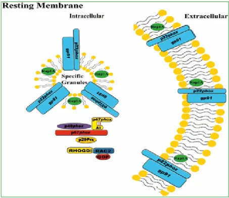
THE NADPH OXIDASE: MEMBRANE AND CYTOSOLIC COMPONENTS
In 1933, it was observed that phagocytic cells, mainly the polymorphonuclear neutrophil (PMN), demonstrated markedly increased oxygen consumption, or a respiratory burst, during phagocytosis. From then on it comes a long way. The NADPH oxidase deficiency was identified as the cause of CGD. NADPH oxidase is a multicomponent complex localized in the phagosomal and plasma membrane composed of a membranous component,
and Rap1A (Figure 2). Cytochrome b558, the redox center of the
NADPH oxidase complex, is a heterodimer consisting of a large flavocytochrome NOX2 (formerly gp91phox) and a small protein p22phox which acts to stabilise the complex and to dock the cytosolic partner p47phox.
Forest R. Sheppard et al., 2005
Figure 2. Resting membrane of human PMNs. Representative picture of a resting PMN and the location of the NADPH oxidase components, including the cytosolic
components (p47phox, p67phox, p40phox, Rac2, and p29) and the membrane-bound
components (gp91phox, p22phox, and Rap1A)
NOX2 contains six transmembrane domains, cytosolic NADPH and FAD binding sites, and two intramembranous haemes that are necessary for catalysing the reduction of molecular O2 to generate
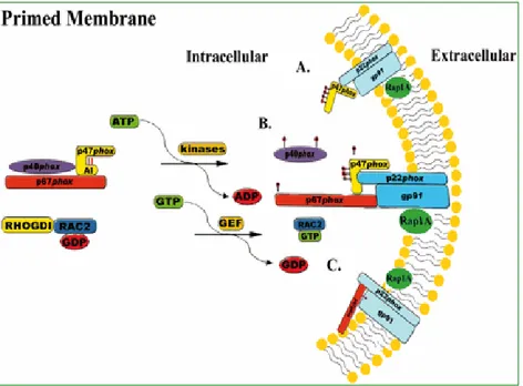
the superoxide anion in the phagosome or the extracellular space. In unstimulated cells, the cytosolic components of the NADPH oxidase, p40phox, p47phox, and p67phox, are associated with a 1:1:1
stoichiometry in the cytosol. Rac2 is complexed in the cytoplasm with Rho-guanine nucleotide dissociation inhibitor. Upon activation, both p47phox and p67phox are phosphorylated and translocate with p40phox to membrane-bound cytochrome b558. Rac2 binds GTP and
migrates to the membrane independently of the p67phox– p47phox complex (Figure 3).
Forest R. Sheppard et al., 2005
Figure 3. Primed membrane of human PMNs. Representative image of the phosphorylation and translocation of the oxidase components with external
priming agents. Depending on the stimulus, only p47phox translocates to the
plasma or granular membrane (A)or both, p47phox-p67phox translocate to the
plasma (granular) membrane (B). Another possibility is that p67phox, but not
p47phox, translocates to the membrane with a stimulus.
Then, in its complexed activated form, NADPH oxidase is able to transfer electrons from cytosolic NADPH to external molecular oxygen (23) (Figure 4).
Forest R. Sheppard et al., 2005
Figure 4. Activated membrane of human PMNs. Representative image of the complete translocation of the oxidase components to the plasma or granular membrane and the electron transfer across the membrane, characteristic of activation of the NADPH oxidase.
Recently, the research on non-phagocyte NADPH oxidase led to the discovery of two families of NOX homologs, NADPH oxidase (NOX) and dual oxidase (DUOX), expressed in several tissues and cells, and involved in different pathological processes. All NOX family members contain a core structure consisting of six transmembrane domains (in which four heme-coordinating histidine residues are located) and a C-terminal cytosolic region (which contains heavily conserved binding sites for FAD and NADPH) (24-25).
The superoxide anion (O2-) in the phagosome or in the extracellular
1) oxidizes a variety of aromatic compounds (R-H) by electron transfer, yielding substrate radicals;
2) oxidizes chloride ions to the non-radical oxidant hypochlorous acid, the PMNs most potent bactericidal product;
3) converts to the reactive hydroxyl radical by the Fenton reaction between the H2O2 and a transition metal catalyst (Fe3-/Fe2-);
4) generates singlet oxygen, an additional, albeit minor, by-product of O2-, which is an extremely energetic form of oxygen, capable of
attacking double bonds;
5) produces reactive nitrogen species from nitric oxide (NO) (23). NOX2 deficiency has been seen predominantly as a decrease in host defence, with an inability to mount an inflammatory response. However, there is increasing evidence for hyperinflammatory, non-infectious complications of CGD. Indeed, in CGD patients exuberant chronic granuloma formation, often without an apparent infectious agent, is a frequent feature. However, presently available data suggest that both increased and decreased NOX2 activity may lead to inflammatory complications. The situation is most puzzling for arthritis and inflammatory bowel disease, which have been classified as diseases caused by increased NOX activity by some, but as diseases associated with a lack of ROS generation by others. In general, the association of increased NOX2 activity with inflammation has been widely discussed, in contrast, the relationship between decreased NOX2 activity and inflammation remains poorly understood (26).
CGD DIAGNOSTIC TESTS
There are several diagnostic tests to evaluate the functionality of NADPH oxidase, mainly: the nitroblue tetrazolium test (NBT), the luminol or lucigenine chemiluminescence technique, the flow cytometry method, the superoxide-inhibitable cytochrome c reduction test by spectrophotometer. The NBT test and the FACS can evaluate intracellular ROS production after soluble or particulate stimuli activation (all reviewed in 1).
The simplest and the least expensive screening test is the reduction of the NBT. This is easily performed by exposing neutrophils to NBT together with a soluble stimulus such as phorbol myristate acetate, formyl peptide, or with a particulate stimulus such as opsonized latex beads, bacteria, or zymosan (to test the ability of cells to phagocytize at the same time). Then, the yellow watersoluble NBT dye is reduced to dark blue insoluble crystals of formazan in the activated cells (Figure 5). This test, is valid with total blood or purified white cells or purified neutrophils, even if the blood sample is transported and preserved (<48 h).
Figure 5. The nitroblue tetrazolium (NBT) test: activated neutrophils are tested for their ability to phagocytose and reduce NBT. a) Reduced NBT is seen as dark-blue crystals. b) Unstimulated neutrophils, or stimulated neutrophils from patients who have CGD fail to reduce NBT. c) Blood from carriers of CGD contains a mixture of normal and abnormal neutrophils.
Healty control CGD patient CGD
The flow cytometry method (fluorescent-activated cell sorting [FACS]) is available using fluorescent probes (dihydrorhodamine-1,2,3 [DHR] or 2′ 7′- dichlorofluorescein diacetate) to measure intracellular ROS production, but the equipment is sophisticated and expensive and the blood sample must be freshly drawn (Figure 6).
Figure 6. The dihydrorhodamine (DHR) 123 test, utilizes flow cytometry to detect the oxidation of dihydrorhodamine 123 in activated neutrophils. The X-linked form is typically associated with an absence of detectable oxidative burst activity, while in X-linked CGD carriers are usually characterized by the presence of two populations of cells: with or without detectable oxidative burst activity.
https://online.epocrates.com/u/2931703/Chronic+granulomatous+disease/Diagnosis/Approach
The missing protein of the NADPH oxidase components (except for rare CGD variants where all the oxidase proteins are expressed) has to be determined using Western blot in the CGD patient’s neutrophils or ebv transformed lymphoblastoid cell lines, with specific antibodies (Figure 7).
Figure 7. Western blot of human neutrophils from a normal subject and a patient
with X-CGD using a gp91-phox monoclonal antibody.
Meanwhile, NOX2 is clearly involved if there is evidence of X-linked transmission, either in the familial history or by the detection of female carriers in relatives. A history of consanguinity in the parents and/or a female CGD patient can evoke an AR inheritance. Definitive evidence of molecular lesions used for genetic counseling and in prenatal diagnosis is achieved by sequencing the appropriate gene. Since the location of genetic mutations is never known (except for the majority of A470 CGD cases), usually, RTPCR from the corresponding mRNA and sequencing of the resulting PCR product are the easiest first steps in determining the genetic lesion in the majority of cases. This cannot be applied for large deletions neither insertions nor other mutations that cause unstable mRNA. Single-strand conformation polymorphism (SSCP) and the more recent Denaturing High Performance Liquid Chromatography (DHPLC) can accelerate specific diagnosis at the DNA level by limiting the need for direct sequencing to a few abnormal polymerase chain reaction (PCR) products identified in the prescreeningprocedure. However, the genetic defect must always be confirmed at the genomic level by sequencing.
DHPLC (DENATURING HIGH PERFORMANCE LIQUID CHROMATOGRAPHY)
The "gold standard" for mutation analysis, direct sequencing, is expensive and labour intensive for high throughput mutation screening, emphasising the need for alternative simple, sensitive, and cost effective methods for mutation analysis. Today the most frequently used methods for prescreening disease genes for mutations are single strand conformation polymorphism analysis (SSCP) and denaturing high performance liquid chromatography (DHPLC). Both methods accelerate specific diagnosis at the DNA level by limiting the need for direct sequencing to a few abnormal polymerase chain reaction (PCR) products identified in the prescreening procedure.
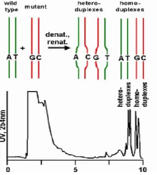
The tecnique of SSCP analysis is based on the principle thatchanges in nucleic acid composition affect the conformation of single stranded DNA and thereby the mobility of the fragmentwhen it is subjected to electrophoresis under non-denaturing conditions. The technique of DHPLC has been developed more recently and relies on different retention times of homoduplex and heteroduplex DNA on ion-pair reverse phase chromatography. Partial heat denaturation of samples decreases the retention time of mismatched DNA fragmentscompared with their intact double stranded counterparts (Figure 8). The main advantages of DHPLC are its high sensitivity and specificity (from 96 to 100%), its low cost of running and speed
www.uni-saarland.de/fak8/huber/dhplc.htm
Figure 8. Schematic representation of DHPLC procedure. Samples and controls are amplified, mixed in equimolar quantity, partially heat denaturated, rinaturated to consent the formation of heteroduplex and homoduplex molecules and finally analyzed through a ion pair reverse phase chromatography.
GENETICS OF CHRONIC GRANULOMATOUS DISEASE
CGD is a heterogeneous disease caused by mutations in any of the following four genes encoding subunits of the superoxide-generating phagocyte NADPH oxidase, including NOX2 and p22phox (cytochrome b558 subunits), p47phox and p67phox (cytosolic components). To date, any mutation has been found in the gene encoding p40 phox. All ethnic group are equally affected. In 1986, the X-linked defective gene was the first gene involved in CGD cloned (27). Subsequently, the cDNAs encoding p47phox
(the NCF1 gene) and p67phox (the NCF2 gene) were cloned and sequenced (28-30) and later was established the chromosomal location (31). Finally Dinauer et al., reported the cloning and chromosomal location of the CYBA gene encoding p22 phox subunit (32).
The most frequent form of CGD (about the 65% of all cases) is the X-linked recessive transmission type of CGD, caused by mutations in the CYBB gene encoding NOX2. The second most common form of CGD is autosomal recessive and is caused by deletion of a GT from a GTGT tandem repeat at the first splice junction in the NCF1 gene encoding p47phox accountig for about 25% of the cases. Mutations in the CYBA and NCF2 genes encoding p22phox and p67phox, respectively, each accounting for less than 5% of CGD cases (see table 3).
subtype GENE PROTEIN INHERITANCE FREQUENCY
X91° CGD ~65% X91- CGD X91+ CGD CYBB gp91phox XR A22° CGD ~ 5% A22+ CGD CYBA p22phox AR ~ 1% A47° CGD NCF1 p47phox AR 25% A67° CGD NCF2 P67phox AR ~ 5%
X-linked CGD
The description of a patients who suffered from CGD but also Duchenne muscolar dystrophy and retinitis pigmentosa made it possible a linkage analysis to localize the CYBB gene (33). Two databases gathered more than 200 mutations on the CYBB gene: the Human Gene Mutation Database (HGMD, http://www.hgmd.cf.ac.uk/ac/index.php) at the Institute of Medical Genetics in Cardiff (Wales) with 282 mutations reported to date (34); the Immunodeficiency database (IDbases, http://bioinf.uta.fi/CYBBbase/) of the Institute of Medical Technology–Bioinformatics in Tampere (Finland), reporting 244 mutations (35).
According to the HGMD database single-nucleotide substitutions represent the 58% of the defects, while small deletions, insertions and insertion-deletions the 26% and large deletions (more frequent) and insertions the 14% of the defects. Mutations in the regulatory region of the CYBB promoter and complex rearrangements represent respectively the 1.5 an the 0.5%. Only three polymorphisms caused by missense mutations are located in the encoding region of the CYBB gene have been reported (36). Most of the mutations in the CYBB gene lead to a lack of NOX2 expression because of the instability of the corresponding mRNA or protein. This phenotype is called X910 CGD, it is the most
frequent and can be caused by all type of mutations.
Non sense mutations, which introduce a stop codon, affect the mRNA at various degrees but usually the truncated NOX 2 protein is never immunodetected by Western Blot. This type of
mutation accounts for about 50% of point mutations in the coding CYBB region (34-37). Approximately 50% of the point mutations in the 13 exons of CYBB gene are missense mutations. These mutations do not usually affect mRNA stability but act on the level of NOX2 expression in phagocytic cells, leading to either X910, X91+ or X91- CGD variants. NADPH oxidase activity is always totally abolished except for some X91- that can show a residual activity (38).
Twenty-six mutations have been found in X91- CGD patients,
they result from structural disorganization, leading to an incomplete loss of protein, partial disfunction or both.
Most of the X91- CGD patients had disease resulting from missense mutations, the rest consists in deletions, mutations in the promoter region and one splice mutation. Of interest, the mutations in the promoter region, are all in a region between the “CCAAT” and the “TATA” boxes. These mutations inhibit the binding of both Elf-1 and PU.1 with consequent low level of NOX2 expression. The amount of superoxide produced by neutrophils from these patients, is probably not suffucient to protect against infections even if X91- CGD patients are usually diagnosed later in life than X910 CGD patients (38-39).
Nineteen mutations have been found to cause X91+ CGD. Most
of them are missense mutations, two are small deletions, and one is a deletion-insertion leading to a normal expression of mutated NOX2 proteins. They are principally located in the COOH terminus cytosolic tail of NOX2, confirming the important role of
Autosomal recessive CGD
Autosomal recessive CGD is caused by genetic defects in one of the three genes CYBA, NCF1, and NCF2 encoding, respectively, p22phox, p47phox, and p67phox. It affects female and male children alike. These three forms collectively account for approximately 30–40% of all CGD cases. Most of the mutations in the three genes are reported in the HGMD databases,.ac.uk/ac/index.php and http://bioinf.uta.fi/ (34-35). It should be noted that polymorphisms in the encoding regions of CYBA, NCF1, and NCF2 are more frequent than in CYBB (41).
The most frequent AR CGD form is caused by mutations in the
NCF1 gene. This gene (OMIM number 233700) has been
mapped to 7q11.23. It possesses 11 exons. A common mutation has been identified in approximately 95% of affected alleles analyzed worldwide. This mutation is a GT deletion (∆GT) in a GTGT tandem repeat, corresponding to the first four bases of exon 2 (42). Most patients have a homozygous GT deletion, which predicts a frameshift within a premature stop codon at amino acid 51, leading to a complete absence of p47phox protein from the patients’ neutrophils (A470 CGD). Of approximately 100 patients investigated to date, only 19 patients had mutations other than the classical ∆GT, they were small mutations (nonsense, missense mutations, and small deletion) and always led to A470 CGD. The reason that the ∆GT mutation predominates is that most normal individuals (>95%) have two NCF1 pseudogenes (ΦNCF1) on each allele, which exhibit the ∆GT deletion, with more than 99% identity with the NCF1 gene and they are physically close to the functional gene at 7q11.23. These ΦNCF1
are the best-conserved unprocessed pseudogenes known (43). Recent studies have demonstrated that the predominance of the
∆GT arises from recombination events between NCF1 and its highly homologous pseudogene ΦNCF1(43-45). Because of the presence of these ΦNCF1 and the extreme homology between them and NCF1, it is hardly possible to detect carriers for A470 CGD by normal PCR and sequencing methods (45). In addition, the p47phox protein level and the NADPH oxidase activity in the phagocytes of carriers are indistinguishable from normal individuals. However, there are some differences between the pseudogenes and the functional gene, therefore, gene and pseudogene-specific PCR, starting from cDNA or from genomic DNA, have been used (44-46). A gene scan method based on the presence of the ∆GT to assess the ratio of NCF1 genes to pseudogenes has also been developed (46-47).
Mutations in the CYBA gene encoding p22phox are extremely rare (frequency <5%). The CYBA gene (OMIM number 233690) mapped to 16q24 has 6 exons. The last update of CYBA mutations done in 2000 by Cross et al. (41) showed 26 different mutations. Most of the mutations are missense or nonsense. It seems that since 2000 no more than two new mutations have been described in CYBA (48-50). Missense mutations leading to A220 CGD are principally located in the potential transmembrane passages of p22phox. Perhaps some of these regions are possible interaction sites with NOX2. Finally, the only missense mutation, Pro156Gln, leading to the unique A22+CGD, is located in the
and p67phox translocation from the cytosol to the plasma membranes. Most likely, binding of p47phox is disturbed because p47phox is thought to interact first with cytochrome b558 (and p22phox more precisely) (52-53).
The last and extremely rare autosomal recessive form of CGD is the form caused by mutations in the NCF2 gene encoding p67phox, accounting for approximately 5% of CGD cases. The NCF2 gene (OMIM number 233710) mapped to 1q25 possesses 16 exons. Its promoter region has been well defined. Because of the homologous cis-element in the CYBB and NCF2 genes, they are regulated by common transcription factors [54-56]. The latest update of NCF2 mutations revealed 18 different mutations (41). Most A67 CGD patients had no expression of the p67phox protein with normal levels of mRNA. Deletions, insertions, missense and nonsense mutations were described as were three splice-site mutations. All the missense mutations led to A670 CGD and were located in the tetratricopeptide repeat (TPR) domains of p67phox. Only one patient (A67− CGD) expressing half-normal amounts of one amino-acid-deleted (Lys58) p67phox has been reported (57). The Lys58 deletion led to the mutated p67phox synthesis but disturbed its interaction with Rac. In this patient, p47phox and p67phox did not translocate to the plasma membrane upon cell activation, indicating that an interaction between p67phox and Rac is essential for the translocation of these cytosolic proteins. Three new mutations were recently described, resulting in 21 different mutations found in the NCF2 gene. An interesting point to underline is that Borgato et al. and our group (58, 60) observed that the absence of p67phox protein expression leads to the absence
or the reduction of p40phox expression. This confirms that p67phox and p40phox protein stability are related (60). However, in the 21 cases reported, p40phox expression was rarely documented.
AIM OF THE STUDY
This study represents the first molecular characterization of a large cohort of italian patients affected by CGD. Its aim was to definetely define the type of molecular lesion of our patients, an indispensable tool for genetic counseling and prenatal diagnosis of CGD.
To date, since the location of genetic mutations is never known a priori, RTPCR from the corresponding mRNA and sequencing of the resulting PCR product were the best approach in determining the genetic defect in the majority of cases.
In our study we used, as a mutational screening method, for the first time in this pathology, the DHPLC (Denaturing High Performance Liquid Chromatography) to detect the presence of mutations in the thirteen exons of the CYBB gene. Moreover we used the same approach to detect the ∆GT deletion in the NCF1 gene. This method appeared a high performance system, sensitive and specific, that identified the 100% of mutations in our cohort. This method improved the CGD diagnosis and the management of CGD patients, who are now protected against severe and life-threatening infections earlier than before.
Moreover we identify 9 novel mutations, now inserted in the international CYBBbase. Our results corroborate research findings and contributed to furthering and disseminating the knowledge of this rare disease.
PATIENTS
Thirty-four male patients, from the Italian registry of Chronic Granulomatous Disease which is part of the Italian Primary Immunodeficiency Network (IPINET), were tested for mutations in the CYBB gene. Patients were diagnosed as having CGD on the basis of their clinical history and the inability of their phagocytes to generate reactive oxygen species (ROS) by standard procedures as previously described (20). Age at diagnosis ranged from 1 month to 49 years (mean 7.16 years, median 3 years). Carrier detection was performed in 23 patients’ mothers and 16 female relatives by means of genetic analysis and through fluorescent in situ hybridization (FISH) of metaphase chromosomes from lymphoblastoid cell lines when needed. All relevant clinical features, immunological and therapeutic data have been already described in a previous report by the IPINET (20). All patients were given antiinfective prophylaxis with trimethoprim/sulphomethoxazole and itraconazole early after diagnosis. Some subjects (patients 2, 4, 6, 8, 17, 20, 23, 24, 26– 28, 32) received discontinuous administration of IFNγ.
Informed consent was obtained from each studied subject or parents of each child and all procedures used were in accordance with the guidelines of the Helsinki Declaration on Human Experimentation.
MATERIAL AND METHODS
Cell lines
Neutrophils were purified by discontinuous Percoll density gradient centrifugation from peripheral blood of patients 17, 33 and 34 as previously described (61). EBV transformed lymphoblastoid cell lines (LCL) were obtained by incubating approximately 5×10⁶ PBMC, with 2ml of supernatant from the EBV-secreting cell line B95-8 for 1 h at 37◦C. Infected cells were cultured at 1×10⁶ cells/ml in RPMI 1640 containing 20% fetal bovine serum, 2mM l-glutamine and 1µg/ml of cyclosporin A in 24-well culture plates. Clumps of cells were usually observed at the end of the first or second week and stable EBV-lymphoblastoid cell lines were established in 3–4 weeks.
Molecular studies
Genomic DNA isolated from peripheral blood of patients and healthy controls (QIAamp DNA Blood kit by QIAGEN GmbH, Hilden, Germany) was amplified using primers (available upon request) flanking the 13 coding exons and the promoter region of the CYBB gene (NG 009065). PCR reactions were carried out in a volume of 50µl containing 100 ng of genomic DNA, 200µM of each dNTP, 0.4µM of each primer (Table 3) and 0.5U of GoTaq DNA polymerase (Promega, Madison, WI). The samples were denatured at 95◦C for 5min followed by 30–40 cycles at 95◦C for 30s, annealing at exon specific temperature for 45s, 72◦C for 30s with a final 5min extension at 72◦C.
Table 3. CYBB specific primer pairs for DHPLC and sequencing.
DHPLC was performed on a WAVE analysis system (Transgenomic, Omaha, NE) using different temperature conditions. Briefly, PCR products were mixed in approximately equimolar proportions with an amplified sample known to contain a wild-type CYBB fragment, then denatured at 94 ◦C for 5min and then cooled at room temperature, to allow heteroduplex formation. Each sample were run on a DnaSept.M column (Transgenomics) and monitored by ultraviolet light (260 nm). Optimum DHPLC temperatures were detected using software-predicted melting profile as a starting point. Samples were run at a mean of three temperatures due to the heterogeneity in the distribution of GC-rich regions (62). Each elution chromatographic profile was compared with profiles associated with homozygous wild-type sequence (Figure 8). Samples with an
Exon 5' primer 3' primer Annealing
Temperature
DHPLC Temperature
(°C)
Prom. cagcaaggctatgaatgctgttcc ctctgacttctcgggttatcttg 56°C Direct sequencing
1 tgtagttgttgaggtttaaag ctctgacttctggggttatcttg 56°C 55,5/58,3/59,3 2 tgactccagtcttctgtggaatc tcccatgcaatattggctgg 60°C 56,3/58,3/59,9 3 tggtgtgtggcctcatgctaag cctctaattttcaaaggccatc 60°C 60,3/61,3/63,3 4 gactgcatctctctctgaacctcagt aaacttggaaccagggagttccct 60°C 56,2/57,5/58,3 5 gttcatacccttcattctctttg ctctaggccattacaattgaggac 60°C 56/56,7/57,9 6 ttgcgcacatgtgtgtgtgtgtgt agcctgtctgctaagggaatgtga 65°C 58,6/59,6/61,3 7 tcctattactaaatgatctggac gttgttattacagtttcattactgac 54°C 55,6/57,6/60,6 8 ctccctctgaatattttgttatc taattactagtggtcagtgtgtaac 56°C 53,4/56,4/58,4/(60,4)
9 ccatatgacactaaaaaggc tcaggaaaatggcactaaatagc 56°C Direct sequencing
10 acatctctgtaactatctcctcc ctttcatttatcggagggcc 60°C 58,8/59,8/61,3
11 gaattccacatggtaatgctgatag gtccttccatagtgacagggct 60°C 58,1/61,1/59,1/(62,1)
12 atccagcagggtgccttggttagaat tttctcaacctgcctgtcccaagt 60°C 59,2/59,7
Figure 8. Examples of DHPLC analysis. In the upper panel the analysis revealed the presence of a mutation in patient BA. In the lower panel has been analyzed the villus from a CGD carrier. The analysis revealed the presence of a mutation.
Direct sequencing was performed after purifying PCR products with QIAquick PCR purification kit (QIAGEN, Hilden, Germany). Both strands were sequenced using the BigDye Terminator v3.1 Cycle Sequencing Kit (Applied Biosystems, Foster City, CA) and analyzed on an ABIPRISM 3130 and 310 automated sequencers (Applied Biosystems, Foster City, CA). For the detection of GT homozygous deletion in exon 2 of NCF-1
Prenatal Prenatal Prenatal
(63), which do not differentiate between NCF-1 and _NCF1. DHPLC analysis revealed the homozygous profile of the patient 20 and the data was confirmed by sequence.
RT-PCR
When required, total RNA was isolated from either mononuclear cells or EBV-transformed B lymphocytes of both CGD patients and healthy individuals by using Trizol procedure (Invitrogen, Life Technologies, Milano, Italy). Reverse transcription was performed using a single reaction kit (Superscript, Invitrogen, Life Technologies, Milano, Italy) according to the manufacturer’s instructions. Three overlapping following primer pairs were used for amplification of the cDNA (NM 000397.3): 1for: 5’-ACTGGGCTGTGAATGAGGG-3’; 5rev: 5’-CCATTCCAC A TTAAATAGATGTGC-3 ’; 3 fo r: 5’-GAAATCTGCTG TCCTTCCTCAG-3’; 8rev :5 ’CCTTC TGTTGAGATCGC CA-3’; 7for: 5’GGAATGCCC AATCCCT CAG-3’; 13 rev: 5’-GGGCC AGACTC AGAGTTGG-3’. For the XK gene we used the following primer pairs: XK for: 5’-CAACATGTTCTGCTGGTCTGCTGT3’; XK rev: 5’AGAGAA GG CTG TGCAGTACCCAA-3’.
Immunoblot analysis
Lymphoblastoid cell lines or neutrophils from patients and healthy controls were lysed on ice cold JS lysis buffer (50mMTris/HCl pH 8, 150mM NaCl, 1.5mM MgCl2, 5mM EGTA, 1% Triton-X, 10% glycerol, 1mMPMSF, aprotinin 1mg/ml, leupeptin 1mg/ml, pepstatin 1mg/ml, 1mM DTT) for
SDS-PAGE gel and then transferred to nitrocellulose membrane (Protran by Schleicher & Schuell-Bioscience, Dassel, Germany). Membranes were blocked in 5% milk for 1 h at room temperature and then incubated, for 1 h at room temperature, with primary antibody (mAb48 kindly provided by Dr. D. Roos). The specific binding of the antibody to protein band was detected using goat anti-mouse IgG conjugated with horseradish peroxidase and visualized by ECL (LiteAblot by Euroclone SpA, Switzerland) (Figure 9).
Figure 9. Western Blot Analysis of CGD patients. The analysis was performed by testing the lysates from lymphoblastoid cell lines. IF, ID and CL are three CGD patients of our cohort. CM is the CL’s mother. Ctr is an healthy control.
FISH
FISH was performed on metaphase lymphoblastoid cell lines preparations using the CGD region probe directly labelled with Spectrum Orange and an alpha X chromosome control probe (Qbiogene) labelled with fluoroscein. The CYBB probe was DNA prepared by a long PCR method using from exons 2–4, 4–8, 8–13 of the CYBB region (64). DNAs were directly labelled with Spectrum Orange (Vysis, Downer’s Grove, IL) by nick translation and mixed with COT-1 and all PCR products were mixed as a probe cocktail for hybridizations. Ten metaphase chromosome preparations with two positive X chromosome control probes were scored for the presence of the test CGD probe. Images were analyzed with a Zeiss Axioskop (New York, NY) microscope equipped with a Cytovision imaging system
(Applied Imaging, Pittsburgh, PA). The imaging system allows for separate exposures of the fluorochromes that assists in visualization of smaller red signal relative to the larger control green fluorochrome.
Statistical analysis
Participating centers were required to register all consecutive CGD cases by a web-base data-base created by Oracle and protected by IANUS® Technology, implemented at Interuniversity Computing Center (CINECA), in Bologna. Data were stored in a central database, organized at the Associazione Italiana Ematologia Oncologia Pediatrica (AIEOP). Standard statistical descriptions of parameters were used to characterize the data. Fisher’s exact test was used to compare differences in percentages for categorical variables, whereas Student’s t-test was adopted to compare means for continuous variables. The infection’s incidence rate per patient-year of follow-up for main infection categories was calculated overall and for each group of mutation. The measure of association used was the relative risk as estimated by the odds ratio. Summary relative risks were computed by the maximum likelihood method; a 95% confidence interval was calculated by use of a test-based estimation procedure. A two-tailed significance test for trend was computed by the Mantel extension of the Mantel–Haenszel procedure. All P values were two-sided and values less than 0.05 were considered statistically significant. Analysis used December 31, 2007 as the reference date, i.e., the day at which all centers locked data on
RESULTS and DISCUSSION
Thirty-three out of the 34 CGD patients included in the present study had mutations in the CYBB gene (NG 009065), the remaining CYBB negative-CGD patient, had a GT homozygous deletion in exon 2 of NCF-1 gene encoding the p47phox protein, due to recombination events between NCF-1 and its two pseudogenes (ΦNCF1) that contain GT deletion (63). All mutations are detailed in Tables 4-7 (all data are published; 65). Mutations are described using the HGVS gene numbering system where nucleotide +1 corresponds to the A of the ATG initiation codon. The level of protein expression was documented in the most of patients and indicated in the following tables.
Table 4. Non sense mutations. In bold patients with novel mutations are shown.m: mother; st: sister
pt Age at diagnosis Carrier’s status Western blot analysis Exon Intron
cDNA change Protein change 1 3y m carrier X910 Ex 2 c.83 G>A p.W28X 2 7y - X910 Ex 2 c.127A>T p. R43X 3 3y m carrier X910 Ex 5 c.388C>T p.R130X 4 10y - X910 Ex 5 c.388C>T p.R130X 5 8mts m carrier X910 Ex 5 c.469C>T p.R157X 6 3y - X910 Ex 7 c.691C>T p.Q231X 7 10mts m carrier + st carrier X910 Ex 8 c.868C>T p.R290X 8 13y - X910 Ex 9 c.1006G>T p.E336X 9 3y m + a carriers n.d. Ex 9 c.1123G>T p.E375X 10 2y m carrier X910 Ex 9 c.1123G>T p.E375X
Table 5 Missense mutations In bold patients with novel mutations are shown.m: mother; s: siblings; c: cousins, a:aunt; st: sister,
Table 6 Deletion-Duplication. In bold patients with novel mutations are shown.m: mother; s: siblings; c:cousins, st: sister, *:dead.
pt Age at diagnosis Carrier’s status Western blot analysis Exon Intron
cDNA change Protein change
11 6y m carrier +
st carrier
X910 Ex 1 c.1 A>G p.M1V
12 2y - X910 Ex 2 c.58 G>A p.G20R
13 s 6y m carrier X91- Ex 9 c.925G>A p.E309K
14 s 1y m carrier X91- Ex 9 c.925G>A p.E309K
15 a 49y m carrier X91- Ex 9 c.925G>A p.E309K
16 a 12y m + 1st + 3 nieces carrier X91- Ex 9 c.925G>A p.E309K 17 2y m not carrier X91- Ex 9 c.1076G>C p.G359A 18 s 6y m carrier + st not carrier X910 Ex 11 c.1357T>A p.W453R 19 s 3y m carrier + st not carrier X910 Ex 11 c.1357T>A p.W453R pt Age at diagnosis Carrier’s status Western blot analysis Exon Intron
cDNA change Protein change
20* 5y A470 Ex2 c.73_74del p.V25VfsX51
21* 16y m carrier + st1 + st2 carrier X910 Ex 3 c.184delT p.F62SfsX66 22 4y m carrier n.d. Ex 6 c.484-100_674+291del p.N162_E225del 23 4y - X910 Ex 7 c.675+?_804+?del p.R226_M268del 24 2y - X910 Ex 7 c.755delG p.G252EfsX254
25 3y m carrier X910 Ex10 c.1287delT +
c.1290delC p.C428fs 26 c 1y - n.d. Ex 1-3 c.1+?_252+?del p.M1_A84del 27 c 1mt - n.d. Ex 1-3 c.1+?_252+?del p.M1_A84del 28 2y m carrier + st carrier X910 Ex 1-13 Del ≥550Kb + XK gene - 29 4y m not carrier X910 Ex 1-13 Del∼500 Kb - 30 2y m carrier n.d. Ex 1 c.42_45dupCATT p.L16HfsX35
Table 7. Splice site mutations. In bold patients with novel mutations are shown. m: mother; st: sister.
The mutations were widely distributed over the CYBB gene and affected both coding and non-coding regions. Nine out of 26 unique mutations found, were novel and submitted to XCGD database (http://www.uta.fi/imt/bioinfo/CYBBbase/), the other 17 had been previously published. All known mutations appear in the X-CGD database with the relative accession number referred to each detected patient (65).
Out of 33 mutations found in the CYBB gene, 23 were single nucleotide substitutions (69.7%): 10 non-sense, 9 missense and 4 splice mutations. 6 were small deletions or duplications (18,2%). 4 were large deletions (12,1%) (Figure 10 – 11).
Type of mutations
70% 18%
12% Single nucleotidesubstitutions
Small deletions and duplications Large deletions
Figure 10. Percentage of mutations found. Most of them are single nucleotide substitutions. pt Age at diagnosi s Carrier’s status Western blot analysis Exon Intron cDNA change Protein change
31 10 mts m carrier X910 Int 2 c.141+5G>T r.46_141del
32 6y m carrier n.d. Int 2 c.142-1G>A r.142_252
del + wt 33 38y m carrier; st1 + st2 + st3 not carriers; st4 carrier X910 Ex 3 c.252G>A r.142_252del
Single nucleotide substitutions 44% 39% 17% Non-sense Missense Splice site mt
Figure 11. Type of substitutions. Non-sense and missense mutations are the majority.
The following nine novel mutations were identified:
c.58G>A: missense mutation in exon 2, resulting in the already
described p.G20R amino acid change in the N-term domain, was detected in the patient 12 (Table 5). Of interest this patient displayed an extremely severe phenotype characterized by 2 episodes of brain abscess and pneumonia, 1 liver and 1 gluteal abscess and a high recurrence of dermatitis and gingivitis (21 and 20 episodes, respectively). Furthermore at the age of 12 years he was diagnosed having celiac disease.
c.1123G>T: non-sense mutation in exon 9, resulting in the
p.E375X amino acid change, introducing a premature stop codon in the FADBD functional domain, was found in two unrelated patients (pts. 9 and 10) (Table 4).
c.1357T>A: missense mutation in exon 11, resulting in the
already described W453R amino acid change in the NADPHBD (caused by c.1357T>C), was detected in two brothers (pts. 18 and 19). This mutation causes the substitution of a non-polar
diagnosed at the age of 3 years because of severe and atypical infections while his older brother (pt. 18) was diagnosed later on, only on the basis of biochemical and molecular analysis but without symptoms of CGD (66).
c.1076G>C: missense mutation in exon 9, causing the amino acid
change p.G359A in the FADBD functional domain, was found in the patient 17 (Table 5). This mutation was found to be de novo. Phylogenetic comparison underlined that G359 is a highly conserved residue (Rattus norvegicus, Gallus gallus, Xenopus tropicalis, Ciona intestinalis, Danio rerio) reinforcing its involvement in the disease. In this patient, the expression of gp91 subunit, tested in neutrophils and lymphoblastoid cell lines, was highly reduced but not absent, although residual ROS production was not observed.
c.184delT in exon 3, resulting in a frameshift at F62 with a
premature stop codon at L66 (p.F62SfsX66), was found in patient 21 (Table 6). The patient’s twin sisters were carriers of the same mutation. In one of them the prenatal diagnosis allowed the identification of a newborn female carrier.
c.1287delT and c.1290delC: deletions of two non-contiguous
nucleotides was found in the patient 25. His mother was heterozygous for both deletions in DNA isolated from peripheral leucocytes (Table 6). The analysis of the affected family pedigree revealed that the maternal grandmother, grandfather and maternal aunt were not carriers. These two non-contiguous base pairs, are located within a region with no evidence of repeated or palindromic sequences. Furthermore, neither the T deletion nor the A deletion have been reported in CYBBbase. This evidence,
excludes the possibility of a mutational hot spot in this site. We hypothesized that a de novo mutation in the patient’s mother, or a single or double deletion in healthy maternal grandfather germ line might have occurred (67-69). Paternity was confirmed using commercial STRs kit (AMPF1STR®IdentifilterTM, Applied Biosystem). Microsatellite analysis indicated that affected allele is present in proband’s mother, maternal aunt and maternal grandfather. However, the analysis of the DNA from maternal grandfather’s spermatozoa failed to detect the presence of the single or double deletion, thus allowing possible a de novo mutation in the patient’s mother (figure12)
Figure 12. Microsatellite analysis indicated that affected allele is present in proband’s mother (M), maternal aunt (MA) and maternal grandfather(MGF). Nevertheless, only the mother is carrier.
I:2 2 1 2 3 1 2 2 1 DXS1214 DXS8090 DXS1068 DXS8102 II:1 1 2 1 2 1 1 1 2 DXS1214 DXS8090 DXS1068 II:2 1 1 1 2 1 1 1 2 DXS1214 DXS8090 DXS1068 DXS8102 II:3 2 3 2 3 DXS1214 DXS8090 DXS1068 DXS8102 III:1 1 1 1 1 DXS1214 DXS8090 DXS1068 DXS8102 I:1 1 1 1 1 DXS1214 DXS8090 DXS1068 DXS8102 MGF MGM MA M F P : c.1287delTand c.1290delC DXS8102
c.141+5G>T: splice-site mutation in intron 2, resulting in the
skipping of exon 2, was found in the patient 31 (Table 7). In this patient, the expression of the gp91 subunit in lymphoblastoid cell lines was absent.
c.142-1G>A: splice-site mutation in intron 2 was found in the
patient 32. This mutation causes the skipping of exon 3 that does not generate coding frameshift (Table7). cDNA analysis revealed the presence of the exon 3 deleted cDNA form and a small amount of wild-type cDNA deriving from a residual normal mRNA splicing. However, the patient’s family history revealed that two brothers deceased at the age of 2 months and 3 years, due to pulmonary disease and a third affected brother died at the age of 28 years due to lung aspergillosis. This mutation was excluded in 100 healthy donors.
c.42-45dupCATT: mutation in the 3’ end of exon 1 was detected
in the patient 30 (Table 6). His mother was heterozygous for the same mutation. cDNA analysis demonstrated that this duplication did not affect the splicing of exon 1 but was conserved in the transcript causing a frameshift at L16 with a premature stop codon at I35.
The remaining mutations have been previously reported and indicated in the X-CGD mutation database (70-71). Particularly, we found six cases of deletions (all in Table 6): two unrelated cases (pts. 28 and 29) harbouring a deletion encompassing the whole gene, two cousins (pts. 26 and 27) with a deletion including the exons 1–3 (the upstream deletion breakpoint is under investigation), one case (pt. 22) with a 581 bp deletion including the exon 6, one case where the deletion c.755delG resulted in a
frameshift with a premature stop codon at I254 (pt. 24) and the last one that included exon 7 (pt. 23). The deletion breakpoints in the patients 28 and 29 have not been characterized, however in patient 28 the deletion was large enough to affect from the adjacent XK gene (OMIM +314850) whose lack is responsible of the Mc Leod syndrome, until the SRPX gene (OMIM 300187), hypothetically involved in the pathogenesis of retinitis pigmentosa. The length of the deletions was deduced amplifying specific genomic regions of the genes included in the short arm of X chromosome at Xp21 where CYBB, XK and SRPX genes reside (72) (Figure 13). Of note, the patient 28 did not present signs or symptoms suggestive of retinitis pigmentosa neither of Mc Leod phenotype. He only displayed weak expression of Kell erythrocyte antigens, differently fromits normal expression in pt. 29, and red cell acanthocytosis (72-73).
Figure 13. Partial characterization of the deletions at CYBB gene locus for cases 28 and 29. The deleted region is denoted in blu and red double arrows.
With respect to the point mutations, we described a c.925G>A missense mutation (p.E309K) in exon 9 in a large family with 4 affected members (pts. 13–16). This mutation caused an X91−
pz 28
missense mutation at the start codon of the gene (pt. 11). We found seven non-sense mutations all reported in Table 1. We described a total of seven CG→TG and CG→CA (pts. 3, 4, 5, 7, 13–16 related, 33, 34) transitions out of 19 single nucleotide substitutions (36.8%). These mutations are among the most frequent causes of human genetic disease phenotypes due to the well known mechanism, restricted to CpG dinucleotides, of methylation–deamination of cytosine which leads to formation of thymidine. In our cohort, the mutations c.388C>T, c.469C>T, c.868C>T, c.925G>A that involve CpG nucleotides, are respectively, already found in 16, 16, 20 and 7 patients reported in the CYBBbase. Moreover the mutations c.388C>T and c.252G>A are recurrent in our relatively little cohort, thus reinforcing the probability of a common molecular mechanism in the onset of these mutations (70, 74)). The splice-site mutation c.252G>A, in exon 3 adjacent to intron 3 of the CYBB gene, was found in two unrelated patients (pts. 33 and 34). This mutation, leading to exon 3 skipping in the most of transcripts, was already described (70, 74-76).
In 20 families, after the identification of the patient’s mutation, we analyzed the X-CGD carrier status in mothers and female family members. Carrier detection revealed the mutated gene in 90% of the mothers with two de novo mutations representing 10% of the studied cases, similarly as seen in previous report (77). The identification of carrier status in mothers of large deletion patients 28 and 29, was not carried out by sequencing because of amplification of normal copy of the gene in the homologous X chromosome. This issue was addressed by FISH. As shown in