314 Letters to the Editor
toxic T-cell phenotype. J Invest Dermatol 1997; 109: ing, T-cell-receptor gene rearrangement and electron microscopic studies. Br J Dermatol 1992; 126: 404–408. 636–640.
7. El Shabrawi-Caelen L, Cerroni L, Kerl H. The clinicopa- 10. Berti E, Tomasini D, Vermeer MH, Meijer CJ, Alessi E, Willemze R. Primary cutaneous CD8-positive epidermo-thologic spectrum of cytotoxic lymphomas of the skin.
Semin Cutan Med Surg 2000; 19: 118–123. tropic cytotoxic T cell lymphomas. A distinct clinicopatho-logical entity with an aggressive clinical behavior. Am J 8. Boulland ML, Wechsler J, Bagot M, Pulford K, Kanavaros
P, Gaulard P. Primary CD30-positive cutaneous T-cell Pathol 1999; 155: 483-492.
11. Felgar RE, Salhany KE, Macon WR, Pietra GG, Kinney lymphomas and lymphomatoid papulosis frequently
express cytotoxic proteins. Histopathology 2000; 36: MC. The expression of TIA-1
+
cytolytic-type granules and other cytolytic lymphocyte-associated markers in 136–144.9. Kikuchi A, Sakuraoka K, Kurihara S, Akiyama M, CD30
+
anaplastic large cell lymphomas (ALCL): correla-tion with morphology, immunophenotype, ultrastructure, Shimizu H, Nishikawa. CD8+
cutaneous anaplasticlarge-cell lymphoma: report of two cases with immunophenotyp- and clinical features. Hum Pathol 1999; 30: 228–236.
Disseminated Pagetoid Reticulosis Presenting as Cytotoxic CD4/CD8 Double Negative
Cutaneous T-cell Lymphoma
G. Pagnanelli1, L. Bianchi1, M. Cantonetti2, A. Orlandi3, M. C. Fargnoli4, L. M. Muscardin5 and S. Chimenti1
1Departments of Dermatology, 2Hematology and 3Pathology, University of Tor Vergata, Osp. S. Eugenio, P. le dell’Umanesimo, 10, IT-00144 Rome, Italy, 4Department of Dermatology, University of L’Aquila, L’Aquila, and 5San Gallicano Institute, IRCCS, Rome, Italy. E-mail: [email protected]
Accepted April 30, 2002.
Sir, CASE REPORT
Disseminated pagetoid reticulosis (DPR) is a rare form A 35-year-old man presented with a 1-year history of general-of cutaneous T-cell lymphoma (CTCL) originally ized, painful, erythematous, some ulcerated and exudative described by Ketron & Goodman in 1931 (1). This patches, plaques and nodules (Fig. 1). No hepatosplenomegaly or lymphadenopathy was detected. Past medical history and lymphoproliferative disorder usually presents as multiple
physical examination were unremarkable, and laboratory erythematous, squamous patches, plaques, nodules, investigations were within normal limits. Staging procedures ulcerated skin tumours and, not infrequently, runs an (total computed tomographic scans and bone marrow aspirate) aggressive course with dissemination of the lesions and showed no abnormalities. The patient’s serum was negative for anti-HTLV-1 and anti-EBV antibodies and the levels of progression to a fatal outcome (1, 2).
sIL-2 receptor and sTNF-a were within normal limits. Biopsy Cytotoxic cutaneous lymphomas are uncommon and
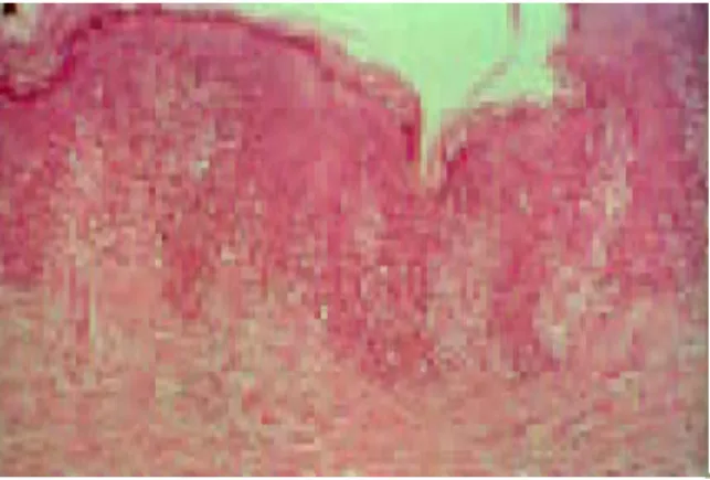
specimens from lesional skin were routinely processed for usually express a CD8 and/or CD56 positive phenotype. formalin xation and paraYn embedding. Histopathologic They represent a heterogeneous group of lymphomas examination showed a dense intraepidermal in ltrate of showing various features with regard to clinicopathol- medium/large neoplastic lymphoid cells with clear, abundant cytoplasm, hyperchromatic nucleus and prominent nucleoli, ogic pro le, immunophenotypic features, clinical course
scattered in the basal and suprabasal layers of the epidermis and prognosis (3). All cytotoxic lymphocytes express a (Fig. 2). A few atypical lymphoid cells were also present set of toxic proteins, e.g. perforins, granzymes A (GrA) around the blood vessels of the papillary dermis. The pheno-and B (GrB), pheno-and the T-cell intracellular antigen-1 typical pro le of the intraepidermal lymphocytes was as fol-(TIA-1) (4), which are reliable markers of cells with
activated cytotoxic function (2).
Gemcitabine is a nucleoside antimetabolite with estab-lished activity against several solid tumours showing promising results in the treatment of lymphoproliferative malignancies. Gemcitabine is a cytosine analogue that causes less myelosuppression as well as immuno-suppression compared with other available nucleoside analogues (5).
We describe here a 35-year-old patient with a primary cutaneous T-cell lymphoma presenting with clinicopa-thologic features of DPR and showing a CD4/CD8 double negative, TIA-1/granzyme B cytotoxic positive phenotype. Furthermore, we report the eYcacy of
gemci-tabine treatment in this aggressive lymphoproliferative Fig. 1. Erythemato-violaceous plaques and nodules at time of presentation.
disorder.
Letters to the Editor 315 up or limited genotypic and/or phenotypic characteriza-tion (2, 6, 7–12). In the past, there had been debate concerning the origin of the atypical cells in pagetoid reticulosis, up until their T-lymphocytic nature was de n-itively established through immunophenotypic and geno-phenotypic studies (13). Another controversial aspect of DPR concerns its association with mycosis fungoides based on their clinical and histopathologic similarities. Therefore many authors consider DPR as an aggressive variant of mycosis fungoides (11). Nevertheless, there are several clinical, histopathologic and immunophenotypic features that distinguish these two entities (14). DPR seems to be an aggressive clonal CTCL with distinctive
Fig. 2. A dense intraepidermal pagetoid in ltrate of medium/large clinicopathological ndings and heterogeneous
immuno-neoplastic lymphoid cells scattered in the basal and suprabasal layers phenotype including CD4
+
T-helper, or CD8+
cyto-of the epidermidis. (Haematoxylin-eosin stain; original magni cationtoxic/suppressor or CD4/CD8 double-negative phenotype ( ´ 50.)
together with ab or cd TCR expression (6–8). Our patient’s neoplastic cells clearly expressed the above-lows: bF-1
+
(T-cell Sciences, Cambridge, USA), CD3+
mentioned cytotoxic proteins, strongly suggesting their (Ylem, L’Aquila, Italy), TIA-1+
(Immunotech, Marseille, origin from an activated cytotoxic T-cell subset, although France) (Fig. 3), GrB+
(Chemicon, Tamecula, USA), CD4-, they did not express CD8 antigen on their cell surface. CD8-, and CD-56-, (Neomarkers, Freemont, USA),CD30-Furthermore, they did not express markers characteristic (Dako, Glostrup, Denmark), CD34-, TdT-, CD20- (Ylem).
of natural killer cells or cd T cells. The negative staining Based on the clinicopathological ndings, primary cutaneous
cytotoxic T-cell lymphoma was diagnosed. The patient was for TdT and CD34 rules out their derivation from a initially treated with IFN-a 3,000,000 I.U. 3 times weekly, T-lymphocyte precursor lineage. According to the subcutaneously, and etretinate 25 mg/day for 3 months, but EORTC classi cation, our patient should t the diagnosis without bene t. The patient then underwent chemotherapy
of CD30 negative pleomorphic large T-cell cutaneous with dexamethasone, cytarabine and cisplatin (DHAP) for 3
lymphoma (15). Lack of CD8 antigen does not allow us cycles each, every 28 days, but again with no improvement.
Therefore, therapy with slow intravenous infusion of gemcitab- strictly to classify our case among the so-called cytotoxic ine at a dosage of 1,250 mg/m2 on days 1, 8 and 15 of a 28-day CTCLs (2), which are characterized by a distinctive schedule (8) was started. Initial improvement of the lesions combination of clinical, histopathological and immuno-was observed after the second cycle of treatment and a
phenotypical features (bF1
+
, CD3+
, CD8+
, CD7+
, complete response was obtained after 6 courses. However, theCD45RA
+
, TIA-1/GMP-17+
) and which usually run cutaneous lesions worsened 3 months after the last gemcitabinecycle and the patient refused any further therapy. He died of an aggressive clinical course (2).
systemic disease 18 months after the initial diagnosis. Our patient was unresponsive to IFN-a in association with etretinate and DHAP therapies and because no eVective standardized cure is available for DPR, we DISCUSSION
started treatment with gemcitabine while waiting for a Few studies on DPR have been reported so far and most bone marrow transplant. Gemcitabine led to a rapid of them include only a few patients, short-term follow- improvement of the skin lesions in our patient, although it did not prevent relapse of the disease 3 months after the end of therapy.
In conclusion, we believe that our case could contrib-ute to the knowledge on the relationship between DPR, cytotoxic cutaneous lymphomas and other CTCLs. Furthermore, we believe that our experience in the use of gemcitabine could contribute to new modalities in the treatment of cytotoxic cutaneous lymphomas, since aggressive therapeutical approaches are often ineVective and therefore new strategies are needed.
REFERENCES
1. Ketron LW, Goodman MH. Multiple lesions of the skin
Fig. 3. The cytotoxic phenotype of the neoplastic cells is demonstrated apparently of epithelial origin resembling clinically mycoses fungoides. Arch Dermatol Syph 1931: 24: by the expression of T-cell intracellular antigen-1. (Haematoxylin
counterstain; original magni cation ( ´ 400.) 758–785.
316 Letters to the Editor
2. Berti E, Tomasini D, Vermeer MH, Meijer CJ, Alessi E, 9. Ralfkiaer E, Thomsen K, Agdal N, Hou-Jensen K, Wantzin GL. The development of a Ki-1-positive large Willemze R. Primary cutaneous CD8-positive
epidermo-tropic cytotoxic T-cell lymphomas. Am J Pathol 1999; cell non-Hodgkin’s lymphoma in pagetoid reticulosis. Acta Derm Venereol 1989; 69: 206–211.
155: 483–492.
3. Santucci M, Pimpinelli N, Massi D, Kadin ME, Meijer 10. Luther H, Bacharach-Buhles M, Schultz-Ehrenburg U, Altmeyer P. Pagetoide retikulose von typ Ketron-CJLM, Muller-Hermelink HK, et al. Cytotoxic/natural
killer cell cutaneous lymphomas: a clinicopathological Goodman. Hautarzt 1989; 40: 530–535.
11. Lacour JP, Juhlin L, El Baze P, Barety M, Ortonne JP. study of 48 cases from the EORTC cutaneous lymphoma
study group. Blood 2002; in press. Disseminated pagetoid reticulosis associated with mycosis fungoides: immunomorphologic study. J Am Acad 4. Krenacs L, Wellmann A, Sorbara L, Himmelmann AW,
Bagdi E, JaVe ES, et al. Cytotoxic cell antigen in anaplastic Dermatol 1986; 5: 898–901.
12. Bukulmez G, Atakan N, Taskin M, Bilezikci B, Uner A. large cell lymphomas of T- and null-cell type and
Hodgkin’s disease: evidence for distinct cellular origin. Disseminated pagetoid reticulosis: plaques and tumoral lesions occurring simultaneously in the same patient. J Blood 1997; 89: 980–989.
5. Zinzani PL, Baliva G, Magagnoli M, Bendandi M, Eur Acad Dermatol Venereol 2001; 15: 59–61.
13. Slater D, Goepel J, Walker A, Corbett P. Lymphocyte Modugno G, Gherlinzoni F, et al. Gemcitabine treatment
in pretreated cutaneous T-cell lymphoma: experience in subsets in pagetoid reticulosis. Br J Dermatol 1984; 11: 244–246.
44 patients. J Clin Oncol 2000; 18: 2603–2606.
6. Haghighi B, Smoller BR, LeBoit PE, Warnke RA, Sander 14. Woringer FR, Kolopp P. Le´sion e´rythe´mato-squameuse polycyclique de l’avant-bras e´voluant depuis 6 ans chez CA, Kohler S. Pagetoid reticulosis ( Woringer-Kolopp
disease): an immunophenotypic, molecular, and clinicopa- un garc¸onnet de 13 ans: histologiquement in ltrat intra-epidermique d’apparence tumorale. Ann Dermatol Syph thologic study. Mol Pathol 2000; 13: 502–510.
7. Mielke V, WolV HH, Winzer M, Sterry W. Localized and 1939; 10: 945–948.
15. Willemze R, Kerl H, Sterry W, Berti E, Cerroni L, disseminated pagetoid reticulosis: diagnostic
immuno-phenotypical ndings. Arch Dermatol 1989; 125: 402–406. Chimenti S, et al. EORTC classi cation for primary cutaneous lymphomas: a proposal from the cutaneous 8. Berti E, Cerri A, Cavicchini S, Delia G, Soligo D, Alessi
E, et al. Primary cutaneous gamma/delta T-cell lymphoma lymphoma study group of the European Organization for Research and Treatment of Cancer. Blood 1997; 90: presenting as disseminated pagetoid reticulosis. J Invest
Dermatol 1991; 96: 718–723. 354–371.
Eosinophilic Pustular Folliculitis Induced by Allopurinol and Timepidium Bromide
Hideki Maejima1, Hideki Mukai2 and Eto Hikaru11Department of Dermatology, St. Luke’s International Hospital, 9-1 Akashi-cho, Chuo-ku Tokyo, Japan 104-8560, and the 2Division of Dermatology, Yokohama Rosai Hospital, Yokohama, Japan. E-mail [email protected]
Accepted April 22, 2002.
Sir,
We describe a woman with numerous papules and pustules on her face and upper trunk induced by allo-purinol and timepidium bromide. The histopathology showed the destruction of hair follicles and the in ltra-tion of eosinophils, which we diagnosed as eosinophilic pustular folliculitis.
CASE REPORT
A 57-year-old Japanese woman was treated with oral allopurinol and timepidium bromide for ureterolith since April 1998. One month later, she presented with an
Fig. 1. Clinical ndings at rst examination: numerous papules and
eruption on her face, followed by numerous rice-sized pustules on the patient’s face. papules and pustules on her face and upper trunk, a
fever and bilateral cervical lymphoadenopath y were also
present (Fig. 1). The woman visited our hospital for vic transaminase, 65 IU/ml; gammaglutamyltrans -peptidase, 159 IU/ml; alkaline phosphatase, 436 IU/ml ) examination in June 1998. Laboratory studies revealed
eosinophilia (white blood cell, 7,400/mm3; eosinophile, and a strong in ammatory reaction (C-reactive protein, 8.3 mg/dl ). No elevations were found in her serum viral 25%, 1,850/mm3), mild liver dysfunction (glutamic
oxaloacetic transaminase, 36 IU/ml; glutamic pyru- titers (human herpes simplex virus, Epstein-Barr virus