Heart Failure
Aerobic Training Decreases
B-Type Natriuretic Peptide Expression and
Adrenergic Activation in Patients With Heart Failure
Claudio Passino, MD,*† Silvia Severino, MS,* Roberta Poletti, MD,* Massimo F. Piepoli, MD,‡ Chiara Mammini, MD,* Aldo Clerico, MD,*† Alessandra Gabutti, MD,* Guido Nassi, MS,* Michele Emdin, MD, PHD*Pisa and Piacanza, Italy
OBJECTIVES We sought to evaluate the effect of physical training on neurohormonal activation in patients with heart failure (HF).
BACKGROUND Patients with HF benefit from physical training. Chronic neurohormonal activation has detrimental effects on ventricular remodeling and prognosis of patients with HF.
METHODS A total of 95 patients with HF were assigned randomly into two groups: 47 patients (group T) underwent a nine-month training program at 60% of the maximal oxygen uptake (VO2),
whereas 48 patients did not (group C). The exercise load was adjusted during follow-up to achieve a progressive training effect. Plasma assay of B-type natriuretic peptide (BNP), amino-terminal pro-brain natriuretic peptide (NT-proBNP), norepinephrine, plasma renin activity, and aldosterone; quality-of-life questionnaire; echocardiogram; and cardiopulmonary stress test were performed upon enrollment and at the third and ninth month.
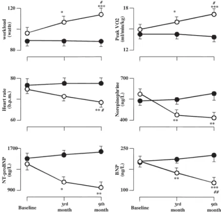
RESULTS A total of 85 patients completed the protocol (44 in group T, left ventricular ejection fraction [EF] 35 ⫾ 2%, mean ⫾ SEM; and 41 in group C, EF 32 ⫾ 2%, p ⫽ NS). At the ninth month, patients who underwent training showed an improvement in workload (⫹14%, p ⬍ 0.001), peak VO2(⫹13%, p ⬍ 0.001), systolic function (EF ⫹9%, p ⬍ 0.01), and quality of
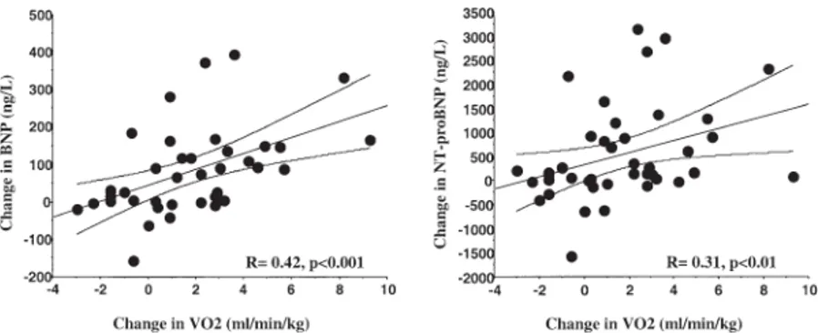
life. We noted that BNP, NT-proBNP, and norepinephrine values decreased after training (⫺34%, p ⬍ 0.01; ⫺32%, p ⬍ 0.05; ⫺26%, p ⬍ 0.01, respectively). Increase in peak VO2with
training correlated significantly with the decrease in both BNP/NT-proBNP level (p⬍ 0.001 and p⬍ 0.01, respectively). Patients who did not undergo training showed no changes. CONCLUSIONS Clinical benefits after physical training in patients with HF are associated with blunting of
adrenergic overactivity and of natriuretic peptide overexpression. (J Am Coll Cardiol 2006; 47:1835–9) © 2006 by the American College of Cardiology Foundation
Chronic heart failure (HF) is characterized by fatigue, dyspnea, abnormal peak oxygen uptake (peak VO2), and
ventilatory efficiency to exercise measured by the slope of increase in ventilation over carbon dioxide output (VE/VCO2
slope) (1). Despite pharmacologic therapy, the prognosis is poor. Aerobic physical training is known to improve exercise tolerance (2–5), ventilatory efficiency (5), quality of life (QOL) (4), and to counteract sympathetic overactivation (3). Finally, rates of mortality and hospitalization decrease after patients undergo training independently from medical treatment (6).
Neurohormonal activation is a hallmark of HF (7). The production of B-type natriuretic peptide (BNP) by ventric-ular cardiomyocytes correlates with left ventricventric-ular dysfunc-tion (8,9). Thus, the assay of either BNP or amino-terminal brain natriuretic peptide (NT-proBNP) has been pro-posed as diagnostic and prognostic tool (9). However, studies concerning the influence of training on BNP
secre-tion early after myocardial infarcsecre-tion (10) and in patients with chronic HF (11,12) have given conflicting results. In this study, we evaluated the effect of home-based aerobic physical training, with adjustment of the exercise load during follow-up for achieving a progressive training effect, on neurohormonal activation in patients in HF.
METHODS
Subjects. We screened 158 consecutive patients with HF from 2002 to 2004. Inclusion criteria were impaired left ventricular systolic function (ejection fraction [EF] ⬍45%) and exercise capacity (peak VO2⬍25 ml/min/kg).
Exclu-sion criteria were New York Heart Association (NYHA) functional class IV, myocardial infarction or unstable angina within six months before the examination, exercise-limiting diseases, and severe pulmonary or renal disease. Of the 158 patients, 35 did not match these criteria, whereas 28 refused to sign the informed consent approved by the institutional ethical committee. Ultimately, 95 patients (80 men and 15 women, mean age 60 ⫾ 1 years) were enrolled. All 95 patients were on stable (i.e., ⬎1 month) optimal pharma-cologic treatment (Table 1). A significant change in
treat-From the *Cardiovascular Medicine Department, CNR Institute of Clinical Physiology, Pisa, Italy; †Scuba Superiore S. Anna, Pisa, Italy; and ‡G. da Saliceto Hospital, Piacenza, Italy.
Manuscript received September 21, 2005; revised manuscript received December 5, 2005, accepted December 13, 2005.
ment or hospitalization resulted in withdrawal from the study.
Study design. This was a prospective, randomized, con-trolled study. Patients were assigned randomly into two groups. The training group (group T) underwent a nine-month train-ing program, whereas control patients (group C) continued their usual lifestyle. The two groups did not differ as to age, gender, NYHA functional class, EF, pharmacologic treatment, or HF etiology (Table 1).
Patients underwent neurohormonal and clinical evalua-tion and a maximal exercise test with gas exchange (cardio-pulmonary stress test [CPT]) within the same morning upon enrollment and at the third and ninth month. An echocardiographic study was performed, and patients rated their QOL on entry and at the end of the study.
Physical training. The training program consisted of cy-cling on a bike for a minimum of 3 days per week, 30 min per day. Patients were instructed to exercise at 60 rpm, keeping heart rate constantly monitored at 65% of peak VO2
heart rate, by a wearable device. At the enrollment, they
participated to in-hospital training sessions. Their compli-ance to the program was checked by a physiotherapist and at the first, second, third, sixth, and ninth month. At the third-month visit, workload was adjusted according to a new CPT. Patients in group C underwent follow-up visits at the third and ninth month to exclude changes in their usual lifestyle and physical activity.
Cardiopulmonary exercise test, echocardiographic study, QOL, and plasma assays. A CPT was performed by exercise on a bicycle ergometer using a ramp protocol with increments of 10 W/min. We measured VO2, CO2
produc-tion, and minute ventilation using breath-to-breath gas analysis (Vmax, Sensormedics, Conshohocken, Pennsylva-nia). Peak VO2(the highest value at end-exercise, as a 20-s
average) and ventilatory efficiency on exercise (slope of the ventilation vs. VCO2relation in its linear part) were
deter-mined. The same physician performed all the CPTs and was unaware of the results of blood sampling. The echocardio-graphic studies were all performed by the same physician.
Quality of life was evaluated by means of the Minnesota Living With Heart Failure Questionnaire; BNP, plasma catecholamines, plasma renin activity, and aldosterone were assayed as described elsewhere (9); and NT-proBNP was measured with an automated electrochemiluminescent immunoassay.
Statistics. Because neurohormone values are not distrib-uted normally, natural logarithmic transformation of data was used for parametric statistical analysis, including two-way repeated-measure analysis of variance, Bonferroni post-hoc test, and linear regression analysis. Skewness and Kurtosis tests were used to determine whether data were normally distributed. Chi-square and unpaired t tests were used to evaluate differences between the two groups. Mauchly’s test to assess the sphericity assumption was performed. To assess the significance of the interaction between groups, we applied the Greenhouse-Geisser correction factor when Mauchly’s test was significant (13). The results are ex-pressed as mean values⫾ SEM, and p values were consid-ered significant when⬍ 0.05.
RESULTS
Three patients in group T did not complete the program: one required treatment changes, one refused to continue after the first month, and one withdrew a after three weeks because of a stroke. Conversely, seven patients in group C did not complete the program: two were lost at follow-up; two required hospitalization for decompensation; two needed treatment changes; and one withdrew after a diagnosis of lung cancer. A total of 85 patients completed the protocol (44 in group T, 41 in group C); of these, 5 patients (2 in group T and 3 in group C), included in data analysis, required only minimal treatment adjustments (change of furosemide dosage by 25 mg).
Table 1. Baseline Characteristics of the Study Patients Assigned Randomly to Group T or C Group T (nⴝ 44) Group C (nⴝ 41) Age (yrs) 60⫾ 2 61⫾ 2 Males/females (n) 39/5 35/6 BMI (kg/m2) 25.8⫾ 0.3 25.1⫾ 0.8 Idiopathic/postischemic (n) 17/27 18/23 LVEF (%) 35.3⫾ 1.6 32.3⫾ 2.2 Sinus rhythm/AF (n) 41/3 39/2
NYHA functional class I/II/III (n⫽ mean ⫾ SEM) 6/28/10 (2.1⫾ 0.1) 8/23/10 (2.1⫾ 0.1) Treatment (%) Furosemide 82 85 Beta-blocker 72 73 ACE inhibitor 78 79 ARB 7 5 Spironolactone 35 38
No significant differences were found for any of the comparisons.
ACE⫽ angiotensinogen-converting enzyme; AF ⫽ atrial fibrillation; ARB ⫽ angiotensin receptor blocker; BMI⫽ body mass index; Group C ⫽ control group; Group T⫽ training group; LVEF ⫽ left ventricular ejection fraction; NYHA ⫽ New York Heart Association.
Abbreviations and Acronyms
BNP ⫽ B-type natriuretic peptide CPT ⫽ cardiopulmonary stress test EF ⫽ ejection fraction
HF ⫽ heart failure
NT-proBNP⫽ amino-terminal pro-brain natriuretic peptide
NYHA ⫽ New York Heart Association QOL ⫽ quality of life
VE/VCO2 ⫽ ventilation/carbon dioxide production
ratio VO2 ⫽ oxygen uptake
Baseline neurohormones and functional capacity. As expected, patients from both group T and C showed neurohormonal activation, depressed functional capacity, and a slightly reduced ventilatory efficiency, with no differ-ent baseline values (Tables 1and2).
Significant relations were found among exercise variables and neurohormone plasma levels in both groups, for in-stance, in group T, peak VO2was inversely correlated with
BNP (R⫽ 0.55, p ⬍ 0.001), NT-proBNP (R ⫽ 0.55, p ⬍ 0.001), and norepinephrine (R⫽ 0.43, p ⬍ 0.001), whereas VE/VCO2slope directly correlated with BNP (R⫽ 0.61, p ⬍
0.001), NT-proBNP (R ⫽ 0.54, p ⬍ 0.001), and norepi-nephrine (R ⫽ 0.43, p ⬍ 0.001).
Effects of physical training on left ventricular volumes and function, functional capacity, and QOL. In group T, EF had increased by the nine-month visit, with a reduction
Figure 1. Changes in exercise variables, resting heart rate, and neurohormones for trained (open circles) and untrained patients (closed circles) at baseline, three, and nine months. *p⬍ 0.05, **p ⬍ 0.01, ***p ⬍ 0.001 versus baseline (significant values assessed by Bonferroni post-hoc test performed on three groups); #p⬍ 0.05, ##p ⬍ 0.01 versus third month. BNP ⫽ brain natriuretic peptide; b.p.m. ⫽ beats/min; NT-proBNP ⫽ amino-terminal pro-brain natriuretic peptide; VO2⫽ oxygen uptake.
Table 2. Echocardiographic, Functional, and Neurohormonal Profile at Baseline and at End of Follow-Up
Group T Group C
Baseline Ninth Month Baseline Ninth Month
Ejection fraction (%) 35⫾ 2 38⫾ 2§ 32⫾ 2 31⫾ 2† LV-EDVi (ml/m2) 111⫾ 8 96⫾ 6‡ 116⫾ 7 120⫾ 8† LV-ESVi (ml/m2) 72⫾ 6 58⫾ 5‡ 78⫾ 6 80⫾ 7† Peak VO2(ml/min/kg) 15⫾ 1 17⫾ 1储 14⫾ 1 13⫾ 1† Workload (W) 99⫾ 6 114⫾ 6储 85⫾ 6 80⫾ 5† VE/VCO2slope 34⫾ 1 33⫾ 1 36⫾ 1 37⫾ 1* BNP (ng/l) 187⫾ 29 123⫾ 23§ 188⫾ 27 201⫾ 32† NT-proBNP (ng/l) 1,370⫾ 234 929⫾ 206‡ 1,581⫾ 251 1,677⫾ 291† Norepinephrine (ng/l) 607⫾ 55 447⫾ 39§ 515⫾ 34 609⫾ 67† PRA (ng/ml/h) 3.04⫾ 0.66 2.96⫾ 0.62 4.26⫾ 1.21 2.45⫾ 0.56 Aldosterone (ng/l) 198⫾ 27 186⫾ 24 195⫾ 26 216⫾ 25
*p⬍ 0.05 vs. group T, †p ⬍ 0.001 vs. group T, ‡p ⬍ 0.05, §p ⬍ 0.01 vs. respective baseline, 储p ⬍ 0.001 vs. respective baseline. BNP⫽ B-type natriuretic peptide; EDVi ⫽ end-diastolic volume normalized by body surface area; ESVi ⫽ end-systolic volume normalized by body surface area; LV⫽ left ventricular; NT-proBNP ⫽ N-terminal probrain natriuretic peptide; PRA ⫽ plasma renin activity; VE⫽ ventilation; VCO2⫽ carbon dioxide production; VO2⫽ oxygen uptake.
in end-diastolic and end-systolic left ventricular volumes (Table 2). Conversely, echocardiographic parameters did not change in group C.
Group T progressively increased tolerance to effort from baseline up to the third and the ninth month (peak VO2F⫽
10.35, p⬍ 0.001, and maximum workload F ⫽ 13.26, p ⬍ 0.001) (Fig. 1). Resting heart rate decreased by the end of the follow-up from 75⫾ 2 beats/min to 69 ⫾ 2 beats/min (p⫽ 0.002). No changes were found in these parameters in group C (Fig. 1). By the end of follow-up, VE/VCO2slope
did not change in either of the two groups, and only patients in group T showed an improvement in their QOL (score from 54 ⫾ 5 to 32 ⫾ 4 in group T, p ⬍ 0.01; from 52 ⫾ 6 to 53⫾ 5 in group C, p ⫽ NS).
Effects of physical training on BNP and neurohormones. During the follow-up, BNP, NT-proBNP, and norepi-nephrine progressively decreased in group T (F⫽ 7.49, p ⬍ 0.01; F ⫽ 4.84, p ⬍ 0.01; and F ⫽ 9.88, p ⬍ 0.001, respectively) but not in group C (Fig. 1). No changes were found in either group with respect to plasma renin activity and aldosterone. At the end of the program, peak VO2 in
group T inversely correlated to BNP (R⫽ 0.52, p ⬍ 0.001), NT-proBNP (R ⫽ 0.51, p ⬍ 0.001), and norepinephrine levels (R⫽ 0.49, p ⬍ 0.001) at a similar extent, as compared with baseline. A similar correlation was found between VE/VCO2 slope and the levels of BNP (R ⫽ 0.61, p ⬍
0.001), NT-proBNP (R ⫽ 0.60, p ⬍ 0.001), and norepi-nephrine (R⫽ 0.59, p ⬍ 0.001). The change in peak VO2
at the end of the program was correlated with BNP and NT-proBNP changes (R⫽ 0.42, p ⬍ 0.001 and R ⫽ 0.31, p ⬍ 0.01, respectively), but not with norepinephrine changes (Fig. 2). Conversely, no correlation was found between the decrease in BNP, NT-proBNP, and norepi-nephrine values and the absolute 3% increase in EF in group T. Finally, a relationship was found between the change in either BNP or NT-proBNP level and the norepinephrine change (R⫽ 0.46, p ⫽ 0.002 and R⫽ 0.36, p ⫽ 0.02, respectively).
DISCUSSION
The present study demonstrates that progressively adjusted aerobic physical training on a nine-month period
progres-sively improves functional capacity, QOL, as well as left ventricular function in patients with HF. These beneficial changes are associated with reduction in plasma levels of proBNP and norepinephrine. Hence, BNP/NT-proBNP plasma concentration appears to be a valuable marker of positive clinical response to training.
Previous studies have demonstrated that training im-proves cardiac function, peak oxygen uptake, and QOL (2– 6,11,12). However, long-term training programs per-formed with a constant intensity of exercise do not seem to guarantee a continuous improvement in functional capacity (4). Conversely, a workload adjusted according to the third-month CPT produced a progressive increase in peak VO2,
paralleled by a progressive decrease in neurohormone level in the present study.
The lack of improvement of the VE/VCO2slope could be
ascribed to the quite-preserved exercise capacity and venti-latory efficiency in our population (Table 2) as compared with a previous study (14). The reduction in plasma BNP/ NT-proBNP in group T might be an expression of im-proved cardiac systolic function (8), sympathetic deactiva-tion (12), and improvement in tissue oxygenation (15). The decrease in plasma norepinephrine level might be explained by the positive modulation of the autonomic balance by physical training (3).
Study limitations. A control group of healthy subjects was not included because it is unlikely that training could affect their BNP/NT-proBNP plasma levels under physiologic conditions (8). Furthermore, women accounted for only 15% of the patients, a figure which reflects the female prevalence of larger HF studies, among which the Extra-MATCH meta-analysis on training in HF (6).
Conclusions. The BNP/NT-proBNP and norepinephrine plasma values progressively decrease after training, parallel-ing functional and clinical improvement. The present find-ings suggest the usefulness of the BNP/NT-proBNP assay as an effective tool for the follow-up of patients with HF undergoing physical training.
Acknowledgments
The authors thank Maria Giovanna Mazzei for QOL questionnaire interpretation, Concetta Prontera for neuro-Figure 2. Relation between the changes in peak oxygen uptake (VO2) and in plasma concentration of cardiac natriuretic peptides after training in group T patients. BNP⫽ brain natriuretic peptide.
hormonal assaying, Luc Zyw for statistical advice, and Manuella Walker for linguistic assistance.
Reprint requests and correspondence: Dr. Claudio Passino,
CNR Institute of Clinical Physiology, Via Moruzzi 1, 56124 Pisa, Italy. E-mail: [email protected].
REFERENCES
1. Francis DP, Shamim W, Davies LC, et al. Cardiopulmonary exercise testing for prognosis in chronic heart failure: continuous and indepen-dent prognostic value from VE/VCO(2)slope and peak VO(2). Eur Heart J 2000;21:154 – 61.
2. Sullivan MJ, Higginbotham MB, Cobb FR. Exercise training in patients with severe left ventricular dysfunction. Hemodynamic and metabolic effects. Circulation 1988;78:506 –15.
3. Coats AJS, Adamopoulos S, Radaelli A, et al. Controlled trial of physical training in chronic heart failure; exercise performance, hemodynamics, venti-lation and autonomic function. Circuventi-lation 1992;85:2119–31.
4. Belardinelli R, Georgiou D, Cianci G, Purcaro A. Randomized, controlled trial of long-term moderate exercise training in chronic heart failure: effects on functional capacity, quality of life, and clinical outcome. Circulation 1999;99:1173– 82.
5. Piepoli M, Clark AL, Volterrani M, Adamopoulos S, Sleight P, Coats AJ. Contribution of muscle afferents to the hemodynamic, autonomic, and ventilatory responses to exercise in patients with chronic heart failure: effects of physical training. Circulation 1996;93:940 –52.
6. Piepoli MF, Davos C, Francis DP, Coats AJ, for the ExTraMATCH Collaborative. Exercise training meta-analysis of trials in patients with chronic heart failure (ExTraMATCH). BMJ 2004;328:189 –92. 7. Packer M. The neurohormonal hypothesis: a theory to explain the
mechanisms of disease progression in heart failure. J Am Coll Cardiol 1992;20:248 –54.
8. Clerico A, Emdin M. Diagnostic accuracy and prognostic relevance of the measurement of cardiac natriuretic peptides: a review. Clin Chem 2004;50:33–50.
9. Emdin M, Passino C, Prontera C, et al. Cardiac natriuretic hormones, neuro-hormones, thyroid hormones and cytokines in normal subjects and patients with heart failure. Clin Chem Lab Med 2004;42:627–36. 10. Yamamoto A, Tanabe K, Yokoyama Y, Itoh H, Murayama M. Influence of aerobic exercise training on brain natriuretic peptide secretion in patients in the chronic phase of myocardial infarction. Jpn Circ J 1998;62:658 – 64.
11. Braith RW, Welsch MA, Feigenbaum MS, Kluess HA, Pepine CJ. Neuroendocrine activation in heart failure is modified by endurance exercise training. J Am Coll Cardiol 1999;34:1170 –5.
12. Conraads VM, Beckers P, Vaes J, et al. Combined endurance/ resistance training reduces NT-proBNP levels in patients with chronic heart failure. Eur Heart J 2004;25:1797– 805.
13. Chen X, Zhao PL, Zhang J. A note on ANOVA assumptions and robust analysis for a cross-over study. Stat Med 2002;21:1377– 86. 14. Davey P, Meyer T, Coats A, et al. Ventilation in chronic heart failure:
effects of physical training. Br Heart J 1992;68:473–7.
15. Baxter GF. Natriuretic peptides and myocardial ischaemia. Basic Res Cardiol 2004;99:90 –3.