BACKGROUND: Prenatal diagnosis of coarctation of the aorta (CoA) is still
challenging and affected by high rates of false-positive diagnoses. The aim
of this study was to ascertain the strength of association and to quantify
the diagnostic accuracy of different ultrasound signs in predicting CoA
prenatally.
METHODS: Medline, Embase, CINAHL, and Cochrane databases were
searched. Random-effects and hierarchical summary receiver operating
characteristic model meta-analyses were used to analyze the data.
RESULTS: Seven hundred ninety-four articles were identified, and 12 (922
fetuses at risk for CoA) articles were included. Mean mitral valve diameter
z score was lower (P<0.001) and the mean tricuspid valve diameter z
score was higher in fetuses with CoA than in those without CoA (P=0.01).
Mean aortic valve diameter z score was lower in fetuses with CoA than in
healthy fetuses (P≤0.001), but the ascending aorta diameter, expressed
as z score or millimeters, was similar between groups (P=0.07 and 0.47,
respectively). Mean aortic isthmus diameter z scores measured either
in sagittal (P=0.02) or in 3-vessel trachea view (P<0.001) were lower
in fetuses with CoA. Conversely, the mean pulmonary artery diameter z
score, the right/left ventricular and pulmonary artery/ascending aorta
diameter ratios were higher (P<0.001, P=0.02, and P=0.02, respectively)
in fetuses with CoA in comparison with controls, although aortic isthmus/
arterial duct diameter ratio was lower in fetuses with CoA than in those
without CoA (P<0.001). The presence of coarctation shelf and aortic
arch hypoplasia were more common in fetuses with CoA than in controls
(odds ratio, 26.0; 95% confidence interval, 4.42–153; P<0.001 and
odds ratio, 38.2; 95% confidence interval, 3.01–486; P=0.005), whereas
persistent left superior vena cava (P=0.85), ventricular septal defect
(P=0.12), and bicuspid aortic valve (P=0.14) did not carry an increased
risk for this anomaly. Multiparametric diagnostic models integrating
different ultrasound signs for the detection of CoA were associated with
an increased detection rate.
CONCLUSIONS: The detection rate of CoA may improve when a
multiple-criteria prediction model is adopted. Further large multicenter studies
sharing the same imaging protocols are needed to develop objective
models for risk assessment in these fetuses.
Risk Factors for Coarctation of the Aorta on
Prenatal Ultrasound
A Systematic Review and Meta-Analysis
© 2016 American Heart Association, Inc.
Correspondence to: Francesco D’Antonio, MD, PhD, Department of Clinical Medicine, Faculty of Health Sciences, UiT - The Arctic University of Norway, Hansine Hansens veg 18, 9019 Tromsø, Norway. E-mail francesco. [email protected]
Sources of Funding, see page 784 Key Words: aortic coarctation ◼ heart defects, congenital ◼ fetal echocardiography ◼ prenatal diagnosis
Alessandra Familiari, MD
Maddalena Morlando, MD
Asma Khalil, MD
Sven-Erik Sonesson,
MD, PhD
Carolina Scala, MD
Giuseppe Rizzo, MD
Gelsomina Del Sordo, MD
Chiara Vassallo, MD
Maria Elena Flacco, MD
Lamberto Manzoli, MD
Antonio Lanzone,
MD, PhD
Giovanni Scambia,
MD, PhD
Ganesh Acharya, MD, PhD
Francesco D’Antonio,
MD, PhD
ORIGINAL RESEARCH
AR
TICLE
C
oarctation of the aorta (CoA) is one of the most
common congenital heart defects in the pediatric
population, accounting for 5% to 8% of children
with congenital heart defects.
1It has been classically
de-fined as a discrete narrowing of the aorta in the region of
the ligamentum arteriosum, although more diffuse forms
of the disease may involve the arch or isthmus to varying
degrees.
2The importance of prenatal diagnosis of CoA relies
on the fact that the burden of mortality and morbidity
associated with this anomaly is significantly higher when
prenatal detection is missed.
3CoA does not cause fetal
circulatory compromise in utero because the aortic
isth-mus is not an essential component of the fetal
circula-tion; however, after birth and ductal closure, a critical
coarctation will result in poor perfusion of the lower body
and acidemia that, together with an increase in left
ven-tricular afterload, might result in acute circulatory shock.
Cases with a less narrow CoA can be completely
asymp-tomatic, develop arterial collateral vessels that bypass
the aortic obstruction, and remain asymptomatic until
they are diagnosed with hypertension.
Prenatal diagnosis of CoA allows planning delivery in a
center with pediatric cardiology service, starting
prosta-glandin infusion immediately after birth to maintain
duc-tal patency, and performing surgery electively. Although
the current rate of mortality and morbidity for this
condi-tion is lower than in the past, lifelong follow-up is needed
in view of the high rates of hypertension and need for
reintervention later in life.
4Prenatal detection of CoA has been reported to be
poor, in general, and this anomaly is usually not suspected
until the third trimester of pregnancy when ventricular or
vascular disproportion is detected.
5,6However, because
the fetal heart has a normal physiological right-sided
dom-inance that increases with gestation, the use of
cardio-vascular disproportion alone has an overall low diagnostic
accuracy that is even lower during the third trimester.
Several ultrasound signs have been proposed to
po-tentially improve the detection rate of prenatal diagnosis
for CoA.
The primary aim of this systematic review was to
iden-tify the ultrasonographic cardiovascular parameters
asso-ciated with the occurrence of CoA. The secondary aim was
to develop a prediction model combining these ultrasound
predictors to improve the prenatal diagnosis of CoA.
METHODS
Protocol, Eligibility Criteria, Information
Sources, and Search
This review was performed according to an a priori designed pro-tocol using methods recommended for systematic reviews and meta-analysis.7,8 Medline, Embase, CINAHL, and Cochrane
data-bases were searched electronically on May 2, 2016 by using com-binations of the relevant medical subject heading (MeSH) terms, key words, and word variants for “coarctation of aorta,” “prenatal diagnosis,” and “ultrasound” (online-only Data Supplement Table I). The search and selection criteria were restricted to the English language. PRISMA guidelines were followed.9
The study was registered with the PROSPERO database (Registration number: CRD42016038845).
Study Selection, Data Collection, and Data Items
Two authors (A.F., M.M.) reviewed all abstracts indepen-dently. Agreement regarding potential relevance was reached by consensus. Full-text copies of those articles were obtained, and the same 2 reviewers independently extracted relevant data regarding study characteristics and pregnancy outcome. If >1 study was published for the same cohort with identical end points, the report containing the most comprehensive information on the population was included to avoid overlapping populations. Excluded studies and the reasons for exclusions are listed in online-only Data Supplement Table II.Quality assessment of the included studies was per-formed using the Newcastle-Ottawa Scale. According to the Newcastle-Ottawa Scale, each study is judged on 3 broad per-spectives: the selection of the study groups, the comparability of the groups, and the ascertainment outcome of interest.10
According to the Newcastle-Ottawa Scale, a study can be awarded a maximum of 1 star for each numbered item within the Selection and Outcome categories. A maximum of 2 stars can be given for Comparability.10
Risk of Bias, Summary Measures, and Synthesis
of the Results
We explored the strength of association between different ultrasound parameters in fetuses in comparison with those
Clinical Perspective
What Is New?
• This systematic review of studies on fetuses with cardiovascular disproportion showed that those with the postnatal diagnosis of coarctation of the aorta (CoA) had significant differences in several cardiac morphological parameters in comparison with cases without CoA.
• The presence of a coarctation shelf or hypoplastic arch was associated with a significantly increased risk of CoA.
• Multiparametric diagnostic models were associated with an increased detection rate.
What Are the Clinical Implications?
• Assessment of left inflow and outflow tracts mayhelp in stratifying the risk of CoA prenatally.
• Future large prospective studies are needed to ascertain the diagnostic performance of ultrasound in detecting CoA prenatally.
without CoA that had their diagnosis confirmed or refuted at postnatal imaging or surgery. The analyzed population included fetuses suspected to be at risk for CoA on the basis of cardiovascular disproportion, defined as a discrepancy in size of either cardiac chambers or great vessels, detected at the scan.
The following ultrasound parameters were assessed: • Inflow tracts: tricuspid valve (TV) z score, mitral valve
(MV) z score
• Outflow tracts: aortic valve (AoV) z score, ascending aorta (AAo) z score, AAo diameter, aortic isthmus (AoI)
z score, AoI diameter, pulmonary valve (PV) z score,
main pulmonary artery (MPA) z score, MPA diameter, arterial duct (AD) z score, AoV growth rate, AoI growth rate.
• Ratios: Right ventricular/left ventricular (RV/LV) diam-eter, RV/LV length, RV/LV area, RV/LV volume, TV/MV, MV/TV, PV/AoV, AoV/PV, MPA/AAo, descending aorta/ AoI angle, MPA/AoI, AoI/AD, AD/AoI diameter.
• Doppler signs: AoI pulsatility index, AoI peak systolic velocity, reversed or mixed flow at the aortic arch, bidi-rectional flow at the foramen ovale.
• Other signs: Persistent left superior vena cava (PLSVC), ventricular septal defect, bicuspid aortic valve (BAV), coarctation shelf, arch hypoplasia, left common carotid to left subclavian artery distance, the ratio of the aortic arch diameter at the left subclavian artery and the distance between the left carotid artery and the left subclavian artery, namely the carotid sub-clavian index, AAo/descending aorta angle, trans-verse aortic arch/descending aorta angle, AoI-AD angle.
All z scores, computed on gestational age (GA) and femur length, were considered suitable for inclusion.
Only case-control studies including fetuses undergoing echocardiography for suspected CoA on the basis of car-diovascular disproportion were considered suitable for the inclusion in this study.5,6 Only full-text articles were
consid-ered eligible for the inclusion and all the studies addressing differences in ultrasonographically measured continuous variables in fetuses with CoA in comparison with those without.
The following studies were excluded:
• Studies with missing prenatal information/diagnosis. • Studies reporting the detection rate of prenatal
ultra-sound in diagnosing CoA at the time of the routine anomaly scan without providing a clear description of the ultrasound criteria used.
• Studies performed in the first trimester of pregnancy. • Autopsy-based studies, because fetuses undergoing
termination of pregnancy are more likely to have other associated major structural and chromosomal anoma-lies, thus potentially increasing the detection rate of this condition.
• Studies published before 2000 as advances in prenatal imaging techniques have improved the diagnosis of fetal cardiac anomalies.
• Studies not providing a clear classification of the anomaly • Case reports, conference abstracts, and case series
with <3 cases of CoA.
Statistical Analysis
We performed traditional head-to-head meta-analyses combin-ing individual study´s means of the parameters obtained from fetuses with and without CoA. We used the random-effects model and computed a summary mean difference, its 95% confidence interval (CI), and the relative intrastudy heterogene-ity (which was quantified using the I2 metric). Then we used
ran-dom-effects meta-analysis to compute a summary odds ratio (OR) of the likelihood of detecting categorical cardiovascular anomalies in fetuses with or without CoA.
For each anomaly, we used the hierarchical summary receiver operating characteristic model to compute sum-mary estimates of sensitivity, specificity, positive and nega-tive likelihood ratios (LR+ and LR–), and diagnostic OR.11,12
Rutter and Gatsonis hierarchical summary receiver operat-ing characteristic parameterization was used because its hierarchical modeling strategy can be used when there is variability in threshold between studies.13 However, when
the number of studies is small, the uncertainty associated with the estimation of the shape parameter could be very high, and models may fail to converge. Thus, for all diagnos-tic-test meta-analyses in which <4 study estimates could be pooled, the DerSimonian-Laird random-effects model was used.
RevMan 5.3 (The Cochrane Collaboration, 2014), Stata command metandi (Stata Corp: 2013), and Meta-Disc 1.4 were used to analyze the data.
RESULTS
Study Selection and Characteristics
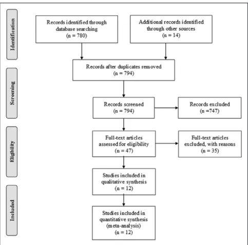
Seven hundred ninety-four articles were identified, 58
were assessed with respect to their eligibility for
inclu-sion, and 12 studies were included in the systematic
review (Table 1, Figure 1).
14–25Nine hundred
twenty-two fetuses undergoing echocardiography for the
sus-picion of CoA were included; of these, 283 (30.69%,
95% CI, 27.7–33.8) were confirmed to have a CoA
postnatally.
Ventricular disproportion was defined as a ratio
be-tween the right and left ventricles >1.5, 1.6, and 1 in
3 studies, respectively, whereas the majority did not
report any specific cutoff.
16,21,25Three studies
17,19,21reported a cutoff of ≥1.6 in the ratio between the PV
and AoV. When plotted together, PV/AoV ≥1.6 was
as-sociated with a significantly increased risk for CoA (OR,
15.11; 95% CI, 6.80–33.6; P≤0.001, I
2, 0%); however,
when this figure was translated into a predictive
mod-el, it gave only a moderate diagnostic accuracy and
was affected by a high false-positive rate (sensitivity:
86.2%; 95% CI, 77.5–92.4; specificity: 51.8%; 95%
CI, 46.1–57.4; LR+: 3.01; 95% CI, 1.09–8.33; LR:
0.20, 95% CI, 0.08–0.54; diagnostic OR: 15.1; 95%
CI, 6.80–33.5).
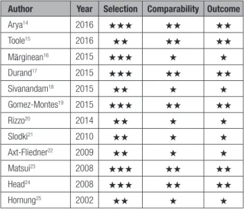
Results of quality assessment of the included
stud-ies using Newcastle-Ottawa Scale for cohort studstud-ies are
presented in Table 2.
ORIGINAL RESEARCH
AR
TICLE
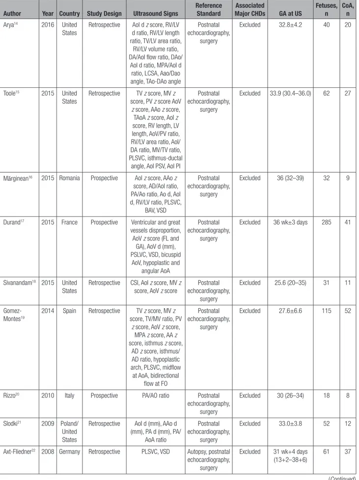
Table 1.
General Characteristics of the Studies Included in the Systematic Review
Author Year Country Study Design Ultrasound Signs Reference Standard Major CHDsAssociated GA at US Fetuses, n CoA, n
Arya14 2016 United
States
Retrospective AoI d z score, RV/LV d ratio, RV/LV length ratio, TV/LV area ratio,
RV/LV volume ratio, DA/AoI flow ratio, DAo/
AoI d ratio, MPA/AoI d ratio, LCSA, Aao/Dao angle, TAo-DAo angle
Postnatal echocardiography, surgery Excluded 32.8±4.2 40 20 Toole15 2015 United States Retrospective TV z score, MV z score, PV z score AoV
z score, AAo z score,
TAoA z score, AoI z score, RV length, LV length, AoV/PV ratio, RV/LV area ratio, AoI/ DA ratio, MV/TV ratio, PLSVC, isthmus-ductal
angle, AoI PSV, AoI PI
Postnatal echocardiography,
surgery
Excluded 33.9 (30.4–36.0) 62 27
Mărginean16 2015 Romania Prospective AoI z score, AAo z
score, AD/AoI ratio, PA/Ao ratio, Ao d, AoI d, RV/LV ratio, PLSVC, BAV, VSD Postnatal echocardiography, surgery Excluded 36 (32–39) 32 9
Durand17 2015 France Prospective Ventricular and great
vessels disproportion, AoV z score (FL and
GA), AoV d (mm), PSLVC, VSD, bicuspid
AoV, hypoplastic and angular AoA Postnatal echocardiography, surgery Excluded 36 wk±3 days 285 41 Sivanandam18 2015 United States
Retrospective CSI, AoI z score, MV z score, AoV z score
Postnatal echocardiography, surgery Excluded 25.6 (20–35) 31 11 Gomez-Montes19
2014 Spain Retrospective TV z score, MV z score, TV/MV ratio, PV
z score, AoV z score,
MPA z score, AA z score, isthmus z score,
AD z score, isthmus/ AD ratio, hypoplastic arch, PLSVC, midflow at AoA, bidirectional flow at FO Postnatal echocardiography, surgery Excluded 27.6±6.6 115 52
Rizzo20 2010 Italy Prospective PA/AO ratio Postnatal
echocardiography, surgery Excluded 30 (26–34) 18 8 Slodki21 2009 Poland/ United States
Retrospective AoI d (mm), AAo d (mm), PA d (mm), PA/ AoA ratio Postnatal echocardiography, surgery Excluded 33.0±3.8 52 12
Axt-Fliedner22 2008 Germany Retrospective PLSVC, VSD Autopsy, postnatal
echocardiography, surgery Excluded 31 wk+4 days (13+2–38+6) 61 37 (Continued )
Synthesis of the Results
The mean MV diameter z score was significantly lower in
fetuses with CoA than in those without CoA (mean
differ-ence [MD], –0.97; 95% CI, –1.43 to –0.51; P<0.001),
whereas the mean TV diameter z score was significantly
higher in fetuses with CoA than in controls (MD, 0.40;
95% CI, 0.09–0.71; P=0.01) (Figure 2).
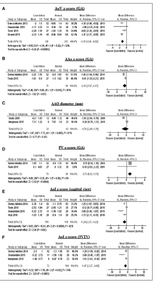
The mean AoV diameter z score for GA was
signifi-cantly lower in fetuses with CoA than in healthy fetuses
(MD, –1.19; 95% CI, –1.56 to –0.82; P≤0.001),
where-as the mean AAo diameters expressed where-as z score or
mm (P=0.07 and 0.47, respectively; Table 3), were not
different between cases and controls, although these
pa-rameters were assessed only in 2 studies. The mean AoI
Matsui23 2008 United
Kingdom
Retrospective Vascular disproportion, PLSVC, VSD, BAV,
shelf, Doppler anomalies, AoI z score,
AoI/AD ratio Autopsy, postnatal echocardiography, surgery Excluded 22 wk+0 days (15+4–38+4) 44 20 Head24 2004 United Kingdom
Retrospective VSD, PLSVC Autopsy, postnatal echocardiography,
surgery
Excluded Not stated 144 43
Hornung25 2001 United
Kingdom
Retrospective VSD, BAV Postnatal
echocardiography, surgery
Excluded 29 (16–38) 38 3
Ao indicates aorta; AAo, ascending aorta; AD, arterial duct; AoA, aortic arch; AoI, aortic isthmus; BAV, bicuspid aortic valve; CHD, congenital heart defect; CoA, coarctation of the aorta; CSI, carotid subclavian index; d, diameter; DAo, descending aorta; GA, gestational age; LCSA, left common carotid–to–left subclavian artery distance; LV, left ventricle; MV, mitral valve; PA, pulmonary artery; PI, pulsatility index; PLSVC, persistent left superior vena cava; PV, pulmonary valve; RV, right ventricle; TAoA, transverse aortic arch; TV, tricuspid valve; US, ultrasound; and VSD, ventricular septal defect.
Table 1.
Continued
Author Year Country Study Design Ultrasound Signs Reference Standard Major CHDsAssociated GA at US Fetuses, n CoA, n
Figure 1.
Systematic review
flowchart.
ORIGINAL RESEARCH
AR
TICLE
diameter z scores measured either in sagittal view (MD,
–1.24; 95% CI, –2.27 to –0.22; P=0.02) or in 3 vessels
and trachea view (MD, 1.47; 95% CI, –2.27 to –0.68;
P<0.001) were significantly lower in fetuses with CoA.
Conversely, mean PA diameter z score was significantly
higher in fetuses with CoA than in controls (MD, 0.73;
95% CI, 0.32–1.13; P<0.001) (Figure 3). Mean
differ-ence of ultrasound parameters which were reported only
in single studies could not be plotted in a quantitative
synthesis are reported in
online-only Data Supplement
Table III
.
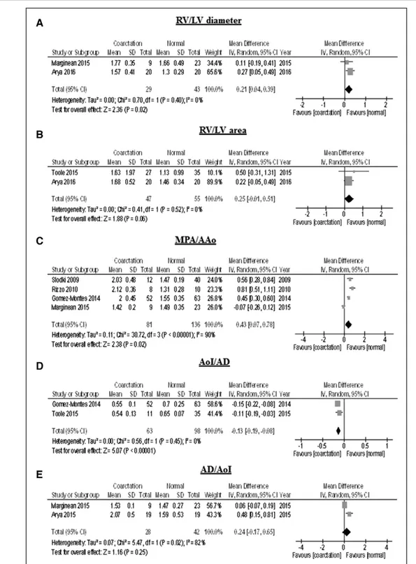
Few studies reported the ratios between different
continuous cardiovascular morphological parameters,
and a quantitative synthesis could be performed for
only 5 parameters (Table 3). RV/LV and PA/AAo
diam-eters were significantly higher in fetuses with CoA than
in controls (MD, 0.21; 95% CI, 0.04–0.39; P=0.02 and
MD, 0.43; 95% CI, 0.07–0.78; P=0.02, respectively),
whereas AoI/AD diameter was lower in fetuses with CoA
than in those without CoA (MD, –0.13, 95% CI, –0.19 to
–0.08; P<0.001) (Figure 4).
The majority of the signs detected at fetal
echocar-diography were reported by single studies and thus
could not be integrated in the quantitative synthesis
(
online-only Data Supplement Table IV
).
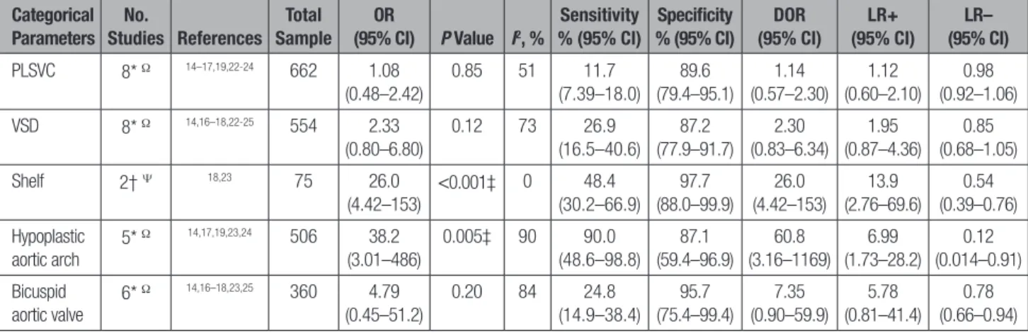
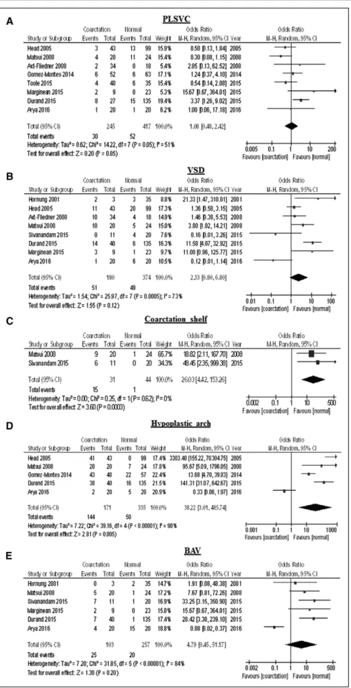
Quantitative data synthesis was possible for 5
categori-cal variables: PLSVC, ventricular septal defect, coarctation
shelf, hypoplastic aortic arch, and BAV. PLSVC (P=0.85),
ventricular septal defect (P=0.12), and BAV (P=0.20) were
not associated with an increased risk of CoA, whereas the
presence of coarctation shelf was significantly more
com-mon in fetuses with CoA than in controls (OR, 26.0; 95%
CI, 4.42–153; P<0.001) (Table 4, Figure 5). Last,
hypo-plastic aortic arch, defined as a subjective observation,
was independently associated with the occurrence of CoA
(OR, 38.2; 95% CI, 3.01–486; P=0.005)
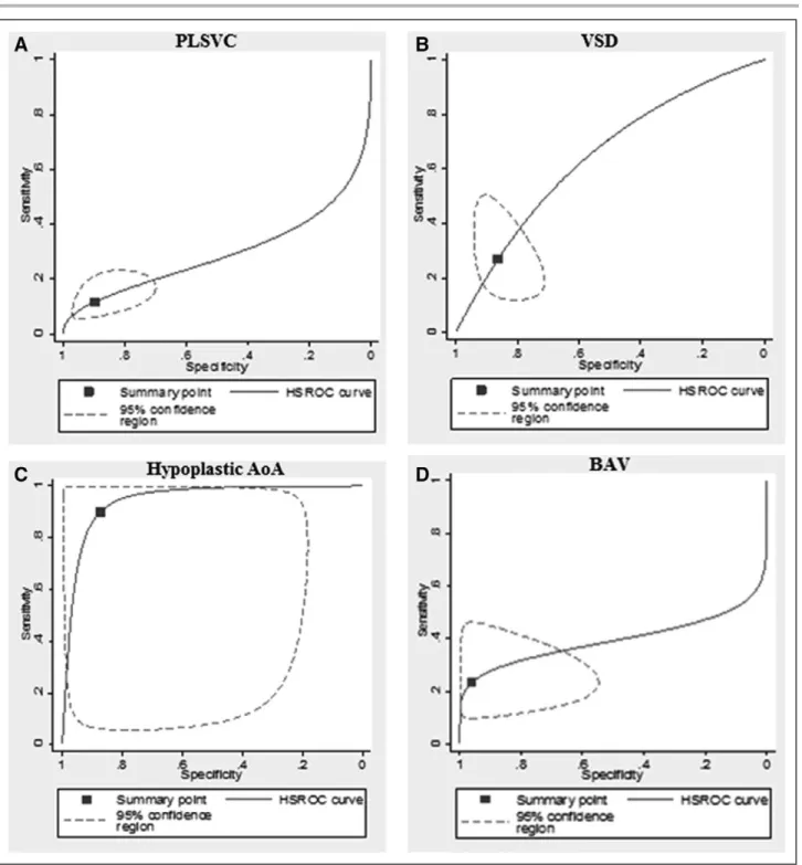
Summary estimates of sensitivity, specificity, LR+,
LR–, and diagnostic OR to predict CoA were computed by
using the hierarchical summary receiver operating
char-acteristic model for all categorical variables presented
in Table 4 (Figure 6). The presence of hypoplastic aortic
arch showed the overall best diagnostic performance in
detecting CoA in fetuses with vascular disproportion with
a sensitivity of 90.0%; 95% CI, 48.6 to 98.8; a
speci-ficity of 87.1%; 95% CI, 59.4 to 96.9; a LR+ of 6.99,
95% CI, 1.73 to 28.2; a LR– of 0.12, 95% CI, 0.014
to 0.91; and a diagnostic OR of 60.8; 95% CI, 3.16 to
1169. Coarctation shelf had a high specificity (97.7%;
95% CI, 88.0–99.9), but was affected by a low sensitivity
Table 2.
Quality Assessment of the Included Studies
According to the Newcastle-Ottawa Scale
Author Year Selection Comparability Outcome
Arya14 2016 ★★★ ★★ ★★ Toole15 2016 ★★ ★★ ★★ Mărginean16 2015 ★★★ ★ ★ Durand17 2015 ★★★ ★★ ★★ Sivanandam18 2015 ★★ ★ ★ Gomez-Montes19 2015 ★★★ ★★ ★★ Rizzo20 2014 ★★ ★ ★ Slodki21 2010 ★★ ★ ★ Axt-Fliedner22 2009 ★★ ★ ★ Matsui23 2008 ★★★ ★★ ★★ Head24 2008 ★★★ ★★ ★★ Hornung25 2002 ★★ ★ ★
A study can be awarded a maximum of 1 star for each numbered item in the Selection and Outcome categories. A maximum of 2 stars can be given for Comparability.
Figure 2.
MD-AV valves.
Results of the meta-analysis comparing the mean tricus-pid valve (TV) (A) and mitral valve (MV) (B) z scores of fetuses with CoA in compari-son with those without CoA. AV indicates aortic valve; CI, confidence limit; CoA, coarcta-tion of the aorta; IV, inverse-variance approach; MD, mean difference; and SD, standard deviation.
(Table 4). All other parameters in isolation had an overall
poor diagnostic accuracy in detecting CoA prenatally.
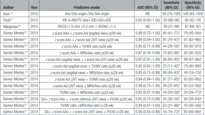
Multiparametric diagnostic models integrating
dif-ferent ultrasound signs for the detection of CoA were
reported only by 4 studies
14–16,19(Table 5). Because all
these models integrate different variables with different
cutoffs, it was not possible to perform a quantitative
data synthesis. In the study by Toole et al
15a multiple–
risk factors model incorporating MV diameter, MV/TV
ratio, isthmus/ductal diameter ratio, and
isthmus–duc-tal angle had an area under the curve of 0.92 (95% CI,
0.80–1.00) with a sensitivity of 85% and a specificity
of 60%, best predictive accuracy was accomplished by
AAo z score + AoI z score (3 vessels and trachea view)
before 28 weeks of gestations with an area under the
curve of 0.98 (95% CI, 0.94–1.0) in the study by Gomez
Montes et al.
19In the study by Arya et al,
14the best
com-bination of sensitivity and specificity was accomplished
by a predictive model integrating the angle between the
ascending aorta and descending aorta and that between
the transverse aorta and descending aorta. In the study
by Mărginean et al
16a combination of RV/LV<1.5, AoI
<4.2 mm, and AD/AoI >1.4 gave the overall best
predic-tive accuracy for CoA, although it was affected by a low
sensitivity (55.56%; 95% CI, 21.2–86.3) (Table 5).
DISCUSSION
Main Findings
The findings of this systematic review show that
fe-tuses with CoA have significant differences in several
parameters, particularly in the left inflow (mean MV
diameter z score) and outflow tracts (mean AoV and
AoI diameter z scores, and RV/LV, PA/AAo, and AoI/
AD ratios). The presence of a coarctation shelf or
hypoplastic arch were associated with a significantly
increased risk of CoA (OR, 26.0; 95% CI, 4.42–153
and OR, 38.2; 95% CI, 3.01–486, respectively). The
prenatal detection rate of CoA was significantly
in-creased when a multiple-criteria prediction model was
adopted.
Large multicenter prospective studies including
fe-tuses with different risk factors for CoA are needed to
ascertain the actual diagnostic performance of fetal
echocardiography in diagnosing CoA.
Strengths and Limitations
Retrospective design, small number of included
cas-es, different GA at scan, imaging protocols adopted,
and lack of definition of the optimal cutoff for many
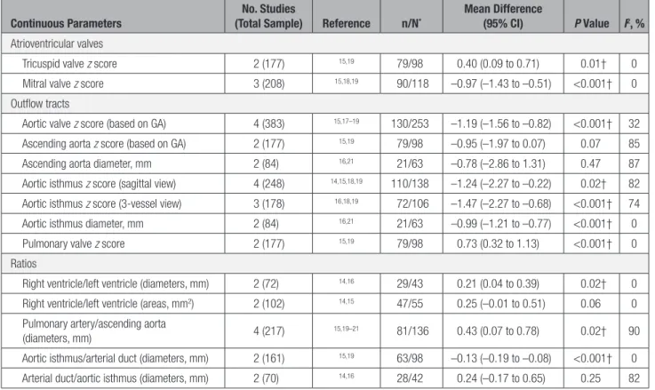
Table 3.
Results of the Meta-Analyses Comparing the Echocardiographic Parameters of Different Cardiac
Structures in Fetuses With Diagnosis of Coarctation of the Aorta Versus Normal Fetuses
Continuous Parameters
No. Studies
(Total Sample) Reference n/N*
Mean Difference
(95% CI) P Value I2, %
Atrioventricular valves
Tricuspid valve z score 2 (177) 15,19 79/98 0.40 (0.09 to 0.71) 0.01† 0
Mitral valve z score 3 (208) 15,18,19 90/118 –0.97 (–1.43 to –0.51) <0.001† 0
Outflow tracts
Aortic valve z score (based on GA) 4 (383) 15,17–19 130/253 –1.19 (–1.56 to –0.82) <0.001† 32
Ascending aorta z score (based on GA) 2 (177) 15,19 79/98 –0.95 (–1.97 to 0.07) 0.07 85
Ascending aorta diameter, mm 2 (84) 16,21 21/63 –0.78 (–2.86 to 1.31) 0.47 87
Aortic isthmus z score (sagittal view) 4 (248) 14,15,18,19 110/138 –1.24 (–2.27 to –0.22) 0.02† 82
Aortic isthmus z score (3-vessel view) 3 (178) 16,18,19 72/106 –1.47 (–2.27 to –0.68) <0.001† 74
Aortic isthmus diameter, mm 2 (84) 16,21 21/63 –0.99 (–1.21 to –0.77) <0.001† 0
Pulmonary valve z score 2 (177) 15,19 79/98 0.73 (0.32 to 1.13) <0.001† 0
Ratios
Right ventricle/left ventricle (diameters, mm) 2 (72) 14,16 29/43 0.21 (0.04 to 0.39) 0.02† 0
Right ventricle/left ventricle (areas, mm2) 2 (102) 14,15 47/55 0.25 (–0.01 to 0.51) 0.06 0
Pulmonary artery/ascending aorta
(diameters, mm) 4 (217) 15,19–21 81/136 0.43 (0.07 to 0.78) 0.02† 90
Aortic isthmus/arterial duct (diameters, mm) 2 (161) 15,19 63/98 –0.13 (–0.19 to –0.08) <0.001† 0
Arterial duct/aortic isthmus (diameters, mm) 2 (70) 14,16 28/42 0.24 (–0.17 to 0.65) 0.25 82
CI indicates confidence interval; CoA, coarctation of the aorta; and GA, gestational age.
*n indicates the overall number of fetuses affected by CoA; and N, the overall number of fetuses not affected by CoA. †Statistically significant value.
ORIGINAL RESEARCH
AR
TICLE
of the included variables represent the major
limita-tion of this systematic review. Because the included
cases are fetuses at high risk of CoA, it is possible
that the figures we reported may not reflect the actual
association between a given sign and the occurrence
of the disease. The majority of the ultrasound signs
associated with CoA were reported only by single
studies and thus a comprehensive quantitative data
Figure 3.
MD-outflow tracts.
Results of the meta-analysis comparing the mean aortic valve (AoV) z score (A), ascending aorta (AAO) z score (B), ascend-ing aorta (AAO) diameter (mm) (C), pulmonary valve (PV) z score (D), and aortic isthmus (AoI) z score in the sagittal view and 3 vessels and trachea view (3VTV), respectively (E and F) of fetuses with CoA in comparison with those without CoA. CI indicates confidence limit; CoA, coarctation of the aorta; GA, gestational age; IV, inverse-variance approach; MD, mean difference; and SD, standard deviation.
synthesis could not be performed. Despite all these
limitations, this review represents the most up-to-date
overall assessment of fetal echocardiography in
de-tecting CoA prenatally, potentially being the basis for
prenatal counseling.
Implications for Clinical Practice
Accurate prenatal diagnosis of CoA allows a preplanned
management of the condition, thus reducing the burden
of short- and long-term morbidities associated with this
anomaly.
3Despite this, prenatal diagnosis of CoA is
chal-Figure 4.
MD ratios.
Results of the meta-analysis comparing the mean ratios of RV/LV diameter (A), RV/LV area (B), MPA/AAo (C), AoI/AD (D), and AD/AoI (E) of fetuses with CoA in comparison with those without CoA. AAo indicates ascending aorta; AD, arterial duct; AoI, aor-tic isthmus; CI, confidence limit; CoA, coarctation of the aorta; IV, inverse-variance approach; MD, mean difference; MPA, main pulmonary artery; RV/LV, right ventricular/left ventricular; and SD, standard deviation.
ORIGINAL RESEARCH
AR
TICLE
lenging. The overall detection rate of prenatal ultrasound
in identifying this anomaly has been reported to be poor
at the time of the routine anomaly scan.
5The overall
diag-nostic performance of cardiovascular disproportion
dur-ing a third trimester scan is poor and is associated with a
high false-positive rate. Moreover, routine third-trimester
scan is not universally performed, unless fetal or
mater-nal complications are suspected, and it is usually
per-formed almost exclusively to assess fetal growth. In this
scenario, the presence of cardiovascular disproportion
may be easily overlooked, thus explaining the reported
low detection rate for CoA. The definition of
cardiovas-cular disproportion is usually subjective, and structurally
normal fetal hearts in the third trimester of pregnancies
exhibit a slight degree of physiological disproportion.
25Conversely, disproportion detected in the late second or
early third trimester of pregnancies carries an increased
risk for the occurrence of CoA. In the current review, a
disproportion of PV/AoV ratio >1.6 was significantly
as-sociated with CoA with an OR of 15.11 (95% CI, 6.80–
33.6). However, when translated into a predictive model,
it had a good sensitivity (86.2%; 95% CI, 77.5–92.4), but
a low specificity (51.8%; 95% CI, 46.1–57.4).
The findings of this systematic review show that, in
fetuses at risk, detailed assessment of several cardiac
parameters might help in stratifying the risk for CoA.
These results are mainly applicable to fetuses with
cardiovascular disproportion on the third-trimester
ultra-sound, and, therefore, the actual performance of
prena-tal ultrasound when applied on an unselected population
needs further evaluation.
26Coarctation shelf refers to a prominent posterior
in-folding in the vessels media, which may extend around
the entire circumference of the aorta. It is more
com-monly detected after birth when ductal tissue believed
to encircle the aorta constricts during ductal closure.
On the basis of the concept that abnormal insertion of
the ductus arteriosus into the descending aorta
dur-ing development might not only play an important role
in the development of CoA, but also affect the shape
of the aortic arch, evaluation angle between segments
of the aortic arch, and the ductus arteriosus has been
suggested to be useful for diagnosing CoA.
14,15The
presence of the shelf had a high specificity but a low
sensitivity for CoA, which is explained in part by
dif-ficulties in the visualization at prenatal
echocardiog-raphy. In the present review, hypoplastic aortic arch,
mainly assessed in fetuses with ventricular
dispropor-tion, showed the best combination of sensitivity and
specificity for CoA. Specific cutoffs for defining the
arch as hypoplastic have not been reported yet, and
the diagnosis is mostly subjective.
PLSVC has been associated with the occurrence of
CoA in other prenatal series.
27,28In our study, PLSVC did
not significantly increase the risk for CoA but this
find-ing might be influenced by the nature of the population
reported in the included studies, and PLSVC may
repre-sent an independent risk factors in for CoA in fetuses not
showing any suspicious sign of CoA.
BAV is commonly associated with CoA in the postnatal
series.
29Although its occurrence was higher in fetuses with
CoA in the current review, BAV was not associated with
a significantly increased risk for this anomaly and was
af-fected by an overall poor diagnostic performance (Table 4).
Spectral and color Doppler ultrasound are commonly
used in clinical practice to confirm CoA, but our review
Table 4.
Likelihood of Presenting Each Cardiovascular Anomaly in Fetuses With a Diagnosis of Coarctation of
the Aorta Versus Normal Fetuses
Categorical Parameters No. Studies References Total Sample OR (95% CI) P Value I2, % Sensitivity % (95% CI) Specificity % (95% CI) DOR (95% CI) LR+ (95% CI) LR– (95% CI) PLSVC 8* Ω 14–17,19,22-24 662 1.08 (0.48–2.42) 0.85 51 11.7 (7.39–18.0) 89.6 (79.4–95.1) 1.14 (0.57–2.30) 1.12 (0.60–2.10) 0.98 (0.92–1.06) VSD 8* Ω 14,16–18,22-25 554 2.33 (0.80–6.80) 0.12 73 26.9 (16.5–40.6) 87.2 (77.9–91.7) 2.30 (0.83–6.34) 1.95 (0.87–4.36) 0.85 (0.68–1.05) Shelf 2† Ψ 18,23 75 26.0 (4.42–153) <0.001‡ 0 48.4 (30.2–66.9) 97.7 (88.0–99.9) 26.0 (4.42–153) 13.9 (2.76–69.6) 0.54 (0.39–0.76) Hypoplastic aortic arch 5* Ω 14,17,19,23,24 506 38.2 (3.01–486) 0.005‡ 90 90.0 (48.6–98.8) 87.1 (59.4–96.9) 60.8 (3.16–1169) 6.99 (1.73–28.2) 0.12 (0.014–0.91) Bicuspid aortic valve 6* Ω 14,16–18,23,25 360 4.79 (0.45–51.2) 0.20 84 24.8 (14.9–38.4) 95.7 (75.4–99.4) 7.35 (0.90–59.9) 5.78 (0.81–41.4) 0.78 (0.66–0.94) For each parameter, summary estimates of sensitivity, specificity, positive and negative likelihood ratios (LR+ and LR–), and diagnostic odds ratio (DOR) to predict coarctation of the aorta were also computed. Depending on the number of studies, computations were based on the DerSimonian-Laird random-effects or hierarchical summary receiver operating characteristic (HSROC) model. CI indicates confidence interval; OR, odds ratio; PLSVC indicates persistent left superior vena cava; and VSD, ventricular septal defect
*HSROC model.
†DerSimonian-Laird random-effects model. ‡Statistically significant value.
Figure 5.
Odds ratios.
Results of the meta-analysis comparing the risk of PLSVC (A), VSD (B), coarctation shelf (C), hypoplastic arch (D) and BAV (E) in fetuses with CoA in compari-son with those without CoA. BAV indicates bicuspid aortic valve; CI, confidence limit; CoA, coarc-tation of the aorta; M-H, Mantel-Haenszel test; PLSVC, persistent left superior vena cava; and VSD, ventricular septal defect.
ORIGINAL RESEARCH
AR
TICLE
could not quantify their role because only 1 study
re-ported their use (
online-only Data Supplement Table V
).
GA at scan represents another relevant issue. It has been
reported
19that the diagnostic accuracy of ultrasound in
de-tecting CoA prenatally may be improved by using different
cutoffs according to the GA at scan but, in the present
re-view, it was not possible to perform the analysis stratifying
by GA. Further studies are needed to ascertain the
contribu-tion of GA at ultrasound in the prenatal diagnosis of CoA.
Assessment of fetal hemodynamics using prenatal
cardiac MRI has been recently suggested to add useful
information in fetuses affected by left-sided congenital
Figure 6.
Hierarchical summary receiver operating characteristic.
Hierarchical summary receiver-operating characteristics (HSROC) curves of the diagnostic performance of persistent left superior vena cava (PLSVC) (A), ventricular septal defect (VSD) (B), hypoplastic aortic arch (AoA) (C), and bicuspid aortic valve (BAV) (D) detected on ultrasound for the detection of coarctation of the aorta. Curves from the HSROC model contain a summary operating point (◼) representing summarized sensitivity and specificity point estimates for individual study estimates (dotted lines: 95% confidence interval).
heart defects and to correlate with lung and brain
de-velopment.
30Ascertaining the role of fetal cardiac MRI
as a potential diagnostic tool for CoA is challenging, but
it might help to confirm or refute the diagnosis in some
cases of ventricular or great vessel disproportion.
CONCLUSION
Detailed fetal echocardiography can stratify the risk for
CoA in fetuses with a suspected diagnosis. Prenatal
de-tection rate of CoA may improve when a multiple-criteria
prediction model is adopted. Further large multicenter
studies sharing the same imaging protocols are
need-ed to develop objective models for risk assessment in
these fetuses, and to ascertain the actual diagnostic
performance of prenatal ultrasound in detecting this
anomaly.
SOURCES OF FUNDING
None.DISCLOSURES
None.AFFILIATIONS
From Department of Maternal-Fetal Medicine, Catholic Univer-sity of the Sacred Heart, Rome, Italy (A.F., G.D.S., C.V., A.L., G.S.); Department of Neuroscience, Reproductive Sciences and Dentistry, School of Medicine, University of Naples Fed-erico II, Italy (M.M.); Fetal Medicine Unit, Saint George’s Hospi-tal, London, United Kingdom (A.K.); Pediatric Cardiology Unit, Department of Women’s and Children’s Health, Karolinska In-stitute, Stockholm, Sweden (S.S.-E.); Department of Obstet-rics and Gynecology, IRCCS San Martino Hospital University of Genoa, Italy (C.S.); Department of Obstetrics and Gynaecol-ogy, University of TorVergata, Rome, Italy (G.R.); Department of Medicine and Aging Sciences, University of Chieti-Pescara, Italy (M.E.F.); Department of Medical Sciences, University of Ferrara, Italy (L.M.); Department of Clinical Science, Interven-tion and Technology, Karolinska Institute, Stockholm, Sweden (G.A.); and Department of Clinical Medicine, UiT- The Arctic Uni-versity of Norway and Department of Obstetrics and Gynecol-ogy, University Hospital of Northern Norway, Tromsø (F.D’A.).
FOOTNOTES
Received June 18, 2016; accepted December 14, 2016. The online-only Data Supplement is available with this arti-cle at http://circ.ahajournals.org/lookup/suppl/doi:10.1161/ CIRCULATIONAHA.116.024068/-/DC1.
Circulation is available at http://circ.ahajournals.org.
Table 5.
Predictive Models for Coarctation of the Aorta Integrating Multiple Risk Factors
Author Year Predictive model AUC (95% CI) Sensitivity (95% CI) Specificity (95% CI)
Arya 14 2016 AAo-DAo angle+TAo-DAo angle NS 95 (75–100) 100 (83–100)
Toole15 2015 MV d+MV/TV ratio+ IDD+IDA+IDD 0.92 (0.80–1.00) 85 (66–96) 60 (42–76)
Mărginean16 2015 RV/LV<1.5+AoI <4.2 mm + AD/AoI >1.4 NS 56 (21–86) 87 (66–97)
Gomez-Montes19 2014 z score AAo + z score AoI (sagittal view) (≤28 wk) 0.88 (0.72–1.00) 60 (41–77)* 78 (45–94)†
Gomez-Montes19 2014 z score AAo + z score AoI (3VT view) (≤28 wk) 0.98 (0.94–1.00) 91 (76–97)* 91 (62–98)†
Gomez-Montes19 2014 z score AAo + TV/MV ratio (≤28 wk) 0.85 (0.71–0.99) 44 (29–59)* 69 (42–87)†
Gomez-Montes 19 2014 z score AAo + MPA/AAo ratio (≤28 wk) 0.87 (0.76–0.99) 78 (63–88)* 62 (36–82)†
Gomez-Montes19 2014 z score AoI (sagittal view) + z score AoI (3VT view) (≤28 wk) 0.97 (0.91–1.00) 86 (65–95)* 89 (57–98)†
Gomez-Montes19 2014 z score AoI (sagittal view) + TV/MV ratio (≤28 wk) 0.82 (0.63–1.00) 23 (11–42)* 70 (40–89)†
Gomez-Montes 19 2014 z score AoI (sagittal view) + MPA/AAo ratio (≤28 wk) 0.85 (0.72–0.98) 68 (48–83)* 44 (19–73)†
Gomez-Montes19 2014 z score AoI (3VT view) + TV/MV ratio (≤28 wk) 0.94 (0.84–1.00) 87 (71–95)* 83 (55–95)†
Gomez-Montes19 2014 z score AoI (3VT view) + MPA/AAo ratio (≤28 wk) 0.89 (0.75–1.00) 48 (32–65)* 82 (52–95)†
Gomez-Montes19 2014 TV/MV ratio + MPA/AAo ratio (≤28 wk) 0.82 (0.67–0.96) 44 (29–59)* 54 (29–77)†
Gomez-Montes19 2014 GA+ z score AAo + z score isthmus (3VT view) + PV/AV (≤28 wk) 0.85 (0.73–0.98) 40 (26–54)* 64 (39–84)†
Gomez-Montes19 2014 TV/MV ratio +MPA/AAo ratio (>28 wk) 0.84 (0.67–1.00) 63 (31–86)* 43 (30–58)†
Gomez-Montes19 2014 GA+ z score AAo + z score AoI (3VT view) + PV/AV (>28 wk) 0.90 (0.83–0.98) 44 (19–73)* 82 (69–90)†
AAo indicates ascending aorta; AD, arterial duct; AoI, aortic isthmus; AUC, area under the curve; AV, aortic valve; CHD, congenital heart defect; CI, confidence interval; d, diameter; DAo, descending aorta; GA, gestational age; IDA, isthmus-ductal angle; IDD, isthmus:ductal diameter; LV, left ventricle; MPA, main pulmonary artery; MV, mitral valve; PV, pulmonary valve; RV, right ventricle; TAoA, transverse aortic arch; TV, tricuspid valve; and 3VT, 3 vessels and trachea.
*For 10% false-positive rate. †For 10% false-negative rate.
ORIGINAL RESEARCH
AR
TICLE
REFERENCES
1. Rosenthal E. Coarctation of the aorta from fetus to adult: curable condition or life long disease process? Heart. 2005;91:1495– 1502. doi: 10.1136/hrt.2004.057182.
2. Reifenstein GH, Levine SA, Gross RE. Coarctation of the aorta; a review of 104 autopsied cases of the adult type, 2 years of age or older. Am Heart J. 1947;33:146–168.
3. Franklin O, Burch M, Manning N, Sleeman K, Gould S, Archer N. Prenatal diagnosis of coarctation of the aorta improves survival and reduces morbidity. Heart. 2002;87:67–69.
4. Brown ML, Burkhart HM, Connolly HM, Dearani JA, Cetta F, Li Z, Oliver WC, Warnes CA, Schaff HV. Coarctation of the aorta: lifelong surveillance is mandatory following surgical repair. J Am Coll Car-diol. 2013;62:1020–1025. doi: 10.1016/j.jacc.2013.06.016. 5. Tegnander E, Williams W, Johansen OJ, Blaas HG, Eik-Nes SH.
Prenatal detection of heart defects in a non-selected population of 30,149 fetuses–detection rates and outcome. Ultrasound Obstet Gynecol. 2006;27:252–265. doi: 10.1002/uog.2710.
6. Hornberger LK, Sahn DJ, Kleinman CS, Copel J, Silverman NH. Antenatal diagnosis of coarctation of the aorta: a multicenter ex-perience. J Am Coll Cardiol. 1994;23:417–423.
7. Henderson LK, Craig JC, Willis NS, Tovey D, Webster AC. How to write a Cochrane systematic review. Nephrology (Carlton). 2010;15:617–624. doi: 10.1111/j.1440-1797.2010.01380.x. 8. NHS Centre for Reviews and Dissemination. Systematic Reviews:
CRD’s Guidance for Undertaking Reviews in Health Care. York, UK: University of York, York Publishing Services Ltd; 2009.
9. Moher D, Shamseer L, Clarke M, Ghersi D, Liberati A, Petticrew M, Shekelle P, Stewart LA; and PRISMA-P Group. Preferred reporting items for systematic review and meta-analysis protocols (PRISMA-P) 2015 statement. Syst Rev 2015;4:1. http://www.prisma-state-ment.org/ Accessed November 20, 2015.
10. Wells GA, Shea B, O’Connell D, Peterson J, Welch V, Losos M, Tugwell P. Newcastle-Ottawa Scale (NOS) for assessing the quality of nonrandomised studies in meta-analyses. The Ottowa Hospital Research Institute. http://www.ohri.ca/programs/clinical_epide-miology/oxford.asp. Accessed February 10, 2016.
11. Macaskill P, Gatsonis C, Deeks JJ, Harbord RM, Takwoingi Y. Chap-ter 10: Analysing and presenting results. In: Deeks JJ, Bossuyt PM, Gatsonis C, eds. Cochrane Handbook for Systematic Reviews of Diagnostic Test Accuracy Version 1.0. The Cochrane Collabora-tion; 2010. http://srdta.cochrane.org/. January 15, 2016. 12. Glas AS, Lijmer JG, Prins MH, Bonsel GJ, Bossuyt PM. The
diag-nostic odds ratio: a single indicator of test performance. J Clin Epidemiol. 2003;56:1129–1135.
13. Rutter CM, Gatsonis CA. A hierarchical regression approach to meta-analysis of diagnostic test accuracy evaluations. Stat Med. 2001;20:2865–2884.
14. Arya B, Bhat A, Vernon M, Conwell J, Lewin M. Utility of novel fetal echocardiographic morphometric measures of the aortic arch in the diagnosis of neonatal coarctation of the aorta. Prenat Diagn. 2016;36:127–134. doi: 10.1002/pd.4753.
15. Toole BJ, Schlosser B, McCracken CE, Stauffer N, Border WL, Sachdeva R. Importance of relationship between ductus and isth-mus in fetal diagnosis of coarctation of aorta. Echocardiography. 2016;33:771–777. doi: 10.1111/echo.13140.
16. Mărginean C, Mărginean CO, Muntean I, Togănel R, Voidăzan S, Gozar L. The role of ventricular disproportion, aortic, and ductal isthmus ultrasound measurements for the diagnosis of fetal aortic
coarctation, in the third trimester of pregnancy. Med Ultrason. 2015;17:475–481.
17. Durand I, Deverriere G, Thill C, Lety AS, Parrod C, David N, Barre E, Hazelzet T. Prenatal detection of coarctation of the aorta in a non-selected population: a prospective analysis of 10 years of ex-perience. Pediatr Cardiol. 2015;36:1248–1254. doi: 10.1007/ s00246-015-1153-1.
18. Sivanandam S, Nyholm J, Wey A, Bass JL. Right ventricular enlarge-ment in utero: is it coarctation? Pediatr Cardiol. 2015;36:1376– 1381. doi: 10.1007/s00246-015-1168-7.
19. Gómez-Montes E, Herraiz I, Gómez-Arriaga PI, Escribano D, Mendoza A, Galindo A. Gestational age-specific scoring systems for the prediction of coarctation of the aorta. Prenat Diagn. 2014;34:1198–1206. doi: 10.1002/pd.4452.
20. Rizzo G, Arduini D, Capponi A. Use of 4-dimensional sonography in the measurement of fetal great vessels in mediastinum to dis-tinguish true-from false-positive coarctation of the aorta. J Ultra-sound Med. 2010;29:325–326.
21. Slodki M, Rychik J, Moszura T, Janiak K, Respondek-Liberska M. Measurement of the great vessels in the mediastinum could help distinguish true from false-positive coarctation of the aorta in the third trimester. J Ultrasound Med. 2009;28:1313–1317. 22. Axt-Fliedner R, Hartge D, Krapp M, Berg C, Geipel A, Koester S,
Noack F, Germer U, Gembruch U. Course and outcome of fetuses suspected of having coarctation of the aorta during gestation. Ultra-schall Med. 2009;30:269–276. doi: 10.1055/s-2008-1027556. 23. Matsui H, Mellander M, Roughton M, Jicinska H, Gardiner HM.
Morphological and physiological predictors of fetal aortic coarcta-tion. Circulacoarcta-tion. 2008;118:1793–1801. doi: 10.1161/CIRCULA-TIONAHA.108.787598.
24. Head CE, Jowett VC, Sharland GK, Simpson JM. Timing of pre-sentation and postnatal outcome of infants suspected of having coarctation of the aorta during fetal life. Heart. 2005;91:1070– 1074. doi: 10.1136/hrt.2003.033027.
25. Hornung TS, Heads A, Hunter AS. Right ventricular dilatation in the fetus: a study of associated features and outcome. Pediatr Cardiol. 2001;22:215–217. doi: 10.1007/s002460010206. 26. Quarello E, Trabbia A. High-definition flow combined with
spatio-temporal image correlation in the diagnosis of fetal coarctation of the aorta. Ultrasound Obstet Gynecol. 2009;33:365–367. doi: 10.1002/uog.6270.
27. Berg C, Knüppel M, Geipel A, Kohl T, Krapp M, Knöpfle G, Germer U, Hansmann M, Gembruch U. Prenatal diagnosis of persistent left superior vena cava and its associated congenital anomalies. Ultrasound Obstet Gynecol. 2006;27:274–280. doi: 10.1002/ uog.2704.
28. Gustapane S, Leombroni M, Khalil A, Giacci F, Marrone L, Bascietto F, Rizzo G, Acharya G, Liberati M, D’Antonio F. Systematic review and meta-analysis of persistent left superior vena cava on prenatal ultrasound: associated anomalies, diagnostic accuracy and post-natal outcome. Ultrasound Obstet Gynecol. 2016;48:701–708. doi: 10.1002/uog.15914.
29. Siu SC, Silversides CK. Bicuspid aortic valve disease. J Am Coll Car-diol. 2010;55:2789–2800. doi: 10.1016/j.jacc.2009.12.068. 30. Sun L, Macgowan CK, Sled JG, Yoo SJ, Manlhiot C, Porayette P,
Grosse-Wortmann L, Jaeggi E, McCrindle BW, Kingdom J, Hickey E, Miller S, Seed M. Reduced fetal cerebral oxygen consumption is associated with smaller brain size in fetuses with congenital heart disease. Circulation. 2015;131:1313–1323. doi: 10.1161/ CIRCULATIONAHA.114.013051.