Research Article
Persistent Unresolved Inflammation in the
Mecp2-308 Female
Mutated Mouse Model of Rett Syndrome
Alessio Cortelazzo,
1,2,3Claudio De Felice,
4Bianca De Filippis,
5Laura Ricceri,
5Giovanni Laviola,
5Silvia Leoncini,
1,6Cinzia Signorini,
6Monica Pescaglini,
3Roberto Guerranti,
2,3Anna Maria Timperio,
7Lello Zolla,
7Lucia Ciccoli,
6and Joussef Hayek
11Child Neuropsychiatry Unit, University Hospital Azienda Ospedaliera Universitaria Senese (AOUS),
Viale M. Bracci 16, 53100 Siena, Italy
2Department of Medical Biotechnologies, University of Siena, Via A. Moro 2, 53100 Siena, Italy 3Clinical Pathology Laboratory Unit, University Hospital AOUS, Viale M. Bracci 16, 53100 Siena, Italy 4Neonatal Intensive Care Unit, University Hospital AOUS, Viale M. Bracci 16, 53100 Siena, Italy
5Centre for Behavioural Sciences and Mental Health, Istituto Superiore di Sanità (ISS), Viale Regina Elena 299, 00161 Rome, Italy 6Department of Molecular and Developmental Medicine, University of Siena, Via A. Moro 6, 53100 Siena, Italy
7Department of Ecological and Biological Sciences, University of Tuscia, Largo dell’Università, snc, 01100 Viterbo, Italy
Correspondence should be addressed to Alessio Cortelazzo; [email protected] Received 6 December 2016; Accepted 19 April 2017; Published 16 May 2017 Academic Editor: Chiara De Luca
Copyright © 2017 Alessio Cortelazzo et al. This is an open access article distributed under the Creative Commons Attribution License, which permits unrestricted use, distribution, and reproduction in any medium, provided the original work is properly cited.
Rett syndrome (RTT) is a rare neurodevelopmental disorder usually caused by mutations in the X-linked gene methyl-CpG-binding protein 2 (MECP2). Several Mecp2 mutant mouse lines have been developed recapitulating part of the clinical features. In particular, Mecp2-308 female heterozygous mice, bearing a truncating mutation, are a validated model of the disease. While recent data suggest a role for inflammation in RTT, little information on the inflammatory status in murine models of the disease is available. Here, we investigated the inflammatory status by proteomic 2-DE/MALDI-ToF/ToF analyses in symptomatic Mecp2-308 female mice. Ten differentially expressed proteins were evidenced in the Mecp2-308 mutated plasma proteome. In particular, 5 positive acute-phase response (APR) proteins increased (i.e., kininogen-1, alpha-fetoprotein, mannose-binding protein C, alpha-1-antitrypsin, and alpha-2-macroglobulin), and 3 negative APR reactants were decreased (i.e., serotransferrin, albumin, and apolipoprotein A1). CD5 antigen-like and vitamin D-binding protein, two proteins strictly related to inflammation, were also changed. These results indicate for thefirst time a persistent unresolved inflammation of unknown origin in the Mecp2-308 mouse model.
1. Introduction
Rett syndrome (RTT, MIM 312750) is a progressive neurode-velopmental disorder, affecting almost exclusively the female gender. With a frequency of approximately 1 : 10,000 live births, it is a leading cause of severe intellectual disability and autistic features in females [1, 2]. The classical clinical picture of the disease [3] is characterized by a period of 6 to 18 months of apparently normal neurodevelopment followed by an early neurological regression, with a progressive loss of acquired cognitive, social, and motor skills in a typical
4-stage neurological regression pattern [4, 5]. Other known features include stereotypic hand movements, communication dysfunction, seizures, postural hypotonia,
tremors, autonomic dysfunction, microcephaly, and
growth failure [1]. It has become apparent that there is a spectrum of severity in RTT, as some patients may present with atypical features, sometimes overlapping with autism
spectrum disorders [3–5]. RTT is known to be caused in
the overwhelming majority of cases by sporadic de novo loss-of-function mutations in the X-linked CpG-binding protein 2 (MECP2) gene [6] encoding
CpG-binding protein 2 (MeCP2), a nuclear protein that binds to methylated CpGs and regulates gene expression [7, 8]. Different types of mutations within MECP2 are known to cause RTT, including missense, nonsense, dele-tions, and insertions [9]. It should be emphasised that
most of the proteins identified in mouse serum have
orthologs in humans and other mammals [10].
Identification of Mecp2 as the disease-causing gene led
rapidly to the development of mouse models of RTT that recapitulate, to varying degrees, the underlying molecular and genetic defects and symptoms of the human disease [11]. Mouse models range from Mecp2-null mutations to specific point mutations mimicking those observed in humans, phenocopying several motor and cognitive features
of RTT patients [12–16]. Although mice cannot model all
aspects of the human RTT, certainly they recapitulate many features of the disease and are generally accepted as excellent tools to study MeCP2 function [17]. An emerging role for
inflammation in human RTT is reported [18–22]. Although
MECP2 is highly expressed in several organs and tissues besides the central nervous system (CNS), including the spleen [16], most of the studies focused on the CNS and little information exists on the inflammatory status in the murine models of the disease, with few exception regarding neuroin-flammation processes related to microglia and macrophage function in Mecp2-null mice [23]. Furthermore, prior reports provide a link for MECP2-RTT to increase eryth-rocyte sedimentation rate and enhance expression of acute-phase response (APR) proteins and cytokine
dysreg-ulation [18–22]. APR is a complex systemic early-defense
system activated by trauma, infection, stress, neoplasia, and inflammation [24]. Although nonspecific, it serves as a core of the innate immune response involving physical and molecular barriers and responses that serve to prevent infection, clear potential pathogens, initiate inflammatory processes, and contribute to resolution and the healing process. APR proteins, an integral part of the APR process, have been a focus of many applications in human
diagnostic medicine and recently have been identified in
common animal species [24]. The number of protein changes was found to be proportional to the severity of
the mutation. Our findings revealed for the first time the
presence of a subclinical chronic inflammatory status related to the severity carried by the MECP2 gene mutation. There-fore, inflammation seems to be a previously unrecognized feature in RTT, which could play a role in the evolution of the pathology and its severity. Although the genetic mecha-nisms of RTT have been explored to an extraordinary extent, to date, the details of the biological mechanisms linking the MECP2 gene mutation to protein expression as a function
of clinical phenotype are yet to be clarified.
2-DE/MALDI-ToF/ToF is extremely helpful in order to understand global protein landscape changes in a given
biological fluids and/or tissues for a given condition and is
considered to be the“gold standard” technique for
identify-ing and characterizidentify-ing proteins whenfitting a series of
differ-ent parameters, such as peptide matches, sequence coverage (%), MOWSE score, and pI/Mr (kDa) [25]. In particular, with the single exception of two proteomic studies on mouse
models [26, 27], very little information exists on possible Mecp2 mouse model-related proteome changes. In the present study, a validated mouse model of RTT was used, that is, the Mecp2-308 mutated model. This model bears a truncating mutation, leading to the expression of a protein truncated at amino acid 308 [16, 28]. Several lines of evidence suggest that the truncated Mecp2 protein retains at least some of its functions (i.e., its ability to regulate BDNF gene expression, a Mecp2 target gene) [29], thus contributing to the milder neurobehavioral phenotype of those mice. Based on the previous data demonstrating a clear phenotype in Mecp2-308 females [30], the present study was carried out in symptomatic heterozygous female mice, the genetic and hormonal milieus that, from a translational point of view, is closer to the ones of RTT patients; indeed, among RTT patients, MECP2 mutations are missense and truncated mutations, not deletions [2, 11].
The aim of the present study was to investigate in plasma
the occurrence of an inflammatory status in the experimental
Mecp2-308 mutated Rett female mouse model by using a pro-teomic 2-DE/MALDI-ToF/ToF approach.
2. Materials and Methods
2.1. Animals. The experimental subjects were 10
–12-month-old Mecp2-308 heterozygous (Het) female mice [B6.129S-MeCP2tm1Hzo/J, stock number: 005439] and wild-type littermates. Mice were housed in polycarbonate transparent
cages (33× 13 × 14 cm) with sawdust bedding and kept on a
12 h light-dark schedule (lights off at 8:00). Temperature
was maintained at 21± 1°C and relative humidity at 60
± 10%. Animals were provided ad libitum with a complete pellet diet (Altromin, Germany). All procedures were carried out in accordance with the European Communities Council Directive (10/63/EU) and formally approved by the Italian Ministry of Health.
2.2. Blood Sampling. Blood samples were centrifuged at 2400g
for 15 min at 4°C and plasma was collected. All the
manipu-lations were carried out within 2 h after sample collection.
2.3. Electrophoretic Separation of Plasma Proteins after Albumin and IgG Depletion. Albumin and IgG were removed using the ProteoPrep Immunoaffinity Albumin & IgG Deple-tion Kit (Sigma-Aldrich), and two-dimensional gel electro-phoresis (2-DE) was performed according to Görg et al.
[31]. Samples containing 60μg of protein as determined by
Bradford [32] were denatured with 10 ml of a solution containing 10% of SDS and 2.3% of dithiothreitol (DTT). Afterwards, samples were combined with 350 ml of solubiliz-ing buffer containsolubiliz-ing 8 M urea, 2% of 3-[(3-cholamidopro-pyl)-dimethylammonium]-1-propane sulfonate, 0.3% DTT, and 2% of immobilized pH gradient (IPG) buffer, loaded into 18 cm IPG strips (pH 3–10) nonlinear on an Ettan IPGphor Apparatus system (GE Healthcare), and rehydrated for 7 h. IEF was carried out for a total of 32 kVh. After focusing, the
strips were equilibrated with the buffer containing 50 mM
Tris-HCl (pH 8.8), 6 M urea, 2% w/v SDS, 30% v/v glycerol, and 1% w/v DTT for 15 min. Subsequently, strips were
equilibrated again with the same equilibration buffer described above, except that it contained 4% w/v iodoaceta-mide instead of DTT and a trace of bromophenol blue. IPG strips and a molecular weight standard were embedded at the top of a 1.5 mm thick vertical polyacrylamide gradient
gel (8–16% T) using 0.5% w/v agarose and run at a constant
current of 40 mA/gel at 20°C. The second dimension was
per-formed on an Ettan Daltsix Electrophoresis system (GE Healthcare). Each sample was carried out in triplicate under the same conditions.
2.4. Protein Identification. After mass spectrometry-compatible silver staining, the preparative gel was matched to the master gel in the analytical gel match set [33]. A spot-picking list was generated and exported to Ettan Spot Picker (GE Healthcare). The spots were excised and delivered into 96-well microplates where they were destained and dehydrated with acetonitrile (ACN) for subsequent rehydra-tion with trypsin solurehydra-tion. Tryptic digesrehydra-tion was carried out overnight at 37°C. Each protein spot digest (0.75 ml) was spotted into the MALDI instrument target and allowed to dry. Then, 0.75 ml of the instrument matrix solution
(satu-rated solution of α-cyano-4-hydroxycinnamic acid in 50%
ACN and 0.5% v/v trifluoroacetic acid) was applied to dried samples which were dried again. Mass spectra were obtained
using an ultrafleXtreme MALDI-ToF/ToF (Bruker
Corpora-tion, Billerica, MA, USA), as previously described [34]. After
tryptic peptide mass acquisition, massfingerprint searching
was carried out in Swiss-Prot/TREMBL and NCBInr data-bases using MASCOT (Matrix Science, London, UK, http:// www.matrixscience.com). A mass tolerance of 100 ppm was allowed and only one missed cleavage site was accepted. Alkylation of cysteine by carbamidomethylation was
assumed as afixed modification, whereas oxidation of
methi-onine was considered a possible modification. Criteria used
to accept identifications included the extent of sequence
cov-erage, number of matched peptides, and probabilistic scores. Tryptic digests that did not produce MALDI-TOF
unambig-uous identifications were subjected to ESI-TRAP MS/MS
peptide sequencing on a nanospray/LCQ Deca ion trap mass spectrometer (Thermo Finnigan, San Jose, CA, USA).
2.5. Image and Data Analysis. Gel imaging was performed by using ImageMaster 2D Platinum v7.0 software (GE Health-care). A reference gel for each group was defined for the comparative analyses. The background was subtracted from all gels using the average on-boundary method. Spot volume was expressed as a ratio of the percentage volume (%V)
detected from the entire gel to minimize differences between
samples (i.e., normalization). Unmatched spots or spots with significantly different %V were considered differently
expressed. Data were expressed as mean± SD/SEM or
medians [95% CI for median] as appropriate. Statistical analysis of protein variations was performed using multiple t-test with a false discovery rate (q) of 0.05. Differences between groups were tested using Kruskal-Wallis test or
one-way ANOVA, with Dunn’s or Holm-Sidak’s
multiple-comparisons tests for post hoc analyses. A two-tailedP value
of less than 0.05 was considered statistically significant. The
statistical software GraphPad Prism v6.01 (GraphPad Software Inc., La Jolla, CA, USA) and MedCalc v12.1.4 soft-ware package (MedCalc Softsoft-ware, Mariakerke, Belgium) were used.
3. Results
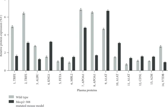
3.1. Protein Expression Profile Differences between Mecp2-308 and Wild-Type Mouse Models. Expression changes for a total of 10 differentially expressed proteins were identified in the Mecp2-308 mutated model, relative to wild type, and are shown in 2-DE maps (Figure 1). The proteins were
subse-quently identified by mass spectrometry. Protein
identifica-tion as well as peptide matches, sequence coverage, and the probabilistic score was obtained using the MASCOT
soft-ware (Table 1). A total of 14 spots were identified as follows:
serotransferrin (TRFE spots 1 and 2), albumin (ALBU, spot 3 as a fragment), kininogen-1 (KNG1, spot 4), alpha-fetoprotein (FETA, spot 5), mannose-binding protein C (MBL2, spot 6), apolipoprotein A1 (APOA1, spots 7 and 8), alpha-1-antitrypsin (A1AT, spots 9, 10, and 11), CD5 antigen-like (CD5L, spot 12), alpha-2-macroglobulin (A2M, spot 13), and vitamin D-binding protein (VTDB, spot 14).
A two-way analysis of variance showed significant
between-subjects effects (proteins, F: 2157.84, DF: 13, P < 0 001;
mouse category (mutated versus wild type), F: 994.35, DF: 1, P < 0 001; and proteins∗mouse category, F: 866.18, DF: 13,P < 0 001).
KNG1, FETA, MBL2, A1AT, and A2M were overex-pressed in the examined Mecp2-308 mutated mouse model (Figures 1 and 2, Supplementary material, Supplementary Table 1 available online at https://doi.org/10.1155/2017/ 9467819). In particular, KNG1 and A2M correspond to frag-ments (KNG1, experimental MW: 12.6 kDa; A2M, experi-mental MW: 64.2 kDa), both considered products of the
entire proteins. Interestingly, all the identified A1AT spots
were significantly overexpressed (P < 0 001) in the Mecp2-308 mutated mouse model.
TRFE, ALBU, and APOA1 were found to be underex-pressed in the mutant mouse model. In particular, ALBU was identified as a fragment (experimental MW: 22.6 kDa), a product of the entire protein (theoretically mapped at MW 70.7 kDa). Of note, the CD5L spot 12 was overexpressed (P < 0 05), while the VTDB spot 14 was significantly
under-expressed (P < 0 001) in the plasma pattern of the
Mecp2-308 mutated murine model.
3.2. Biological Functions and APR Role of Differentially Expressed Plasma Proteins in the Mecp2-308 Female Mouse Model. Functions of differentially expressed proteins were evaluated in greater detail specifically for mice with the use of ExPASy, a bioinformatics resource portal operated by the Swiss Institute of Bioinformatics (SIB) (https://www .expasy.org/). The majority of the proteins can be catego-rized as either positive APR reactants (i.e., KNG1, FETA, MBL2, A1AT, and A2M) or negative APR proteins (i.e., TRFE, ALBU, and APOA1). KNG1, A1AT, and A2M, three
protease inhibitors, are strictly related to inflammatory
FETA, MBL2, ALBU, APOA1, and VTDB are known to be mainly binding and transport proteins for bioactive molecules and/or inflammatory mediators [38–43]. CD5L
is a key regulator of inflammatory response mechanisms
and is a macrophage-secreted glycoprotein mainly
expressed in lymphoid and inflamed tissues, regulating
the tissue macrophage homeostasis [44, 45]. A summary of the biological functions and APR role of the
differentially expressed plasma proteins in the Mecp2-308 female model is shown in Table 2.
4. Discussion
Ourfindings indicate for the first time the presence of a
per-sistent unresolved inflammatory status in the Mecp2-308 female mutated mouse model of RTT. The inflammatory
1, TRFE 1, TRFE 2, TRFE 2, TRFE 13, A2M 13, A2M 3, ALBU 3, ALBU 4, KNG1 4, KNG1 8, APOA1 8, APOA1 7, APOA1 7, APOA1 12, CD5L 12, CD5L 9, A1AT 9, A1AT 10, A1AT 10, A1AT 6, MBL2 6, MBL2 5, FETA 5, FETA 14, VTDB 14, VTDB 11, A1AT 11, A1AT
Wild type Mecp2‒308 mutated mouse model 150 100 75 50 37 25 20 15 MW (kDa) 150 100 75 50 37 25 20 15 MW (kDa) pI 3‒10 nonlinear pI 3‒10 nonlinear
Figure 1: Silver-stained 2-DE of proteins from wild-type and Mecp2-308 mutated mouse models. A total of 60 μg of protein from albumin-depleted plasma samples was subjected to the first dimension electrophoresis on IPG strips, with nonlinear pH ranging from 3 to 10 (pI, isoelectric point), followed by SDS-polyacrylamide gradient gel (8–16% T) electrophoresis. Molecular mass (MW, kDa) and pI markers are indicated along the gels. Numbers followed by short names denote the mass spectrometry-identified protein spots listed in Tables 1 and 2.
Table 1: Summary of proteins identified in the Mecp2-308 plasma proteome.
Spot SwissProt code Protein name Short name Theoretical pI/Mr (kDa) matchesPeptide coverage (%)Sequence MOWSE score
1 Q921I1 Serotransferrin TRFE 6.81/79.2 5/8 8 154
2 Q921I1 Serotransferrin TRFE 6.81/79.2 9/20 16 272
3 P07724 Albumin ALBU 5.75/70.7 17/38 26 483 4 O08677 Kininogen-1 KNG1 6.05/74.1 6/9 9 219 5 P02772 Alpha-fetoprotein FETA 5.47/48.7 5/8 11 148 6 P41317 Mannose-binding protein C MBL2 4.96/26.3 3/6 14 93 7 Q00623 Apolipoprotein A1 APOA1 5.64/30.5 11/35 31 280 8 Q00623 Apolipoprotein A1 APOA1 5.64/30.5 10/35 36 239 9 P07758 Alpha-1-antitrypsin A1AT 5.33/46.0 4/10 9 81 10 P07758 Alpha-1-antitrypsin A1AT 5.33/46.0 15/50 30 804 11 P07758 Alpha-1-antitrypsin A1AT 5.33/46.0 6/11 19 143 12 Q9QWK4 CD5 antigen-like CD5L 5.01/40.3 4/7 13 133 13 Q61838 Alpha-2-macroglobulin A2M 6.27/167.0 11/25 8 370
14 P21614 Vitamin D-binding protein VTDB 5.26/54.6 14/25 7 86
protein pattern was found to be mainly characterized by an attenuated APR, with evident similarities to that of the human disease. The attenuated APR pattern, observed in the Mecp2-308 mutated murine model, shares some relevant features to the protein changes in the plasma protein pattern from RTT girls [18, 46–48]. In particular, plasma from Mecp2-308 mutant mouse shows increased levels of the positive APR reactant A1AT, a multifunctional protein
involved in anti-inflammatory and tissue protective
prop-erties. More broadly, A1AT plays an important role in
modulating immunity, inflammation, apoptosis, and
possi-bly cellular senescence programs [36].
Interestingly, 4 well-known positive APR reactants (i.e., KNG1, FETA, MBL2, and A2M), not previously evidenced in the plasma from RTT patients, specifically characterize the Mecp2-308 inflammatory response. Apart from their typ-ical APR function, these proteins are also involved in the coagulation process (i.e., KNG1 and A2M) [35, 37], fatty acid and metal binding (i.e., FETA) [39], and innate immune defense and inflammatory modulation (i.e., MBL2) [40]. In particular, KNG1 and A2M are two well-known protease inhibitors. KNG1 fragments possess biological activity with
moderate to strong correlation with specific diseases (i.e.,
early progressive renal function decline with type 1 diabetes) Table 2: Biological functions and APR role of the differentially expressed plasma proteins in the Mecp2-308 female mouse model.
Protein name Short name Biological functions (source: ExPASy) APR proteins
Serotransferrin TRFE Iron binding and transport (−)
Albumin ALBU Transport and regulation of colloidal osmotic pressure (−)
Kininogen-1 KNG1 Mediator of inflammation and coagulation and protease inhibitor (+)
Alpha-fetoprotein FETA Inflammation and fatty acid and metal binding (+)
Mannose-binding protein C MBL2 Innate immune defense and inflammation modulation (+)
Apolipoprotein A1 APOA1 Lipid transport and metabolism (−)
Alpha-1-antitrypsin A1AT Inflammation, coagulation, and protease inhibition (+)
CD5 antigen-like CD5L Regulation of mechanisms in inflammatory responses NA
Alpha-2-macroglobulin A2M Inflammation, coagulation, and protease inhibition (+)
Vitamin D-binding protein VTDB Vitamin D sterol carrier NA
APR: acute phase response; (+): positive APR proteins; (−): negative APR proteins; NA: not applicable.
8 Rela ti ve p ro tein exp re ssio n ( % V) 6 4 2 0
1, TRFE 2, TRFE 3, ALB
U 4, KN G1 5, FET A 6, MBL2 7, APO A1 8, APO A1 9, A1A T 10, A1A T 11, A1A T 12, CD5L 13, A2M 14, V TD B Wild type Mecp2‒308
mutated mouse model
Plasma proteins
Figure 2: Expression analysis of plasma proteins from wild-type and Mecp2-308 female mutated mouse models. Expression changes of proteins are reported as mean± SEM and are statistically significant. Numbers followed by short names refer to proteins reported in Figure 1.
[35], while those of murine A2M increase during inflamma-tory responses and tumor growth [37]. Likewise, FETA shows close connections with the modulation of the proin-flammatory response of human keratinocytes [39]. Of note, MBL2 comprises a cysteine-rich domain at the N-terminus followed by a collagen-like domain, a neck region, and a car-bohydrate recognition domain at the C-terminus [40]. This domain recognizes and binds to chemical patterns, which include D-mannose, L-fucose, and GlcNAc, carbohydrate residues found on the surface of many pathogens. Further-more, its structural features suggest a possible role in opsonisation, activating a complement through the lectin complement pathway [40].
A key novel finding for the Mecp2-308 plasma protein
pattern is represented by the increase in CD5L coupled with the decrease in VTDB. These two proteins, although not
strictly APR reactants, are closely related to inflammatory
processes [43–45].
CD5L expression appears to be increased in activated macrophages thus regulating inflammatory response pro-cesses [49, 44, 45]. In particular, CD5L is known to prevent
Toll-like receptor-induced TNF-α and IL-1β secretion, with
a concomitant increase in IL-10 levels, thereby downregulat-ing the tissue macrophage inflammatory reaction [45]. An increased CD5L expression was recently evidenced in plasma
from RTT girls in the presence of an inflammatory APR
pro-tein response [48]. CD5L induction or upregulation were also
reported in response to inflammatory stimuli depending on
the variety of physiological situations [49, 50]. However, the basic mechanism of the CD5L expression induction could depend on a microenvironment (i.e., cell-cell interaction between a specific type of cells and macrophages) [49]. Therefore, also cytokines produced by lymphocytes could contribute to the upregulation of CD5L expression [49]. Interestingly, circulating levels of several cytokines including
macrophage-related cytokines such as TNF-α, 6,
IL-12p70, IL-10, TGF-β1, IL-8, and RANTES were found to be
abnormal in RTT girls [22]. In particular, a strongly increased release of IL-10 was evidenced [22]. The evidenced positive correlation between CD5L and IL-10 suggests a gen-eral hyperactivity of the macrophage component of RTT [48]. Several of the observed cytokine pattern changes in MECP2-RTT girls [22] appear to reflect a likely macrophage dysregulation/dysfunction as previously suggested for Mecp2-null mice [19], demonstrating that MeCP2 acts as a regulator of the response to inflammatory stimuli in the microglia and macrophages. Further study will precisely clarify the elements required for CD5L expression induction in RTT. Of note, VTDB levels are reported to be decreased in
acute inflammatory conditions [43]. Moreover, VTDB
impacts on 25-hydroxyvitamin D levels under different
phys-iologic and pathologic conditions [43]. Furthermore, three negative reactants (i.e., TRFE, ALBU, and APOA1) signifi-cantly changed in their expression, in line with our prior study [18]. Alterations in the TRFE levels may lead to abnor-mal iron metabolism in RTT [38]. Expression of TRFE, pre-viously reported to be involved in autism pathophysiology,
decreases during inflammation [38]. Thus, the observed
underexpression of TRFE in RTT, as well as in the
Mecp2-308 mutant murine model, would suggest once again that
an inflammatory process could play a key role in the
patho-genesis of the disease. ALBU is involved in the regulation of the osmotic blood pressure, ions, hormones, and fatty acid binding [41]. Finally, APOA1, a principal component of high-density lipoproteins, shows an impressive portfolio of anti-inflammatory mechanisms at the interface of vascular inflammation and inflamed tissue [42]. Its list of properties includes inhibition of endothelial cell adhesion molecule expression, myeloid lineage cell proliferation, and expression of chemokine receptors and cytokines in synovial lining cells,
and neutrophils ingress into inflamed tissues [42].
Very little is known on inflammation and the APR pro-cess in Mep2-308 mutated mouse model, as well as in other experimental mice. In Mecp2-308 male mouse brains, an
abnormal profile of cytokines has been observed (i.e., IL-6
was dramatically overexpressed, while TNF-α was
downreg-ulated), thus suggesting that the morphological deficits of
astrocytes are accompanied by aberrant functionality. On the other hand, no data are available on plasma cytokine levels in this Mecp2 mouse line. Recently, high levels of TNF-α, IL-6, and IL-3 have been reported in an in vitro human peripheral blood mononuclear cell (PBMC) experi-mental setting in which the MECP2 gene was silenced [21].
Clinical studies support the presence of differences in the
clinical manifestation of the syndrome in RTT patients
carry-ing different mutations in the MECP2 gene. From a
transla-tional point of view, this is consistent with the profound
differences in the symptom gravity and in the age of their
onset exhibited by the various mouse models developed. Indeed, different RTT models are characterized by different genetic mutations and by consequent different degrees of protein integrity [17]. We have chosen to study the Mecp2-308 RTT mouse model since among RTT patients, MECP2 mutations are missense and truncated mutations, so this model more closely resembles the clinical phenotype shown by the majority of the patients compared to null models. In particular, the Mecp2-308 model is character-ized by a delayed onset of symptoms and a prolonged life-span [16, 28] in comparison with models in which the gene is deleted. Importantly, the behavioral phenotype of Mecp2-308 recapitulates most of the clinical phenotype shown by RTT patients carrying C-terminal deletions of the MECP2 gene [51].
Reduction in several plasma proteins, potentially
affecting cholesterol transport and inhibiting oxidation
phe-nomena, is known to occur during inflammation. These
pro-teins include cholesterol ester transfer protein, hepatic lipase, and apolipoproteins. It is thought that reduction in these proteins, associated with an increase in positive APR proteins, may change the high-density lipoprotein from anti-inflammatory into proinflammatory particles [52].
A recent report, analyzing the response of astrocytes during activation by proinflammatory stimulation, proposes that the protective phenotype against iron-mediated oxida-tive stress in cell death involves a complex change in the expression and activity of several genes involved in the control of the cellular redox state [53]. Therefore, it is plausible that abnormal redox status and unrecognized
proinflammatory stimuli would converge in RTT patients to functionally damage the astrocytes within the CNS. There is a mounting evidence for a persistent redox
imbal-ance in RTT [54–56]. This abnormal redox homeostasis
could be related to the wider context of an unresolved
inflammatory process whose fine mechanisms remain to
be elucidated.
Inflammation plays an important role in the
patho-physiology of several common diseases, such as Alzhei-mer’s and atherosclerosis. Thus, the cellular mechanisms by which resolution occurs and the key biochemical path-ways associated with the return to homeostasis/catabasis (return from disease) clearly open many new avenues for potential therapeutic interventions in a wide range of
diseases associated with unresolved inflammation [57].
RTT appears to be more complex than previously thought. A gene sequence analysis has indicated several hun-dreds of gene mutations associated with the MECP2 gene mutation and, therefore, to be considered potential disease modifiers [58].
MECP2 levels start to increase postnatally with the protein being abundant in the mature nervous system [59]. MeCP2 protein expression is not uniform in different neuronal cell populations and parts of the brain [60] and is known to change with age [61]. MeCP2 is a
multifunc-tional protein influencing gene expression and metabolism
on many levels [62]. MeCP2 is currently considered a global, genome-wide transcriptional regulator (i.e., mainly repressor) whose function is well correlated with DNA methylation density for a large proportion of unusually long brain-specific genes [62]. Likely, a loss of MeCP2 function causes a transcriptional imbalance leading to a suboptimal brain homeostasis [63]. A clear cut proof-of-concept demonstration for the effects of MeCP2 gene mutations on the homeostasis of the brain is the absence of major phenotypes associated with Rett-like mice when levels of the central nervous system MeCP2 are normal [64]. MECP2 dampens neuronal transcription globally, thus allowing activity-related responses essential for the
learning process and specific synapsis formation [65, 66].
Although the genetic mechanisms of RTT have been explored to an extraordinary extent, to date the details of the biological mechanisms linking the MECP2 gene mutation to protein expression as a function of clinical phenotype have yet to be clarified. The findings of the present study add a fur-ther phenotypical feature, thus expanding our knowledge on the molecular cascade of events that lead to the disease phenotype.
5. Conclusion
Our results indicate for thefirst time the occurrence of a
per-sistent unresolved inflammation of unknown origin in the
Mecp2-308 female mouse model of RTT, which appears to be helpful to elucidate in greater detail the importance of
the inflammatory status, disentangle the role of inflammation
in RTT on the phenotype, and contribute in developing inflammation-targeted therapeutic strategies.
Abbreviations
A1AT: Alpha-1-antitrypsin A2M: Alpha-2-macroglobulin ALBU: Albumin APOA1: Apolipoprotein A1 CD5L: CD5 antigen-like FETA: Alpha-fetoprotein KNG1: Kininogen-1 MBL2: Mannose-binding protein C TRFE: SerotransferrinVTDB: Vitamin D-binding protein.
Conflicts of Interest
The authors declare that they have no conflict of interest.
Authors
’ Contributions
Alessio Cortelazzo and Claudio De Felice contributed equally to this work.
Acknowledgments
The authors heartily thank the internationally recognized illustrator Roberto Innocenti (http://www.robertoinnocenti .com) for the continued support and the sensitization work towards Rett syndrome and Dr. Roberto Faleri (Central Medical Library) for the online bibliographic assistance. This research was supported by grant from the Jerome Lejeune Foundation (France) and AIRETT (Italy) to Giovanni Laviola and by the IRSF HeART Award (no. 3107) to Bianca De Filippis. Special thanks go to Rett girls and their families who represent the ultimate goal of our research.
References
[1] M. Chahrour and H. Y. Zoghbi,“The story of Rett syndrome: from clinic to neurobiology,” Neuron, vol. 56, no. 3, pp. 422– 437, 2007.
[2] L. S. Weaving, C. J. Ellaway, J. Gecz, and J. Christodoulou, “Rett syndrome: clinical review and genetic update,” Journal of Medical Genetics, vol. 42, no. 1, pp. 1–7, 2005.
[3] A. Rett,“On a unusual brain atrophy syndrome in hyperam-monemia in childhood,” Wiener Medizinische Wochenschrift, vol. 116, no. 37, pp. 723–726, 1966.
[4] B. Hagberg,“Clinical manifestations and stages of Rett syn-drome,” Mental Retardation and Developmental Disabilities Research Reviews, vol. 8, no. 2, pp. 61–65, 2002.
[5] J. L. Neul, W. E. Kaufmann, D. G. Glaze et al., “Rett syn-drome: revised diagnostic criteria and nomenclature,” Annals of Neurology, vol. 68, no. 6, pp. 944–950, 2010. [6] R. E. Amir, I. B. Van den Veyver, M. Wan, C. Q. Tran, U.
Francke, and H. Y. Zoghbi, “Rett syndrome is caused by mutations in X-linked MECP2, encoding methyl-CpG bind-ing protein 2,” Nature Genetics, vol. 23, no. 2, pp. 185–188, 1999.
[7] M. Chahrour, S. Y. Jung, C. Shaw et al., “MeCP2, a key contributor to neurological disease, activates and represses transcription,” Science, vol. 320, no. 5880, pp. 1224–1229, 2008.
[8] P. L. Jones, G. J. Veenstra, P. A. Wade et al., “Methylated DNA and MeCP2 recruit histone deacetylase to repress transcription,” Nature Genetics, vol. 19, no. 2, pp. 187– 191, 1998.
[9] T. Bienvenu and J. Chelly, “Molecular genetics of Rett syn-drome: when DNA methylation goes unrecognized,” Nature Reviews. Genetics, vol. 7, no. 6, pp. 415–426, 2006.
[10] R. Wait, G. Chiesa, C. Parolini et al.,“Reference maps of mouse serum acute-phase proteins: changes with LPS-induced inflammation and apolipoprotein A-I and A-II transgenes,” Proteomics, vol. 5, no. 16, pp. 4245–4253, 2005.
[11] D. M. Katz, J. E. Berger-Sweeney, J. H. Eubanks et al., “Preclin-ical research in Rett syndrome: setting the foundation for translational success,” Disease Models & Mechanisms, vol. 5, no. 6, pp. 733–745, 2012.
[12] R. Z. Chen, S. Akbarian, M. Tudor, and R. Jaenisch, “Defi-ciency of methyl-CpG binding protein-2 in CNS neurons results in a Rett-like phenotype in mice,” Nature Genetics, vol. 27, no. 3, pp. 327–331, 2001.
[13] P. Moretti, J. A. Bouwknecht, R. Teague, R. Paylor, and H. Y. Zoghbi,“Abnormalities of social interactions and home-cage behavior in a mouse model of Rett syndrome,” Human Molecular Genetics, vol. 14, no. 2, pp. 205–220, 2005. [14] J. D. Picker, R. Yang, L. Ricceri, and J. Berger-Sweeney,“An
altered neonatal behavioral phenotype in Mecp2 mutant mice,” Neuroreport, vol. 17, no. 5, pp. 541–544, 2006. [15] M. Santos, A. Silva-Fernandes, P. Oliveira, N. Sousa, and P.
Maciel,“Evidence for abnormal early development in a mouse model of Rett syndrome,” Genes, Brain, and Behavior, vol. 6, no. 3, pp. 277–286, 2007.
[16] M. Shahbazian, J. Young, L. Yuva-Paylor et al.,“Mice with truncated MeCP2 recapitulate many Rett syndrome features and display hyperacetylation of histone H3,” Neuron, vol. 35, no. 2, pp. 243–254, 2002.
[17] L. Ricceri, B. De Filippis, and G. Laviola,“Mouse models of Rett syndrome: from behavioural phenotyping to preclinical evaluation of new therapeutic approaches,” Behavioural Phar-macology, vol. 19, no. 5-6, pp. 501–517, 2008.
[18] A. Cortelazzo, C. De Felice, R. Guerranti et al.,“Subclinical inflammatory status in Rett syndrome,” Mediators of Inflam-mation, vol. 2014, Article ID 480980, 13 pages, 2014. [19] J. C. Cronk, N. C. Derecki, E. Ji et al.,“Methyl-CpG binding
protein 2 regulates microglia and macrophage gene expression in response to inflammatory stimuli,” Immunity, vol. 42, no. 2, pp. 679–691, 2015.
[20] N. C. Derecki, E. Privman, and J. Kipnis,“Rett syndrome and other autism spectrum disorders-brain diseases of immune malfunction?” Molecular Psychiatry, vol. 15, no. 4, pp. 355– 363, 2010.
[21] C. M. O’Driscoll, M. P. Lima, W. E. Kaufmann, and J. P. Bress-ler, “Methyl CpG binding protein 2 deficiency enhances expression of inflammatory cytokines by sustaining NF-κB signaling in myeloid derived cells,” Journal of Neuroimmunol-ogy, vol. 283, pp. 23–29, 2015.
[22] S. Leoncini, C. De Felice, C. Signorini et al.,“Cytokine dys-regulation in MECP2- and CDKL5-related Rett syndrome: relationships with aberrant redox homeostasis, in flamma-tion, and ω-3 PUFAs,” Oxidative Medicine and Cellular Longevity, vol. 2015, Article ID 421624, 18 pages, 2015. [23] M. Horiuchi, L. Smith, I. Maezawa, and L. W. Jin,“CX(3)CR1
ablation ameliorates motor and respiratory dysfunctions and
improves survival of a Rett syndrome mouse model,” Brain, Behavior, and Immunity, vol. 60, pp. 106–116, 2017. [24] C. Cray, J. Zaias, and N. H. Altman,“Acute phase response in
animals: a review,” Comparative Medicine, vol. 59, no. 6, pp. 517–526, 2009.
[25] N. L. Anderson and N. G. Anderson,“The human plasma pro-teome: history, character, and diagnostic prospects,” Molecular and Cellular Proteomics, vol. 2, no. 1, p. 50, 2003.
[26] V. Matarazzo and G. V. Ronnett, “Temporal and regional differences in the olfactory proteome as a consequence of MeCP2 deficiency,” Proceedings of the National Academy of Sciences of the United States of America, vol. 101, no. 20, pp. 7763–7768, 2004.
[27] A. Cortelazzo, C. De Felice, R. Guerranti et al.,“Abnormal N-glycosylation pattern for brain nucleotide pyrophosphatase-5 (NPP-5) in Mecp2-mutant murine models of Rett syndrome,” Neuroscience Research, vol. 105, pp. 28–34, 2016.
[28] B. De Filippis, L. Ricceri, and G. Laviola, “Early postnatal behavioral changes in the Mecp2-308 truncation mouse model of Rett syndrome,” Genes, Brain, and Behavior, vol. 9, no. 2, pp. 213–223, 2010.
[29] L. Lyon, P. W. Burnet, J. N. Kew et al., “Fractionation of spatial memory in GRM2/3 (mGlu2/mGlu3) double knock-out mice reveals a role for group II metabotropic glutamate receptors at the interface between arousal and cognition,” Neuropsychopharmacology, vol. 36, no. 13, pp. 2616–2628, 2011.
[30] B. De Filippis, M. Musto, L. Altabella, E. Romano, R. Canese, and G. Laviola,“Deficient purposeful use of forepaws in female mice modelling Rett syndrome,” Neural Plasticity, vol. 2015, Article ID 326184, 13 pages, 2015.
[31] A. Görg, C. Obermaier, G. Boguth et al.,“The current state of two-dimensional electrophoresis with immobilized pH gradi-ents,” Electrophoresis, vol. 21, no. 6, pp. 1037–1053, 2000. [32] M. M. Bradford, “A rapid and sensitive method for the
quantitation of microgram quantities of protein utilizing the principle of protein-dye binding,” Analytical Biochemis-try, vol. 72, no. 1-2, pp. 248–254, 1976.
[33] E. Mortz, T. N. Krogh, H. Vorum, and A. Görg,“Improved sil-ver staining protocols for high sensitivity protein identification using matrix-assisted laser desorption/ionization-time offlight analysis,” Proteomics, vol. 1, no. 11, pp. 1359–1363, 2001. [34] U. Hellman, C. Wernstedt, J. Góñez, and C. H. Heldin,
“Improvement of an “In-Gel” digestion procedure for the micropreparation of internal protein fragments for amino acid sequencing,” Analytical Biochemistry, vol. 224, no. 1, pp. 451– 455, 1995.
[35] I. Isordia-Salas, R. A. Pixley, I. M. Sáinz, C. Martínez-Murillo, and R. W. Colman,“The role of plasma high molecular weight kininogen in experimental intestinal and systemic in flamma-tion,” Archives of Medical Research, vol. 35, no. 5, pp. 369– 377, 2004.
[36] J. M. Hunt and R. Tuder,“Alpha 1 anti-trypsin: one protein, many functions,” Current Molecular Medicine, vol. 12, no. 7, pp. 827–835, 2012.
[37] T. Jinbo, T. Sakamoto, and S. Yamamoto, “Serum α2-macroglobulin and cytokine measurements in an acute inflammation model in rats,” Laboratory Animals, vol. 36, no. 2, pp. 153–157, 2002.
[38] D. A. Loeffler, J. R. Connor, P. L. Juneau et al., “Transferrin and iron in normal Alzheimer’s disease, and Parkinson’s
disease brain regions,” Journal of Neurochemistry, vol. 65, no. 2, pp. 710–716, 1995.
[39] G. J. Mizejewski,“Alpha-fetoprotein (AFP) and inflammation: is AFP an acute and/or chronic phase reactant?” Journal of Hematology & Thromboembolic Diseases, vol. 3, no. 1, pp. 1– 9, 2015.
[40] M. M. Dean, R. M. Minchinton, S. Heatley, and D. P. Eisen, “Mannose binding lectin acute phase activity in patients with severe infection,” Journal of Clinical Immunology, vol. 25, no. 4, pp. 346–352, 2005.
[41] J. P. Nicholson, M. R. Wolmarans, and G. R. Park,“The role of albumin in critical illness,” British Journal of Anaesthesia, vol. 85, no. 4, pp. 599–610, 2000.
[42] D. Nguyen, M. Nickel, C. Mizuguchi, H. Saito, S. Lund-Katz, and M. C. Phillips,“Interactions of apolipoprotein A-I with high-density lipoprotein particles,” Biochemistry, vol. 52, no. 11, pp. 1963–1972, 2013.
[43] P. Yousefzadeh, S. A. Shapses, and X. Wang, “Vitamin D binding protein impact on 25-Hydroxyvitamin D levels under different physiologic and pathologic conditions,” International Journal of Endocrinology, vol. 2014, Article ID 981581, 6 pages, 2014.
[44] L. Sanjurjo, N. Amézaga, C. Vilaplana et al.,“The scavenger protein apoptosis inhibitor of macrophages (AIM) potentiates the antimicrobial response against Mycobacterium tuberculo-sis by enhancing autophagy,” PloS One, vol. 8, no. 11, article e79670, 2013.
[45] L. Sanjurjo, N. Amézaga, G. Aran et al.,“The human CD5L/ AIM-CD36 axis: a novel autophagy inducer in macrophages that modulates inflammatory responses,” Autophagy, vol. 11, no. 3, pp. 487–502, 2015.
[46] A. Cortelazzo, R. Guerranti, C. De Felice et al.,“A plasma pro-teomic approach in Rett syndrome: classical versus preserved speech variant,” Mediators of Inflammation, vol. 2013, Article ID 438653, 10 pages, 2013.
[47] C. De Felice, A. Cortelazzo, C. Signorini et al.,“Effects of ω-3 polyunsaturated fatty acids on plasma proteome in Rett syn-drome,” Mediators of Inflammation, vol. 2013, Article ID 723269, 9 pages, 2013.
[48] A. Cortelazzo, C. De Felice, S. Leoncini et al.,“Inflammatory protein response in CDKL5-Rett syndrome: evidence of a sub-clinical smouldering inflammation,” Inflammation Research, vol. 66, no. 3, pp. 269–280, 2017.
[49] I. Haruta, Y. Kato, E. Hashimoto et al.,“Association of AIM, a novel apoptosis inhibitory factor, with hepatitis via supporting macrophage survival and enhancing phagocytotic function of macrophages,” The Journal of Biological Chemistry, vol. 276, no. 25, pp. 22910–22914, 2001.
[50] L. Sanjurjo, G. Aran, N. Roher, A. F. Valledor, and M. R. Sarrias, “AIM/CD5L: a key protein in the control of immune homeostasis and inflammatory disease,” Journal of Leukocyte Biology, vol. 98, no. 2, pp. 173–184, 2015. [51] S. Díaz de León-Guerrero, G. Pedraza-Alva, and L.
Pérez-Martinez, “In sickness and in health: the role of methyl-CpG binding protein 2 in the central nervous system,” The European Journal of Neuroscience, vol. 33, no. 9, pp. 1563–1574, 2011.
[52] B. J. Van Lenten, S. T. Reddy, M. Navab, and A. M. Fogelman, “Understanding changes in high density lipoproteins during the acute phase response,” Arteriosclerosis, Thrombosis, and Vascular Biology, vol. 26, no. 8, pp. 1687–1688, 2006.
[53] R. Macco, I. Pelizzoni, A. Consonni et al.,“Astrocytes acquire resistance to iron-dependent oxidative stress upon proinflam-matory activation,” Journal of Neuroinflammation, vol. 10, no. 1, p. 130, 2013.
[54] C. De Felice, C. Signorini, S. Leoncini et al.,“The role of oxida-tive stress in Rett syndrome: an overview,” Annals of the new York Academy of Sciences, vol. 1259, pp. 121–135, 2012. [55] C. Signorini, C. De Felice, T. Durand et al.,“Isoprostanes and
4-hydroxy-2-nonenal: markers or mediators of disease? Focus on Rett syndrome as a model of autism spectrum disorder,” Oxidative Medicine and Cellular Longevity, vol. 2013, Article ID 343824, 10 pages, 2013.
[56] C. De Felice, F. Della Ragione, C. Signorini et al.,“Oxidative brain damage in Mecp2-mutant murine models of Rett syn-drome,” Neurobiology of Disease, vol. 68, pp. 66–77, 2014. [57] C. N. Serhan, S. D. Brain, C. D. Buckley et al.,“Resolution of
inflammation: state of the art, definitions and terms,” FASEB Journal, vol. 21, no. 2, pp. 325–332, 2007.
[58] E. Grillo, C. Lo Rizzo, L. Bianciardi et al., “Revealing the complexity of a monogenic disease: Rett syndrome exome sequencing,” PloS One, vol. 8, no. 2, article e56599, 2013. [59] N. Kishi and J. D. Macklis,“MECP2 is progressively expressed
in post-migratory neurons and is involved in neuronal matu-ration rather than cell fate decisions,” Molecular and Cellular Neurosciences, vol. 27, no. 3, pp. 306–321, 2004.
[60] K. Sugino, C. M. Hempel, B. W. Okaty et al., “Cell-type-spe-cific repression by methyl-CpG-binding protein 2 is biased toward long genes,” The Journal of Neuroscience, vol. 34, no. 38, pp. 12877–12883, 2014.
[61] D. Jain, K. Singh, S. Chirumamilla et al.,“Ocular MECP2 pro-tein expression in patients with and without Rett syndrome,” Pediatric Neurology, vol. 43, no. 1, pp. 35–40, 2010.
[62] M. J. Lyst and A. Bird,“Rett syndrome: a complex disorder with simple roots,” Nature Reviews. Genetics, vol. 16, no. 5, pp. 261–275, 2015.
[63] R. Ruth and A. P. Bird, “MeCP2 mutations: progress towards understanding and treating Rett syndrome,” Genome Medicine, vol. 9, no. 1, p. 7, 2017.
[64] P. D. Ross, J. Guy, J. Selfridge et al.,“Exclusive expression of MeCP2 in the nervous system distinguishes between brain and peripheral Rett syndrome-like phenotypes,” Human Molecular Genetics, vol. 25, no. 20, pp. 4389–4404, 2016. [65] L. M. Monteggia and E. T. Kavalali,“Rett syndrome and the
impact of MeCP2 associated transcriptional mechanisms on neurotransmission,” Biological Psychiatry, vol. 65, no. 3, pp. 204–210, 2009.
[66] F. Ehrhart, S. L. Coort, E. Cirillo, E. Smeets, C. T. Evelo, and L. M. Curfs,“Rett syndrome - biological pathways leading from MECP2 to disorder phenotypes,” Orphanet Journal of Rare Diseases, vol. 11, no. 1, p. 158, 2016.