Hydrocephalus onset after microsurgical or endovascular treatment for acute
subarachnoid hemorrhage. Retrospective Italian Multicenter Study
Michelangelo Gangemi
1, MD, Luigi Maria Cavallo
1, MD, PhD, Alberto Di Somma
1, MD,
Grazia
Marina Mazzucco
2, MD, Paolo Sebastiano Bono
3, MD, Giovanni Ghetti
4, MD,
Giampaolo Zambon
5,
MD
1Department of Neurosciences and Reproductive and Odontostomatological Sciences Division of Neurosurgery, Università
degli Studi di Napoli Federico II, Naples, Italy
2Department of Neurosurgery, Treviso Regional Hospital-University of Padova, Treviso, Italy 3
Department of Human Pathology and Oncology, University of Florence, Units of Neurosurgery Careggi Hospital, Florence, Italy
4
Ospedale S. Maria della Misericordia- S. Andrea delle Fratte, Università degli Studi di Perugia, Perugia, Italy
5Department of Neuroscience and Neurosurgery, San Bortolo Hospital, Vicenza, Italy Corresponding Author: Alberto Di Somma, MD ([email protected])
Abstract - Background: Chronic shunt-dependent
hydrocephalus is a complication of aneurysmal
subarachnoid hemorrhage (aSAH). Its incidence and
risk factors have been described while the
hydrocephalus onset in terms of days after treatment
(microsurgical or endovascular) has not been yet
analyzed.
Materials and Methods: 45 patients, treated for
aSAH in 4 Italian Neurosurgical Departments, were
retrospectively analyzed. It was calculated the time
that elapses between treatment and hydrocephalus
onset in 36 patients.
Results: Of the 45 shunted patients, 15 (33.3%)
were included in the microsurgical group (group A)
and 30 (66.6%) were in the endovascular one (group
B). There was no difference of the hydrocephalus
onset between the two groups (24,1 days, group A
vs. 27,7 days, group B). The presence of
intracerebral hematoma (ICH) caused a delay in the
hydrocephalus onset after endovascular treatment in
terms of 11,5 days compared to microsurgical group
as well the absence of vasospasm determined a
delay of 13,7 days (not statistically significant).
Conclusion:
No
difference
in
terms
of
hydrocephalus
onset
after
microsurgical
or
endovascular treatment
has been demonstrated
.
Only the presence of ICH or the absence of
vasospasm can cause a slight delay in the time of
hydrocephalus onset in the endovascular series (not
statistically significant). Long-term follow-up
studies involving higher numbers of subjects are
needed to better demonstrate this issue.
Keywords: Hydrocephalus, Intracranial Aneurysm, Microsurgical Treatment, Endovascular Treatment, Multicenter Study.
1. INTRODUCTION
Hydrocephalus, a well-known complication of aneurysmal subarachnoid hemorrhage (aSAH), may occur through obstructive mechanisms when blood products or adhesions block cerebrospinal fluid (CSF) circulation within the ventricular system or may result from problems due to impaired CSF absorption at the arachnoid granulations [1-6]. In particular, chronic shunt-dependent hydrocephalus is a recognized complication of aneurysmal subarachnoid hemorrhage [7].Many authors reported poorer neurological outcomes and cognitive deficits as some of the most common adverse outcomes of hydrocephalus.
Several reports have analyzed the institutional incidence and putative risk factors for shunt-dependent hydrocephalus after aSAH and compared the rate of shunt-dependent hydrocephalus in patients treated using microsurgical or endovascular techniques [2-8, 11]. While hydrocephalus incidence and risk factors have been well described, the time that elapses between the treatment (microsurgical or endovascular) and the onset of hydrocephalus and the factors that may play a relevant role on the time of its appearance have not been widely reported.
In the present study, we retrospectively analyzed a cohort of 45 patients with shunt-dependent hydrocephalus after aSAH treated with endovascular or microsurgical
technique in four different Italian Neurosurgical Departments (Florence, Perugia, Treviso, Vicenza). II. MATERIAL AND METHODS
Patients' data collection
This retrospective multicenter study included 45 patients who developed shunt-dependent hydrocephalus after subarachnoid hemorrhage due to cerebral aneurysm rupture.
Four Italian neurosurgical Departments were enrolled: "Careggi" University Hospital (Florence); Treviso Regional Hospital, University of Padova (Treviso); University of Perugia, "Santa Maria della Misericordia" Hospital (Perugia); "San Bortolo" Hospital (Vicenza). Patients with shunt-dependent hydrocephalus were retrospectively assigned to two groups, according to the treatment they received (microsurgical, group A; endovascular, group B) and the main characteristics were compared each other.
The following data were collected for both group: age, sex, aneurysm location, Hunt-Hess grade, Fisher grade, presence of cerebral and/or intraventricular hemorrhage, vasospasm, external ventricular and/or lumbar drainage, lamina terminalis fenestration (in microsurgical group), type of hydrocephalus and, finally, the time that elapses between treatment and onset of hydrocephalus.
We renamed this time window as "Hydrocephalus onset after treatment"; it was possible to calculate it, from the clinical records we received, on 13 and 23 cases of group A and group B, respectively, for a total of 36 cases. Nine patients were excluded from this sub-analysis: in seven cases we received wrong or no information about the hydrocephalus onset after treatment, while in two cases patients developed hydrocephalus very late (after 1 year and 4 year, respectively, from the endovascular treatment). These two groups were analyzed in respect to the following parameters: Hunt-Hess grade, presence of cerebral hemorrhage, intraventricular hemorrhage and vasospasm.
Statistical Analysis
Data were compiled in a Microsoft Excel database, and comparison between subgroups was made with t-Student test. Statistical significance was accepted at the 95% confidence level (P<0.05).
III. RESULTS
Of the 45 shunted patients, 15 (33.3%) were included in the microsurgical group and 30 (66.6%) in the endovascular one (tab.1). Groups were almost homogeneous for age and sex.
TABLE 1. PRINCIPAL CHARACTERISTICS IN MICROSURGICAL AND
ENDOVASCULAR GROUP PATIENTS.
No. of patients Clipping Group (%) Coiling Group (%) Total No. of patients 15 (100%) 30 (100%) 45 Age <50 0 (0%) 3 (10%) 3 >50 15 (100%) 27 (90%) 42 Sex Male 5 (33%) 8 (29%) 13 Female 10 (67%) 22 (71%) 32
Hunt and Hess grade
I 2 (13%) 9 (30%) 11 II 1 (7%) 2 (7%) 3 III 3 (20%) 10 (33%) 13 IV 5 (33%) 7 (23%) 12 V 3 (20%) 2 (7%) 5 N.A. 1 (7%) - - 1 Fisher grade 1 0 (0%) 0 (0%) 0 2 3 (20%) 7 (23%) 10 3 3 (20%) 7 (23%) 10 4 9 (60%) 16 (54%) 25 Location of Aneurysms AcomA Complex 5 (33%) 11 (37%) 16
Middle Cerebral Artery 8 (53%) 6 (20%) 14
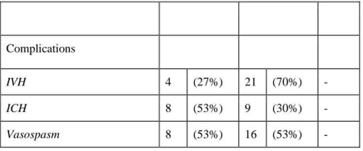
Complications
IVH 4 (27%) 21 (70%)
-ICH 8 (53%) 9 (30%)
-Vasospasm 8 (53%) 16 (53%)
-With regard to Hunt-Hess grade, patients treated with endovascular technique presented most frequently with a lower clinical severity score, i.e. Hunt-Hess grade I, II and III (70%), compared to patients treated with microsurgical approach (40%). On the other hand, Hunt-Hess grade IV and V were the most probable clinical presentation in patients treated with microsurgical approach (53%), compared to patients treated with endovascular technique (30%) (tab.2).
TABLE 2. DIFFERENT CLINICAL PRESENTATION IN PATIENTS TREATED VIA MICROSURGICAL OR ENDOVASCULAR TECHNIQUE
No. of patients Clipping Group (%) Coiling Group (%) Total
Hunt and Hess grade
I + II + III 6 (40%) 21 (70%) 27
IV + V 8 (53%) 9 (30%) 17
N.A. 1 (7%) - - 1
Fisher grade IV was the most frequent neuroradiological finding in both groups (60% group A vs. 54% group B). The rate of intraventricular hemorrhage (IVH) in the group A was significantly lower than in the group B (27% vs. 70%) whereas the rate of intracerebral hematoma (ICH) was significantly higher in the group A rather than in the group B (53% vs. 30%). The presence of vasospasm after treatment was homogeneous for both groups (53%, group A vs. 53%, group B).
External ventricular drainage was applied in the majority
of cases in both groups and the most frequent type of hydrocephalus was tetraventricular. Malfunctioning shunt systems were reported in few cases in both groups (20%, group A vs. 10%, group B) (tab.3).
TABLE 3. CHARACTERISTICS AND TREATMENTS OF THE HYDROCEPHALUS IN MICROSURGICAL AND ENDOVASCULAR GROUPS No. of patients Clipping Group (%) Coiling Group (%) Total
External Ventricular Drainage 11 (73%) 26 (87%)
-Lumbar Drainage 1 (7%) 6 (20%)
-Type of Hydrocephalus
Triventricular 1 (7%) 1 (3%) 2
Tetraventricular 14 (93%) 29 (97%) 43
Shunt Malfunctioning 3 (20%) 3 (10%) 6
According to the literature, the type of treatment to be adopted is strongly driven by the aneurysm location. As a matter of fact, our data showed that posterior cerebral circulation aneurysms were usually treated via an endovascular modality (13%, group A vs. 43%, group B) while middle cerebral artery aneurysms were preferably selected for microneurosurgical treatment (53%, group A vs. 20%, group B). Anterior cerebral artery complex aneurysms were treated either by neurosurgical or endovascular approaches (33%, group A vs. 37%, group B).
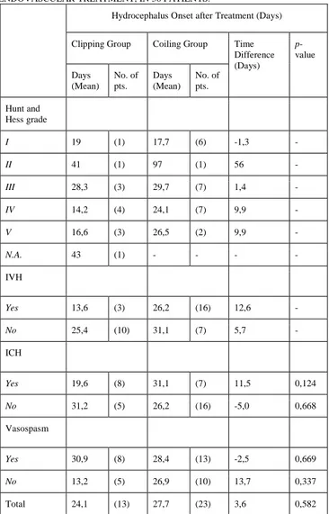
The time between the treatment (either surgical or endovascular) and the development of hydrocephalus, i.e. "hydrocephalus onset after treatment", was not different between the two groups.
This data was calculated on 13 cases of patients treated with microsurgical techniques and 23 cases of patients underwent endovascular procedure, for a total of 36 cases.
Patients treated with endovascular technique developed hydrocephalus after an average of 27,7 days. On the other hand, patients who underwent surgery developed hydrocephalus after 24,1 days (tab.4). This data was not statistically significant (p=0,582).
TABLE 4. HYDROCEPHALUS ONSET AFTER MICROSURGICAL OR ENDOVASCULAR TREATMENT, IN 36 PATIENTS.
Hydrocephalus Onset after Treatment (Days)
Clipping Group Coiling Group Time Difference (Days) p-value Days (Mean) No. of pts. Days (Mean) No. of pts. Hunt and Hess grade I 19 (1) 17,7 (6) -1,3 -II 41 (1) 97 (1) 56 -III 28,3 (3) 29,7 (7) 1,4 -IV 14,2 (4) 24,1 (7) 9,9 -V 16,6 (3) 26,5 (2) 9,9 -N.A. 43 (1) - - - -IVH Yes 13,6 (3) 26,2 (16) 12,6 -No 25,4 (10) 31,1 (7) 5,7 -ICH Yes 19,6 (8) 31,1 (7) 11,5 0,124 No 31,2 (5) 26,2 (16) -5,0 0,668 Vasospasm Yes 30,9 (8) 28,4 (13) -2,5 0,669 No 13,2 (5) 26,9 (10) 13,7 0,337 Total 24,1 (13) 27,7 (23) 3,6 0,582
Accordingly, group A and group B were divided into subgroups on the basis of clinical presentation and neuroradiological features. In many cases, such as Hunt-Hess grades and presence/absence of intraventricular hemorrhage, the sample size was not homogeneous due to the poor number of cases, so none conclusions could be drawn.
On the other hand, in subgroups with enough number of patients, the analysis showed that the presence of intracerebral hematoma caused a delay in the hydrocephalus onset after treatment in terms of 11,5 days in patients treated with endovascular technique compared to patients treated with microsurgical technique. This data was not statistically significant (p=0,124).
Moreover, the absence of vasospasm determined a delay of 13,7 days in patients treated with endovascular technique compared to patients treated with microsurgical technique.
This data was not statistically significant (p=0,337). IV. DISCUSSION
Development of hydrocephalus after subarachnoid hemorrhage is a well-known complication in ruptured cerebral aneurysms. Such hydrocephalus may be acute or chronic: acute hydrocephalus occurs immediately after subarachnoid hemorrhage and can determine catastrophic results if not promptly treated. This type of hydrocephalus is caused by mechanical obstruction of cerebrospinal fluid pathways.
On the other hand, chronic hydrocephalus may occur between 2 and 6 weeks post-SAH and may cause a significant setback in the patient’s clinical course. This type is caused by impaired cerebrospinal fluid absorption as a result of arachnoidal adhesions after SAH. However, the exact origin of chronic communicating hydrocephalus following subarachnoid hemorrhage is not well understood and recently some authors postulate that proliferation of arachnoidal cells, triggered by the inflammatory reaction or blood clotting products, could result in obstruction of CSF flow through arachnoid villi into the venous sinuses [12].
The consequences of chronic hydrocephalus have been extensively studied. Hydrocephalus can result in long-term memory problems and the development of psycho-organic disorders, and cognitive deficits have been noted after even slight temporary ventricular dilation. Indeed, the development of chronic hydrocephalus can be associated with almost doubling of the permanent neurological morbidity rate after aneurysmal rupture. Chronic hydrocephalus has been reported to range between 6% and 37% patients with aSAH [2,3,8,9,13-15]. Numerous papers have been published addressing the relationship between the clinical procedure (clipping or coiling) used to treat the aneurysms and the development of chronic hydrocephalus. This issue is still debated in the current literature.
However, whether there is a significant difference in chronic hydrocephalus onset in terms of days after treatment in patients who underwent endovascular treatment or microsurgical clipping has not yet been determined. The aim of our study was to rule out an answer to this topic.
With regard to data collected, in our series the majority of patients were over 50 years of age in both treatment groups and there was a clear female preponderance in both groups (tab.1). Different authors reported the relationship between age and initial condition in patients with aneurysms who develop hydrocephalus, and older patients have been found to be more likely to develop this condition [14,16].
According to previous studies [8], our data showed that the location of aneurysms was different between the two treatment groups, with significantly more coiled aneurysms of the posterior circulation and significantly more clipped aneurysms of the middle cerebral artery. This is in accordance with current treatment rules and the
specific advantages of coiling versus clipping [17,18]. As shown by other authors [8], IVH was more often present in the coil group than in the clip group while ICH generally led to microsurgical treatment. On the other hand, different from previous report [8], with regard to Fisher grades on computed-tomography scan, the majority of patients were admitted to our Institutions with Grade IV.
In our series has been demonstrated no difference in terms of hydrocephalus onset after endovascular or microsurgical treatment.
Only analyzing some variables associated to the hydrocephalus onset in the two groups some differences have been highlighted, albeit these data did not reach statistic significance.
The presence of intracerebral hematoma determines a non-statistical significant delay in the time of hydrocephalus onset in patients treated with endovascular technique compared to patients treated by microsurgery and the absence of vasospasm development as a complication of aSAH causes a similar non-statistical significant delay in the endovascular group.
In our series, coiling group patients had a better clinical presentation than patients who underwent microsurgical procedure (tab.2).
Even if these data do not reach statistical significance, we might speculate that patients who underwent minimal-invasive endovascular intervention and were admitted with lower Hunt and Hess grade were possibly subjected to a less close follow-up thus resulting in a slight delay in the diagnosis of hydrocephalus as compared to microsurgical group, which were admitted to our Institutions with severe neurological status.
V. CONCLUSION
Our findings suggest that there is no difference in terms of hydrocephalus onset after microsurgical or endovascular treatment. Only the presence of intracerebral hematoma or the absence of vasospasm can cause a slight, although not statistically significant, delay in the time of hydrocephalus onset in the endovascular group patients. Better clinical presentation may lead to a delay in the diagnosis of hydrocephalus in the endovascular group. However, long-term follow-up studies involving higher numbers of subjects are needed to better demonstrate this possible relationship.
ACKNOWLEDGMENT
This project has been supported by Johnson & Johnson Medical.
REFERENCES
[1] Kim SH, Chung PW, Won YS, Kwon YJ, Shin HC, Choi CS. Effect of cisternal drainage on the shunt dependency following aneurysmal subarachnoid hemorrhage. J Korean Neurosurg Soc 2012;52:441-6. [2] Dehdashti AR, Rilliet B, Rufenacht DA, de Tribolet N. Shunt-dependent hydrocephalus after rupture of intracranial aneurysms: a prospective study of the influence of treatment modality. J Neurosurg 2004;101:402-7.
[3] Brisman JL, Berenstein A. Factors related to hydrocephalus after aneurysmal subarachnoid hemorrhage. Neurosurgery 2004;54:1031.
[4] Dorai Z, Hynan LS, Kopitnik TA, Samson D. Factors related to hydrocephalus after aneurysmal subarachnoid hemorrhage. Neurosurgery 2003;52:763-9; discussion 9-71.
[5] Kwon JH, Sung SK, Song YJ, Choi HJ, Huh JT, Kim HD. Predisposing factors related to shunt-dependent chronic hydrocephalus after aneurysmal subarachnoid hemorrhage. J Korean Neurosurg Soc 2008;43:177-81. [6] Auer LM, Mokry M. Disturbed cerebrospinal fluid circulation after subarachnoid hemorrhage and acute aneurysm surgery. Neurosurgery 1990;26:804-8; discussion 8-9.
[7] O'Kelly CJ, Kulkarni AV, Austin PC, Urbach D, Wallace MC. Shunt-dependent hydrocephalus after aneurysmal subarachnoid hemorrhage: incidence, predictors, and revision rates. Clinical article. J Neurosurg 2009;111:1029-35.
[8] de Oliveira JG, Beck J, Setzer M, et al. Risk of shunt-dependent hydrocephalus after occlusion of ruptured intracranial aneurysms by surgical clipping or endovascular coiling: a single-institution series and meta-analysis. Neurosurgery 2007;61:924-33; discussion 33-4. [9] Gruber A, Reinprecht A, Bavinzski G, Czech T, Richling B. Chronic shunt-dependent hydrocephalus after early surgical and early endovascular treatment of ruptured intracranial aneurysms. Neurosurgery 1999;44:503-9; discussion 9-12.
[10] Komotar RJ, Hahn DK, Kim GH, et al. The impact of microsurgical fenestration of the lamina terminalis on shunt-dependent hydrocephalus and vasospasm after aneurysmal subarachnoid hemorrhage. Neurosurgery 2008;62:123-32; discussion 32-4.
[11] Chohan MO, Carlson AP, Hart BL, Yonas H. Lack of functional patency of the lamina terminalis after fenestration following clipping of anterior circulation aneurysms. J Neurosurg 2013.
[12] Massicotte EM, Del Bigio MR. Human arachnoid villi response to subarachnoid hemorrhage: possible relationship to chronic hydrocephalus. J Neurosurg 1999;91:80-4.
[13] Kassell NF, Torner JC, Jane JA, Haley EC, Jr., Adams HP. The International Cooperative Study on the Timing of Aneurysm Surgery. Part 2: Surgical results. J Neurosurg 1990;73:37-47.
[14] Pietila TA, Heimberger KC, Palleske H, Brock M. Influence of aneurysm location on the development of chronic hydrocephalus following SAH. Acta Neurochir (Wien) 1995;137:70-3.
[15] Vale FL, Bradley EL, Fisher WS, 3rd. The relationship of subarachnoid hemorrhage and the need for postoperative shunting. J Neurosurg 1997;86:462-6. [16] Graff-Radford NR, Torner J, Adams HP, Jr., Kassell NF. Factors associated with hydrocephalus after subarachnoid hemorrhage. A report of the Cooperative Aneurysm Study. Arch Neurol 1989;46:744-52.
[17] Molyneux AJ, Kerr RS, Yu LM, et al. International subarachnoid aneurysm trial (ISAT) of neurosurgical clipping versus endovascular coiling in 2143 patients with ruptured intracranial aneurysms: a randomised comparison of effects on survival, dependency, seizures, rebleeding, subgroups, and aneurysm occlusion. Lancet 2005;366:809-17.
[18] Molyneux A, Kerr R, Stratton I, et al. International Subarachnoid Aneurysm Trial (ISAT) of neurosurgical clipping versus endovascular coiling in 2143 patients with ruptured intracranial aneurysms: a randomised trial. Lancet 2002;360:1267-74.