Multimodal treatment of gastric cancer in the west: Where
are we going?
Daniele Marrelli, Karol Polom, Giovanni de Manzoni, Paolo Morgagni, Gian Luca Baiocchi, Franco Roviello
Daniele Marrelli, Karol Polom, Franco Roviello, Departmentof Medicine, Surgery and Neurosciences, University of Siena, 53100 Siena, Italy
Giovanni de Manzoni, Department of Surgery, University of Verona, 37134 Verona, Italy
Paolo Morgagni, Depertment of Surgery, “Morgani-Pierantoni” Hospital, 47100 Forlì, Italy
Gian Luca Baiocchi, Surgical Clinic, University of Brescia, 25100 Brescia, Italy
Author contributions: Marrelli D and Polom K contributed equally to this manuscript; Marrelli D and Roviello F were responsible for study conception and design; Marrelli D and Polom K wrote the paper; Marrelli D and Polom K analyzed the data; de Manzoni G, Morgagni P and Baiocchi GL carried out the research; de Manzoni G, Morgagni P, Baiocchi GL and Roviello F performed critical revision of the manuscript.
Conflict-of-interest statement: No potential conflicts of interest relevant to this article were reported.
Open-Access: This article is an open-access article which was selected by an in-house editor and fully peer-reviewed by external reviewers. It is distributed in accordance with the Creative Commons Attribution Non Commercial (CC BY-NC 4.0) license, which permits others to distribute, remix, adapt, build upon this work non-commercially, and license their derivative works on different terms, provided the original work is properly cited and the use is non-commercial. See: http://creativecommons.org/ licenses/by-nc/4.0/
Correspondence to: Daniele Marrelli, MD, Associate Professor, Department of Medicine, Surgery and Neurosciences, University of Siena, 55 Via Banchi di Sotto, 53100 Siena,
Italy. [email protected] Telephone: +39-577-585882 Fax: +39-577-233337 Received: January 28, 2015
Peer-review started: January 28, 2015 First decision: March 10, 2015 Revised: March 25, 2015
EDITORIAL
DOI: 10.3748/wjg.v21.i26.7954 © 2015 Baishideng Publishing Group Inc. All rights reserved.Accepted: May 2, 2015 Article in press: May 4, 2015 Published online: July 14, 2015
Abstract
The incidence of gastric cancer (GC) is decreasing worldwide, especially for intestinal histotype of the distal third of the stomach. On the contrary, proximal location and diffuse Lauren histotype have been reported to be generally stable over time. In the west, no clear improvement in long-term results was observed in clinical and population-based studies. Results of treatment in these neoplasms are strictly dependent on tumor stage. Adequate surgery and extended lymphadenectomy are associated with good long-term outcome in early-stage cancer; however, results are still unsatisfactory for advanced stages (Ⅲ
and Ⅳ), for which additional treatments could provide a survival benefit. This implies a tailored approach to GC. The aim of this review was to summarize the main multimodal treatment options in advanced resectable GC. Perioperative or postoperative treatments, including chemotherapy, chemoradiotherapy, targeted therapies, and hyperthermic intraperitoneal chemotherapy have been reviewed, and the main ongoing and completed trials have been analyzed. An original tailored multi-modal approach to non-cardia GC has been also proposed.
Key words: Epidemiology; Hyperthermic intraperitoneal
chemotherapy; Chemotherapy; Radiotherapy; Gastric cancer; Targeted therapy
© The Author(s) 2015. Published by Baishideng Publishing
Group Inc. All rights reserved.
Core tip: In advanced gastric cancer (GC), multimodal
treatment is currently an option in the west. Adequate surgery and extended lymphadenectomy, together with
survival worsened over time for patients with non-cardiac tumors, whereas the risk of death decreased for patients with cardiac tumors[12].
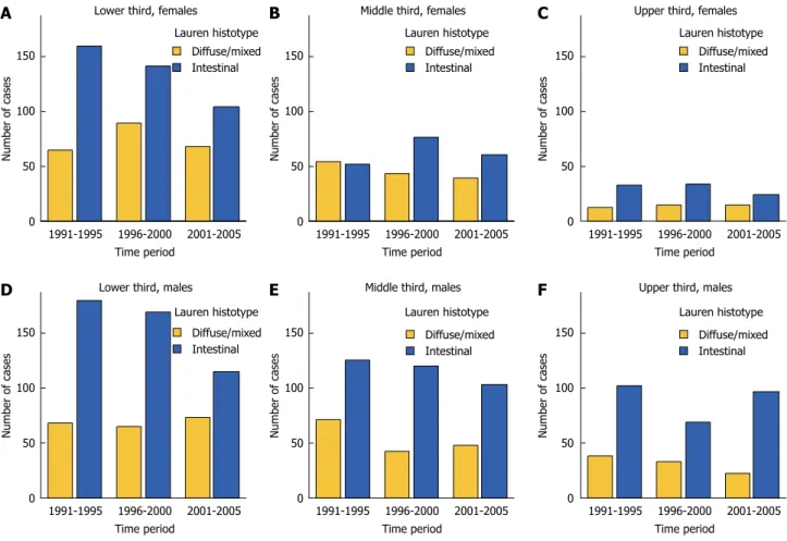
Different epidemiological trends in the intestinal type (IT) and diffuse type (DT) Lauren histotypes have also been observed. The declining incidence of GC has been linked to the decreasing number of ITs; on the contrary, the incidence of DT is generally stable throughout the world[10,13-15]. As most proximal tumors
are IT, it is important, when evaluating epidemiological trends, to group data according to histotype and location. In a recent study from the Italian Research Group for Gastric Cancer (GIRCG), a decreasing number of IT tumors of the distal stomach was observed; on the contrary, IT located in the proximal third, and DT, at any location, were stable over time[9]. As a
conse-quence, the DT neoplasms showed a relative increase with time (Figure 1).
Recent studies have also reported different trends of GC incidence in young patients; declining rates were observed for subjects aged 40-84 years, whereas for younger cohorts, the incidence rates increased over time[16]. Recent reports from Europe also confirm
these findings[17]. The higher prevalence of DT in
young patients may explain the epidemiological trends described for specific histotypes of GC.
As for GC prevention, two potential strategies are proposed. Primary prevention is possible due to eradication of H. pylori, and secondary prevention by detection of GC in mass screening[4]. Primary
prevention is based on the fact that H. pylori is the strongest known factor associated with distal IT GC. It is possible to eradicate the infection using antibiotics in association with an antisecretory agent. It is proposed to offer prophylactic eradication for high-risk individuals, or for patients in high-risk areas.
For secondary prevention, mass screening is performed in countries with the highest incidence of GC. In Japan or South Korea the screening pro-grams seem to be effective, with the higher rate of early GC detection, improved 5-year survival, and improved proportion of localized GC at diagnosis[4,17].
The main screening methods are barium X-ray, combination of barium digital radiography together with serum pepsinogen testing, and endoscopy with photofluorography. However, mass screening is hard to promote and organize in low-risk areas, where few but more advanced GC cases, mainly with proximal location or DT tumors, are generally observed in clinical practice[4].
CLINICAL IMPLICATIONS OF CHANGING
EPIDEMIOLOGY
The above-mentioned epidemiological trends could have important clinical implications. Indeed, the overall number of newly diagnosed GC cases is decreasing, but the relative percentage of proximal locations and
modern chemotherapy, radiotherapy, targeted therapies, and a combination of all could possibly improve survival in advanced GC. A tailored multimodal approach is strictly necessary in the light of treatment results and recent epidemiological trends, which indicate a relative increase of more aggressive forms, such as proximal location and diffuse Lauren histotype in the west. The main ongoing and completed clinical trials regarding multimodal approach to GC have been reviewed, and an original tailored multimodal protocol to non-cardia GC has been proposed.
Marrelli D, Polom K, de Manzoni G, Morgagni P, Baiocchi GL, Roviello F. Multimodal treatment of gastric cancer in the west: Where are we going? World J Gastroenterol 2015; 21(26): 7954-7969 Available from: URL: http://www.wjgnet.com/1007-9327/full/v21/ i26/7954.htm DOI: http://dx.doi.org/10.3748/wjg.v21.i26.7954
CHANGING EPIDEMIOLOGY OF GASTRIC
CANCER
Despite the reported declining incidence, gastric cancer (GC) is one of the most common causes of cancer mortality worldwide[1-3]. It represents the fourth most
common cancer after lung, breast and colorectal cancer, and the second most common cause of cancer-related death after lung cancer. Geographic variability of GC is also well known: highest incidence rates are observed in East Asia, Central Asia, Eastern Europe, and the Pacific Coast of South and Central America, whereas the lowest incidence rates are found in Northern Europe and North America[4]. Even within the
same country, there can be wide variation in geographic incidence: for example, in Italy, mortality is high in the central region, especially along the Central Apennine Mountains, and very low in Southern Italy[5,6]. Even
if partly obscured by population aging, a decreasing incidence of GC has been reported worldwide in recent decades. This epidemiological trend has been attributed to several factors, such as the increased consumption of vegetables and fruit instead of cured meat, and changed methods of food conservation (refrigeration, instead of salt preservation)[7]. The
decreased prevalence of Helicobacter pylori (H. pylori) infection has also had a role. However, decreasing rates are more evident in high-risk areas, whereas in low-risk areas, the rates have fallen slowly, with a trend to become stable over time[5,6,8,9].
Certain subtypes of GC demonstrate different epidemiological features. Tumors located in the distal third of the stomach have shown the most evident decrease in incidence, whereas proximal tumors are stable or even increasing[10,11]. This trend has been
confirmed in some recent studies: the incidence decreased among men and women, but the proportion of cardiac tumors remained stable over time; 5-year
DT is increasing. Proximal tumors, including those involving the esophagogastric junction (EGJ), are associated with higher clinical aggressiveness and worse prognosis[9,18-21]. The relative increase in the
proportion of proximal tumors could lead to a general decrease in survival probability.
Another important consequence of epidemiological trends is the relative increase in DT tumors (Figure 2). Besides histomorphometrical characteristics, IT and DT histotypes show evident differences in epide-miological, clinical and molecular features[22]. IT type
is more common in males and older patients, whereas DT type usually affects younger patients with a lower male-female ratio. Environmental factors seem to be involved in the pathogenesis of IT tumors, and they usually follow the sequence of chronic atrophic gastritis, intestinal metaplasia, and dysplasia. On the contrary, DT tumors usually originate from healthy gastric mucosa or non-atrophic gastritis, and are more related to genetic factors. A further characteristic of the DT is their greater biological aggressiveness. The risk of lymph node metastasis is higher in the DT than the IT, at the same T stage. Indeed, the DT is a strong risk factor for lymph node metastasis in early GC[23], but an increased risk is also present in more
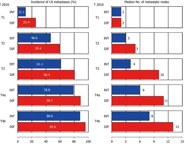
advanced pT stages. The correlation between lymph node metastasis and Lauren histotype, stratified for pT stage, has been evaluated in 2090 non-cardia GC
patients from the GIRCG database (Figure 3). The incidence and number of lymph node metastases were notably higher in the DT than IT groups at the same pT stage. Furthermore, DT is also a risk factor for lymph node metastases in extra-regional nodal locations (such as para-aortic nodes)[24,25].
The higher probability of lymph node metastases in DT could be an indication for more extended lympha-denectomy or neoadjuvant treatment. In contrast, clinical diagnosis, by radiological imaging, of lymph node metastasis may be more difficult in the DT. It has been reported that, in this histotype, the size of involved nodes may be smaller than the commonly used cut-off values[22].
Besides the lymph node involvement, DT tumors also show a greater propensity to peritoneal spread. Indeed, several studies have demonstrated a higher risk of peritoneal recurrence in DT tumors; mainly when the tumor has serosal involvement[22,26].
In a GIRCG follow-up study, the 5-year risk of peritoneal recurrence has been calculated to be 69% in DT GC with serosal involvement, vs 20% for IT cases at the same pT stage. It has been demonstrated that the clinical impact of extended surgery, including D2/D3 lymphadenectomy, is of low value in serosally exposed forms at risk of peritoneal recurrence[27,28].
The chance of cure in patients with peritoneal recurrence of GC is low: in a GIRCG follow-up study,
Figure 1 Changing number of patients in three subperiods, stratified according to tumor location and Lauren histotype (GIRCG database).
Number of cases Number of cases 150 100 50 0
Lower third, females
1991-1995 1996-2000 2001-2005 Time period 150 100 50 0
Lower third, males
1991-1995 1996-2000 2001-2005 Time period Number of cases 150 100 50 0
Middle third, females
1991-1995 1996-2000 2001-2005 Time period Number of cases 150 100 50 0
Middle third, males
1991-1995 1996-2000 2001-2005 Time period Number of cases 150 100 50 0
Upper third, females
1991-1995 1996-2000 2001-2005 Time period Number of cases 150 100 50 0
Upper third, males
1991-1995 1996-2000 2001-2005 Time period
A
B
C
D
E
F
Diffuse/mixed Intestinal Diffuse/mixed Intestinal Diffuse/mixed Intestinal Diffuse/mixedIntestinal Diffuse/mixedIntestinal
Diffuse/mixed Intestinal Lauren histotype Lauren histotype Lauren histotype
Intestinal type Diffuse type
Decreasing incidence Relative increase
Figure 2 Images of intestinal (A, C) and diffuse type (B, D) tumors of the stomach. The arrow in D indicates the infiltrative growth of the diffuse histotype in the
gastric wall.
A
B
C
D
Figure 3 Incidence of lymph node metastases according to Lauren histotype stratified for pT stages (GIRCG database).
Incidence of LN metastases (%) Median No. of metastatic nodes INT DIF INT DIF INT DIF INT DIF INT DIF T1 T2 T3 T4a T4b INT DIF INT DIF INT DIF INT DIF INT DIF T1 T2 T3 T4a T4b 0 20 40 60 80 100 0 3 6 9 12 15 T 2010 11.1 25.4 46.6 59.4 61.1 80.4 78.8 88.7 88.0 95.6 2 2 3 5 4 10 6 11 8 13 T 2010
5-year survival probability in 221 patients with metachro-nous peritoneal carcinomatosis (PC) was only 3% (Figure 4)[29]. As such, prevention of peritoneal recurrence,
more than treatment after its occurrence, may be the only potential chance of cure in high-risk cases[30].
Late-phase DT GC can evolve into diffuse infiltra-tion, thickening and stiffening of the gastric wall with reactive fibrosis, also named gastric linitis plastica. This is a subset of GC with a large propensity to diffuse infiltration, massive lymph node metastasis, and peritoneal seeding[31]. The rate of radical resection
in this form of GC is < 30%, and, even after R0 resection, the 5-year survival probability does not exceed 5%. Some population-based studies from Europe, along with the decreased incidence of GC, have reported a significant increase of gastric linitis plastica with time[32]. These data are consistent with
previously mentioned epidemiological trends.
Pathological characteristics of different histotypes of GC may explain epidemiological and survival data reported in large European studies. Recent data from 49 cancer registries in 18 European countries (EUROCARE-4 Working Group) have revealed a notable survival increase in Europe over the period 1988-1999 for several cancer sites, in particular, for prostate, colorectal and breast cancer. However, for GC, the increase was small (from 22% to 24%), despite potential time-related improvements in diagnosis, surgical and medical treatment[33]. Survival
improvement was higher for men (4.1%) than women (1.4%). The declining incidence of cancers of the distal stomach could help to explain these survival trends. Indeed tumors of the cardia or fundus are usually diagnosed in older patients, at an advanced stage, and with diffuse/signet ring cell morphology. Other population-based and clinical studies reported similar results. In the previously mentioned French study, the global prognosis of GC did not improve significantly over a 12-year period of observation[32]. Recent
studies from the Netherlands have also confirmed the decreasing incidence of GC but stable survival rates over time[34].
These data seem to be consistent with the findings of a previous GIRCG study: along with the decreasing number of distal IT tumors and the relative increase of DT forms with time, a lack of improvement of cancer-related survival probability, and a significant increase of peritoneal recurrence after surgery were observed[9].
In particular, survival rates decreased in the more recent period in the group of patients with serosal involvement, in women, and in distal tumors, whereas an increasing trend was observed in proximal tumors. All these data may fit with the hypothesis that the relative increase in DT tumors may have contributed to the absence or small improvement of treatment of GC in western countries.
TREATMENT OF EARLY FORMS
Surgical treatment with adequate lymphadenectomy could offer a high probability of cure even in western patients. Survival rates in early stages reported from specialized western centers are similar to those obtained in Japan and South Korea[21,27,35].
Selected forms of early GC can be treated by endoscopic mucosal resection or endoscopic submu-cosal dissection, in accordance with the standard criteria described by the Japanese Gastric Cancer Association (JGCA), with acceptable results even in the west[36,37]. The resection is judged as curative when
all of the following conditions are fulfilled: en bloc resection, tumor size not greater than 2 cm, histology of intestinal-differentiated-type, pT1a, negative horizontal (lateral) margin, negative vertical margin, and no lymphovascular invasion.
Although endoscopic approaches to early forms of GC are increasing in specialized centers in the west, they are still far from becoming a clinical standard. Early forms not treatable by endoscopic resection should be submitted to surgical resection with lymphadenectomy. According to the JGCA treatment guidelines[36], D1 lymphadenectomy may be adequate
for early GC with clinically negative lymph nodes. However, we should underline that a proportion of early GC in the west is DT, which is associated with a higher risk of lymph node metastases and greater lymph node spread, especially when submucosa is involved. Furthermore, in the west, endoscopic resection, which can be considered as a treatment as well as a staging procedure, is performed less frequently than in East Asia, and the clinical diagnosis of lymph node metastasis by imaging procedures still has low accuracy[38]. As such, the Italian guidelines
advise standard D2 lymphadenectomy in early forms of GC[39]. Only in selected cases (high-risk patients,
early forms with favorable pathological characteristics, not treatable by endoscopic resection) should more
Months after diagnosis of peritoneal recurrence 100 90 80 70 60 50 40 30 20 10 0 0 6 12 18 24 30 36 42 48 54 60
Figure 4 Survival rate of patients with peritoneal recurrence of gastric cancer.
limited procedures be considered (D1 plus).
Early forms of GC could also be treated by mini-mally invasive (laparoscopic or robotic) approaches, which demonstrated non-inferior oncological results compared with open surgery[40,41]. However, it should
be emphasized that oncological criteria regarding resection margin and lymph node dissection need to be carefully followed in minimally invasive procedures.
TREATMENT OF ADVANCED
RESECTABLE FORMS
In advanced resectable forms of GC, it is now well established that adequate surgical treatment is a key factor in obtaining acceptable long-term results. As for the extent of resection, subtotal gastrectomy offers low postoperative morbidity and mortality risk, and better quality of life, without affecting long-term oncological results, when an adequate resection margin can be obtained (R0 resection)[42]. A proximal
margin of at least 3 cm is recommended for T2 or deeper tumors with an expansive growth pattern, and 5 cm is recommended for DT and tumors with infiltrative growth pattern. In all other cases, total gastrectomy should be the preferred procedure. In early GC, a resection margin of 2 cm may be enough[39]. Total gastrectomy with splenectomy should
be also recommended for tumors located along the greater curvature. Splenectomy should be performed only when macroscopic involvement of lymph nodes at the splenic hilum is present.
The extent of lymphadenectomy is crucial. Even if some randomized studies have failed to demonstrated a significant advantage for overall survival, a re-evaluation of the Dutch trial showed a reduced cancer-related survival in the long term and a higher incidence of late recurrence of GC in patients submitted to limited (D1) lymphadenectomy[43].
It is important to ensure that good early post-operative results in terms of morbidity and mortality are achieved. This is consistent with the reports of observational nonrandomized studies from specialized centers[44,45].
Nowadays, D2 lymphadenectomy is generally accepted as the standard approach in most national guidelines[39,46]. The correct procedure for
lympha-denectomy involves the removal of nodal stations from 1 to 12, with some variations depending upon the extent of gastric resection[36]. Special attention should
be paid upon to the complete removal of infrapyloric nodes (station 6), right paracardial nodes (station 1), left gastric artery nodes (station 7), celiac axis (station 9), hepatic artery (station 8a), splenic artery (station 11), and hepatoduodenal ligament nodes (station 12a).
More extended lymphadenectomies (D2+) can be performed in selected cases at risk of metastasis to posterior (stations 8p, 12p, 12b and 13), mesenteric
(station 14) or para-aortic (stations 16a2 and b1) lymph nodes, in specialized centers and in the setting of clinical studies[22,23]. In particular, proximal or DT
tumors are particularly prone to metastasis to distant nodes, and in our opinion they may benefit from super-extended lymphadenectomy[25,28]. However, it
should be emphasized that in more advanced stages (UICC TNM stages ⅢA and more) the results of surgery, even with adequate lymphadenectomy, are still unsatisfactory in western patients[35]. As such,
additional treatments should be planned to improve long-term survival in these patients.
MULTIMODAL TREATMENT OF GASTRIC
CANCER
Neoadjuvant treatment seems to be a good option in advanced GC. The term advanced should be understood as a T3, T4 and/or N+ and/or with positive peritoneal cytology. The majority of patients who are diagnosed at this stage might receive benefits from perioperative treatment.
Even though dietary changes and the use of antibiotics to treat chronic H. pylori infection have helped to reduce steadily the number of new cases of GC, the progress in GC treatment is still limited[47].
Surgery remains the only treatment with curative intent in locoregional disease. From an oncological point of view the issue is to resect the cancer with a negative resection margin (R0), and with adequate lymph node dissection. The biggest problem, especially in the west, is diagnosis of patients with locally advanced disease. Advanced disease is associated with a higher rate of locoregional recurrence. For locally advanced forms, additional multimodal treatment in the preoperative, perioperative and postoperative phases has been proposed. Nowadays, we can observe geographic differences in multimodal treatment of GC. In Asia, the most commonly used treatment is adjuvant chemotherapy; in the United States, the favored treatment is chemoradiotherapy (CRT); and in Europe, neoadjuvant therapy is mostly used.
Advanced GC still has a poor survival (< 30% 5-year survival probability for stage Ⅲ). Cunningham et al[48]
and Ychou et al[49] have demonstrated the advantage
of starting multimodal treatment with preoperative chemotherapy over surgery alone, therefore, this seems to be a good treatment option. In the trial by Schuhmacher et al[50], neoadjuvant therapy improved
R0 resection rate even though it did not improve overall survival (OS). In the study by Stahl et al[51],
neoadjuvant CRT showed a higher rate of complete responders, and in the study by van Hagen et al[52],
improved OS was observed.
In Asian countries in contrast, the greatest interest lies in postoperative oral chemotherapy, which is associated with improved OS compared with surgery alone[53,54]. However, these results have not been
reproduced in western countries.
In the United States, CRT has been used routinely since 2001, after the trial of MacDonald et al[55].
Neoadjuvant chemotherapy
The neoadjuvant approach is currently recommended across Europe based on the Magic and FNLCC/FFCD trial[48,49]. Other benefits of neoadjuvant chemotherapy
(NC), discussed by Ott et al[56], for potentially
resectable GC are higher rate of R0 resection achieved by downstaging of a primary tumor, and probable effect on micrometastases and isolated tumor cells in lymph nodes. Ott et al emphasized also that the neoadjuvant setting is more often proposed for younger patients and those in general good health.
In the Magic trial, chemotherapy consisted of three cycles of intravenous (i.v.) epirubicin, cisplatin and 5-fluorouracil (FU) preoperatively and three cycles postoperatively[48]. NC was not associated with
worse postoperative complications and 30-d mortality than surgery alone, thus overturning the argument that neoadjuvant therapy may be more dangerous for patients. From the main results, 5-year survival rate was 36% vs 23% in favor of perioperative chemotherapy. Also, OS and progression-free survival (PFS) were significantly better. Only 49.5% of patients received the full perioperative chemotherapy treatment, therefore, this was one of the main issues criticized by some investigators. This issue was investigated in the study by Mirza et al[57] in which it
was checked in patients using the same regimen as in the Magic trial. The full perioperative regimen had a beneficial effect on DFS but not on OS. It may be concluded that administrating the adjuvant part of this regimen postponed tumor recurrence rather than helping in prevention.
The FNLCC/FFCD trial proved the beneficial effect of perioperative chemotherapy for gastric and esophageal adenocarcinoma[49]. In the preoperative period,
two or three cycles of i.v. cisplatin and 5-FU were administered, and after surgery, chemotherapy was continued when response to treatment was observed. A higher rate of R0 resection in NC in comparison with surgery alone was observed, as well as improved OS and DFS. The 5-year survival rates were 38% vs 24% in favor of NC.
In a meta-analysis by Ronellenfitsch et al[58] OS
was 9% better after neoadjuvant therapy. This effect was seen 18 mo after surgery and lasted at least 10 years. R0 resection was achieved 1.4 times more often after neoadjuvant treatment. Importantly, side effects of neoadjuvant therapy, such as postoperative morbidity or mortality, as well as prolonged hospital stay, were not increased significantly compared with surgery alone. Another interesting aspect was that no benefit of neoadjuvant therapy was seen in elderly patients. The subgroup of patients with EGJ cancer had the greatest benefit in OS. One of the unanswered
questions is the age of patients recruited to the trial. Most trials excluded patients aged > 70 years. This issue is currently under investigation by a study in Germany. Another subgroup of patients of particular interest is those with signet ring cell carcinoma. They seem not to benefit from neoadjuvant treatment[59].
The response rate differs also according to pathological features. In DT tenors, a good pathological response was only observed in 14.5% of patients[60].
In Asian countries, neoadjuvant treatment is also beginning to play an important role. Currently several trials (JCOG 0210, JCOG 0501, JCOG 1002, and PRODIGY) are under way. In Italy, a GIRCG phase Ⅱ trial recruited patients with non-cardiac GC who underwent accurate pretreatment clinical staging with diagnostic laparoscopy and peritoneal washing, followed in all cases by standard D2 gastrectomy. This trial aims to answer whether preoperative or perioperative chemotherapy plays a role in advanced GC treatment (NCT01876927).
Neoadjuvant CRT
As NC proved to be safe for preoperative treatment, the addition of radiotherapy to preoperative treatment has gained interest. The German POET trial compared NC vs CRT for locally advanced EGJ cancers[51]. In one
arm, two courses of cisplatin, 5-FU and folic acid (PLF), followed by 3 wk of combined CRT (30 Gy in 3 wk with cisplatin/etoposide), and surgery were administered, vs 2.5 courses of PLF with surgery. This trial was closed early, and showed no significant difference in survival: 33.1 mo vs 21.1 mo in favor of CRT, but with higher mortality in the CRT arm: 10.2% vs 3.8% (P = 0.26). Results regarding 3-year survival showed an improvement from 28% to 48% in the CRT arm.
In a study by Burmeister et al[61] on 75 patients,
the addition of radiotherapy increased the rate of pathological complete remission (13% vs 0%, P = 0.02), and reduced the rate of R1 resection (0% vs 4%, P = 0.04). Analyzing 5-year OS and PFS, only a trend was observed in favor of CRT, without statistical significance (OS 45% vs 36%, P = 0.6).
In the CROSS trial from the Netherlands, patients with esophageal and EGJ cancers were assigned to CRT (carboplatin, paclitaxel and 41.4 Gy radiotherapy in 23 fractions) followed by surgery, vs surgery alone[52]. The surgery alone arm showed R0 resection
in 69% of patients with a median survival time of 24.2 mo, whereas in the neoadjuvant CRT arm, R0 resection was achieved in 92% (P < 0.001), with complete pathological response rate in up to 29% of patients; however, it is noteworthy that in case of squamous cell carcinoma the complete response rate was better (49%) than for adenocarcinoma (23%). The median survival time was 49.4 mo (P = 0.003), and 5-year survival improved from 34% to 47%. Postoperative complications rate and in-hospital mortality were similar in both arms (4%).
The neoadjuvant regimen also reduced locoregional recurrence rate (34% to 14%; P < 0.001), and the probability of PC (14% to 4%; P < 0.001). Distant metastases also showed a difference between both arms (35% vs 29%, P = 0.025). This treatment protocol is now recommended for neoadjuvant CRT in patients with EGJ adenocarcinoma in the US. The currently ongoing TOPGEAR trial is investigating CRT vs chemotherapy in EGJ and stomach cancers. In the chemotherapy arm, three courses of epirubicin, cisplatin and flurouracil (ECF) are given preoperatively, and in the CRT arm, two courses of ECF followed by 45 Gy, or radiation with concurrent 5-FU. Patients in both arms receive three courses of ECF after surgery.
A meta-analysis by Sjoquist et al[62] reviewed trials
with localized gastroesophageal adenocarcinoma with preoperative CRT and chemotherapy alone. The hazard ratio for OS was 0.75.
Adjuvant chemotherapy
Analyzing data from different countries, the results of adjuvant chemotherapy after gastrectomy in western studies are less convincing than in Asian studies. In a Japanese study (ACTS-GC trial), oral fluoropyrimidine (S-1) was given after surgery for 1 year, and results were compared with surgery alone. The 5-year OS was 70.1% vs 61.1%[53,54]. This trial
was stopped earlier because of significantly better OS in the S-1 group. It needs to be underlined that the high rates of OS in both arms were due to excellent surgery, as D2 lymphadenectomy was confirmed in all cases. The problem in translating this trial into a Caucasian population is that Tegafur, present in S-1 as a precursor of 5-FU, is transformed in the body by cytochrome P450 to 5-FU. The probable difficulties observed in Caucasians are due to polymorphism of CYP2A6 gene, and subsequent complications[63]. In the
FLAGS trial, comparison of cisplatin + S-1 and cisplatin + 5-FU for palliative therpy showed significantly better tolerance in patients with the addition of S-1[64].
In the CLASSIC trial, adjuvant chemotherapy with capecitabine and oxaliplatin after curative D2 gastrectomy was compared with surgery alone[65].
This Asian trial showed significant improvements in 3-year disease-free survival (DFS; 74% vs 59%, P < 0.0001), and OS (83% vs 78%, P = 0.0493). This trial was stopped earlier as the benefit of using this chemotherapy regimen was demonstrated. In the chemotherapy arm, oxaliplatin-induced peripheral neuropathy occurred in 56% of patients, but grade 3/4 only occurred in 2% of cases. It seems that this regimen might be an alternative to the S-1 regimen. A meta-analysis on 17 trials of adjuvant chemotherapy after gastrectomy showed a small but significant benefit for 5-FU-based chemotherapy[66]. Adjuvant
chemotherapy increased OS by 6%, and reduced the risk of death by 18%. A meta-analysis by Zhang et al[67] showed that four chemotherapy regimens may
be effective: 5-FU + mitomycin C + adriamycin; 5-FU + mitomycin C; tegafur; and mitomycin C. Other proposed regimens seem to be not so effective: 5-FU + carmustine, 5-FU + methyl-semustine, 5-FU + cisplatin, 5-FU + anthracyclines, and 5-FU + mitomycin C + cytarabine. Another meta-analysis by the GASTRIC group (Global Advanced/Adjuvant Stomach Tumor Research International Collaboration) showed significant improvement in OS after 5-FU-based chemotherapy[66]. The same group in another
meta-analysis on advanced GC concluded that experimental arms of chemotherapy are responsible for modest improvement in OS and DFS (hazard ratio 0.88 and 0.81). The median survival was below 1 year and none of the new regimens can be used as a standard[68].
Adjuvant CRT
The results of the INT-0116 trial by MacDonald et al[55]
show that adjuvant CRT plays an important role in GC treatment. The problem of additional radiotherapy is the increased toxicity rate. Grade 3/4 hematological toxicity occurred in 54%, and gastrointestinal toxicity in 33% of patients. The toxic effect was also responsible for stopping the treatment in many cases. In patients with diffuse histology, the addition of radiotherapy did not confer any additional benefit. The biggest concern is about radiation of a large area of gastrointestinal mucosa. Current studies are focused on using 3D conformal and intensity-modulated radiation therapy (IMRT), and also new, safer radiotherapy techniques[69-71]. In a phase Ⅱ trial with 3D-CRT/IMRT,
grade 3 or 4 nausea and vomiting (14.5%), decreased appetite (11.8%), leukopenia/neutropenia (9.1%) and fatigue (6.4%) were observed, and it proved to be a safe procedure[71]. We also need to mention that
in the MacDonald et al[55] trial, an increased number
(but not significant) of secondary malignancies after additional CRT were reported. The biggest challenge is to prove whether addition of radiotherapy to the regimen is better than chemotherapy alone. This issue was analyzed in the ARTIST trial[72]. No difference
in 3-year DFS was observed between those two arms, but analyzing subgroups with lymph node metastases, 3-year DFS was improved in the CRT arm (77.5% vs 72.3%, P = 0.0365). This was also seen when adjusting for tumor stage. No difference in case of local or distal recurrence rate was observed. No OS results were reported in the 3-year analysis. The ARTIST-Ⅱ trial will investigate the influence of chemotherapy or CRT in patients with lymph-node positive GC. One particularly interesting aspect is that, in the INT-0116 trial, D2 resection was performed in only 10% of cases, whereas in most Asian studies, it is close to 100%. Indeed, local recurrences were observed in 29% of cases in the INT-0116 trial, vs 2.8% in the Japanese ACTS-GC trial. It seems that the addition of radiotherapy confers a potential benefit to patients with a suboptimal surgical approach. This was
proved by a Dutch study, showing a reduction in local recurrences after CRT in patients with D1 resection, whereas this effect was not seen in the D2 resection group[73].
The problem of GEJ region radiotherapy is descri-bed later. The main difficulty is that these patients are subgroups in esophageal cancer and GC trials. Some of these problems were mentioned above, for example, in a neoadjuvant setting as in the CROSS trial[52]. After GEJ surgery, additional CRT is based on
the INT-0116 trail (approximately 20% of patients in this trial had a GEJ location)[55]
. In the current AJCC staging, GEJ tumors are staged as esophageal and not as gastric. The only trial exclusively for GEJ tumors was done in Germany, analyzing neoadjuvant CRT vs chemotherapy alone[51]. There was a higher rate of
complete pathological response (15.6% vs 2%), and a trend towards improved 3-year survival (47% vs 28%, P = 0.07) in favor of neoadjuvant CRT[51].
The German trial also tried to identify those patients who would benefit from neoadjuvant therapy using positron emission tomography/computed tomography[74]. The MUNICON study tried to predict
response after 2 wk of NC in GEJ cancer. Non-responders to chemotherapy underwent surgery, sparing them from unnecessary toxicity, as well as undergoing surgery earlier. It should also be noted that most GEJ tumors are fluorodeoxyglucose (FDG) sensitive, but in 30% they do not take up FDG[75]
. The solution might be to use radioisotopes such as fluorothymidine for GC[76]. The most important studies from multimodal GC
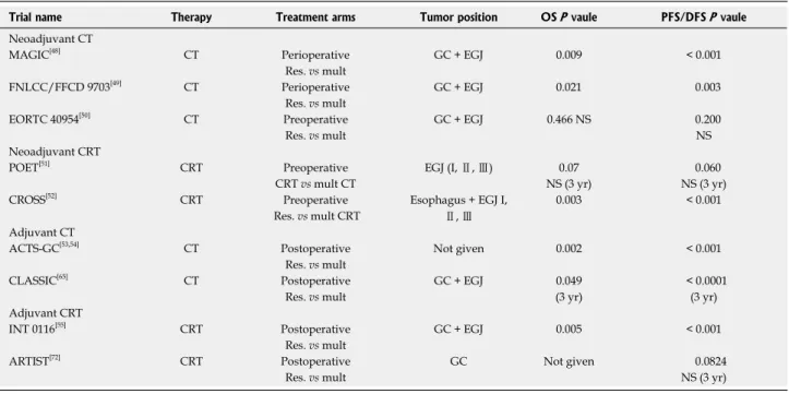
treatment are presented in Table 1.
Targeted therapies
The new drugs that may be used in targeted thera-pies probably play an increasing role in modern treatment of GC. Epidermal growth factor receptor (EGFR) is overexpressed in most GC. The trials that used anti-EGFR antibody cetuximab (EXPAND trial), and panitumumab (REAL3 trial) failed to improve survival in GC patients[77,78]
. In REAL3, panitumumab was shown to actually worsen survival of treated patients[78]
. Another antibody tested in an adjuvant setting in GC is bevacizumab against vascular endothelial growth factor A. In the AVAGAST trial, this antibody did not improve OS when added to standard chemotherapy[79]
. Overexpression of human epidermal growth factor receptor-2 (HER-2/neu) is present in > 20% of patients with GC. An antibody against this receptor - trastuzumab - showed signi-ficant improvement in OS in metastatic gastric and GEJ cancers in the ToGA trial[80]
. The oral antibody lapatinib is currently being investigated for HER-2-positive GC in the LOGIC trial. Currently, we await the results of ongoing trials using molecular-targeted drugs in GC: LOGIC (lapatinib), TYTAN (lapatinib), RAINBOW (ramucirumab), GRANITE-1 and GRANITE-2 (everolimus). We also need to mention that currently many drugs are being tested in phase Ⅰ and Ⅱ trials, such as the recently finished phase Ⅱ trial of apatinib, with promising results[81,82]
. From the molecular point of view, the greatest interest lies in drugs that will
Table 1 Main trials regarding adjuvant and neoadjuvant therapy for gastric cancer reported in literature
Trial name Therapy Treatment arms Tumor position OS P vaule PFS/DFS P vaule
Neoadjuvant CT
MAGIC[48] CT Perioperative GC + EGJ 0.009 < 0.001 Res. vs mult
FNLCC/FFCD 9703[49] CT Perioperative GC + EGJ 0.021 0.003 Res. vs mult
EORTC 40954[50] CT Preoperative GC + EGJ 0.466 NS 0.200
Res. vs mult NS
Neoadjuvant CRT
POET[51] CRT Preoperative EGJ (I, Ⅱ, Ⅲ) 0.07 0.060 CRT vs mult CT NS (3 yr) NS (3 yr) CROSS[52] CRT Preoperative Esophagus + EGJ I,
Ⅱ, Ⅲ
0.003 < 0.001 Res. vs mult CRT
Adjuvant CT
ACTS-GC[53,54] CT Postoperative Not given 0.002 < 0.001 Res. vs mult
CLASSIC[65] CT Postoperative GC + EGJ 0.049 < 0.0001 Res. vs mult (3 yr) (3 yr) Adjuvant CRT
INT 0116[55] CRT Postoperative GC + EGJ 0.005 < 0.001 Res. vs mult
ARTIST[72] CRT Postoperative GC Not given 0.0824 Res. vs mult NS (3 yr)
CT: Chemotherapy; CRT: Chemoradiotherapy; GC: Gastric; EGJ: Esophageal gastric junction; OS: Overall survival; PFS/DFS: Progression-free survival/ disease-free survival; Mult: Multimodal treatment; Res: Surgical resection alone.
be effective against VEGR2, c-MET, FGFR1, 2, HER2, HER3, and members of the PI3K/AKT/mTOR pathway.
Hyperthermic intraperitoneal chemotherapy
In advanced cases PC of gastric origin is a condition with poor prognosis, with a mean survival range of 2.2-8.8 mo and no 5-year survival probability[30].
The peritoneal surface is a preferential site of GC dissemination. The current lack of efficient systemic therapy has led many clinicians to combat this localized disease by intraperitoneal administration of cytotoxic agents (intraperitoneal chemotherapy; IPEC). Other possible delivery options have been described, like perioperative normothermic intraperitoneal chemotherapy (NIPEC), hyperthermic intraperitoneal chemotherapy (HIPEC), early postoperative intra-peritoneal chemotherapy (EPIC), and delayed postoperative intraperitoneal chemotherapy (DIPEC)[83].
As Spratt in 1980 proposed HIPEC with additional cytoreductive surgery, this new therapeutic option began to play an important role in advanced GC[84]. The
advantage of HIPEC in comparison with other ways of delivering IPEC is the combined effect of cytostatic drug and heat, which results in a greater cytotoxic effect on the cancer cells[30]. Neoadjuvant as well as adjuvant
treatment showed a potential benefit in decreasing rates of PC[85]. Initial IPEC studies showed that
patients receiving chemotherapy intraperitoneally with mitomycin C, but also cisplatin and 5-FU had better OS after curative resection of locally advanced GC[86]. After
the first report by Fujimoto et al[87] regarding HIPEC
in patients with secondary PC, others have used that technique for PC of GC origin. In one of the biggest studies on 107 patients treated with HIPEC, Yonemura et al[88] showed that patients who underwent complete
resection had better 5-year survival than those with residual disease (13% vs 2%). The completeness of resection was an independent prognostic factor[89,90]. A
French multi-institutional study on 159 patients showed that radical resection and HIPEC were associated with a 5-year survival rate of 23%[83]. However, it should
be emphasized that only a small proportion of patients who underwent complete macroscopic cytoreduction (R0 or R1) had a chance of survival in that study.
Another issue is PC after radical gastrectomy. The peritoneal surface is the most common site of GC recurrence after surgery. After curative resection, PC may occur in 20%-50% of cases, and rises up to 80% in cases with positive peritoneal cytology[91,92].
The biggest problem is that adjuvant intravenous chemotherapy or radiotherapy does not improve survival in patients at high risk of PC. Only IPEC may prevent the development of PC, and addition of hyperthermia synergistically with some drugs increases the depth of penetration into the tissue[30].
At least two meta-analyses have studied IPEC. In the first by Xu et al[93] of 11 randomized clinical trials,
seven compared surgery + HIPEC vs surgery alone.
IPEC was superior after curative surgery vs surgery alone, and combination of HIPEC and activated carbon particles was significantly better than other drug combinations. The second meta-analysis, by Yen et al[94], reviewed all clinical trials of IPEC. Among 13
trials, four of them investigated the efficacy of HIPEC, five NIPEC, two EPIC, two combined HIPEC and EPIC, and finally, two trials reported the combined effects of DIPEC. All data form 1648 patients showed a significant difference in survival of patients treated with HIPEC, or HIPEC together with EPIC. A trend toward survival improvement was observed with NIPEC. No benefit was seen using EPIC or DIPEC. In our opinion, the addition of HIPEC may provide a survival benefit in patients at high risk of PC after gastrectomy, such as patients with diffuse-mixed type, serosal invasion, or positive peritoneal cytology. HIPEC is an effective treatment in patients with free cancer cells and cancer microfoci, but becomes less effective as the tumor size increases, and the disease is disseminated[30].
A new trial is ongoing to prove the effectiveness of HIPEC during curative gastrectomy in case of positive peritoneal cytology (GASTRICHIP trial). This new perspective can probably assist wider usage of HIPEC to prevent further PC.
Metastatic GC
GC is often diagnosed as an advanced disease, especially in western countries where no screening for early diagnosis is used. The surgical resection of all pathological tissues is essential for curative treatment, and in most cases of advanced disease, it is not possible. Palliative chemotherapy for stage Ⅳ GC is a treatment of choice. Because of improvement of modern chemotherapy, better response, and usage of surgical techniques, survival of stage Ⅳ GC has improved during recent decades. The biggest question is who will benefit from more aggressive treatment, especially keeping in mind that extended survival is important, as well as patients’ quality of life (QoL)[95]. The role of surgery even in primarily
incurable disease has increased because some patients who respond well to chemotherapy might be restaged and eventually undergo surgery. Unfortunately, the outcomes measured in most studies are limited to survival. Surgical palliation should be defined as a treatment that relieves symptoms or improves QoL[96].
Surgical resection that does not remove all pathological masses should be named as noncurative rather than palliative. In the SEER database of 23830 patients with stage Ⅳ disease, surgery was offered to 45.7% of patients. Overall, the median survival was only 4 mo. The surgical approach is associated with some survival advantages compared with other palliative treatments. In the study by Li et al[97] on a group of
253 synchronous GC metastases, 5-year survival was 6.5% for patients with resection vs 0% without surgery. Multivariate analysis proved that patients
with liver metastases, peritoneal dissemination, and those without resection deteriorated. The survival difference between groups with or without resection was only seen with those who had single site peritoneal dissemination. The Cochrane review found that chemotherapy improved survival over best supportive
[98]
stated an advantage for combination chemotherapy over single agent approaches. The improvement in tumor response after multimodal treatment again raises a question about the surgical approach. In a Japanese study of 28 patients who responded well to S-1-based chemotherapy, there was a 93% rate of
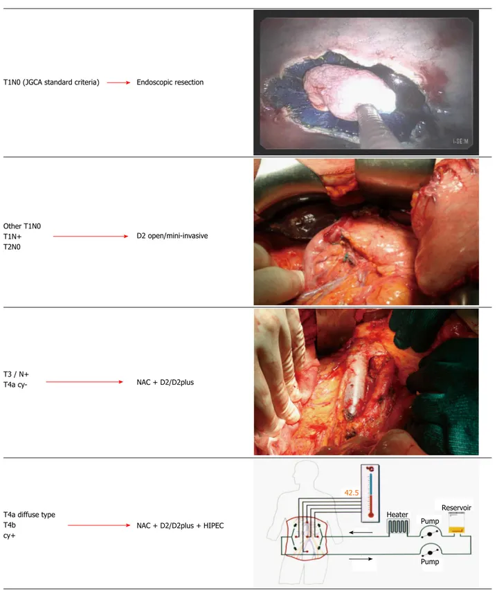
Figure 5 Proposal of a tailored multimodal approach in resectable non-cardiac gastric cancer. JGCA: Japanese Gastric Cancer Association; NAC: Neoadjuvant
chemotherapy; HIPEC: Hyperthermic intraperitoneal chemotherapy.
Adjuvant chemotherapy should be also considered, according to final pathological report Endoscopic resection
T1N0 (JGCA standard criteria)
D2 open/mini-invasive Other T1N0
T1N+ T2N0
NAC + D2/D2plus
NAC + D2/D2plus + HIPEC T3 / N+
T4a
cy-T4a diffuse type T4b cy+ 42.5 Heater Pump Reservoir Pump
patients, and the median survival was 29 mo, with 34% 5-year survival[99]. In the French FREGAT study,
palliative gastrectomy was performed because of solid organ metastases (5.6%), localized PC (4.6%), diffuse PC (2.3%) or incomplete tumor resection (12.8%)[100].
Median survival of patients with resection was better than in the non-resection group (11.9 mo vs 8.5 mo, P < 0.001). Multivariate analysis proved that factors associated with survival were: ASA score Ⅱ-Ⅳ, localized PC, diffuse PC, and signet ring histology. Patients with ASA Ⅰ/Ⅱ and incomplete resection without metastasis or PC, one-site solid organ metastasis without PC, or localized PC without signet ring cell histology, showed the highest benefit from surgery. This subgroup of patients had median survival from 12 to 18.3 mo. Analyzing surgical treatment in the case of distant metastases, we must also mention treatment of liver metastases from GC. No trials have been performed in this field, and a recent review by Grimes et al[101] was
based on 17 retrospective studies. The solitary disease patients had better OS than those with metachronous disease, and patients with metachronous disease had better prognosis than those with synchronous disease. Hepatectomy in these patients is a safe procedure with about 2% perioperative mortality, and morbidity from 17% to 60%. The authors state that metachronous metastatic disease limited only to the liver, with the possibility of surgical resection, should be consider in a clinical trial.
In the latest GIRCG study on synchronous hepatic metastases in cases of GC, it was clear that clinical criteria could be used to select candidates for curative surgery. The surgical approach has an impact on survival especially when adjuvant chemotherapy is added[102].
Multivisceral resection of advanced forms
The role of multivisceral resection, in the setting of locally advanced GC, has been evaluated in several studies. Most of them reported a higher risk for perioperative morbidity and mortality, with limited objective benefit in terms of survival, but a potential advantage of extended resection for some subgroups[103]. In a recent GIRCG study, 206 patients
with a clinical T4b carcinoma were evaluated[104].
One hundred and twelve patients underwent combined resection of the adjacent organs for clinical T4b stage disease. Postoperative mortality and complication rates were acceptable, and overall 5-year survival rate was 27.2%. The completeness of resection and lymph node invasion were independent prognostic parameters at multivariate analysis. At present, even if a chance of cure with an extended surgical approach could be obtained in subgroups of patients with invasion of adjacent organs, a multimodal approach should include neoadjuvant treatment, followed by extended surgery in responders. The addition of HIPEC should be considered.
CONCLUSION
Results of treatment in specialized western centers are good in early stage (Ⅰ/Ⅱ) GC, but are still unsatis-factory in more advanced stages (ⅢB and higher), when compared with eastern studies. Treatment options have changed in recent years from a standard to a tailored approach. Different individualized procedures can range from endoscopic resection, D2 with open or minimally invasive approach, to neoadjuvant therapy followed by extended surgery (Figure 5). In more advanced stages, a combined approach with the inclusion of HIPEC may represent a new frontier for multimodal treatment of resectable GC. It should be also emphasized that tailored treatment of GC involves appropriate pretreatment clinical staging of the disease. Clinicians should expect to face, in the future, fewer GC cases, but with higher biological aggressiveness, due to the relative increase of proximal and DT tumors. The high propensity of DT to lymph node metastasis and peritoneal dissemination makes multimodal treatment, in particular including NC and HIPEC, a modern and necessary approach to this still fatal disease.
REFERENCES
1 Ferlay J, Shin HR, Bray F, Forman D, Mathers C, Parkin DM.
Estimates of worldwide burden of cancer in 2008: GLOBOCAN 2008. Int J Cancer 2010; 127: 2893-2917 [PMID: 21351269 DOI: 10.1002/ijc.25516]
2 La Vecchia C, Bosetti C, Lucchini F, Bertuccio P, Negri E, Boyle
P, Levi F. Cancer mortality in Europe, 2000-2004, and an overview of trends since 1975. Ann Oncol 2010; 21: 1323-1360 [PMID: 19948741 DOI: 10.1093/annonc/mdp530]
3 Jemal A, Bray F, Center MM, Ferlay J, Ward E, Forman D. Global
cancer statistics. CA Cancer J Clin 2011; 61: 69-90 [PMID: 21296855 DOI: 10.3322/caac.20107]
4 Verlato G, Di Leo A, Maria Rossi G, de Manzoni G. Epidemiology
of Gastric Cancer and Screening Programs. In: de Manzoni G, Roviello F, Siquini W, editors. Surgery in the multimodal management of gastric cancer. Milan: Springer, 2012: pp. 1-7 5 Marrelli D, Pedrazzani C, Corso G, Neri A, Di Martino M, Pinto
E, Roviello F. Different pathological features and prognosis in gastric cancer patients coming from high-risk and low-risk areas of Italy. Ann Surg 2009; 250: 43-50 [PMID: 19561483 DOI: 10.1097/ SLA.0b013e3181ad6487]
6 Inghelmann R, Grande E, Francisci S, Verdecchia A, Micheli
A, Baili P, Capocaccia R, De Angelis R. Regional estimates of stomach cancer burden in Italy. Tumori 2007; 93: 367-373 [PMID: 17899867]
7 Crew KD, Neugut AI. Epidemiology of gastric cancer. World J
Gastroenterol 2006; 12: 354-362 [PMID: 16489633 DOI: 10.3748/ wjg.v12.i3.354]
8 Bertuccio P, Chatenoud L, Levi F, Praud D, Ferlay J, Negri E,
Malvezzi M, La Vecchia C. Recent patterns in gastric cancer: a global overview. Int J Cancer 2009; 125: 666-673 [PMID: 19382179 DOI: 10.1002/ijc.24290]
9 Marrelli D, Pedrazzani C, Morgagni P, de Manzoni G, Pacelli
F, Coniglio A, Marchet A, Saragoni L, Giacopuzzi S, Roviello F. Changing clinical and pathological features of gastric cancer over time. Br J Surg 2011; 98: 1273-1283 [PMID: 21560122 DOI: 10.1002/bjs.7528]
10 Wu H, Rusiecki JA, Zhu K, Potter J, Devesa SS. Stomach carcinoma incidence patterns in the United States by histologic type and anatomic site. Cancer Epidemiol Biomarkers Prev 2009;
18: 1945-1952 [PMID: 19531677 DOI: 10.1158/1055-9965.
EPI-09-0250]
11 Verdecchia A, Corazziari I, Gatta G, Lisi D, Faivre J, Forman D. Explaining gastric cancer survival differences among European countries. Int J Cancer 2004; 109: 737-741 [PMID: 14999783 DOI: 10.1002/ijc.20047]
12 Dassen AE, Lemmens VE, van de Poll-Franse LV, Creemers GJ, Brenninkmeijer SJ, Lips DJ, Vd Wurff AA, Bosscha K, Coebergh JW. Trends in incidence, treatment and survival of gastric adenocarcinoma between 1990 and 2007: a population-based study in the Netherlands. Eur J Cancer 2010; 46: 1101-1110 [PMID: 20219351 DOI: 10.1016/j.ejca.2010.02.013]
13 Laurén PA, Nevalainen TJ. Epidemiology of intestinal and diffuse types of gastric carcinoma. A time-trend study in Finland with comparison between studies from high- and low-risk areas. Cancer 1993; 71: 2926-2933 [PMID: 8490820]
14 Kaneko S, Yoshimura T. Time trend analysis of gastric cancer incidence in Japan by histological types, 1975-1989. Br J Cancer 2001; 84: 400-405 [PMID: 11161407 DOI: 10.1054/bjoc.2000.1602] 15 Henson DE, Dittus C, Younes M, Nguyen H, Albores-Saavedra J. Differential trends in the intestinal and diffuse types of gastric carcinoma in the United States, 1973-2000: increase in the signet ring cell type. Arch Pathol Lab Med 2004; 128: 765-770 [PMID: 15214826 DOI: 10.1043/1543-2165(2004)128]
16 Anderson WF, Camargo MC, Fraumeni JF, Correa P, Rosenberg PS, Rabkin CS. Age-specific trends in incidence of noncardia gastric cancer in US adults. JAMA 2010; 303: 1723-1728 [PMID: 20442388 DOI: 10.1001/jama.2010.496]
17 Hamashima C, Shibuya D, Yamazaki H, Inoue K, Fukao A, Saito H, Sobue T. The Japanese guidelines for gastric cancer screening. Jpn J Clin Oncol 2008; 38: 259-267 [PMID: 18344316 DOI: 10.1093/jjco/hyn017]
18 Correa P. Gastric cancer: two epidemics? Dig Dis Sci 2011; 56: 1585-1586; author reply 1586 [PMID: 21394461 DOI: 10.1007/ s10620-011-1642-x]
19 Marrelli D, De Stefano A, de Manzoni G, Morgagni P, Di Leo A, Roviello F. Prediction of recurrence after radical surgery for gastric cancer: a scoring system obtained from a prospective multicenter study. Ann Surg 2005; 241: 247-255 [PMID: 15650634 DOI: 10.1097/01.sla.0000152019.14741.97]
20 Kattan MW, Karpeh MS, Mazumdar M, Brennan MF. Postoperative nomogram for disease-specific survival after an R0 resection for gastric carcinoma. J Clin Oncol 2003; 21: 3647-3650 [PMID: 14512396 DOI: 10.1200/JCO.2003.01.240]
21 Han DS, Suh YS, Kong SH, Lee HJ, Choi Y, Aikou S, Sano T, Park BJ, Kim WH, Yang HK. Nomogram predicting long-term survival after d2 gastrectomy for gastric cancer. J Clin Oncol 2012; 30: 3834-3840 [PMID: 23008291 DOI: 10.1200/JCO.2012.41.8343] 22 Marrelli D, Roviello F, de Manzoni G, Morgagni P, Di Leo A,
Saragoni L, De Stefano A, Folli S, Cordiano C, Pinto E. Different patterns of recurrence in gastric cancer depending on Lauren’ s histological type: longitudinal study. World J Surg 2002; 26: 1160-1165 [PMID: 12209247 DOI: 10.1007/s00268-002-6344-2] 23 Roviello F, Rossi S, Marrelli D, Pedrazzani C, Corso G, Vindigni
C, Morgagni P, Saragoni L, de Manzoni G, Tomezzoli A. Number of lymph node metastases and its prognostic significance in early gastric cancer: a multicenter Italian study. J Surg Oncol 2006; 94: 275-280; discussion 274 [PMID: 16917863]
24 Marrelli D, Mazzei MA, Pedrazzani C, Di Martino M, Vindigni C, Corso G, Morelli E, Volterrani L, Roviello F. High accuracy of multislices computed tomography (MSCT) for para-aortic lymph node metastases from gastric cancer: a prospective single-center study. Ann Surg Oncol 2011; 18: 2265-2272 [PMID: 21267792 DOI: 10.1245/s10434-010-1541-y]
25 de Manzoni G, Di Leo A, Roviello F, Marrelli D, Giacopuzzi S, Minicozzi AM, Verlato G. Tumor site and perigastric nodal status are the most important predictors of para-aortic nodal involvement in advanced gastric cancer. Ann Surg Oncol 2011; 18: 2273-2280 [PMID: 21286941 DOI: 10.1245/s10434-010-1547-5]
26 Roviello F, Marrelli D, de Manzoni G, Morgagni P, Di Leo
A, Saragoni L, De Stefano A. Prospective study of peritoneal recurrence after curative surgery for gastric cancer. Br J Surg 2003;
90: 1113-1119 [PMID: 12945079 DOI: 10.1002/bjs.4164]
27 Sasako M, Sano T, Yamamoto S, Kurokawa Y, Nashimoto A, Kurita A, Hiratsuka M, Tsujinaka T, Kinoshita T, Arai K, Yamamura Y, Okajima K. D2 lymphadenectomy alone or with para-aortic nodal dissection for gastric cancer. N Engl J Med 2008;
359: 453-462 [PMID: 18669424 DOI: 10.1056/NEJMoa0707035]
28 Roviello F, Pedrazzani C, Marrelli D, Di Leo A, Caruso S, Giacopuzzi S, Corso G, de Manzoni G. Super-extended (D3) lympha-denectomy in advanced gastric cancer. Eur J Surg Oncol 2010; 36: 439-446 [PMID: 20392590 DOI: 10.1016/j.ejso.2010.03.008] 29 Baiocchi GL, Marrelli D, Verlato G, Morgagni P, Giacopuzzi S,
Coniglio A, Marchet A, Rosa F, Capponi MG, Di Leo A, Saragoni L, Ansaloni L, Pacelli F, Nitti D, D’Ugo D, Roviello F, Tiberio GA, Giulini SM, De Manzoni G. Follow-up after gastrectomy for cancer: an appraisal of the Italian research group for gastric cancer. Ann Surg Oncol 2014; 21: 2005-2011 [PMID: 24526547 DOI: 10.1245/s10434-014-3534-8]
30 Roviello F, Caruso S, Neri A, Marrelli D. Treatment and prevention of peritoneal carcinomatosis from gastric cancer by cytoreductive surgery and hyperthermic intraperitoneal chemotherapy: overview and rationale. Eur J Surg Oncol 2013; 39: 1309-1316 [PMID: 24183797 DOI: 10.1016/j.ejso.2013.10.010]
31 Pedrazzani C, Marrelli D, Pacelli F, Di Cosmo M, Mura G, Bettarini F, Rosa F, de Manzoni G, Roviello F. Gastric linitis plastica: which role for surgical resection? Gastric Cancer 2012;
15: 56-60 [PMID: 21717092 DOI: 10.1007/s10120-011-0063-z]
32 Fayçal J, Bessaguet C, Nousbaum JB, Cauvin JM, Cholet F, Bideau K, Robaszkiewicz M, Gouérou H. Epidemiology and long term survival of gastric carcinoma in the French district of Finistere between 1984 and 1995. Gastroenterol Clin Biol 2005; 29: 23-32 [PMID: 15738892 DOI: 10.1016/S0399-8320(05)80690-6] 33 Verdecchia A, Guzzinati S, Francisci S, De Angelis R, Bray F,
Allemani C, Tavilla A, Santaquilani M, Sant M. Survival trends in European cancer patients diagnosed from 1988 to 1999. Eur J Cancer 2009; 45: 1042-1066 [PMID: 19124239 DOI: 10.1016/ j.ejca.2008.11.029]
34 Dassen AE, Dikken JL, Bosscha K, Wouters MW, Cats A, van de Velde CJ, Coebergh JW, Lemmens VE. Gastric cancer: decreasing incidence but stable survival in the Netherlands. Acta Oncol 2014;
53: 138-142 [PMID: 23829603 DOI: 10.3109/0284186X.2013.789
139]
35 Marrelli D, Morgagni P, de Manzoni G, Coniglio A, Marchet A, Saragoni L, Tiberio G, Roviello F. Prognostic value of the 7th AJCC/UICC TNM classification of noncardia gastric cancer: analysis of a large series from specialized Western centers. Ann Surg 2012; 255: 486-491 [PMID: 22167003 DOI: 10.1097/ SLA.0b013e3182389b1a]
36 Japanese Gastric Cancer Association. Japanese gastric cancer treatment guidelines 2010 (ver. 3). Gastric Cancer 2011; 14: 113-123 [PMID: 21573742 DOI: 10.1007/s10120-011-0042-4] 37 Catalano F, Trecca A, Rodella L, Lombardo F, Tomezzoli A,
Battista S, Silano M, Gaj F, de Manzoni G. The modern treatment of early gastric cancer: our experience in an Italian cohort. Surg Endosc 2009; 23: 1581-1586 [PMID: 19263148 DOI: 10.1007/ s00464-009-0350-5]
38 Morgagni P, Petrella E, Basile B, Mami A, Soro A, Gardini A, Calzolari F, Garcea D, Bertocco M. Preoperative multidetector-row computed tomography scan staging for lymphatic gastric cancer spread. World J Surg Oncol 2012; 10: 197 [PMID: 23006343 DOI: 10.1186/1477-7819-10-197]
39 De Manzoni G, Baiocchi GL, Framarini M, De Giuli M, D’Ugo D, Marchet A, Nitti D, Marrelli D, Morgagni P, Rinnovati A, Rosati R, Roviello F, Allieta R, Berti S, Bracale U, Capelli P, Cavicchi A, Di Martino N, Donini A, Filippini A, Francioni G, Frascio M, Garofalo A, Giulini SM, Grassi GB, Innocenti P, Martino A, Mazzocconi G, Mazzola L, Montemurro S, Palasciano N, Pantuso G, Pernthaler H, Petri R, Piazza D, Sacco R, Sgroi G, Staudacher C, Testa M, Vallicelli C, Vettoretto N, Zingaretti C, Capussotti L,
Morino M, Verdecchia GM. The SIC-GIRCG 2013 Consensus Conference on Gastric Cancer. Updates Surg 2014; 66: 1-6 [PMID: 24523031 DOI: 10.1007/s13304-014-0248-1]
40 Hyun MH, Lee CH, Kim HJ, Tong Y, Park SS. Systematic review and meta-analysis of robotic surgery compared with conventional laparoscopic and open resections for gastric carcinoma. Br J Surg 2013; 100: 1566-1578 [PMID: 24264778 DOI: 10.1002/bjs.9242] 41 El-Sedfy A, Brar SS, Coburn NG. Current role of minimally
invasive approaches in the treatment of early gastric cancer. World J Gastroenterol 2014; 20: 3880-3888 [PMID: 24833843 DOI: 10.3748/wjg.v20.i14.3880]
42 De Manzoni G, Verlato G, Roviello F, Di Leo A, Marrelli D, Morgagni P, Pasini F, Saragoni L, Tomezzoli A. Subtotal versus total gastrectomy for T3 adenocarcinoma of the antrum. Gastric Cancer 2003; 6: 237-242 [PMID: 14716518 DOI: 10.1007/ s10120-003-0261-4]
43 Songun I, Putter H, Kranenbarg EM, Sasako M, van de Velde CJ. Surgical treatment of gastric cancer: 15-year follow-up results of the randomised nationwide Dutch D1D2 trial. Lancet Oncol 2010; 11: 439-449 [PMID: 20409751 DOI: 10.1016/S1470-2045(10)70070-X]
44 Roviello F, Marrelli D, Morgagni P, de Manzoni G, Di Leo A, Vindigni C, Saragoni L, Tomezzoli A, Kurihara H. Survival benefit of extended D2 lymphadenectomy in gastric cancer with involvement of second level lymph nodes: a longitudinal multicenter study. Ann Surg Oncol 2002; 9: 894-900 [PMID: 12417512 DOI: 10.1245/ASO.2002.02.002]
45 Verlato G, Roviello F, Marchet A, Giacopuzzi S, Marrelli D, Nitti D, de Manzoni G. Indexes of surgical quality in gastric cancer surgery: experience of an Italian network. Ann Surg Oncol 2009;
16: 594-602 [PMID: 19118437 DOI: 10.1245/s10434-008-0271-x]
46 Brar S, Law C, McLeod R, Helyer L, Swallow C, Paszat L, Seevaratnam R, Cardoso R, Dixon M, Mahar A, Lourenco LG, Yohanathan L, Bocicariu A, Bekaii-Saab T, Chau I, Church N, Coit D, Crane CH, Earle C, Mansfield P, Marcon N, Miner T, Noh SH, Porter G, Posner MC, Prachand V, Sano T, van de Velde C, Wong S, Coburn N. Defining surgical quality in gastric cancer: a RAND/ UCLA appropriateness study. J Am Coll Surg 2013; 217: 347-57.e1 [PMID: 23664139 DOI: 10.1016/j.jamcollsurg.2013.01.067] 47 Lordick F, Allum W, Carneiro F, Mitry E, Tabernero J, Tan P, Van
Cutsem E, van de Velde C, Cervantes A. Unmet needs and challenges in gastric cancer: the way forward. Cancer Treat Rev 2014; 40: 692-700 [PMID: 24656602 DOI: 10.1016/j.ctrv.2014.03.002] 48 Cunningham D, Starling N, Rao S, Iveson T, Nicolson M, Coxon F,
Middleton G, Daniel F, Oates J, Norman AR; Upper Gastrointestinal Clinical Studies Group of the National Cancer Research Institute of the United Kingdom. Capecitabine and oxaliplatin for advanced esophagogastric cancer. N Engl J Med 2008; 358: 36-46 [PMID: 18172173 DOI: 10.1056/NEJMoa073149]
49 Ychou M, Boige V, Pignon JP, Conroy T, Bouché O, Lebreton G, Ducourtieux M, Bedenne L, Fabre JM, Saint-Aubert B, Genève J, Lasser P, Rougier P. Perioperative chemotherapy compared with surgery alone for resectable gastroesophageal adenocarcinoma: an FNCLCC and FFCD multicenter phase III trial. J Clin Oncol 2011; 29: 1715-1721 [PMID: 21444866 DOI: 10.1200/ JCO.2010.33.0597]
50 Schuhmacher C, Gretschel S, Lordick F, Reichardt P, Hohenberger W, Eisenberger CF, Haag C, Mauer ME, Hasan B, Welch J, Ott K, Hoelscher A, Schneider PM, Bechstein W, Wilke H, Lutz MP, Nordlinger B, Van Cutsem E, Siewert JR, Schlag PM. Neoadjuvant chemotherapy compared with surgery alone for locally advanced cancer of the stomach and cardia: European Organisation for Research and Treatment of Cancer randomized trial 40954. J Clin Oncol 2010; 28: 5210-5218 [PMID: 21060024 DOI: 10.1200/ JCO.2009.26.6114]
51 Stahl M, Walz MK, Stuschke M, Lehmann N, Meyer HJ, Riera-Knorrenschild J, Langer P, Engenhart-Cabillic R, Bitzer M, Königsrainer A, Budach W, Wilke H. Phase III comparison of preoperative chemotherapy compared with chemoradiotherapy in patients with locally advanced adenocarcinoma of the
esophagogastric junction. J Clin Oncol 2009; 27: 851-856 [PMID: 19139439 DOI: 10.1200/JCO.2008.17.0506]
52 van Hagen P, Hulshof MC, van Lanschot JJ, Steyerberg EW, van Berge Henegouwen MI, Wijnhoven BP, Richel DJ, Nieuwenhuijzen GA, Hospers GA, Bonenkamp JJ, Cuesta MA, Blaisse RJ, Busch OR, ten Kate FJ, Creemers GJ, Punt CJ, Plukker JT, Verheul HM, Spillenaar Bilgen EJ, van Dekken H, van der Sangen MJ, Rozema T, Biermann K, Beukema JC, Piet AH, van Rij CM, Reinders JG, Tilanus HW, van der Gaast A. Preoperative chemoradiotherapy for esophageal or junctional cancer. N Engl J Med 2012; 366: 2074-2084 [PMID: 22646630 DOI: 10.1056/NEJMoa1112088] 53 Sakuramoto S, Sasako M, Yamaguchi T, Kinoshita T, Fujii M,
Nashimoto A, Furukawa H, Nakajima T, Ohashi Y, Imamura H, Higashino M, Yamamura Y, Kurita A, Arai K. Adjuvant chemotherapy for gastric cancer with S-1, an oral fluoropyrimidine. N Engl J Med 2007; 357: 1810-1820 [PMID: 17978289 DOI: 10.1056/NEJMoa072252]
54 Sasako M, Sakuramoto S, Katai H, Kinoshita T, Furukawa H, Yamaguchi T, Nashimoto A, Fujii M, Nakajima T, Ohashi Y. Five-year outcomes of a randomized phase III trial comparing adjuvant chemotherapy with S-1 versus surgery alone in stage II or III gastric cancer. J Clin Oncol 2011; 29: 4387-4393 [PMID: 22010012 DOI: 10.1200/JCO.2011.36.5908]
55 Macdonald JS, Smalley SR, Benedetti J, Hundahl SA, Estes NC, Stemmermann GN, Haller DG, Ajani JA, Gunderson LL, Jessup JM, Martenson JA. Chemoradiotherapy after surgery compared with surgery alone for adenocarcinoma of the stomach or gastroesophageal junction. N Engl J Med 2001; 345: 725-730 [PMID: 11547741 DOI: 10.1056/NEJMoa010187]
56 Ott K, Lordick F, Blank S, Büchler M. Gastric cancer: surgery in 2011. Langenbecks Arch Surg 2011; 396: 743-758 [PMID: 21234760 DOI: 10.1007/s00423-010-0738-7]
57 Mirza A, Pritchard S, Welch I. The postoperative component of MAGIC chemotherapy is associated with improved prognosis following surgical resection in gastric and gastrooesophageal junction adenocarcinomas. Int J Surg Oncol 2013; 2013: 781742 [PMID: 24163764 DOI: 10.1155/2013/781742]
58 Ronellenfitsch U, Schwarzbach M, Hofheinz R, Kienle P, Kieser M, Slanger TE, Burmeister B, Kelsen D, Niedzwiecki D, Schuhmacher C, Urba S, van de Velde C, Walsh TN, Ychou M, Jensen K. Preoperative chemo(radio)therapy versus primary surgery for gastroesophageal adenocarcinoma: systematic review with meta-analysis combining individual patient and aggregate data. Eur J Cancer 2013; 49: 3149-3158 [PMID: 23800671 DOI: 10.1016/j.ejca.2013.05.029]
59 Messager M, Lefevre JH, Pichot-Delahaye V, Souadka A, Piessen G, Mariette C; FREGAT working group - FRENCH. The impact of perioperative chemotherapy on survival in patients with gastric signet ring cell adenocarcinoma: a multicenter comparative study. Ann Surg 2011; 254: 684-693; discussion 693 [PMID: 22005144 DOI: 10.1097/SLA.0b013e3182352647]
60 Becker K, Mueller JD, Schulmacher C, Ott K, Fink U, Busch R, Böttcher K, Siewert JR, Höfler H. Histomorphology and grading of regression in gastric carcinoma treated with neoadjuvant chemotherapy. Cancer 2003; 98: 1521-1530 [PMID: 14508841] 61 Burmeister BH, Thomas JM, Burmeister EA, Walpole ET,
Harvey JA, Thomson DB, Barbour AP, Gotley DC, Smithers BM. Is concurrent radiation therapy required in patients receiving preoperative chemotherapy for adenocarcinoma of the oesophagus? A randomised phase II trial. Eur J Cancer 2011; 47: 354-360 [PMID: 21084184 DOI: 10.1016/j.ejca.2010.09.009]
62 Sjoquist KM, Burmeister BH, Smithers BM, Zalcberg JR, Simes RJ, Barbour A, Gebski V; Australasian Gastro-Intestinal Trials Group. Survival after neoadjuvant chemotherapy or chemoradiotherapy for resectable oesophageal carcinoma: an updated meta-analysis. Lancet Oncol 2011; 12: 681-692 [PMID: 21684205 DOI: 10.1016/S1470-2045(11)70142-5]
63 Ajani JA, Faust J, Ikeda K, Yao JC, Anbe H, Carr KL, Houghton M, Urrea P. Phase I pharmacokinetic study of S-1 plus cisplatin in patients with advanced gastric carcinoma. J Clin Oncol 2005; 23: