NEW INSIGHTS ON ANTHRACOTHERIUM MONSVIALENSE DE ZIGNO, 1888 (MAMMALIA, CETARTIODACTYLA) FROM THE LOWER OLIGOCENE OF MONTEVIALE (VICENZA, NORTHEASTERN ITALY)
ELENA GHEZZO1 & LUCA GIUSBERTI2
1Dipartimento di Scienze della Terra, Università di Firenze, Via La Pira 4, Florence, Italy. E-mail: [email protected] 2Dipartimento di Geoscienze, Università di Padova, Via Gradenigo 6, 35131 Padova, Italy.
To cite this article: Ghezzo E. & Giusberti L. (2016) - New insights on Anthracotherium monsvialense De Zigno, 1888 (Mammalia,
Cetartiodacty-la) from the lower Oligocene of Monteviale (Vicenza, Northeastern Italy). Riv. It. Paleontol. Strat. 122(3): 119-140.
Abstract. In Italy, anthracotheres are represented by a few fossils, most of them described during the XIX
century and without a standardized scientific method. Anthracotherium monsvialense De Zigno, 1888 was originally
erected from a fossil discovered in the site of Monteviale (Vicenza, northeastern Italy), whose Rupelian (MP21) lignitic beds yielded the richest lower Oligocene evidence of the genus Anthracotherium in Europe. A. monsvialense
ranges from MP21 to MP23 and its small size has been interpreted as a consequence of the insular environment, at least at Monteviale.
In this study, we summarize the long history of Italian findings providing new descriptions of dental and postcranial morphological features of A. monsvialense, and comparing such small anthracothere with its Asian and
European relatives. Morphometric analyses are also performed on teeth, in order to verify the presence of evolu-tionary trends of the genus Anthracotherium.
Received:January 24, 2016; accepted: September 11, 2016.
Keywords: historical reconstruction, morphology, taxonomy, Anthracotheriidae, Rupelian, northeastern Italy.
I
ntroductIonThe origin and evolutionary history of the Cetartiodactyla (Montgelard et al. 1997) is far to be completely solved. They appeared across the Hol-artic region at the beginning of the Eocene (e.g., Rose 1996; Blondel 2001), from uncertain centers of origin, during the Mammalian Dispersal Event (MDE), one of the key events of the Paleocene/ Eocene boundary (Aubry et al. 2007). The MDE introduced the earliest members of the orders Ce-tartiodactyla, Perissodactyla and Primate (APP taxa) in North America, Asia and Europe during a brief warming event (100-200 kyr) which occurred in the basal Eocene, the “Paleocene/Eocene thermal maximum” (Koch et al. 1992; Bowen et al. 2001; Gingerich 2006). According to recent stratigraphic calibrations, the APP taxa appeared earlier in NW Europe (MDE phase I), in the latest Paleocene, sug-gesting an older evolution of the groups, possibly migrated from mid-latitude Asia (Hooker 2015). It implies that the presence of Diacodexis gigasei, Smith,
Smith and Sudre, 1996 within the latest Paleocene PE I zone in NW Europe represents so far the old-est record of order Cetartiodactyla (Hooker 2015),
being previously unknown before the Eocene (Pro-thero & Foss 2007).
Among Cetartiodactyla, the family Anthra-cotheriidae includes even-toed and non-ruminant herbivores, with five cusps on upper brachyodont molars, a lophed structure which prefigures the se-lenodonty adapted to a frugivorous/folivorous diet (more evident on the higher-crowned lower mo-lars), and separated cuboid and navicular among the tarsals (Janis 1995; Blondel 2001).
Most of the recent interest about anthraco-theres concerns the relationships of this taxon with hippos and cetaceans (Montgelard et al. 1997; Ag-nasson & May-Collado 2008). In fact, while Pick-ford (2008) rejected the hypothesis in favor of a divergence from the suid family of family Palaeo-choeridae, cladistic analyses confirm the affinities between Hyppopotamoidea and anthracotheres, proving the speciation of the former taxon prob-ably from bothriodontines (Boisserie et al. 2011; Li-horeau et al. 2015).
The family Anthracotheriidae is represented by a huge number of well-differentiated species since their appearance in the uppermost middle Eocene Asian records (Pondaung Formation, Tsu-bamoto et al. 2002; TsuTsu-bamoto & Tsogtbaatar 2008; Soe 2008; Ducrocq et al. 2015). Members
of the family have been reported in Asia (Ozan-soy 1962; Tsubamoto & Tsogtbaatar 2008), Europe (Ozansoy 1962; Ducrocq 1995; Legendre 1995; Antoine et al. 2011; Főzy & Szente 2014), Africa (Ducrocq et al. 2001; Miller et al. 2007; Holroyd et al. 2010; Sileem & Hewaidy 2015), North and Cen-tral America (Kron & Manning 1998; Rincon et al. 2013), spreading in Europe since the late Eocene with the genera Elomeryx, Doplopus and Prominathe-rium (Kowalevsky 1893; Hellmund 1991; Meinolf &
Kurt 1994; Kostopoulos et al. 2012; Lihoreau et al. 2009). The single K/Ar dating of 37.7 ± 1.5 Ma (early Priabonian, late Eocene, MP16) for the an-thracotheres from Dětӑn, in Czech Republic (Fej-far 1987; Fej(Fej-far & Kaiser 2005) has been revised by Aguilar et al. (1997), who assigned the fauna to the MP21, the isotopic da-ting being possibly falsified by argon excess (Kelley 2002).
The genus Anthracotherium appeared in Europe
in correspondence of the “Grande Coupure” faunal
turnover (Mammalian Paleogene Zone - MP21) (Stehlin 1910a). In fact, the climatic changes and the formation of land connections at the Eocene-Oligocene boundary (Stehlin 1910b; Lihoreau et al. 2004) through a south-east Balkans-Anatolian way (Ducrocq 1995) facilitated the migration and prob-ably the speciation and divergence of new species of anthracotherids. At the end of Oligocene, the genus completely disappeared in Europe (Lihoreau & Ducrocq 2007; Scherler et al. 2010) with the latest occurrence recorded at Rickenbach (MP29, Swit-zerland) (Becker et al. 2004; Mennecart et al. 2012). This work is focused on the richest lower Oligocene findings of the genus Anthracotherium
in Europe, discovered during mining activity in the surroundings of Monteviale village (Vicenza, northeastern Italy). The species Anthracotherium monsvialense De Zigno, 1888 had been reported in
Italy, dubitatively at Cadibona and in few localities in southern France, western Switzerland, Germany, Spain and Turkey (Lebkuchner 1974, tab. 23-1; Kot-sakis 1986; Sudre 1995; Becker et al. 2004; Scherler 2011, 2013) and spans from early to early “middle” Oligocene (Sudre 1995; Pandolfi et al. 2016). Since about MP25, Anthracotherium magnum Cuvier, 1822
occurred in Europe, becoming quite frequent in the European Oligocene record (Lihoreau & Ducrocq 2007).
Our detailed description of the fossils of A. monsvialense from the type locality provides specific
morphological information of teeth and postcranial anatomy, improving the knowledge about this taxon within the family Anthracotheriidae. In fact, the de-tailed knowledge of dental and postcranial charac-teristics of the species is of primary importance in order to enrich the extant knowledge about anthra-cotheres dispersal pathways in Europe (Lihoreau et al. 2004). The occurrence of A. monsvialense in
the Venetian region at the Grande Coupure suggests
a land connection via an island chain, consisting of micro-continents and volcanic back-arcs between southern Europe and southeastern Asia, where the genus Anthracotherium probably originated
(Tsu-bamoto et al. 2002; Böhme et al. 2014; Pandolfi et al. 2016).
A
novervIewonI
tAlIAnAnthrAcotheres The first record of Italian anthracotheres was reported by Borson (1820), who described a few teeth of a completely unknown animal at the Re-gia Accademia delle Scienze di Torino. Such teeth were found in the lignite deposits near the village of Cadibona (Savona, northwestern Italy), which yield-ed the oldest and richest collection of A. magnum inItaly (Borson 1820; Squinabol 1890a, 1890b; Sieber 1935). Two years after Borson’s report, Georges Cuvier erected the genus Anthracotherium based
on the fossils from Cadibona (Cuvier 1822). The name, formed by the Greek words ἄνϑραξ= ‘coal’ and ϑηρίον = ‘beast’ (Agassiz 1842), was clearly in-spired by the sedimentary context where the fossil was found.
In addition to Cadibona’s specimens, a few remains had been reported from Agnana (Reggio Calabria) by Montagna (1857), and determined as
A. magnum by Flores (1897). Even though the
ecto-style is well developed on M3, suggesting a correct determination of the few teeth remains from A-gnana (Flores 1897; plate 1 fig. 5), the material is in need of a careful systematic revision and standard-ized description. The age of these fossils was esti-mated between late Eocene and early Miocene (Dal Piaz 1929; Esu & Kotsakis 1983; Kotsakis 1986).
Since 1858, several remains of anthracoth-eres have been found in the eastern side of Alps, in the Vicenza province, at Zovencedo and Mon-teviale (De Zigno 1888), and, less than one century later, at Chiuppano (Leonardi 1950; Accordi 1951).
The fossils from Zovencedo were found in a lignite bed with “tortoises and other animals” and had been
described by Francesco Secondo Beggiato (1806-1883) in 1865. Unfortunately, in 1945, an Allied bombing partially destroyed the historical building of the Museo Naturalistico Archeologico of Vicen-za, where the fossils were housed, and the material today does not result in the inventory database of the Museum, indicating that the Zovencedo col-lection is completely lost (Dal Lago, pers comm., 2015). Recently, Bona and Grandi (2014) described a new maxillar fragment of Anthracotheridae
(Prominatherium cf. dalmatinum) from Grancona
(Vi-cenza), suggesting a late Eocene age for the fossil. Finally, the remains of anthracotheres reported by Stehlin (1910a) and Kotsakis (1986) from Tuscany and from Oschiri (Sardinia) at the MN2 (lower Mio-cene) should be considered with caution (Bruijn & Rümke 1974; Kotsakis 1986; Made 1999, 2008).
t
heM
ontevIAlesIte:
geologIcAlAnd pAleontologIcAlcontextGeological context. In the Veneto region
(northeastern Italy), Oligocene sedimentary rocks are well represented along the eastern Lessini Mountains margin and in the Berici Hills (Rasser & Harzhauser 2008). During the early Oligocene, the area was characterized by a shallow-water carbona-te platform, delimicarbona-ted northward by emerged land and southward by a coral reef separating a well-oxygenated lagoon from a deep basin (e.g., Frost 1981) (Fig. 1). This sedimentary environment is represented by the richly fossiliferous
Castelgom-berto Calcarenites, widely outcropping in the study area (e.g. Ungaro 1978; Frost 1981; Mietto 1988). The lagoonal sedimentation of Castelgomberto Platform was locally interrupted by explosive vol-canism, which often lasted for only one paroxistic episode (Mietto 2006). Some explosive volcanic structures emerged from the shallow sea (ca. 20 m deep) forming short-lived islands. Some of these islands, like Zovencedo (Berici Hills) and Monte-viale (Lessini Mountains), were large enough to sustain a rich vegetation and terrestrial vertebra-tes (Pandolfi et al. 2016), as vertebra-testified by their ligni-te fossiliferous deposits. Such ligni-temporary islands were subsequently submerged and covered by new carbonate deposits that eventually filled the lago-on (Mietto 1988, 2006). After subsequent phases of emersion, volcanism and marine sedimentation, the whole area was uplifted by the Alpine orogeny.
The vertebrate fauna of Monteviale.
Du-ring the XIX and the first half of XX century, in the area of Monteviale, the exploitation of lignitic beds led to the discovery of a rich Oligocene ver-tebrate fauna (Mietto 2006 and references therein). The faunal assemblage consists of fishes ( “Lepi-docottus” papyraceous (Agassiz, 1832)), amphibians
(Palaeobatrachus sp.), reptiles (Trionyx italicus von
Schauroth, 1865, Bergouniouxchelys vallesnerii
(Ber-gounioux, 1954), Diplocynodon cf. D. ratelii Pomel,
1847), and mammals, represented by Dugongidae Gray, 1821, pantolestid (Epapheliscus italicus Van
Valen, 1966 and ?Pantolesta incertae sedis), bats (Ar-chaeopteropus transiens Meschinelli, 1903), and
ungu-lates (Epiaceratherium bolcense Abel, 1910, Anthraco-therium monsvialense De Zigno, 1888, Anthracochoerus
Fig. 1 - A) location map of Monteviale with the geologic detail of the Berici Hills (white bricks indicate the area of Castelgomberto Limestone, black zones are urban centers); B) reconstruction of depositional context during the Oligocene (schemas from Frost 1981, modified).
stehlini Dal Piaz, 1931, Anthracochoerus fabianii Dal
Piaz, 1931, and ?Propalaeochoerus paronae Dal Piaz,
1930) (Mietto 2006; Pandolfi et al. 2016).
The remains were collected during coal mi-ning activities and dug by miners, who sold the fossils to contemporary naturalists, private col-lectors and paleontologists (Mietto 2006; Pandolfi et al. 2016). This process prevented the complete recovery of the specimens preserved in the ligni-te beds. Miners probably sold only the best speci-mens they came across, and wasted the ones con-sidered not interesting or not recognized as fossils. As the mining activity stopped in 1952, no further discoveries have been made, and the mines’ tun-nels are now mostly covered. To date, no outcrop of the original lignite beds is known (Mietto 2006).
The anthracotheres from Monteviale.
Anthracotheres from Monteviale had been men-tioned for the first time by Beggiato (1865), who described two isolated teeth (a m3 and a lower incisor) and compared them with Anthracotherium
from Zovencedo. Beggiato (1865) recognized the presence of a small Anthracotherium morphotype in
the site of Monteviale. A palate with the complete dentition of an anthracothere collected at Monte-viale was bought by professor Giovanni Omboni (1829-1910) for the Gabinetto of the Regia Uni-versità di Padova in 1886 and it had been conside-red the most important fossil from the site. This fossil represents the holotype of Anthracotherium monsvialense, originally described by Baron
Achil-le De Zigno (Fig. 2) in 1888, and it is currently
housed at the Museo di Geologia e Paleontologia, Università di Padova (MGP-PD), inventory num-ber MGP-PD 26556 (Fig. 3).
The holotype of A. monsvialense became
quickly the object of strong debate (e.g., Dal Piaz 1932) due to the presence of one supernumerary molar on each upper tooth-series (De Zigno 1888; Flores 1897). Twenty years later, Stehlin (1910a) finally recognized the partial forgery of the spe-cimen, consisting in the addition of two supple-mentary M3s and lower incisors, probably made Fig. 2 - Achille De Zigno (Padova 1813 - Padova 1892). De Zigno,
a famous Italian palaeobotanist, also published fifteen pa-pers about fossil vertebrates of northern Italy and had been member of the Accademia dei Lincei and president of se-veral Italian Institutes (i.e. the Istituto Veneto di Scienze, Lettere ed Arti, the Accademia Galileiana and the Società Geologica Italiana; Roghi et al. 2013). Photo from the family archive of N.H. Alberto Lonigo, Mestrino (Padova).
Fig. 3 - Upper dental series of A. monsvialense MGP-PD 26556: (B) is
the holotype of the species (A: as drawn by De Zigno 1888), despite the additional last molars and the lower incisors, in-serted in the jaw in place of the upper ones (white squares) made by the miner seller in 1886.
by a seller (Fig. 3b). Notwithstanding, the species
A. monsvialense represents today one of the oldest
and smallest European Anthracotherium, and
con-sequently, it is one of the putative links between the European and Asian species after the Asian radiation.
MAterIAlAndMethods
Materials: all the described specimens are housed in the
Museo di Geologia e Paleontologia, Università di Padova (Padova, Italy), being part of the permanent exhibition of the museum. The collection from Monteviale includes numerous fossils (95 specimens listed below in “Referred Material”).
A. monsvialense is here re-described and compared with the
specimens of A. magnum, housed at the Università degli Studi di
Genova (the whole collection from Cadibona) and in the Museo
di Storia Naturale, Università di Firenze (Section of Geology and Paleontology) (casts, inventory number: IGF 5089V; IGF 11615V; IGF 2107V; IGF 11621V; IGF 4167V). Such casts reproduce real fossils from Cadibona (Savona, northwestern Italy) and are labeled as
Anthracotherium gastaldii Squinabol, 1890 (synonymized to A. magnum;
Kotsakis 1986).
For relative wearing stage estimation of the teeth, we num-bered the progressive development of visible inner dentine, identi-fied as follow: 0 = tooth not worn but almost partially emerged; 1= enamel partially worn; 2 = first comparison of dentine in less than half of masticatory surface; 3 = all the cusp/cuspids are worn and show dentine; 4 = dentine of different cusps/cuspids is joined to each other; 5 = the masticatory surface of the tooth is completely worn (Tab. 1). Stage 0 is considered as sub-adult, stages 1-2-3 are considered completely adults and stages 4-5 are senile. The minimal number of individuals (MNI) has been estimated using wear stages of dental series with almost two teeth in sequence.
State of preservation: the fossils from Monteviale show a
characteristic state of preservation: post-depositional events and dia-genesis concentrated high percentages of pyrite in the bone cavities. Usually, this is dangerous for fossil preservation: high humidity con-ditions easily start oxidation, leading to the formation of sulphates with a significant increase of the volume of the mineral. Therefore, it causes the chemical modification of the specimen starting from the inner side, with the formation of cracks on the bone surface until the complete and un-reversible destruction of the fossil (Mikuž & Aničič 2007; Del Favero et al. 2012). In order to isolate and protect the fossils from the external environment, during the first half of XX century all the specimens had been painted using a grey varnish, partially covering specific features. Recently, the remains of A. mon-svialense have been partially restored to their original status (Maritan
2004-2005), allowing to describe them in great detail. A new step of restoration of the Monteviale’s collection has been completed in the spring of 2016.
Measurements and tools: all the measurements were taken
with a vernier caliper (resolution of 0.05 mm) using the protocol pro-posed by Driesch (1976). Statistical test MANOVA has been perfor-med using Past3.02a tools (Hammer & Harper 2001). Pictures have
been taken using a Nikon D300S digital reflex camera; colors and contrasts were modified for a better color and lighting effect.
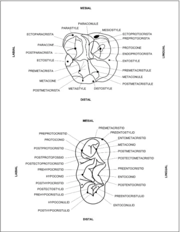
Terminology: taxonomic and morphological nomenclature
follows the standard outlined by Boisserie et al. (2010) for bunodont cetartiodactyls, partially modified by Barone (2006) and Lihoreau & Ducrocq (2007). Dental nomenclature of upper and lower molars is shown in Fig. 4.
Anatomical and measurements abbreviations: I/i =
up-per/lower incisor; C/c = upup-per/lower canine; P/p = upup-per/lower premolar; M/m = upper/lower molar; d = upper/lower deciduous tooth, L = length, W = width.
Institutional abbreviations: MGP-PD, Museo di Geologia
e Paleontologia, Università degli Studi di Padova; MP-FI Museo di Storia Naturale, Università degli Studi di Firenze; abbreviations re-ported in the Appendix 1 are from Tsubamoto et al. (2002).
s
ysteMAtIcsOrder Cetartiodactyla Montgelard,
Catzeflys & Douzery, 1997 Family Anthracotheriidae Leidy, 1869 Subfamily Anthracotheriinae Leidy, 1869
Genus Anthracotherium Cuvier, 1822
Inv. n. upper teeth (MGP-PD) M3 M2 M1 P4 P3 P2 P1 C I3 I2 I1 27361 left 0 0 27361 right 0 0 31495 1 2 31489 2 2+ 31479 2+ 2+ 27363 1 2 4 3- 1 31486 1 3- 3 27359 left 1+ 4 31501 2 3 31413 3 3+ 27364 right 3 4-31502 4 5 27364 left 3 4/5 5 27366 5 4 2 27360 left 2/3 3/4 2/3 27360 right 2/3 2 31503 3 3 Inv. n. lower teeth (MGP-PD) m3 m2 m1 p4 p3 p2 p1 c i3 i2 i1 27378 0 1+ 3 31493 0 3- 3 27359 right 1+ 3+ 3 31498 3+ 3 31481 3/4 31497 4- 4 27377 2 4 27375 3+ 4+ 4+ 3+ 1 27379 3+ 4/5 27382 right 1 0 0 27382 left 1 0 0 27384 left 2/3 2/3 27360 right 3 2/3 27384 right 4/5 2/3 2/3 2/3 31412 4 4++ 31432 4- 4
5-Tab. 1 - Wearing stage of A. monsvialense teeth. The wear was
estima-ted considering the features of the masticatory surface of each tooth, as explained in “Material and methods”.
Anthracotherium monsvialense De Zigno, 1888
Figs 2, 5-19
1865 Anthracotherium magnum Cuv.; von Schauroth: 266, pl.
XXX, fig. 2.
1865 Anthracotherium magnum minore (sic) Cuv.; Beggiato: 8, figs
6, 7.
+1888 Anthracotherium monsvialense Zigno: 37, pl. I.
1897 Anthracotherium magnum Cuv.; Flores: 93.
1910a Anthracotherium monsvialense de Zigno; Stehlin: 176, figs
1-2.
1915 Anthracotherium monsvialense Zigno; Fabiani: 308.
1926 Anthracotherium monsvialense De Zigno; Dal Piaz: 55.
1929 Anthracotherium monsvialense; Dal Piaz: 911.
1932 Anthracotherium monsvialense De Zigno; Dal Piaz: 2, figs
1-4 (text figs), pls I-XVI.
1980 Anthracotherium monsvialense; Altichieri: 173 (text fig.).
1986 Anthracotherium monsvialense Zigno; Kotsakis: 143.
1997 Anthracotherium monsvialense De Zigno; Mietto: 134, fig.
130.
2005 Anthracotherium monsvialense Zigno; Kotsakis et al.: 135,
fig. 18.
2006 Anthracotherium monsvialense De Zigno; Mietto: 41, fig. 25.
2016 Anthracotherium monsvialense Zigno; Pandolfi et al.: 23, figs
16-18.
Holotype: MGP-PD 26556: palate with upper tooth series
(Fig. 3).
Range: Italy, and probably south France, western
Switzer-land and west-middle Germany, Spain and Turkey.
Referred Material: Splancnocranium: MGP-PD 5740,
26556, 27359, 27360, 27382, 31503; upper canines: MGP-PD 27367, 27369, 31490; upper incisors: MGP-PD 27371, 27372, 27373, 27385, 31477a, 31477c, 31478a, 31489, 27370 (cfr. A. monsvialense);
up-per premolars and molars: MGP-PD 31410, 31411, 31492, 31496, 31499, 31500, 31506a, 31506b, 31512; upper tooth series: MGP-PD 5548, 27363, 27364, 27366, 27388, 31413, 31486, 31488, 31491, 31495, 31501, 31502; teeth with fragmented mandibles: MGP-PD 27377, 27378, 27379, 27384, 27386, 27387, 31412, 31432, 31479, 31480, 31493, 31507c; lower canines: MGP-PD 27368; lower inci-sors: MGP-PD 5551, 31415, 31497, 31498; premolars and molars: MGP-PD 5549, 12837, 27376, 27380, 27381, 31414, 31478b, 31482a, 31484, 31494, 31504, 31505, 31513a-c; fragmented teeth: MGP-PD 31477b, 31478c, 31482b, 31483); juvenile deciduous teeth: MGP-PD 27361, 27362, 27383, 31481.
Postcrania are represented by a distal epiphysis of a right hu-merus (A. monsvialense, MGP-PD 27389), complete, but fragmented
hindlimb (MGP-PD 12900); isolated postcranial remains such as a left ulna (MGP-PD 31401), two fragments of tibia (MGP-PD 31507a-b), patella (MGP-PD 27390, 31487), metapods (MGP-PD 27393, 27394, 27395, 27396), tarsals (right astragalus MGP-PD 27391, MGP-PD 31485), and phalanxes (MGP-PD 27397, 27398, 27399).
Description
Upper teeth: molars are well imbricated,
low-crowned and bunodont. Among upper teeth there is not a diastema; incisors are more and more horizontally inserted on the premaxillar with the fist one enlarged and completely horizontally oriented (Fig. 5).
M3: the third upper molar shows a
sub-rec-tangular shape with a prevalent protocone. The mesial margin of the cingulum has a noticeable
me-siostyle, it is not joined to the protocone by the ec-toprotocrista. In addition, the mesiostyle ends with a variably developed bulged cingulum at the lingual
side. The parastyle shows a lingual low crest that reaches the paracone bulge. The lingual side of the tooth is well developed and enlarged, with a dou-ble lobed appearance formed by protocone and metaconule (Fig. 6). On all the M3 of the collec-tion but on MGP-PD 27363, entostyles are really low-crowned and un-developed. The dimensions of all M3s, MGP-PD 27363 and MGP-PD 27359 are larger than the other homologous, nevertheless the entostyle is similar in size in all M3. This allows ruling out the hypothesis of a sex-related cause for the development of the entostyle.
A basal notch isolates the distostyle to the postmetacristule, but the distostyle is ideally con-nected to the metaconule by such crest. Distostyle is Fig. 4 - Scheme of the dental nomenclature of third upper and lower
molars. Other teeth follow the same nomenclature. Ecto-paracristule, preEcto-paracristule, postprotocrista, endoprotofos-sa and endometacristule are not shown in figure, but their position can be inferred by the anatomical description of M3 (schemas from Boisserie et al. (2010) applied to anthra-cotheres).
not joined to the metacone. Metastyle has the same size of distostyle and it is located on the distolabial side. The postmetacrista connects the metastyle to the metacone on MGP-PD 27363 and MGP-PD 27364 (Fig. 6). On MGP-PD 27366 postmetacrista and metastyle are separated, as well as distostyle and postmetaconule. The ectostyle stands out in the la-bial margin with a large columnar structure and wi-thout any labial crests; it is the only one connected to two tips, the metacone and paracone (respecti-vely by the premetacrista and postparacrista). A real ectocristyle is not visible and the crest begins and diverges directly from the ectostyle. The paracone seems to be quite isolated in comparison to other cusps. It has a conical mesiolabial side, whereas the medial side has a more jagged appearance with a variable number of fossae.
All the upper molars exhibit an accessory cusp, the paraconule, which is a characteristic featu-re of all anthracothefeatu-res. It is located on the trigonid, between the paracone and the protocone, and it is connected to the latter with the preprotocrista. The paraconule has two crests: the ectoparacristule that is distally directed to the inner side of the molar and the preparacristule that links it with the preproto-crista in the upper side. The preparacristule reaches also the mesial cingulum, but it does not reach the labial parastyle. The endoprotocrista is connected to
the premetacristule at the centre of the tooth. The postprotocrista is distally directed and completely isolated; it is also separated from the endoprotocri-sta by a thin endoprotofossa. The premetacristule and the endometacristule origin from the metaco-nule; the former is high and joined to the endopro-tocrista, whereas the latter is transversally directed and shows a bi-lobed shape on MGP-PD 27364. Both are not connected to the metacone and a well-defined sagittal valley divides the two distal cusps. The metacone shows a postmetacrista that develops sub-parallel to the sagittal axis (Fig. 6).
M2: the lingual side is characterized by a less
bulged feature of basal protocone and metaconu-le in correspondence to the third molar. Distostymetaconu-le and metastyle are not developed on the distal edge of the tooth. Postmetaconule and postmetacone end as simple crists (they appear truncated). The tooth is about 1/3 smaller than M3 (Tab. 2). The ectostyle is developed as well as the homologous of M3, whereas the entostyle is faint.
M1: only MGP-PD 27363 (Fig. 7)
preser-ves useful morphological features. The tooth is morphologically similar to M2. The quadrangular collar is simplified and undeveloped, the crest on the lingua-mesial edge of the tooth is smaller than in other molars, and the ectostyle is pronunced on the lingual side.
P4: the last premolar displays only two cusps
on the labial (paracone) and lingual (protocone) si-Fig. 5 - A. monsvialense MGP-PD 27360: ventral side of upper and
lower incisors and canines (A) and left upper incisors series and canine in lateral view (B). Upper incisors are canine-like and obliquely inserted on the jaw. Upper canines are labeled by the white arrow.
Fig. 6 - A. monsvialense MGP-PD 27364: fragment of the palate with
des (Fig. 3). A continuous cingulum is slightly
deve-loped on the mesial and lingual edges, whereas it is more developed distally, with a low-crowned meta-style and distometa-style. Parameta-style is on the mesial-labial corner of the tooth. The two cusps are divided by a deep sagittal fossa in the middle of the tooth. The
protocone is bulged on the lingual side and shows a steep and flat surface on the labial one (formed by the preprotocrista and the distally directed endopro-tocrista) (Fig. 7), while the paracone is conical with
several quite developed crests. Distally, the distopro-tocrista is completely worn.
P3: the third premolar is transversally oriented
on the upper jaw, and displays a high-crowned para-cone. Preparacrista and postectoparacrista reach the basal end of the enamel, on the mesio-lingual and disto-labial edge of the tooth, respectively. On the lingual side, the tooth enlarges distally, for the deve-lopment of a distal crest with a metastyle (Fig. 3).
P2: P2s are preserved on the holotype
(MGP-PD 26556) and on MG-PD 27365. P2 is
morpho-logically similar to P3. The tooth is mono-cuspidate and asymmetrical, for the presence of a mesially di-rected and high-crowned paracone. The distal side of the tooth ends with a flat surface. There is a postpa-racrista and a cingulum on the distal edge of the tooth.
The distal side of P2 is larger than the mesial one for the presence of the metastyle and a quite de-veloped postparacrista.
P1: the first premolar on MGP-PD 27365 is
similar to the P2, but differs by having a smaller size, by lacking a real ectoparacrista (only a small tubercle is present) and by having a less pronounced distal fe-ature.
Upper canines: upper canines are well preserved
on MGP-PD 27360 (Fig. 5) and MGP-PD27365 (Fig. 8). They have a conical shape with triangular profi-le in labial view. MGP-PD27360 has a higher crown in comparison to MGP-PD 27365. This difference is commonly related to the sex of the animals as sugge-sted by Lihoreau & Ducrocq (2007), the former pro-bably representing the male. There is not a real diaste-ma, neither before nor after the canines on MGP-PD 27360, nor on MGP-PD 5548, the other putative females; indeed, a short diastema exist on MGP-PD 27365, between the canine and the last incisor.
p value: 1,82E-21
M3 birmanicum crassum magnum monsvialense pangan tenuis birmanicum 2,31E-03 1,62E-08 5,49E-11 2,07E-09 9,73E-03 crassum 2,31E-03 9,75E-06 9,54E-07 1,42E-05 3,22E-04 magnum 1,62E-08 9,75E-06 4,13E-05 5,23E-05 4,10E-06 monsvialense 5,49E-11 9,54E-07 4,13E-05 0,3597 1,31E-09 pangan 2,07E-09 1,42E-05 5,23E-05 0,3597 3,30E-08 tenuis 9,73E-03 3,22E-04 4,10E-06 1,31E-09 3,30E-08
p value: 7,92E-30
m3 birmanicum crassum magnum M-monsvialense monsvialense pangan tenuis birmanicum 0,05557 2,9014E-10 4,3325E-04 4,8567E-06 1,2108E-02 1,5915E-02 crassum 0,05557 4,6034E-12 2,4579E-04 6,7907E-06 1,8268E-02 3,3715E-07 magnum 2,9014E-10 4,6034E-12 4,6148E-04 7,6273E-06 2,4129E-04 3,4301E-20 M-monsvialense 4,3325E-04 2,4579E-04 4,6148E-04 0,17736 0,53433 1,8187E-09 monsvialense 4,8567E-06 6,7907E-06 7,6273E-06 0,17736 0,19662 1,2349E-13 pangan 1,2108E-02 1,8268E-02 2,4129E-04 0,53433 0,19662 1,0362E-06 tenuis 1,5915E-02 3,3715E-07 3,4301E-20 1,8187E-09 1,2349E-13 1,0362E-06
Tab. 2 - MANOVA test among up-per and lower teeth of the compared anthracotheres. Positive results are reported in bold.
Fig. 7 - A. monsvialense: upper P3, P4, and molar series of MGP-PD
27363 on occlusal (A), labial (B) and lingual (C) views. The surface of teeth is coated with ammonium chloride in order to maximize different features. Scale bar 2 cm. See the on-line content of Appendix 2 for the 3d rendering.
Upper incisors: I2 and I3 are canine-like, with a
single cusp and a single root, a convex mesial edge and a longer concave distal edge. A developed me-dial crest runs from the tip to the distal end of the tooth. The enamel-root boundary is oblique and goes down from the mesial side to the distal one. I1s are large and spatular-shaped. Crowns are flat and slightly curved towards the lower jaw, enlarging the enamel surface at the mesial and distal ends of the teeth.
Lower teeth: the cheek teeth are
mesio-di-stally joined to each other and there is not a diastema between molars and premolars, neither among the other teeth of the lower jaw. The trigonid is well-developed on m3, and less well-developed and smaller on m2 and m1. On m1, the trigonid can occupy less than half of the tooth. The premolars, m1, and m2 have two joined roots in correspondence of the trigonid and talonid, whereas m3 has four or five in-dependent roots, under each cuspid and an enlarged one under the distal buldged hypoconulid.
m3: the tooth has a sub-rectangular shape,
en-larged on the mesial edge (Figs 9, 10a, b). Mesially to the metaconid, the preentostylid is present. This stylid occurs on all the analyzed m3 and is located above the enamel-root boundary on the mesio-lin-gual side. On MGP-PD 27378, the postectostylid is little developed and on MGP-PD 27376 (Fig. 10a, b) there are two small postectostylids between the hypoconid and hypoconulid (such teeth are also the largest m3 of the entire collection - Appendix 1).
Premetacristid and postectometacristid are straight and oriented along the sagittal plane of the tooth. Premetacristid is joined to the preprotocristid with multiple (two or three) little tubercles. The entome-tacristid is represented by a short cristid, which is mesio-medially oriented. Postmetacristid and post-protocristid display a U-shaped wall between the tri-gonid and talonid. A well-developed prehypocristid divides the transverse fossae in the inner side of the
m3 into a labial and a lingual valley. Protoconid and hypoconid are more selenodont than the lingual cu-spids; preprotocristid, postprotocristid, prehypocri-stid and posthypocriprehypocri-stid strongly curve toward the lingual side of the tooth and end over the medial plane of the masticatory surface. On the labial side, the above-mentioned cuspids have a bulged neck, forming two developed lobes. The postectopro-tocristid is straight oriented and consists of a low cristid on the distal side of the protoconid; it ends in correspondence of the labial transverse valley without a real junction to the prehypocristid, but forming a triangular postprotofossid.
On the talonid, the prehypocristulid is not fu-sed with the posthypocristid, but they join along the inner side of the tooth, at the distal narrowed side of the tooth. The talonid has a bulged neck. The main tip of the talonid, the hypoconulid, has no ec-tohypocristulid on the labial side. An entoconulid is variably developed on the m3s and usually well de-veloped on the largest specimens. For example, it is a small tip on MGP-PD 27380 (Fig. 10c) and a real Fig. 8 - A. monsvialense MGP-PD 27365: fragment of upper dental
series with the I3, high-crowned canine, P1 and P2.
Fig. 9 - A. monsvialense MGP-PD 31434: dorsal (A) and lingual (B)
views. Such fossil is the better mandibular horizontal branch preserved with molars (m1-m3).
cuspid on MGP-PD 27376 (Fig. 10a, b), respecti-vely 56.4 mm and 49.8 mm of length. When develo-ped, the entoconulid is well-divided from the other cuspid of the talonid and it has a single crest, a pre-entocristulid; the latter occurs on the mesial-lingual side reaching the prehypocristulid, but without a real junction. In fact, the medial valley of the talo-nid is long and continues between the hypoconulid and the entoconulid and the respective cristids.
m2: this tooth is characterized by a smaller
size than m3. The absence of a talonid is substi-tuted by a medial distal stilid (distostylid) and by a general simplify feature, with the repetition of cri-stid morphologies. There is a small preentostilid, a postectoprotocristid and a postectometacristid; the
prehypocristid is the most developed cristid of the tooth.
m1: this tooth is labial-lingually thinner and
shorter if compared to the m2. It has a simplified morphology. The preentostilid is still present, but there are not other stilids or a clear evidence of a distostylid.
p4: the last lower premolar has a single main
protoconid and a distal cingulum with the
broad-ba-sed tubercle of the hypoconid. There is an appre-ciable metaconid on the lingual side of the protoco-nid, aligned to the lingual cuspids of the following molars. The wearing erased most of the crests in all the specimens from Monteviale, but the presen-ce of a preprotocristid, a postprotocristid and an
Fig. 10 - A. monsvialense: lower third
molars with different deve-lopment of entoconulid: lar-ge left m3 (MGP-PD 27376) in lingual (A) and dorsal (B) view; smaller right m3 (MGP-PD 27380) on occlu-sal (C) view.
Fig. 11 - A. monsvialense: left
mandi-ble (MGP-PD 27375) of A. monsvialense on occlusal (A)
and lateral (B) views with p3-m3.
endometactistid could be inferred (Fig. 11). A di-stostylid and an entocristid are visible only on the fragmented p4 of MGP-PD 27378. The paracone is not present in the lingual side of the tooth.
p3: the third premolar also has a single
proto-conid and a distal less developed cingulum. The
to-oth differs from p4 by the presence of two distal tubercles at the lingual and labial side of the tooth; such tubercles emerge at the mid-height of the pro-toconid crown, the lingual one being larger than the labial tubercle (Fig. 11).
p2: p2 is not preserved.
p1: on MGP-PD 27384 (Fig. 12) the first
pre-molar is located on the inner side of the mandibu-lar ramus, due to compaction and diagenesis. The shape of the tooth is similar to that of the canine, with a more developed concave distal edge, a bulged distal side and a single mesially oriented protoconid. Accessorized cristids are not evident.
Lower canines: canines are similar in shape to
the upper homologous, but appear more slender. The single cuspid is mesially oriented and the to-oth is obliquely inserted in the jaw. The specimen MGP-PD 27384 (Fig. 12) probably belongs to a female individual, due to the simple shape and its small size (Tab. 2). The two canines on MGP-PD 27382 (Fig. 13) are noticeably larger and higher than the previous one, with a more conical feature. They probably belong to a male individual.
Lower incisors: lower incisors are spatula-like,
forming a real spade on the rostrum. They are
cylin-drical and dorso-ventrally compressed from the root to the enamel. i3 (Figs 5 and 12) is shorter and larger than i2 and i1. The latter incisor is the smaller of the series.
Mandibular bone: MGP-PD 31432 is a
rela-tively well-preserved left horizontal ramus of A. mon-svialense with m3-m1 in situ (Fig. 9). The preserved
mandible is thin with an enlarged distal side below and behind m3. The height of the horizontal ra-mus behind m3 is 70 mm. The ventral edge of the
mandible curves toward the angular process. Other mandibular fragments do not show useful morpho-logical characters and are not described here.
Postcranial bones: In general, postcranial
bones from Monteviale are poorly preserved; fos-sils suffered the effects of post depositional com-paction and diagenesis; they are partially destroyed by pyrite oxidation, damaged and quite fragmented. Fig. 12 - A. monsvialense MGP-PD
27384: lower jaw with right
m1, fragmented p4-p2, p1 (white arrows), canine and spatular arrangement of left incisors.
Fig. 13 - A. monsvialense: dorsal (A) and ventral (B) view of the
man-dibular rostrum (MGP-PD 27382). Left i1 and i2 have been reconstructed.
The forelimb is represented by a right hu-meral distal end (MGP-PD 27389). The capitum is medial-laterally short and the trochlea is thin and it ends with an angular edge. The medial epicondyle consists of a flat surface (distal width = 52 mm). The olecranon fossa is deep and quite large (epicon-dilar distance = 30 mm) and the lateral epicondyle is medio-laterally thinner than the medial one (18 and 26 mm respectively).
The proximal side of a left ulna (MGP-PD 31401, Fig. 14) shows a well-developed incisure on the apex of the large olecranon (medial-lateral width > 20.5 mm; dorso-ventral dept of the ole-cranon above the anconeal process = 49 mm). The trochlear trough is large and consists of a rounded surface. The diaphysis is broken under the humeral articulation. With the exception of a short junction under the trochlear trough, this ulna was not ossi-fied with the radius.
The hind limb is represented by several bo-nes. MGP-PD 12900 consists of a left leg (Fig. 15),
and includes the distal epiphysis of the femur, ti-bia, tarsals, metatarsals, sesamoids and phalanxes. The bones are fragmented and partially embedded in a black-painted slab: therefore, they can be only partially described and measured. The length of the small cuneiform is 26 mm, whereas the medio-late-ral width is 21.5 mm. The length of the calcaneus is 100 mm (from the talon to the distal edge) and the distal width of the astragalus is 37 mm. Medial phalanxes (of the second and third digit) are 42 mm long and the median width is 18.5 mm; lateral pha-lanxes are about 31.5 mm in length and 8 mm in Fig. 14 - A. monsvialense: schematic drawing of MGP-PD 31401,
pro-ximal fragment of a left ulna on frontal (A) and medial (B) views.
Fig. 15 - Hindlimb of A. monsvialense (MGP-PD 12900) and
identi-fication of anatomical elements of the autopodium (cal. = calcaneus; astr. = astragalus; nav. = navicular; cun. = cunei-form; ses. = sesamoid; met. = metatarsus; ph. = phalanxes I, II, and III).
width. In the collection of MGP-PD, other two un-determined lateral phalanxes measure respectively 39.5x10.5 mm (MGP-PD 27397) and 38.5x10 mm (MGP-PD 27398).
The two patella have a diamond feature with rounded angle; they are medio-laterally enlarged (GL = 66.5 and 62.7 mm; GB = 44.2 and 48.6 mm, respectively), with a rather flat surface. The caudal surface is divided by an eccentric bulged wall in two articular surfaces, the medial one being thinner and
with well-recognizable outline. MGP-PD 31487 shows the reorganization of the lateral articular surface, perhaps due to a rearrangement of lateral muscles (due to articular stress or after a collision).
Metapodial bones are represented by two specimens. Such extremities consist of two distal fragments of lateral digits and two partially joined medial metapods of about 10 cm in length (MGP-PD 27393, 27394, 27395, 27396). Unfortunately, the proximal epiphysis are not preserved and their anatomical position (manus or pes) cannot be
infer-red (Fig. 16).
MGP-PD 31507a-b consists of a proximal and distal portion of a right tibia. The surface of the proximal epiphysis is large: the medial and late-ral articular surfaces are quite externally developed and the lateral intercondylar eminence slightly emer-ges from the articulation surface (the medial one is not preserved). Cranially, the lateral tuberosity starts from the lateral side of the bone, reaching the large and columnar tibial crest (medial-lateral width = 31 mm) in the middle, forming a curved bulge on the medial side (Fig. 17). Such feature leaves a deep ten-dinean groove on the middle side of the tuberosity, usually visible on extant graviportal herbivores (i.e. hippos and elephants). The passage for the extensor muscle is laterally located and is formed by a thin and transversal valley between the lateral articular surface and the cranial tuberosity.
The distal diaphysis of the tibia MGP-PD 31507 has a compressed triangular transverse sec-tion (medial-lateral width > 55 mm, cranial-distal width > 31.6). The distal epiphysis (cranial-distal length = 48 mm) is formed by a single well-deve-loped cranio-distally directed medial coclea; it is parallel to the medial edge of the bone and deep. Distally, the medial malleole is rounded. The dorsal Fig. 16 - Partially joined metapods of A. monsvialense (MGP-PD
27396 and MGP-PD 27394).
Fig. 17 - A. monsvialense:
pro-ximal epiphysis of the right tibia (MGP-PD 31507a); the frontal-right view is shown (A) in order to un-derline the tendinean groove in the middle of the cranial tube-rosity. The schematic drawing is on frontal view (B).
apophysis is moved directly in correspondence of the distal end of the medial coclea, on the lateral side of the bone. The lateral side is partially broken and formed by a concave and medially developed surface.
The well-preserved astragalus (MGP-PD 27391: GL= 64.5 mm, proximal width = 30.5 mm, distal width = 34.7 mm) shows a proximal trochlea with two parallel articular surfaces and a medial deep valley for the tibial articulation; the lateral side is larger and protrudes more dorso-posteriorly than the medial surface. The distal trochlea is partially deflected and distally protrudes with a zigzag edge, caused by an enlargement of the lateral articular fa-cet. The two sides of the astragalus are not aligned to each other, the proximal one being medially di-rected (Fig. 18).
Comparison
Anthracotherium monsvialense from Monteviale.
Evidences of A. monsvialense are rare in Europe,
being probably found only in a few Rupelian (MP21-23) localities, at Bach, Caylux, Quercy, and Villebra-mar in France, Weinheim in Germany, and Vaulruz in Switzerland (Kotsakis 1986; Sudre 1995; Becker et al. 2004; Scherler 2011 and reference therein). So far, Monteviale represents the first site where such anthracothere had been described, yielding the ri-chest collections of the species in Europe.
As in other cetartiodactyls (Lihoreau & Du-crocq 2007), sexual dimorphism of A. monsvialense
is recognizable by the canine features, and by the development and morphology of m3. As described in the previous section, massive m3 could belong to males and more slender m3 to females, while the development of entoconulid varies accordingly to the size of the tooth and cannot represent a sexual character (Fig. 10).
Since the early discoveries, anthracotheres have been considered semi-aquatic animals, due to the hippo-like size and body proportions, and their barrel and massive body mass; for A. monsvialense
the body mass has been estimated to be probably more than 300 kg by Martinez and Sudre (1995; fig.
6) (calculated using the linear regression for mass estimation applied with reserve; the mathematical function is: Y = 3.16X1.482 - where Y is the
estima-ted body mass and X the product between lateral length and medio-lateral width), or about 150-200 kg by Tsubamoto (2014, fig 3A) (both body mass estimations are referred to the astragalus MGP-PD 27391). Anyway, the morphological adaptations to a real amphibian life-style seen for extant hippos were not evolved in A. monsvialense and other Anthracothe-rium species (Pickford 2008).
The presence of lignite beds in the Montevia-le locality attests to a wet habitat. The area was cha-racterized by the presence of humid forests and it was probably located close to a coastal brackish la-goon that was characterized by salinity fluctuations (Pandolfi et al. 2016). The whole fauna from Monte-viale was linked to such kind of habitat, because the taphonomic setting, with several skeletal parts still in articulation (i.e. MGP-PD 27360, 27396, 12900 just for anthracotheres), suggests that the carcasses did not undergo transportation and consequently are not allochthonous. To date, new observations and analyses should be performed to support this hypothesis.
Both permanent and milk teeth (Fig. 19; MGP-PD 27383; dM3L =24-30 mm; dM3W=20-Fig. 18 - A. monsvialense: right astragalus (MGP-PD 27391) on dorsal
28 mm) are present in the collection of Monteviale (NMI = 18), testifying to the presence of different ontogenetic stages in the deposit (young - MGP-PD 27383, sub-adults, adults and senile, considering the dental exploitation) (Fig. 20). Similarly to other mammals, the first molar is the first tooth to become worn (Tab. 1). In the upper jaw, M2 and M3 beca-me worn later, but such pattern can differ from the canonical exploitation (see Tab. 1, MGP-PD 27366 for example). Premolars follow the trend of M2-M3. Unfortunately, dental series with both incisives and cheek teeth are not preserved; rostral teeth are more spotted-distributed and incisors show a simi-lar wearing stage to each others; as a consequence, wearing pattern cannot be evaluated for these teeth. In the lower jaw, the m1 is the first tooth subjected to wearing, followed by the m2. p4 follows the same trend of m2; otherwise m3 starts to be worn when m1-m2 are in the stage 3. In MGP-PD 27384 (Fig. 12), mesial teeth (from p1 to i1) are 2/3 in stage when the m1 is quite completely worn (stage 4/5). Such dental erosion allows us to infer and reiterate a chewing method focused on cheek teeth, in order to manipulate the food (crush and mash) (Tab. 1).
Anthracotheres from Monteviale were browsing animals, as proven by the low-crowned upper and lower molars; the latter being more buno-selenodont. The position and orientation of incisors allow to compare the diet of the species to recent taxa, such as wild boar, peccary and war-thogs. These extant taxa display more bunodont and high-crowned cheek teeth than anthracothe-res. Lower incisors are used like a spatula in recent suids in order to pull up rhizomes, roots, tubers and
fruits, and a similar habit could be inferred for the anthracotheres.
Fossils of A. monsvialense from Monteviale
show a quite homogeneous and fixed morphology of permanent teeth. Only the entostyle on M3 (Fig. 7) and entoconulid vary in size. Entoconulid varies according to the large and small m3 (Fig. 10), but it is always recognizable, also on the most worn surfa-ce of small third molars.
Morphology and size comparison. The genus Anthracotherium is represented by scarce and
scat-tered remains in Europe. Such situation has led to the proliferation of several species which should be put in synonymy if dental intraspecific variability is taken into account (Lihoreau & Ducrocq 2007). To date, the comparison between A. monsvialense and
Fig. 19 - Juvenile right mandible of
A. monsvialense (MGP-PD
27383), with the milk canine, premolar and molar. The di-stal tooth is the m1, almost completely erupted. At the same time, a premolar had been erupting between the deciduous canine (dc) and deciduous first premolar (dp1).
Fig. 20 - Clustered column chart for the estimation of NMI. Tab. 1 has been critically considered in order to divide subadult (sa), adult (a) and senile (s) dental series. Then, upper and lower teeth have been compared obtaining a NMI of 18.
other close chronologically related species is limited to a few characteristics. The difference between the third upper molars of A. monsvialense from
Monte-viale considered as a whole and the homologous to-oth from Dvérce, as drew by Fejfar (1987; fig. 10.6) and determined as Anthracotherium cf. monsvialense,
consists of the presence of a loop in correspon-dence of the labial edge of the transverse valley in the Czech anthracothere. The anthracothere from Dvérce resembles more “cf. Bothriodon sp.” from the
upper Eocene Mongolian remains of the Ergilin Dzo Formation (Tsubamoto & Tsogtbaatar 2008), aged at the late Eocene, but it is still similar to the fossils from Monteviale for the size. Moreover, the-se two European anthracotheres differ from each other by the smaller paracone on M3, both in abso-lute size and relatively to the paraconule, ruling out that they belong to the same species.
The similitude between A. monsvialense and Anthracotherium alsaticum Cuvier, 1822 has been
un-derlined by several researchers (i.e. Stehlin 1910a; Sudre 1995), both for their chronology and abso-lute size of teeth (see below for size comparison). Anyway, differences at the level of incisors (Stehlin 1910a) and the relative less marked difference between length and width of A. monsvialense with
re-spect to A. alsaticum from Lobsann (France)
(Scher-ler 2011) seem to rule out a possible synonymy (Fig. 21).
Compared to A. illyricum Teller, 1886, from
Trbovlje (Slovenia), A. monsvialense (and A. magnum
too) has not a bulged style on the distal-labial side of P4 (Hilber 1919, tab. 2-3). Spillmann (1974) establi-shed the species Anthracotherium frehi from an upper
Oligocene site close to Freinberg (North Austria). Such anthracothere is similar in size to A. monsvia-lense and A. alsaticum (LM3 = 33.5 mm, WmaxM3
= 43.0 mm), but it shows a constricted talon (me-tacone and metaconule) on M3 with a noticeable reduction of the development and number of styles (Spillmann 1974, fig. 3); in fact, if compared to A. monsvialense, teeth of A. frehi appear really simplified.
Compared to A. magnum, A. monvialense shows
a relatively smaller ectostyle and the absence of preectostyle on M3 (and upper molars in general), and it differs from the larger European anthra-cotheres from Rickenbach (MP29, Switzerland), Mouillach and Raynal (France), and Cadibona (Italy) for the relatively smaller size of the paraconule in respect to the protocone and paracone. Such diffe-rent characteristics give to A. monsvialense a
mesio-distally un-compressed upper molar appearance, with similar labial-lingual and mesio-distal lengths. Similarly to A. magnum, the postprotocrista does not
join the premetacristule.
Ducrocq (1995) recognized a possible phyle-tic relationship among Anthracotherium pangan
Pil-grim & Cotter, 1916, Anthracotherium chaimanei
Du-crocq, 1999 from Krabi and A. monsvialense from
Monteviale, underlying the affinities within the genus and the progressive evolution of styles on P4-M3, of lingual cristids on lower premolars and Fig. 21 - Scatter diagram of M3 (A) and m3 (B), lengths (abscissa) and widths (ordinate) of Asian and European species of the genus A
nthraco-therium. Linear regression for A. monsvialense and anthracotheres from Monteviale are drawn as continue lines (large and thin trendlines,
the orientation of P3. Indeed, some features seem to exclude a close affinity between A. magnum and A. monsvialense; the paraconid is not visible on the
lingual side of p4 in A. monsvialense, but it is
deve-loped in A. magnum and such lacking feature seems
to be better related to the anthracotheres from Pon-daung, but not to A. chaimanei (Tsubamoto et al.
2002, fig. 6). Moreover, similarly to A. monsvialense,
anthracotheres from Pondaung show two cuspids variably developed on m3 talonid (Tsubamoto et al. 2002), meanwhile such additional cuspids is always
well developed in A. magnum from Cadibona.
In general, postcranium of the anthracothere from Monteviale is not well-preserved and cannot be compared with European relatives. The two pre-served astragali (MGP-PD 27391 and portion of MGP-PD 12900) show the typical morphology of ancient not-ruminant Cetartiodactyla, characterized by not-aligned proximal and distal trochlea and an enlarged surface for the articulation with the navi-cular bone (Martinez & Sudre 1995).
Metapods of A. monsvialense are thick and
he-avy built, especially the medial ones, with a reduc-tion of lateral digits, including such species within the Anisodactyla (Kowalevsky 1874) together to the younger A. bumbachense Stehlin, 1910a (aged from
MP22 to MP25, Scherler et al. 2010). Such distal conformation of the limbs could be an adaptation to its massive weight, since larger medial metapods cope better with larger support surfaces, which is useful also in a swampy environment (Coughlin & Fish 2009).
Teeth size comparison among Asian anthra-cotheres, A. magnum, and A. monsvialense from
Mon-teviale and other localities reveals a chaotic distribu-tion of measurements within each species, without finite limits among European species and A. pangan
(Fig. 21a-b). Spatial overlaps among species can be partially correct considering different dental cha-racteristics and chronology (the Asian species being older than the European anthracothere evidences). Asian species from the Pondaung Formation have been dated to about 37.2 Ma (Tsubamoto et al. 2002) and have been considered in this analysis for their morphological affinities with European anthracotheres.
Anthracotherium crassum Pilgrim & Cotter, 1916
from Pondaung represents the smallest species in our comparison, having the m3 length within a ran-ge of 32.4-39.5 mm (n. 9) (Fig. 21b). A. pangan has
a diversified distribution through all the groups, and it is noticeable that it matches with the size of A. monsvialense. A. monsvialense has an m3 with a
length varying within a range of 43.1-56.4 mm (n. 10), whereas the largest European anthracothere,
A. magnum, occupies the right side of the figure for
its massive features and larger teeth (range of 52.5-83.5 mm of length, n. 26). A. alsaticum from Ruffels
de Giroussens (France; Astre 1927) falls within the largest A. crassum variability.
The length-width scatter diagram of M3 (Fig. 21a) shows a partially different situation. All the species are within a rigorous trend of increasing size and A. pangan and A. monsvialense are completely
overlapping in size.
In order to verify such correlations, we ap-plied the statistic test MANOVA to both upper and lower third molars (Tab. 2). Assuming that teeth length and width are normally distributed, the null hypothesis have been tested, consisting on the pre-conception that all samples are taken from popu-lations with equal multivariate means (A. alsaticum, A. illyricum and A. frehi had been not considered
be-cause they are represented by only one tooth each) (Hammer & Harper 2010). The p values result lower
Fig. 22 - Increasing size of teeth among different species of anthra-cotheres, shown with a radar diagram: a) A. tenuis b) A. crassum c) A. birmanicum d) A. pangan e) A. alsaticum f) A. monsvialense from Monteviale g) A. monsvialense from other
than 0.05 for both upper and lower third molars. The pairwise comparison reveals no correlation among the species, with the exception of low corre-lation between A. crassum-A. birmanicum for the m3.
Indeed, strong correlation results for A. monsvialen-se-A. pangan, underlying their putative affinity.
Combined distribution of measurements in the multi-dimensional chart (Fig. 22) shows again the intrinsic equivalence of size of A. pangan (d) and A. monsvialense from Monteviale (f). Asian
anthraco-theres have short third molars, whereas the avera-ge measurements of A. magnum shift outward (h).
The trend supports an exponential increase of size when the underlying bone (maxillar and mandible) allows the elongation and widening of the tooth.
d
IscussIonThe faunal assemblage and chronology of Monteviale has been recently analysed and discus-sed by Pandolfi et al. (2016), referring such fauna to the lowermost Rupelian, MP21. The mammal fauna recorded at Monteviale is, as a whole, related to the migration event that follows the Grande Cou-pure. It settled in the area of Monteviale inhabiting a
fresh water humid environment, given the presence of frogs (genus Palaeobatrachus). At the same time,
the presence of Dugongidae in the site testifies to marine transgression into a coastal brackish lagoon environment (Pandolfi et al. 2016).
Among the 15 taxa described at Monteviale,
A. monsvialense is one of the largest species along
with Epiceratherium, and it represents the most
signi-ficant species for dating the site, because its presen-ce in Europe is restricted to the first part of Oligo-cene (MP21-MP23).
While the Monteviale’s fauna includes several endemic species (Pandolfi et al. 2016), A. monsvialen-se can be somehow considered a tile of a different
puzzle, being recorded in other European sites. In fact, the European invasion from Asia corresponds to the acquisition of a larger size for anthracotheres.
Our results support Ducrocq’s (1999) conclu-sions about an evolutive pattern of anthracotheres, with a progressive radiation of new species; from the smaller Asian specimen, we attend to the evolu-tion of midium-sized lower Oligocene animals (in-cluding A. pangan and A. monsvialense) and finally to
the largest A. magnum of MP25-MP29 in Europe.
Along with A. monsvialense, the collection of
Anthracotheridae from Monteviale includes a single m3 (MGP-PD 14757) reported as “Anthracotherium
(Prominatherium) dalmatinum” in the inventory label,
and a small amount of fossils attributed to the little-known Anthracochoerus fabiani Dal Piaz, 1931, and Anthracochoerus stehlini Dal Piaz, 1931. Such fossils
have been recently restored and an ongoing stu-dy will give us the opportunity of a new research about their morphological features (Ghezzo et al. in preparation). At the current state of the art, we can only underline that the presence of A. (Promi-natherium) dalmatinum, if confirmed, could represent
the second Italian occurrence of such species along with the teeth from Grancona (Bona & Grandi 2014).
c
onclusIonsGeological evidence suggests an insular en-vironment for the area around Monteviale and the northestern Venetian region during the earliest Oli-gocene, also supported by the fauna discovered in the lignite deposits of Monteviale and by the pre-sence of several endemic species (Kotsakis 1986; Pandolfi et al. 2016).
Our analysis reveals a wider context scenario, retracing the anthracotheres discoveries in the Ita-lian Peninsula. The putative ancestors of A. mon-svialense, intermediate in size between the late
Eo-cene and late OligoEo-cene species, probably reached Europe across an island-chain formed by the cur-rent Iran, Anatoly and the Balkans (Ducrocq 1995; Böhme et al. 2014, Pandolfi et al. 2016), populating the area of Monteviale. The older Anthracotheri-dae finding at Dvérce (Czech Republic) testifies to
a possible migration wave of the Asian Anthracothe-rium also towards North and East Europe.
A. monsvialense is a distinct and valid species
recognized since the second half of XIX century. The morphological differences with the younger
A. magnum confirm that the two species shared the
same phyletic branch, the lifestyle and a subtropical habitat, but at different times. Probably, the geo-graphical range of A. monsvialense was more related
to islands and relative southern latitudes with re-spect to that A. magnum, and its chronological
di-stribution in Europe was shorter, starting at about the “Grande Coupure” event and completely
disappe-aring around 30 Myr (late early Oligocene).
Our results strongly suggest a progressive lar-ger size acquired by anthracotheres in about 5 Ma, from the late Eocene to the late Oligocene and du-ring the Asia-European migration.
Furthermore, the new description here pro-vided opens new possibilities for morphological comparison among Eurasian anthracotheres, and it emphasizes the importance of anthracotheres in the Alpine High context.
Acknowledgements. We wish to give our acknowledgements to
the curator of the Museo di Geologia e Paleontologia dell’Università di Padova, Mariagabriella Fornasiero, the supervisor of the Geologi-cal and PaleontologiGeologi-cal section of the Museum of Natural History of Florence Elisabetta Cioppi, for the access to the collections and their kindness, Antonio Dal Lago, curator of the Natural History and Archeological Museum of Vicenza for the information about the lost collection of anthracotheres from Zovencedo, and Alberto Lonigo, great nephew of Achille De Zigno, who gave us the ama-zing photo of his forefather. Angela Patuzzo is acknowledged for her support and supervision of the statistical analysis. Thanks also to the anonymous referees that helped us to improve the manuscript. The associate editor Lorenzo Rook is deeply acknowledged for his valuable comments and support.
We thank P. Vianello for assistance in an early phase of stu-dy of Monteviale’s collection. Last but not least, Stefano Castelli is acknowledged for his detailed photographic work on the specimens.
This contribution is framed within the Italian Ministry of Education and Research (MIUR) funds (PRIN 2010X3PP8J_003, re-sponsible D. Rio/E. Fornaciari, Università di Padova).
RefeRences
Abel O. (1910) - Kritische Untersuchungen über die paläo-genen Rhinocerotiden Europas. Abh. k. k. geol. Reich-sanst. Wien, 20(3): 1-52.
Accordi B. (1951) - Resti di antracoterio nell’Oligocene di Chi-uppano (Vicenza). Ann. Univ. Ferrara, n. Ser. Sez. IX - Sci. geol. paleont., 1(1): 1-36.
Agassiz L. (1832) - Untersuchungen über die fossilen Süßwas-ser-Fische der tertiäre Formationen. Jb. Mineral., Geogn., Geol. Petrefaktenkd., 3: 129-138.
Agassiz L. (1842) - Nomenclator Zoologicus, continens no-mina systematica generum Animalium tan viventium quam fossilium, secundum ordinem alphabeticum dis-posita, adjectis auctoribus, libris, in quibus reperiuntur, anno editionis, etymologia et familis, ad quas pertinent, in singulis classibus. Fasciculus I continens Mammalia, Echinodermata et Acalephas. Sumptibus et typis Jent et Gassman, Soloduri. Various pagination.
Agnasson I. & May-Collado L. (2008) - The phylogeny of Cetartiodactyla: the importance of dense taxon samp-ling, missing data, and the remarkable promise of cyto-chrome b to provide reliable species-level phylogenies.
Mol. Phylogenet. Evol., 48: 964-985.
Aguilar J.-P., Legendre S. & Michaux J. (1997) - Actes du Con-grès BiochroM’97. Mém. Trav. E.P.H.E. Inst. Montpellier,
21: 769-805.
Altichieri L. (1980) - Il giacimento di Monteviale (Vicenza). In: AAVV - I vertebrati fossili italiani. Catalogo della Mostra Verona, 1980: 173-174. Tipografica “La Grafica”, Vago (Verona).
Antoine P-O., Métais G., Orliac M.J., Peigné S., Rafaÿ S., Solé F. & Vianey-Liaud M. (2011) - A new late Early Oligo-cene vertebrate fauna from Moissac, South-West France.
C. R. Palevol, 10: 239-350.
Aubry M.-P., Ouda K., Dupuis C., Berggren W.A., Van Cou-vering J.A., Ali J., Brinkhuis H., Gingerich P.R., Heil-mann-Clausen C., Hooker J., Kent D.V., King C., Knox R.W.O.B., Laga P., Molina E., Schmitz B., Steurbaut E. & Ward D.R. (2007) - The Global Standard Stratotype-Sec-tion and Point (GSSP) for the base of the Eocene Series in the Dababiya section (Egypt). Episodes, 30: 271-286.
Barone R. (2006) - Anatomia comparata dei mammiferi do-mestici. Volume 1: Osteologia. Il Sole 24 Ore Edagri-cole.
Beggiato F.S. (1865) - Anthracotherio di Zovencedo e Monte-viale. Mem. Soc. ital. Sci. nat. Milano 1: 1-10, 1 pl.
Becker D., Lapaire F., Picot L., Engesser B. & Berger J-P. (2004) - Biostratigraphie et paléoécologie du gisement à vertébrés de La Beuchille (Oligocène, Jura, Suisse). Rev. Paléobiol., vol. spéc. 9: 179.
Bergounioux F.M. (1954) - Les cheloniens fossiles des terrains tertiaires de la Venetie. Mem. Ist. Geol. Min. Univ. Padova,
18: 1-115.
Blondel C. (2001) - The Eocene-Oligocene ungulates from Western Europe and their environment. Palaeogeogr., Pal-aeoclimatol., Palaeoecol., 168: 125-139.
Boisserie J-R., Lihoreau F., Orliac M., Fischer R.E., Weston E.M. & Ducrocq S. (2010) - Morphology and phyloge-netic relationships of the earliest known hippopotamids (Cetartiodactyla, Hippopotanidae, Kenyapotaminae).
Zool. J. Linnean Soc., 158: 325-366.
Boisserie J.R., Fischer R., Lihoreau F. & Weston E. (2011) - Evolving between land and water: key questions on the emergene and history of the Hippopotamidae (Hippo-potamoidea, Cetancodonta, Cetartiondatyla). Biol. Rev. Cambridge Phil. Soc., 86: 601-625.
Bona F. & Grandi F. (2014) - The geologically oldest terrestrial mammal of the Italian peninsula. XII EAVP, Abstract book: 30.
Borson É. (1820) - Note sur des dents du grand mastodonte trouvées en Piédmont et sur des machoides et dens fos-siles prises dans la Mine de Houille de Cadibona Proghe Savone. Mem. r. Accad. Sci. Torino (Mem. Cl. Sci. fis. nat.),
27: 31-42.
Bowen G.J., Koch P.L., Gingerich P.D., Norris R.D., Bains S. & Corfield R.M. (2001) - Refined isotope stratigraphy across the continental Paleocene-Eocene boundary on Polecat Bench in the northern Bighorn Basin. In: Gin-gerich P.D. (Ed.) - Paleocene-Eocene stratigraphy and biotic change in the Bighorn and Clarks Fork Bains, Wyoming. Univ. Michigan Pap. Paleont., 33: 73-88.