Vol. 120, n. 2: 105-116, 2015
© 2015 Firenze University Press http://www.fupress.com/ijae
ITALIAN JOU R NAL OF ANATOMY AN D EM B RYOLOGY
DOI: 10.13128/IJAE-17798 Research article - Histology and cell biology
Sarcoglycans and gaba
areceptors in rat central nervous
system: an immunohistochemical study
Giuseppina Cutroneo1,2, Placido Bramanti2, Giuseppe Anastasi1,2, Daniele Bruschetta1,2, Angelo
Favaloro1,2, Giovanna Vermiglio1,2,*, Fabio Trimarchi1,2, Debora Di Mauro1,2, Giuseppina Rizzo1,2
1 Department of Biomedical Sciences and Morpho-Functional Imaging, University of Messina, Messina, Italy 2 IRCCS Centro Neurolesi “Bonino-Pulejo”, Messina, Italy
Submitted November 3, 2014; accepted revised February 27, 2015
Abstract
Sarcoglycan subcomplex is a transmembrane glycoprotein system which connects extracellular matrix to cytoskeleton. Although this complex has been found in several non-muscular tissues, no data exist about a sarcoglycan subcomplex in brain. Only the presence of ε-sarcoglycan in brain has been described in detail because its mutation determines Myoclonus Dystonia Syn-drome. Also ζ-, β- and δ-sarcoglycans have been found in brain but only at mRNA level and their distribution in brain is still unknown. Here, we have searched for the expression of all sar-coglycans in specific brain regions of rat as hippocampus, cerebral and cerebellar cortex. Since a correlation between dystrophin glycoprotein complex and γ-amino butyric acid A (GABAA)
receptor was demonstrated, we have investigated also a possible colocalization between sarco-glycans and GABAA receptor. Results have shown that all sarcoglycans are expressed in
neu-rons of all observed regions; these proteins show a spot-like pattern of fluorescence and are mainly localized at soma level. Moreover, each sarcoglycan colocalizes with GABAA receptor.
The present study shows, for the first time, the expression of all sarcoglycans in brain; moreo-ver, the prevalent localization of sarcoglycans at post-synaptic level and the colocalization of these glycoproteins with GABAA receptor suggests that sarcoglycans play a key role in central
nervous system, regulating post-synaptic receptors assembly. Key words
Sarcoglycans, GABAA receptor, cerebellar cortex, neurons, rat Introduction
The sarcoglycan (SG) sub-complex (SGC) is a glycoprotein system, member of dystrophin-glycoprotein complex (DGC), consisting, besides SGs, of several proteins including sarcospan, syntrophins and dystrobrevins which directly or indirectly asso-ciate with dystrophin.
The DGC represents a functional core which mediates the connections between extra-cellular matrix (ECM) and cytoskeleton. In detail, the SG sub-complex, made up of four transmembrane glycoproteins, α-, β-, γ- and δ-SGs, associates with α- and β-dystroglycans (DGs). Dystrophin, a cytoskeletal protein, associates with β-dys-* Corresponding author. E-mail: [email protected]
troglycans which in turns binds to α-dystroglycans. Then, α-dystroglycans binds to laminin, a basal lamina protein.
Sarcoglycans play a key role in stabilizing the molecular axis of DGC but they are functional only when they exist as a complex (Shiga et al., 2006). In fact, it was demonstrated that a mutation in any one of four SG genes determines the disruption of the entire SGC and causes autosomal recessive limb-girdle muscular dystrophies (LGMD) or sarcoglycanopathies (Roberds et al., 1994; Bönnemann et al., 1995; Lim et al., 1995; Nigro et al., 1996; Betto et al., 1999).
A fifth SG, ε-SG, highly homologous to α-SG, has been identified (Ettinger et al., 1997); it is widely expressed in most tissues suggesting that it is not muscle-specif-ic (Ettinger et al., 1997; Chan et al., 1998; Duclos et al., 1998). Then, after identifmuscle-specif-ica- identifica-tion of ε-SG, the study of SGs in muscle was extended also to smooth muscle fibers where it was hypothesized that ε-SG might replace α-SG (Straub et al., 1999). A sixth SG, ζ-SG, has been identified; this glycoprotein presents high homologies with γ- and δ-SGs (Wheeler et al., 2002).
The identification of two novel isoforms suggested that these proteins link to each other to form two different tetrameric complexes: one complex, made up of α-, β-, γ- and δ-SGs, which is characteristic of cardiac and skeletal muscle but is absent from smooth muscle, and a second complex, made up of β-, δ-, ε- and ζ- SGs, which is present in all muscular tissues (Wheeler et al., 2003). Our previous study on smooth muscle have shown the presence of all SGs, suggesting the possibility of the existence of both tetrameric complexes at the same time or an hexameric complex; this struc-ture can present a high or low expression of a single SG, according to the physiologi-cal function of smooth muscle (Anastasi et al., 2007).
Since SGs have been studied largely in muscle tissues and a key role has been hypothesized in signaling function in these tissues, there are few data about their presence in non-muscle tissues. In central nervous system (CNS) only ε-SGs has been investigated in depth because a mutation in its gene (SGCE) determines Myoclo-nus Dystonia Syndrome (MDS; Zimprich et al., 2001); SGCE protein has been found expressed in different human brain regions as cerebellum, dentate nucleus, putamen and motor cortex (Ritz et al., 2011) and in several parts of mouse brain (Chan et al., 2005). Other studies have demonstrated that rat ε-SG mRNA is most abundant in regions with dense neuronal packing such as the hippocampus, cerebellar molecular layer and cerebral cortex (Xiao and LeDoux, 2003).
It has also been demonstrated, by reverse transcription polymerase chain reaction (RT-PCR) technique, that even ζ-SG transcript was especially abundant in brain, sug-gesting that this protein may play a crucial role in the central nervous system (Shiga et al., 2006). Other SGs, as β- and δ-SGs, were detected in the brain at mRNA or pro-tein levels, but their distribution in the brain is unknown.
A previous study of ours has shown the expression of five SGs in human intra-surgical healthy cerebral cortex biopsies, suggesting that the entire SGC is expressed in brain (Anastasi et al., 2012), even if, in previous studies, other Authors considered ε-SG the unique glycoprotein of the SGC in the CNS (Waite et al., 2009).
Since SGCE mutations cause nervous system disorders but not muscular dystro-phies and since mutations in the genes for α-, β-, γ- and δ-SG cause muscular dys-trophies, but not nervous system disorders, a unique function of ε-SG in the central nervous system has been hypothesized (Nishiyama et al., 2004). Because a loss of
ε-SG does not affect the expression levels of α-, δ- and ζ-SGs in the mouse striatum nucleus, it was supposed that in brain ε-SG may be arranged as a unique complex rather than one of two tetrameric complexes (ε-β-γ-δ or ε-β-ζ-δ) as in other muscle and non-muscle tissues (Yokoi et al., 2012).
In contrast, it was hypothesized that ε-SG can form heterotetrameric arrangement (Allikian and McNally, 2007) and that ε-SG and ζ-SG may be members of a brain-spe-cific DGC, called “DGC-like” for its structural differences from the DGC of muscle (Waite et al., 2009). The brain “DGC-like” could be involved in postsynaptic clus-tering and stabilization of some inhibitory γ-amino butyric acid-ergic (GABAergic) synapses and it could play a determinant role in specifying the localization and/or molecular identity of GABAergic synapses. In detail, dystrophin is thought to play as an actin-binding postsynaptic scaffold (Graf et al., 2004; Craig and Kang, 2006)
It has been also demonstrated that other members of DGC, as dystroglycans and dystrobrevins, are correlated GABAARε (Lévi et al., 2002; Briatore et al., 2010).
Although abundant expression of ε- and ζ-SGs and presence of other SGs at mRNA or protein level (α-, β- and δ-SGs) have been shown in brain, no reports describe the presence and distribution of all of six SGs in this tissue. Our previous report evaluated the expression of five SGs in human cerebral cortex but this work was performed in little biopsies extracted from healthy brain region around glioma (Anastasi et al., 2012).
Therefore, we have performed an immunofluorescence and molecular study using the rat brain to investigate the expression of all SGs and their distribution in more than one brain region: hippocampus, cerebral and cerebellar cortex; moreover, we have evaluated the existence of a colocalization between SGs and GABAA receptor, an
issue which has never been investigated yet.
Materials and Methods
Principles of laboratory animal care and national laws on animal use were observed for this study, which was authorized by the Ethical Committee for Animal Research of the University of Messina.
Immunofluorescence
Ten normal male adult Wistar rats were used. The animals were sacrificed after anaesthesia and their cerebrum and cerebellum were extracted and fixed in 3% para-formaldehyde in 0.2 mol/L phosphate buffer, pH 7.4. After numerous rinses in 0.2 mol/L phosphate buffer and phosphate buffered saline (0.2 mol/L phosphate buffer, pH 7.6, with 0.9% NaCl: PBS), the cerebrum and cerebellum were infiltrated with 12% and 18% saccharose, frozen in liquid nitrogen and stored at -20°C. By cryotomy, both cerebrum and cerebellum were cut with coronal cuts, in anterior to posterior direc-tion, in 30 μm sections collected on glass slides coated with 0.5% gelatine and 0.005% chromium potassium sulphate. After the cut we chose the sections corresponding to the hippocampus, cerebral cortex and cerebellar cortex. On these sections two series of indirect immunofluorescence reactions were performed: 1) single localization for all six SGs; 2) double localization for each SG and GABAARε receptor.
To block non-specific sites and to make the membranes permeable, the sections were pre-incubated with 1% bovine serum albumin (BSA) and 0.3% Triton X-100 in PBS at room temperature for 15 min. Finally the sections were incubated with prima-ry antibodies at room temperature for 2 h. The following primaprima-ry antibodies obtained from Santa Cruz Biotechnology (Dallas, TX) were used, all diluted 1:100: goat poly-clonal anti α-SG; goat polypoly-clonal anti β-SG; goat polypoly-clonal anti γ-SG; goat polyclon-al anti δ-SG; goat polyclonpolyclon-al anti ε-SG; goat polyclonpolyclon-al anti ζ-SG. All primary anti-bodies were demonstrated with Texas-Red-conjugated mouse anti goat IgG (1:100; Jackson ImmunoResearch Laboratories, West Grove, PA), applied for 1 h at room temperature. After incubation with the secondary antibodies and after three rinses of 10 min each, the sections dedicated to double labelling were incubated also with rabbit polyclonal anti GABAARε receptor (1:200; Santa Cruz Biotechnology) followed
by FITC conjugated mouse anti rabbit IgG (1:100; Jackson ImmunoResearch Laborato-ries). Then, to label nuclei the sections were incubated with DAPI (Sigma Aldrich, St. Louis, USA) for 10 min at room temperature. Finally, the slides were washed in PBS and sealed with mounting medium.
Samples were observed with a Zeiss LSM 510 confocal microscope (Zeiss, Oberko-chen, Germany) equipped with Argon laser (458 nm and 488 nm λ) and two HeNe laser (543 nm and 633 nm λ). All images were digitized at a resolution of 8 bits into an array of 2048 x 2048 pixels. Optical sections of fluorescence specimens were obtained at 488 nm λ, at 62/s scanning with up to 8 repetitions on average. The pin-hole was set for optimal resolution. Contrast and brightness were established by examining the most brightly labelled pixels and choosing settings that allowed clear visualization of structural details while keeping the highest pixel intensities near 200. Digital images were cropped and figure montages prepared using Photoshop 7.0 (Adobe, San Jose, CA).
Nissl stain
For Nissl staining, brain tissues were immersed in a solution of 10% formalin for 5 days, embedded in paraffin, and cut into 4 micron coronal sections and mounted on Superfrost slides. The sections were deparaffinized in xylene, hydrated in alcool, washed in distilled water and stained in thionin solution (1%). Then, sections were dried and coverslipped. Both sections of cerebrum and cerebellum were observed and photographed with a BH-2 microscope (Olympus, Tokyo, Japan).
The color images were converted into black and white using Adobe Photoshop 7.0. software.
Results
Cerebral cortex
The results of single localization reactions showed that each SG is expressed at protein level in the nervous tissue of rat. In particular, staining for ζ-, β-, and γ-SG was clearly detectable in motor cortex area (Fig. 1). The same was observed for other SGs (data not shown).
Sarcoglycans showed a “spot-like” pattern of fluorescence, with spots of 0,5-2 µm average diameter (Fig. 2). These spots are localized in the soma of cells that morpho-logically correspond to pyramidal neurons and some granular neurons (white arrow in Fig. 2).
Double localization reactions between each SG and GABAAR showed that all SGs
(red channel) and GABAAR (green channel) colocalize at soma level. It was possible
to observe both uniform and spot staining pattern for GABAAR around the
nucle-us; colocalization between SGs and GABA receptor was underlined by yellow spots upon merge between red and green channels (Fig. 1, right column).
Hippocampus
Results of single localization reactions showed that α-, δ-, and ε-SG were expressed in hippocampus (Fig. 3). The same was observed for other SGs (data not shown).
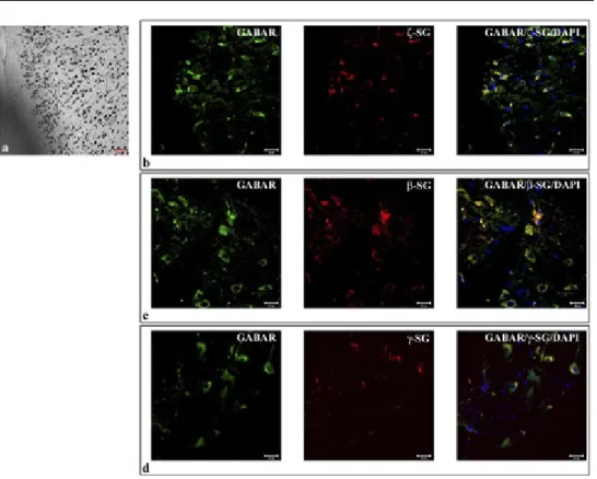
Fig. 1 – Nissl stain shows the pyramidal motor cortex area of rat cerebral cortex (1a). The compound panel of sections shows double localization reactions between ζ- (b), β- (c), γ-SGs (D) (red channel) and GABAARε (green channel in b, c and d) in rat cerebral cortex, motor cortex area. It is possible to observe the soma staining pattern both of GABAARε and ζ-, β-,γ SGs in pyramidal neurons. Their colocalization is underlined by yellow fluorescence (merge of red and green channels in b, c, and d). The nuclei are marked with DAPI (blue channel). The same results have been found for all sarcoglycans (data not shown).
Also in hippocampus SGs showed a “spot-like” staining pattern in the soma (Fig. 4). In detail, SGs staining was localized at soma level of granule cells of den-tate gyrus, in the soma of pyramidal cells of CA1, CA2 and CA3 regions and also
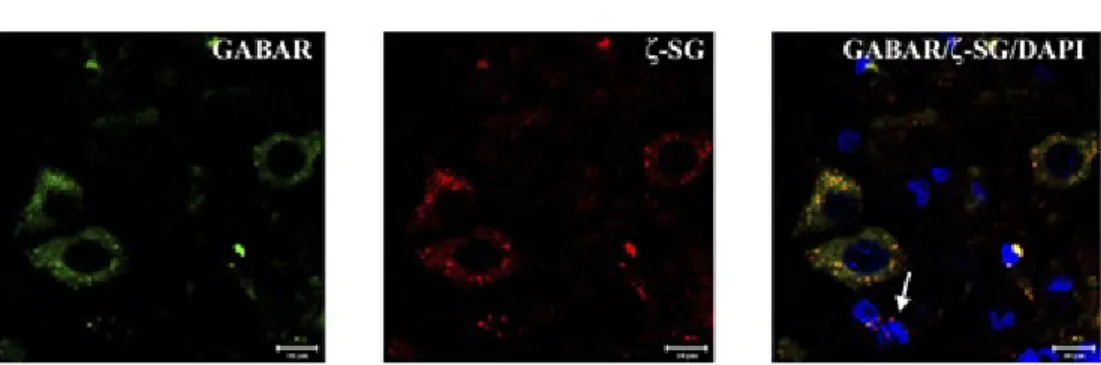
Fig. 2 – High magnification of pyramidal neurons of motor cortex area. ζ-SG (red channel) is expressed by a “spot-like” staining pattern in cellular soma. GABAARε (green channel) doesn’t present the same “spot-like” pattern but is high expressed in cellular soma. Colocalization between ζ-SG and GABAARε is underlined by yellow fluorescence. In this field it is possible to observe some small neurons (probably granular neuron) which are positive both for ζ-SG and GABAARε and some small neurons which are positive only for ζ-SG (white arrow). The same results have been found for all sarcoglycans. The nuclei are marked with DAPI (blue channel).
Fig. 3 – Nissl staining shows dentate gyrus (DG), Purkinje cell layer (PL) (a), Cornu Ammonis (CA)2, CA3 (b), and CA1 (c) regions of rat hippocampus. The compound panel of sections shows double localization reac-tions between α- (d), δ- (e), ε-SGs (f) (red channel) and GABAARε (green channel in d, e, and f) in rat hip-pocampus. The pictures show that α-, δ-, ε-SGs are expressed in all observed regions of hippocampus and they always colocalize with GABAARε. The nuclei are marked with DAPI (blue channel). The same results have been found for all sarcoglycans (data not shown).
in basket neurons of deep, polymorphic layer of the dentate gyrus (white arrow in Fig. 4). SGs were more expressed in pyramidal neuron of CA1, CA2 and CA3 regions; they were also widely expressed in dentate gyrus granular cells which are character-ized by smaller spots than pyramidal neurons. The cells of polymorphic layer present less spots of SGs than neurons of other hippocampus regions. The double localization reactions between each SG and GABAAR receptor showed that in granule cell layer of
dentate gyrus, SGs are expressed alone, with few point of colocalization with GAB-AAR which is almost absent from dentate gyrus granule cells. Instead it was possible
to observe colocalization between SGs and GABAAR in the soma of pyramidal cells of
CA1, CA2 and CA3 regions and in the soma of polymorphic cell layer (yellow fluo-rescence in Fig. 4).
Cerebellar cortex
The single localization reaction of SGs in cerebellar cortex showed that α-, β-, and ε-SGs were present also in this region of brain (Fig. 5). The same was observed for other SGs (data not shown). In particular it was possible to fluorescence for all SGs in all of the three cerebellar cortex layers: molecular, granular and Purkinje cell layer. The “spot-like” staining pattern of all SGs was localized mainly in the soma of Pur-kinje cell layer (Fig. 6). The images show that SGs are less expressed in the molecu-lar and granumolecu-lar layers than in Purkinje cell layer, where there is a higher density of
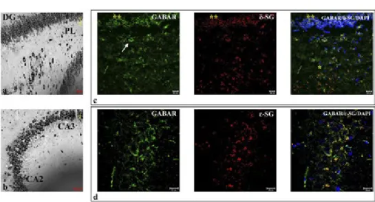
Fig. 4 – Nissl staining shows dentate gyrus, Purkinje cell layer (PL) (a) and Cornu Ammonis (CA)2, CA3 (b) at high magnification. The compound panel of sections shows that δ- (c) and ε-SGs (d) (red channel) are expressed in the soma of granule cells of dentate gyrus and in the soma of polymorphic layer (white arrow in b). They are also expressed in the beginning of CA1, at the bottom of dentate gyrus and Purkinje layer. The colocalization between δ-, ε-SG and GABAARε (green channel in c and d) is highlighted by yellow fluo-rescence. The nuclei are marked with DAPI (blue channel). The same results have been found for all sarcogly-cans (data not shown).
spots. Double localization reaction between SGs and GABAAR showed that SGs
colo-calize mainly at soma level of Purkinje cells (Fig. 5).
Discussion
It is well known that the SG sub-complex plays a key role in muscle tissues, because of its involvement in some forms of hereditary muscular dystrophies as Limb Girdle Muscular Dystrophies (LGMD), and for this reason it is considered as import-ant as dystrophin (Roberds et al., 1994; Bönneman et al., 1995; Nigro et al., 1996; Betto et al., 1999).
Despite these premises, insufficient data exist about SGs in non-muscle tissues. In CNS, only ε-SG has been investigated in detail, demonstrating that its mutation causes
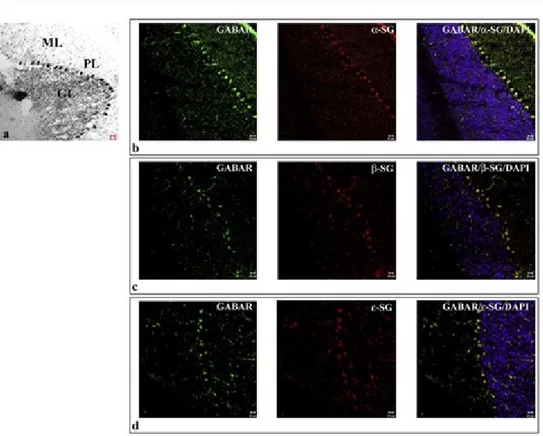
Fig. 5 – Nissl staining shows the three layers of cerebellar cortex (a): molecular layer (ML), granular layer (GL), and Purkinje cell layer (PL). The compound panel of sections shows double localization reactions between α- (b), β- (c), ε-SGs (d) (red channel) and GABAARε (green channel in b, c, and d) in rat cerebellar cortex. It is possible to observe that sarcoglycans are mainly expressed at Purkinje cell layer level. They also show to be lower expressed in molecular and granular layer than Purkinje cell layer. In Purkinje cell soma α-, β-, and ε-SGs colocalize with GABAARε (yellow fluorescence in b, c, and d). The nuclei are marked with DAPI (blue channel). The same results have been found for all sarcoglycans (data not shown).
the Myoclonus Dystonia Syndrome (MDS; Zimprich et al., 2001). Immunohistochem-istry and fluorescent in situ hybridization (FISH) revealed a wide distribution of ε-SG protein and mRNA throughout the human, rat and mouse brain (Xiao and LeDoux, 2003; Nishiyama et al., 2004; Chan et al. 2005; Ritz et al., 2011) suggesting a key role of this glycoprotein in brain. Other studies have also shown that ζ-SG is highly expressed in brain, but this SG was demonstrated only by molecular techniques (Shiga et al., 2006) and its function is still unclear. Then, it is believed that only ε- and ζ-SG are present in brain and some Authors consider the presence of a prototypical tetram-eric SG complex in brain unlikely (Zimprich et al., 2001; Waite et al., 2009).
Other SGs, as α-, β- and δ-SGs, were detected in the brain at mRNA or pro-tein levels, but their distribution in the brain is incompletely known; our previous immunofluorescence and molecular study performed on human surgical biopsies of healthy cerebral cortex has shown the presence of five SGs (α-, β-, γ-, δ- and ε-SGs) (Anastasi et al., 2012) demonstrating that this sub-complex is not muscle specific.
On this basis, in the present report, we have extended our study to confirm our first results about SGs in brain. In particular, here we have investigated each SG in three different regions of central nervous system of rat: 1) cerebral cortex (motor cor-tex); 2) hippocampus; 3) cerebellar cortex.
In the mouse motor cortex, ε-SG mRNA was found most intense in pyramidal neurons of layer 5 but was also observed in layers 2 and 3 (Chan et al., 2005). In human cerebral cortex we have already observed the expression of SGs, particular-ly in large neurons but also in astrocytes (Anastasi et al., 2012). Results of present report, obtained from rat cerebral cortex, confirm our data of human cerebral cor-tex, showing the expression of all SGs, α-, β-, γ-, δ-, ε- and ζ-SGs, with a “spot-like” staining pattern mainly in the soma of pyramidal neurons of motor cortex. In the hip-pocampus, the literature describes moderate levels of ε-SG mRNA in the pyramidal cells of CA1– 3 and dentate gyrus; in dentate gyrus moderate levels of ε-SG mRNA were detected in the granule cell layer and in the polymorphic layer.
However, cells in the polymorphic layer showed a denser immunostaining than those in the granule cell layer (Chan et al., 2005). We have found the same results in
Fig. 6 – Double localization reaction between ζ-SG (red channel) and GABAARε (green channel) on Purkin-je cell layer shows an intense fluorescence for these proteins in cellular soma. The colocalization between these proteins is highlighted by yellow fluorescence. Moreover, it is possible to detect a less intense fluores-cence for ζ-SG and GABAARε in granule cells and in the molecular layer than in Purkinje cells. The nuclei are marked with DAPI (blue channel). The same results have been found for all sarcoglycans (data not shown).
rat hippocampus but for all SGs. In fact, each isoforms shows an high pattern of fluo-rescence in the soma of pyramidal neurons of CA1-3 regions, a moderate fluofluo-rescence pattern in the soma of granular cell layer of dentate gyrus, and a dense staining pat-tern in the polymorphic cell layer. In cerebellar cortex, ε-SG was found in molecular and Purkinje layer but not in granular layer (Chan et al., 2005). Our results show that in rat cerebellum all SGs are specifically expressed in the Purkinje cell bodies. Some cells in the molecular layer also expressed each SGs, possibly representing basket and star cells, and some immunoreactivity was also found in granular layer, probably rep-resenting Golgi interneurons.
For the first time, these data show that all of six components of SG sub-complex are expressed in rat brain suggesting that all isoforms, and not only ε-SG, may play a role in central nervous system.
It has been reported that ε-SG can form heterotetrameric assemblies (Allikian and McNally, 2007) and ε-SG and ζ-SG have been considered members of a brain-specific DGC, called “DGC-like” because of its structural differences compared with the mus-cle DGC (Waite et al. 2009). The brain “DGC-like” may be involved in postsynaptic clustering and stabilization of some inhibitory GABAergic synapses. A characteristic feature of GABAergic synapses in cortical areas, in fact, (including cerebral cortex, hippocampal formation, and cerebellar cortex) is the presence of dystrophin and its associated glycoprotein complex (DGC), notably in perisomatic synapses. In detail, dystrophin is thought to play as an actin-binding postsynaptic scaffold (Graf et al., 2004; Craig and Kang, 2007). Therefore, the DGC might play a determinant role in specifying the localization and/or molecular identity of GABAergic synapses.
Furthermore, one cannot excludes that interactions with GABAAR involve
oth-er memboth-ers of the DGC rathoth-er than dystrophin itself. In fact, also othoth-er proteins of DGC as dystroglycans and dystrobrevins are correlated with GABAergic receptors in cerebellum (Briatore et al., 2010). Even dystroglycan is selectively associated with inhibitory GABAergic synapses although is dispensable for their differentiation (Lévi et al., 2002).
So it is possible to imagine the presence of a brain DGC which is made up by all components of the SG sub-complex. In detail, the prevalent localization of SGs in post-synaptic sites suggest their correlation with the synaptic network, probably regulating post-synaptic receptors assembly. For these reasons, in the present report, we also analyzed the correlation between each SG and GABAAR. Results have shown
that each SG colocalizes with GABAAR in the cellular soma in cerebral cortex,
hyppo-campus and cerebellar cortex of rat.
The results also show that SGs don’t always colocalize with GABAAR, because
cells negative for GABAAR, but positive for SGs, have been found. This suggests that
this glycoprotein system is not exclusively correlated with GABAergic synapses but it could be also involved in other kinds of synaptic networks, as dopaminergic and serotonergic networks. Actually, in several studies about Myoclonus Dystonia Syn-drome a correlation between ε-SG and dopaminergic neurons in substantia nigra and retinal tegmental area has been described (Yokoi et al., 2006).
Our recent results, obtained from a study performed in a model of absence epi-lepsy, WAG/Rij rats, have shown that the absence or altered expression of GABAAR
do not affect SGs expression (data not shown). ε-SG has been also found in serotoner-gic cells (Chan et al., 2005). We have obtained preliminary evidence of colocalization
between SGs and DOPA and serotonin receptors (data not shown) in according with Literature (Chan et al., 2005; Yokoi et al., 2006).
This work for the first time shows the presence of all of six SGs in rat brain and their distribution in specific brain regions as cerebral cortex, hippocampus and cere-bellar cortex; in detail they are localized in post-synaptic sites and for the first time they have been demonstrated in colocalization with GABAAR.
Research in progress might support also the hypothesis that SGs, as other DGC components, colocalize with other kind of post-synaptic receptors (dopamine and serotonine). It will be important, besides, to address if in brain these glycoproteins arrange to form one of the known, typical tetrameric complexes or if they achieve another kind of arrangement, as the hexameric one that we have already hypothe-sized to be present in muscle (Anastasi et al., 2007).
References
Allikian M.J., McNally E.M. (2007) Processing and assembly of the dystrophin glyco-protein complex. Traffic 8: 177-183.
Anastasi G., Cutroneo G., Sidoti A., Rinaldi C., Bruschetta D., Rizzo G., D’Angelo R., Tarone G., Amato A., Favaloro A. (2007) Sarcoglycan subcomplex expression in normal human smooth muscle. J. Histochem. Cytochem. 55: 831-843.
Anastasi G., Tomasello F., Di Mauro D., Cutroneo G., Favaloro A., Conti A., Ruggeri A., Rinaldi C., Trimarchi F. (2012) Expression of sarcoglycans in the human cer-ebral cortex: an immunohistochemical and molecular study. Cells Tissues Organs 196: 470-480.
Betto R., Biral D., Sandona D. (1999) Functional roles of dystrophin and associated proteins. New insights for the sarcoglycans. Ital. J. Neurol. Sci. 20: 371-379.
Bönnemann C.G., Modi R., Noguchi S., Mizuno Y., Yoshida M., Gussoni E., Mc- Nal-ly E.M., Duggan D.J., Angelini C., Hoffman E.P. (1995) β-sarcogNal-lycan (A3b) muta-tions cause autosomal recessive muscular dystrophy with loss of the sarcoglycan complex. Nat. Genet. 11: 266-272.
Briatore F., Patrizi A., Viltono L., Sassoe-Pognetto M., Wulff P. (2010) Quantitative organization of GABAergic synapses in the molecular layer of the mouse cerebel-lar cortex. Plos One 5: e12119.
Chan Y.M., Bonnemann C.G., Lidov H.G., Kunkel L.M. (1998) Molecular organization of sarcoglycan complex in mouse myotubes in culture. J. Cell Biol. 143: 2033-2044. Chan P., Gonzalez-Maeso J., Ruf F., Bishop D.F., Hof P.R., Sealfon S.C. (2005) Epsilon
sarcoglycan immunoreactivity and mRNA expression in mouse brain. J. Comp. Neurol. 482: 50-73.
Craig A.M., Kang Y. (2007) Neurexin–neuroligin signaling in synapse development. Curr. Opin. Neurobiol. 17: 43-52.
Duclos F., Straub V., Moore S.A., Venzke D.P., Hrstka R.F., Crosbie R.H., Durbeej M., Lebakken C.S., Ettinger A.J., Van der Meulen J., Holt K.H., Lim L.E., Sanes J.R., Davidson B.L., Faulkner J.A., Williamson R., Campbell K.P. (1998) Progressive mus-cular dystrophy in alpha-sarcoglycan-deficient mice. J. Cell. Biol. 142: 1461-1471. Ettinger A.J., Feng G., Sanes J.R. (1997) epsilon-sarcoglycan, a broadly expressed
hom-ologue of the gene mutated in limb-girdle muscular dystrophy 2D. J. Biol. Chem. 272: 32534-32538.
Graf E.R., Zhang X.Z., Jin S.X., Linhoff M.W., Craig A.M. (2004) Neurexins induce differentiation of gaba and glutamatepostsynaptic specializations via neuroligins. Cell 119: 1013-1026.
Lévi S., Grady R.M., Henry M.D., Campbell K.P., Sanes J.R., Craig A.M. (2002) Dys-troglycan is selectively associated with inhibitory GABAergic synapses but is dis-pensable for their differentiation. J. Neurosci. 22: 4274-4285.
Lim L.E., Duclos F., Broux O., Bourg N., Sunada Y., Allamand V., Meyer J., Richard I., Moomaw C., Slaughter C., Tome F.M.S., Fardeau M., Jackson C.E., Beckmann J.S., Campbell K.P. (1995) Beta-sarcoglycan: characterization and role in limb-gir-dle muscular dystrophy linked to 4q12. Nat. Genet. 11: 257-265.
Nigro V., Moreira E., Piluso G., Vainzof M., Belsito A., Politano L., Pucca A.A., Pas-sos-Bueno M.R., Zatz M. (1996) Autosomal recessive limb-girdle muscular dystro-phy, LGMD 2F, is caused by a mutation in the g-sarcoglycan gene. Nat. Genet. 14: 195-198.
Nishiyama A., Endo T., Takeda S., Imamura M. (2004) Identification and characteri-zation of epsilon-sarcoglycans in the central nervous system. Mol. Brain Res. 125: 1-12.
Ritz K., van Schaik B.D.C., Jakobs M.E., van Kampen A.H., Aronica E., Tijssen M.A., Baas F. (2011) SGCE isoform characterization and expression in human brain: implications for myoclonus–dystonia pathogenesis? Eur. J. Hum. Gen. 19: 438-444. Roberds S.L., Leturcq F., Allamand V., Piccolo F., Jeanpierre M., Anderson R.D., Lim
L.E., Lee J.C., Tome F.M.S., Romero N.B., Fardeau M., Beckmann J.S., Kaplan J.C., Campbell K.P. (1994) Missense mutations in the adhalin gene linked to autosomal recessive muscular dystrophy. Cell 78: 625-633.
Shiga K., Yoshioka H., Matsumiya T., Kimura I., Takeda S., Imamura M. (2006) Zetasarcoglycan is a functional homologue of gamma-sarcoglycan in the forma-tion of the sarcoglycan complex. Exp. Cell Res. 312: 2083-2092.
Straub V., Ettinger A.J., Durbeej M., Venzke D.P., Cutshall S., Sanes J.R., Campbell K.P. (1999) Epsilon-sarcoglycan replaces alpha-sarcoglycan in smooth muscle to form a unique dystrophin-glycoprotein complex. J. Biol. Chem. 274: 27989-27996.
Waite A., Tinsley C.L., Locke M., Blake D.J. (2009) The neurobiology of the dystrophin associated glycoprotein complex. Ann. Med. 41: 344-359.
Wheeler M.T., McNally E.M. (2003) Sarcoglycans in vascular smooth and striated muscle. Trends Cardiovasc. Med. 13: 238-243.
Wheeler M.T., Zarnegar S., McNally E.M. (2002) Zeta-sarcoglycan, a novel component of the sarcoglycan complex, is reduced in muscular dystrophy. Hum. Mol. Genet. 11: 2147-2154.
Xiao J., LeDoux M.S. (2003) Cloning, developmental regulation and neural localiza-tion of rat q-sarcoglycan. Mol. Brain Res. 119: 132-143.
Yokoi F., Dang M.T., Li J., Li Y. (2006) Myoclonus, motor deficits, alterations in emo-tional responses and monoamine metabolism in epsilon-sarcoglycan deficient mice. J. Biochem. 140: 141-146.
Zimprich A., Grabowski M., Asmus F., Naumann M., Berg D., Bertram M., Scheidt-mann K., Kern P., WinkelScheidt-mann J., Muller-Myhsok B., Riedel L., Bauer M., Mul-ler T., Castro M., Meitinger T., Strom T.M., Gasser T. (2001) Mutations in the gene encoding epsilonsarcoglycan cause myoclonus-dystonia syndrome. Nat. Genet. 29: 66-69.