Noninvasive Follicular Thyroid Neoplasm
With Papillary-Like Nuclear Features
in the Pediatric Age Group
Esther Diana Rossi, MD, PhD, MIAC 1; Swati Mehrotra, MD2; Ayse Irem Kilic, MD2;
Iclal Erdem Toslak, MD3; Jennifer Lim-Dunham, MD3; Maurizio Martini, MD, PhD1; Guido Fadda, MD1,4; Celestino Pio Lombardi, MD4; Luigi Maria Larocca, MD1; and G€uliz A. Barkan, MD, FIAC2
BACKGROUND: The most common malignant thyroid neoplasm in children is papillary thyroid carcinoma (PTC). In 2015, the Endocrine Pathology Society introduced the terminology “noninvasive follicular thyroid neoplasm with papillary-like nuclear features” (NIFTP) to replace the noninvasive follicular variant of PTC. The objective of the current study was to eval-uate previously diagnosed PTC in the pediatric population, reappraise it for NIFTP, and discuss the impact of NIFTP on the risk of malignancy (ROM) for each The Bethesda System for Reporting Thyroid Cytopathology category in the pediatric population. METHODS: The electronic databases of both study institutions were searched for all thyroidectomy specimens in patients aged <19 years from June 1, 2001 through June 1, 2016. The patient’s age, sex, diagnosis, previous fine-needle aspiration cytology diagnosis, and follow-up were tabulated. Slides for available cases were reviewed and cases qualifying as NIFTP were separated. RESULTS: The cohort included 101 resected nodules; cytological diagnoses were available for 95 cases. These cases included diagnoses of nondiagnostic (5 cases; 5.2%), benign (21 cases; 22.1%), atypia/follicular lesion of undetermined significance (9 cases; 9.5%), follicular neoplasm/suspicious for follicular neoplasm (FN/SFN) (25 cases; 26.3%), suspicious for malignancy (7 cases; 7.4%), and malignant (28 cases; 29.5%). On the histological follow-up, 50 cases (49.5%) were benign, 49 cases (48.5%) were malignant, and 2 cases (1.9%) were NIFTP. These NIFTP cases origi-nally were diagnosed as FNs on fine-needle aspiration cytology. The average ROM for FNs with and without NIFTPs was 28% and 25%, respectively CONCLUSIONS: According to our rate of 1.9% for NIFTPs on reappraisal for resected nodules, this entity is likely to be less frequent in the pediatric population due to the higher prevalence of PTCs and/or more aggressive variants. NIFTPs do not appear to affect the ROM for The Bethesda System for Reporting Thyroid Cytopathol-ogy categories in the pediatric population. However, large-scale studies are necessary to determine whether NIFTPs could affect the pediatric population. Cancer Cytopathol 2018;126:27-35.VC 2017 American Cancer Society.
KEY WORDS: BRAF mutation; noninvasive follicular thyroid neoplasm with papillary-like nuclear features (NIFTP); nonin-vasive follicular variant of papillary thyroid carcinoma; pediatric thyroid lesions; thyroid carcinoma.
INTRODUCTION
It is well known that well-differentiated carcinoma of the thyroid and specifically papillary thyroid carcinoma (PTC) and its variants are among the most common endocrine malignancies in both adult and pediatric patients.1–14 Several publications have demonstrated that thyroid lesions in childhood have a 1.6-fold higher
Corresponding author: Esther Rossi, MD, PhD, MIAC, Division of Anatomic Pathology and Histology, “Agostino Gemelli” School of Medicine, Catho-lic University of the Sacred Heart, Largo Francesco Vito, 1, 00168 Rome, Italy; [email protected]
1
Division of Anatomic Pathology and Histology, “Agostino Gemelli” School of Medicine, Catholic University of the Sacred Heart, Rome, Italy;2
Depart-ment of Pathology, Loyola University School of Medicine, Maywood, Illinois;3
Department of Radiology, Loyola University School of Medicine, May-wood, Illinois;4
Division of Endocrine Surgery, “Agostino Gemelli” School of Medicine, University Foundation Polyclinic, Catholic University of the Sacred Heart, Rome, Italy
Part of the data were presented as a poster at the United States and Canadian Academy of Pathology Annual Meeting; March 4-March 10, 2017; San Antonio, TX.
Received: June 28, 2017; Revised: August 28, 2017; Accepted: September 11, 2017 Published online October 12, 2017 in Wiley Online Library (wileyonlinelibrary.com) DOI: 10.1002/cncy.21933, wileyonlinelibrary.com
cancer risk compared with those noted in adulthood.4–6 According to the data from the World Health Organiza-tion, PTC accounts for approximately 90% of all cancers, and for the most part are diagnosed as the classic variant of PTC, especially in children aged >12 years. However, in these last decades, several authors have demonstrated that the follicular variant of PTC (FVPC) includes both encap-sulated/noninvasive (NI-FVPCs) and invasive FVPCs (I-FVPCs), which have a different prognosis and molecular findings.15–34In fact, several publications concluded that I-FVPCs are associated with more aggressive features char-acterized by lymph node metastases, disease recurrence, and the prevalence of genetic alterations whereas NI-FVPCs, which encompass 50% to 70% of NI-FVPCs, have a less aggressive outcome.24
In 2015, the Endocrine Pathology Society introduced the term “noninvasive follicular thyroid neoplasm with papillary-like nuclear features” (NIFTP) to replace NI-FVPC. Not only did the introduction of this entity with a set of specific morphological features impact on histology, but it also involved both the cytological diagnosis and the categories of thyroid lesions.24Since then, the majority of published articles regarding NIFTPs have included series of thyroid lesions in adulthood.15–34 To the best of our knowledge, the incidence of NIFTPs in pediatric patients is not exactly known. Nonetheless, it stands to reason that, especially in a pediatric population, the correct manage-ment and treatmanage-ment rely on an accurate cytological diagno-sis. Given that, the evaluation of the allocation of NIFTPs in the different cytological categories and the impact on the risk of malignancy (ROM) in the different diagnostic categories is crucial for specifying some diagnostic criteria that would lead to appropriate management. In this con-text, the application of the criteria used for identifying NIFTPs in adult thyroid lesions should be adopted for pediatric thyroid nodules.
In this retrospective study, including 2 different insti-tutions, the objective was to evaluate the previously diag-nosed PTCs in our pediatric cohorts. Herein, we report the potential impact of a NIFTP diagnosis on the associ-ated ROM for each category of The Bethesda System for Reporting Thyroid Cytopathology (TBSRTC).
MATERIALS AND METHODS
A retrospective, computerized search of all thyroid fine-needle aspiration cytology (FNAC) cases recorded between
January 2001 and December 2016 at the Agostino Gemelli Hospital of Catholic University in Rome (CU) and at the Loyola University Medical Center (LUMC) in Chicago was performed. A subset of the CU cases also were dis-cussed in a previous publication.11The institutional elec-tronic medical record systems (Sunquest CoPathPlus [version 6.0; Sunquest Information Systems Inc, Tucson, Arizona] at LUMC and Armonia-Metafora [Metafora, Rome, Italy] at CU) were searched for thyroidectomy specimens in the pediatric age group (aged < 19 years). The patient’s age, sex, diagnosis, previous FNAC diagnosis, and follow-up information were tabulated. All available thyroidectomy slides were reviewed. In both institutions, the majority of nodules were evaluated and biopsied under ultrasound guidance by clinicians and radiologists. All FNAC specimens from CU were processed with liquid-based cytology (LBC) using ThinPrep 5000 processing (Hologic Inc, Marlborough, Massachusetts) and Papanico-laou staining, whereas at LUMC, they were processed through the standard smear and respective staining meth-ods (air-dried Diff-Quik smears, alcohol-fixed Papanico-laou-stained smears). In addition, LBC was performed if the initial evaluation indicated low cellularity.
All FNAs (2 to 4 passes for each lesion) were per-formed with 25-gauge to 27-gauge needles at both institu-tions. The cases from CU were collected using 2 passes without rapid on-site evaluation for LBC. At LUMC, rapid on-site evaluation is performed on all pediatric thy-roid FNAC specimens that are obtained by radiologists.
At both institutions, the parents of the patients had been appropriately informed regarding the procedure and written informed consent was provided by them. The study was independently evaluated and approved by the institutional review boards of both institutions. All details regarding the LBC method and ancillary techniques have been described in our previous articles.11,12,14,35–37
The adequacy was reported accordingly to the classifi-cation system of the UK Royal College of Pathologists and Gollner parameter; the lower limit for the adequacy for each sample was established in 6 groups of thyroid follicular epithelial cells within the submitted slide and each of these groups had at least 10 well-visualized epithelial cells.38
Specifically, the cytological cases in the reference period were revised and all were classified according to TBSRTC.39 As reported in TBSRTC, the categories are defined as follows: nondiagnostic (ND), which includes inadequate and cystic-hemorrhagic lesions; benign lesions
(BLs), which include benign follicular nodules, lympho-cytic thyroiditis, etc; atypia of undetermined significance/ follicular lesion of undetermined significance (AUS/ FLUS); follicular neoplasm (FN); suspicious for malig-nancy (SM); and positive for maligmalig-nancy (PM).
In the 2 institutions, all cytological and histological sections were evaluated by cytopathologists and patholo-gists with experience in diagnosing thyroid pathology. Those cases in which the interpretation was equivocal were submitted to the diagnostic judgment of the other patholo-gists until a final agreement was achieved.
Molecular Analysis
Molecular analysis was only performed for 73 of the 95 cases from CU.
DNA was extracted from LBC samples stored in Pre-servCyt solution (Hologic Inc) and from paraffin-embedded tissues. LBC samples were centrifuged and the supernatant fluid was discarded and the cellular pellet proc-essed. The pellet was incubated at 56 8C for 3 hours in 180 lL of ATL lysis buffer and 20 lL of proteinase K (20 mg/mL) from the QIAamp DNA Mini Kit (Qiagen, Hilden, Germany). For histological samples, 10 lm of slide tissue was deparaffinized and, after ethanol treatment, was incubated at 56 8C overnight in 180 lL of ATL lysis buffer and 20 lL of proteinase K (20 mg/mL) from the QIAamp DNA mini kit (Qiagen). DNA was extracted fol-lowing the manufacturer’s protocol, and we spectrophoto-metrically assessed the quantity and quality of the DNA (A260, A260/280 ratio, spectrum 220-320 nm; Biochrom, Cambridge, UK) and by separation on an Agilent 2100 Bioanalyzer (Aligent, Santa Clara, California). Low purity or insufficient DNA samples were extracted a second time. After a first amplification on a Rotor-Gene Q polymerase chain reaction cycler (Qiagen), the mutational analysis of BRAF was achieved using Anti-EGFR MoAb Response (BRAF status) kit (Diatech Pharmacogenetics, Diatech Pharmacogenetics srl, Jesi, Italy) by pyrosequencing via the PyroMark Q96 ID system (Qiagen). The sensitivity of this method was found to be 5% in the CU laboratory.40The exon 15: ACAGT/AGAAA sequence was analyzed. The percentage of disease-specific cells for molecular analysis was at least 50% in all LBC samples.
Histology
All surgical specimens were fixed in 10% buffered formal-dehyde and embedded in paraffin. The 5-mm-thick
sections were stained with hematoxylin and eosin. All tumors were submitted entirely for microscopic examina-tion. The perithyroid adipose tissue, if present, was submit-ted and examined for lymph node research. The diagnosis of PTC was based on the presence of true papillary struc-tures and distinctive nuclear feastruc-tures, whereas the diagnosis of FVPC relied on the detection of entire follicular archi-tecture and the nuclear features of PTC in multiple foci. Encapsulated tumors with either lymphovascular invasion (within the capsule or beyond) or capsular penetration were diagnosed as invasive FVPCs. All cases were classified according to the seventh edition of the American Joint Committee Cancer TNM staging system.41The histologi-cal diagnosis of NIFTP was rendered according to the cri-teria described in the recent article by Nikiforov et al.24 However, in the study institutions, the NIFTP terminol-ogy was used for FNs without any overt papillary struc-tures. The diagnosis of follicular adenoma (FA) was based on evidence of a capsulated nodular lesion with typical fol-licular cells.
Statistical Analysis
Descriptive statistical analysis (including sex, age, laterality, size of the nodule, type of surgery, and histologic diagno-sis) was performed using a commercially available statistical software package (SPSS version 23.0; IBM Corporation, Armonk, New York) for Windows (Microsoft Corpora-tion, Redmond, Washington). The histological diagnoses were considered to be the gold standard for the statistical analysis. Comparison of categorical variables was formed using the Z Test Calculator for 2 population per-centages using a 2-tailed hypothesis test. A P < .05 was considered statistically significant.
RESULTS
The study cohort included 101 surgical cases (73 from CU and 28 from LUMC) and corresponding cases with cyto-logical samples (73 from CU and 22 from LUMC) obtained between January 2001 and December 2016. The entire series included 83 female and 18 male patients with ages ranging from 9 to 18 years (mean, 15 years). For the reference period, the entire pediatric cytological series (100 cases from CU and 80 cases from LUMC) included the following distribution of thyroid diagnoses: 2.8% were ND (ND plus cystic cases), 60.5% were BLs, 4.8% were AUS/FLUS, 12.5% were FN/SFN, 5.7% were SM, and
13.7% were PM. The size of the lesions ranged from 0.4 cm to 6.0 cm in greatest dimension. All subcentimeter lesions were discovered during radiologic screening for causes unrelated to the thyroid glands. There was no signif-icant difference in the size observed among the diagnostic entities.
The clinicopathologic data, including the cytological diagnoses, are summarized in Table 1.
Histologic diagnoses were rendered as benign in 49.5% of cases and malignant in 48.5% of cases with the exclusion of 1.9% of NIFTP cases (Table 2). The surgical pathology follow-up of the 50 BLs included goiter (8 cases), Graves’ disease (2 cases), hyperplastic oncocytic nodules in Hashimoto thyroiditis (3 cases), FA (35 cases), and Hurthle cell adenoma (2 cases). The 51 cases with malignant histological diagnoses included 37 PTC cases, 5 I-FVPC cases, 2 NI-FVPC (NIFTPs) cases, 5 follicular carcinomas (FC) cases, 1 medullary thyroid carcinoma case, and 1 poorly differentiated carcinoma case. Specifi-cally, the 37 PTC cases included 30 classic variant, 2 solid variant, 3 diffuse sclerosing variant, 1 macrofollicular vari-ant, and 1 columnar variant. Table 3 outlines the distribu-tion of the histological variants of PTC among the cytological categories diagnosed according to TBSRTC.
We also evaluated the different cytological categories for these 95 cases including 5 ND cases (5.2%), 21 BLs (22.1%), 9 AUS-FLUS cases (9.5%), 25 FN cases (26.3%), 7 SM cases (7.4%), and 28 PM cases (29.5%); the cytohistological correlation of the 95 cases is depicted in Table 2. The 5 ND cases included 2 goiters and 3 hyper-plastic adenomas with Hashimoto thyroiditis, whereas the 21 BLs with surgical pathology follow-up included 8 goi-ters, 12 FAs, and 1 classic variant of PTC. The 9 AUS/ FLUS cases were 1 goiter, 7 FAs, and 1 FC. The 25 FNs were subclassified as 20 FNs and 5 oncocytic FNs (OFN). The former included 1 hyperplastic oncocytic nodule, 10 FAs, 2 OFNs, 2 NIFTPs (Figs. 1 and 2), 1 I-FVPC, 1 mac-rofollicular variant of PTC, 2 FCs, and 1 poorly differenti-ated carcinoma. The 5 OFN cases were 3 FAs and 2 OFNs. Seven cases diagnosed as SM underwent surgical resection; all of them had malignant follow-up, including 3 I-FVPCs, 2 classic variant of PTC, 1 diffuse sclerosing vari-ant of PTC, and 1 columnar varivari-ant of PTC. Surgical pathology follow-up was available for 28 cases classified as PM on FNAC; all were found to be malignant. The most frequent malignant diagnoses were classic variant of PTC (22 cases), solid variant of PTC (2 cases), diffuse sclerosing variant of PTC (2 cases), 1 case of I-FVPC, and medullary
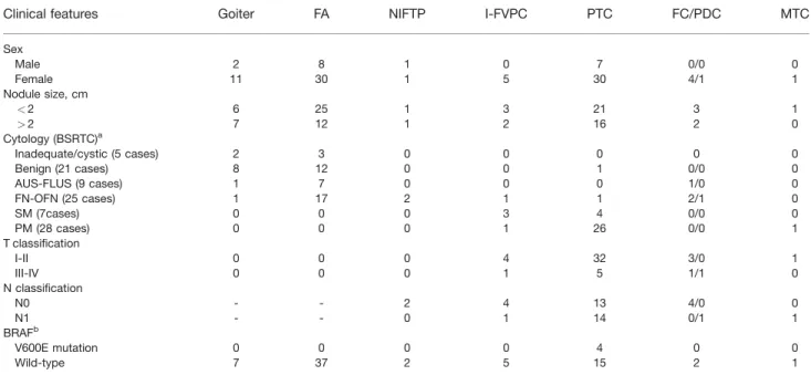
TABLE 1. Clinicopathological Features in the Current Series
Clinical features Goiter FA NIFTP I-FVPC PTC FC/PDC MTC
Sex Male 2 8 1 0 7 0/0 0 Female 11 30 1 5 30 4/1 1 Nodule size, cm <2 6 25 1 3 21 3 1 >2 7 12 1 2 16 2 0 Cytology (BSRTC)a Inadequate/cystic (5 cases) 2 3 0 0 0 0 0 Benign (21 cases) 8 12 0 0 1 0/0 0 AUS-FLUS (9 cases) 1 7 0 0 0 1/0 0 FN-OFN (25 cases) 1 17 2 1 1 2/1 0 SM (7cases) 0 0 0 3 4 0/0 0 PM (28 cases) 0 0 0 1 26 0/0 1 T classification I-II 0 0 0 4 32 3/0 1 III-IV 0 0 0 1 5 1/1 0 N classification N0 - - 2 4 13 4/0 0 N1 - - 0 1 14 0/1 1 BRAFb V600E mutation 0 0 0 0 4 0 0 Wild-type 7 37 2 5 15 2 1
Abbreviations: AUS/FLUS, atypia of undetermined significance/follicular lesion of undetermined significance; BSRTC, Bethesda System for Reporting Thyroid Cytopathology; FA, follicular adenoma; FC/PDC, follicular carcinoma/poorly differentiated carcinoma; FN/OFN, follicular neoplasms/oncocytic follicular neo-plasms; I-FVPC, invasive follicular variant of papillary thyroid carcinoma; MTC, medullary thyroid carcinoma; NIFTP, noninvasive follicular thyroid neoplasm with papillary-like nuclear features; PM, positive for malignancy; PTC, papillary thyroid carcinoma; SM, suspicious for malignancy.
aCytology was available for 95 of 101 cases.
thyroid carcinoma (1 case). In the current series, we had only 2 NIFTPs with preceding FNA diagnoses of FN.
Based on the histopathologic follow-up, there were 2 false-negative results and no false-positive cases. The global evaluation of the 3 indeterminate subcategories (AUS/ FLUS, FN/OFN, and SM) demonstrated 13 malignant diagnoses (31.7%), whereas for each subcategory the ROM was 11.1%, 20%, and 100%, respectively, with the exclu-sion of NIFTPs. The incluexclu-sion of NIFTPs slightly increased the ROM for the FN category from 25% to 28%.
The evaluation of BRAFV600Eanalysis demonstrated that the quality and quantity of DNA obtained from LBC stored material was sufficient for an adequate molecular evaluation and we obtained molecular yields from all the analyzed cases. Specifically, the 2 NIFTPs were wild-type
whereas 4 of the 19 PTCs (21%) had BRAFV600E
tions. In addition, a retrospective evaluation of RAS muta-tions was performed on both LBC stored material and histological slides from the 2 NIFTPs. These 2 cases were both found to be wild-type for RAS mutations.
The statistical analysis was performed with and with-out the inclusion of NIFTPs in the malignant category. The first scenario demonstrated a sensitivity of 77.7%, a specificity of 100%, a negative predictive value of 82%, a positive predictive value of 100%, and a diagnostic accu-racy of 89%, whereas the second scenario resulted in a sen-sitivity of 81.3%, a specificity of 100%, a negative predictive value of 85.4%, a positive predictive value of 100%, and a diagnostic accuracy of 91.1%.
In Table 4, we compared the findings of the current study regarding NIFTPs with those of studies published from adult cohorts.27–29,31,42 We compared the rate of NIFTPs for the entire FNAC series and for the PTC cases. The figures assessed much higher NIFTP rates in adult populations compared with the pediatric cohort herein.
DISCUSSION
In the current study, we aimed to evaluate the prevalence of the new entity of NIFTP in the pediatric population.
TABLE 2. Cytohistological Correlation of 95 Thyroid FNAC Specimens at 2 Institutions Benign Histology
Follow-Up (CU/LUMC)
Malignant Histology Follow-Up (CU/LUMC)
Total FNAC (95 cases) 51 (46/5) 44 (28/16)
Inadequate FNAC (5 cases) 5 (5/0) 0 (0/0)
Benign FNAC (21 cases)a
20 (16/4) 1 (0/1)
Goiter 17 (13/4) 1 (0/1)
Hyperplastic nodules 3 (3/0) 0 (0/0)
Indeterminate lesions (41 cases) 26 (25/1) 15 (13/2)
AUS/FLUS (9 cases) 8 (8/0) 1 (1/0) FN/OFN (25 cases)a 18 (17/1) 7 (5a /2) SM (7 cases) 0 (0/0) 7 (7/0) Malignant (28 cases) 0 (0/0) 28 (15/13)
Abbreviations: AUS/FLUS, atypia of undetermined significance/follicular lesion of undetermined significance; CU/LUMC, Catholic University (Rome)/Loyola Uni-versity Medical Center (Chicago); FN/OFN, follicular neoplasms/oncocytic follicular neoplasms; FNAC, fine-needle aspiration cytology; SM, suspicious for malignancy.
aThe 2 cases of noninvasive follicular thyroid neoplasm with papillary-like nuclear features (NIFTP) were included in the malignant FNs.
TABLE 3. Distribution of the Histological Variants of PTC Among the Cytological Categories Diagnosed According to The BSRTC
Histology Cytology CPTC I-FVPC DSPTC Col-PTC MPTC SPTC
ND 0 0 0 0 0 0 BL 1 0 0 0 0 0 AUS/FLUS 0 0 0 0 0 0 FN/OFN 0 1 0 0 1 0 SM 2 3 1 1 0 0 PM 22 1 2 0 0 2
Abbreviations: AUS/FLUS, atypia of undetermined significance/follicular lesion of undetermined significance; BL, benign lesion; BSRTC, Bethesda System For Reporting Thyroid Cytopathology; Col-PTC, columnar variant of papillary thyroid carcinoma; CPTC, classic variant of papillary thyroid carcinoma; DSPTC, dif-fuse sclerosing variant of papillary thyroid carcinoma; FN/OFN, follicular neoplasms/oncocytic follicular neoplasms; I-FVPC, infiltrative follicular variant of papil-lary thyroid carcinoma; MPTC, macrofollicular variant of papilpapil-lary thyroid carcinoma; ND, nondiagnostic; PTC, papilpapil-lary thyroid carcinoma; SM, suspicious for malignancy; SPTC, solid variant of papillary thyroid carcinoma; PM, positive for malignancy.
In the last few years, several authors have demon-strated that the majority, if not all, of the patients with encapsulated FVPCs do not develop any disease recurrences and metastases after a follow-up of years and/or deca-des.15–24A multi-institutional series including 109 patients with encapsulated FVPCs (NI-FVPCs) documented that none of these individuals developed any disease recur-rence.24As a result, the 2015 Endocrine Pathology Society meeting questioned whether these NI-FVPCs warrant a diagnosis of carcinoma and whether to rename them. Based on their yields, the Endocrine International Working Group recommended the reclassification of these NI-FVPCs as “noninvasive follicular thyroid neoplasm with papillary-like nuclear features” (NIFTP). Specifically, this entity was characterized by a set of morphological findings, including nuclear membrane irregularities, a maximum of 1% papillary architecture, ground glass appearance of the nuclei, nuclear pseudoinclusions, and larger nuclear size in
a context of encapsulated follicular tumor. Thus, according to Nikiforov et al, the recent reclassification of NIFTP may have a significant impact on the ROM for indeterminate/ follicular proliferations, which represent the most frequent cytological diagnoses of NIFTPs.24
In particular, recent articles by authors from different institutions have demonstrated that if NIFTPs were no longer termed “carcinoma,” the ROM for these categories would have a significant decrease of between 20% to 48%.15–34
A growing number of studies have investigated whether these lesions can be identified and diagnosed on either histology or FNAC and, more specifically, their impact on ROM. Several recent articles have been published regarding NIFTPs in the adult population, even though, to the best of our knowledge, the incidence of NIFTPs in pedi-atric patients is not exactly known.15–34,42–44According to Figure 1. (Top) Cytological features of a case of a follicular
neoplasm (liquid-based cytology, H & E; original magnifica-tion 3 400) (Bottom) diagnosed on histology as noninvasive follicular thyroid neoplasm with papillary-like nuclear features (H & E, original magnification 3 40).
Figure 2. (Top) Cytological features of a follicular neoplasm diagnosed as noninvasive follicular thyroid neoplasm with papillary-like nuclear features on histology (liquid-based cytology, H & E; original magnification 3 400). (Bottom) Some histological details of the nodule are shown (H & E, original magnification 3 1000).
Nikiforov et al24 and other series, the initial incidence of NIFTP in adults has been reported to be as high as 16% to 23% of all PTC cases (Table 4).27–29,31,42 In the current series, we reported a rate of 1.9% for the diagnostic category of NIFTP in a pediatric cohort of patients with resected thy-roid nodules. However, considering only the PTC cohort, our rate of NIFTPs was 4.5%. These figures are not com-pletely different from new published data from the adult population.43,44Not surprisingly, recent series have reported a consistently lower incidence than what was speculated by Nikiforov et al,24 most likely due to more rigid evaluation criteria for the definition of encapsulation of the tumor.43,44 In fact, the rates of NIFTP were reported to account for <5% of all PTCs in recent series in which rigid evaluation criteria were the standard of practice.43,44
Nonetheless, in this context, the most valid guideline is the application and adoption in pediatric thyroid nod-ules of the same criteria used for identifying NIFTPs in adult thyroid lesions.
The diagnostic role of FNAC in the evaluation of pediatric thyroid lesions has been thoroughly assessed and reported in the literature.1–14 In fact, numerous articles have emphasized that the majority of thyroid lesions are easily recognized and correctly diagnosed on cytological specimens. For this reason, the objective of the current study was to: 1) appraise the relevance of NIFTPs in a multi-institutional pediatric cytological cohort; and 2) dis-cuss the role of NIFTPs in light of their risk of malignancy and false-negative and false-positive results. To the best of our knowledge, to date TBSRTC and any other thyroid international classification systems have not included any specific evaluation of NIFTPs.38,39Regardless of the age of the patients, the majority of FVPCs (including both non-invasive and non-invasive carcinomas) are diagnosed in the cate-gories of indeterminate proliferations (AUS/FLUS, FN, and SM) due to subtle nuclear/cellular and/or architectural
features that do not allow for a conclusive cytological diag-nosis of malignancy.25–34 Faquin et al demonstrated that the highest impact of the reclassification of NIFTPs led to a decrease in the ROM ranging from 13.6% to 23.5%, espe-cially for AUS/FLUS, for which, most likely due to the inclusion of NIFTP, the ROM appears to be slightly higher than that expected from TBSRTC.28Analyzing the distri-bution of NIFTPs among our cytological categories, we estimated a correlation between NIFTPs and indeterminate categories as demonstrated by the fact that our 2 NIFTP cases (1.9%) were diagnosed as FN whereas the majority of I-FVPCs were classified as SM and PM. These data con-firmed the results from Maletta et al31and Bizzarro et al.27 In fact, these 2 studies highlighted that indeterminate diag-noses in NIFTPs (including both AUS/FLUS and FN) were twice those found in the I-FVPTCs (54.1% vs 29.2%).27,31For this reason, the same authors investigated how some cellular/morphological features are likely to be associated with a diagnosis of NIFTP on FNAC. According to these recent studies, we found that our 2 pediatric NIFTP cases were characterized by 100% microfollicular structures and a nuclear size ranging from 14 to 20 lm; therefore, these morphological recognitions might help to render a cytological diagnosis of “follicular proliferation”/ FN, also including a possible NIFTP diagnosis.25–34
The comparative analysis between the 2 institutions suggested the low prevalence of NIFTPs in the pediatric population and strongly demonstrated the high diagnostic accuracy of cytology regardless of the cytological methods used, including both conventional and LBC preparations.
The exclusion of NIFTPs from the malignant histo-logical lesions resulted in a malignancy rate of 48.5%, which is in perfect alignment with the statement from the American Thyroid Association Task Force on Pediatric Thyroid Cancer, which reported a malignancy rate between 9.2% and 50% for pediatric thyroid nodules.7
TABLE 4. Comparison of the Current Pediatric Series With Some Adult NIFTP Series in the Literature (Cytohistological Series)
Study
Total No. of FNAC Cases With Surgical Follow-Up
Total No. of PTC Cases
No. of NIFTP Cases (% in All FNAC Versus % in PTC)
Current pediatric series 95 44 2 (1.9% vs 4.5%)
Strickland 201529 655 346 85 (12.9% vs 24.6%) Faquin 201628 1827 756 173 (9.4% vs 23%) Maletta 201631 157 120 96 (61% vs 80%) Bizzarro 201627 90 77 24 (26.6% vs 31%) Jiang 201742 302 25 8 (2.6% vs 32%)
Abbreviations: FNAC, fine-needle aspiration cytology; NIFTP, noninvasive follicular thyroid neoplasm with papillary-like nuclear features; PTC, papillary thyroid carcinoma.
Our 2 NIFTP cases also confirmed the lower aggressive behavior reported by Nikiforov et al in their study of NIFTPs24; in fact, the 2 NIFTP cases in the current study did not demonstrate any disease recurrences or lymph node metastases during a follow-up period of 7 years. Fur-thermore, we also found a lack of common somatic muta-tions (ie, BRAFV600Eand RAS) in the 2 NIFTP cases in the current study. Nonetheless, larger and multi-institutional studies might confirm our preliminary yields.
A limitation of the current study was that it is a retro-spective study with a starting point of surgical cases. Another limitation may be the relatively low number of cases compared with similar studies in the adult popula-tion, a limitation that is inherent to studies in pediatric populations.
According to the Surveillance, Epidemiology, and End Results program and the World Health Organization, PTC accounts for approximately 90% of all cancers, and is mainly diagnosed as the classic variant of PTC.1–8 This diagnostic evidence might justify the low prevalence of NIFTPs in pediatric thyroid nodules. For example, accord-ing to the literature, the current series included 71.5% of the classic variant of PTC, 11.9% of I-FVPCs, 7.1% of diffuse sclerosing variants, 4.7% of the solid variant, 2.3% of the columnar variant, and 2.3% of the macrofollicular variant.
The results of the current study demonstrate that this new histological entity might be diagnosed in only a few cases among a pediatric series. Even though a definitive diagnosis of NIFTP cannot be rendered on FNAC, we associated these cases with specific cytological features, including a microfollicular pattern and a nuclear size in between those of FAs and invasive PTCs. In pediatric series as well as in adult cohorts, the role of NIFTP in the defini-tion of surgical treatment remains an ongoing concern even though a conservative approach is the best recom-mended therapy in cases of suspected NIFTP, especially in pediatric patients.
The new terminology of NIFTP might be considered in pediatric series even though further studies, including larger series, are necessary to define the role of this entity in pediatric cohorts, its clinical management, and the long-term follow-up.
FUNDING SUPPORT
No specific funding was disclosed.
CONFLICT OF INTEREST DISCLOSURES
The authors made no disclosures.
AUTHOR CONTRIBUTIONS
Esther Diana Rossi: Conceptualization, methodology, software, validation, formal analysis, investigation, resour-ces, data curation, writing–original draft, writing–review and editing, visualization, supervision, project administra-tion, and funding acquisition. Swati Mehrotra: Conceptu-alization, methodology, investigation, resources, data curation, writing–review and editing, visualization, and supervision. Ayse Irem Kilic: Conceptualization, method-ology, software, validation, formal analysis, investigation, resources, data curation, writing–original draft, writing– review and editing, visualization, supervision, project administration, and funding acquisition. Iclal Erdem Toslak: Conceptualization, methodology, visualization, for-mal analysis, investigation, and resources. Jennifer Lim-Dunham: Conceptualization, methodology, validation, for-mal analysis, investigation, and resources. Maurizio Mar-tini: Software and formal analysis. Guido Fadda: Writing– original draft and writing–review and editing. Celestino Pio Lombardi: Writing–review and editing. Luigi Maria Larocca: Conceptualization, validation, resources, writing– review and editing, and funding acquisition. G€uliz A. Bar-kan: Conceptualization, methodology, formal analysis, resources, writing–original draft, writing–review and edit-ing, visualization, supervision, and project administration. REFERENCES
1. Corrias A, Mussa A, Baronio F, et al; Study Group for Thyroid Diseases of Italian Society for Pediatric Endocrinology and Diabe-tology (SIEDP/ISPED). Diagnostic features of thyroid nodules in pediatrics. Arch Pediatr Adolesc Med. 2010;164:714-719.
2. Zimmermann D. Thyroid carcinoma in children and adolescents: diagnostic implications of analysis of the tumor genome. Curr Opin Pediatr. 2013;25:528-531.
3. Degnan BM, McClellan DR, Francis GL. An analysis of fine nee-dle aspiration biopsy of the thyroid in children and adolescents. J Pediatr Surg. 1996;31:903-907.
4. Wiersinga WM. Management of thyroid nodules in children and adolescents. Hormones. 2007;6:194-199.
5. Monaco SE, Pantanowitz L, Khalbuss WE, et al. Cytomorphologi-cal and molecular genetic findings in pediatric thyroid fine-needle aspiration. Cancer Cytopathol. 2012;120:342-350.
6. Smith M, Pantanowitz L, Khalbuss WE, Benkovich VA, Monaco SE. Indeterminate pediatric thyroid fine needle aspirations: a study of 68 cases. Acta Cytol. 2013;57:341-348.
7. Francis GL, Waguespack SG, Bauer AJ, et al; American Thyroid Association Guidelines Task Force. Management guidelines for children with thyroid nodules and differentiated thyroid cancer: the American Thyroid Association Guidelines Task Force on Pediatric Thyroid Cancer. Thyroid. 2015;25:716-759.
8. Lai SW, Roberts DJ, Rabi DM, Winston KY. Diagnostic accuracy of fine needle aspiration biopsy for detection of malignancy in pediatric thyroid nodules: protocol for a systematic review and meta-analysis. Syst Rev. 2015;24:120.
9. Gupta A, Ly S, Castroneves LA, et al. A standardized assessment of thyroid nodules in children confirms higher cancer prevalence than in adults. J Clin Endocrinol Metab. 2013;98:3238-3245.
10. Vidya V, Hemalatha AL, Rakhi B, et al. Efficacy and pitfalls of FNAC of thyroid lesions in children and adolescent. J Clin Diagn Res. 2014;8:35-38.
11. Rossi ED, Straccia P, Martini M, et al. The role of thyroid fine-needle aspiration cytology in the pediatric population: an institu-tional experience. Cancer Cytopathol. 2014;122:59-67.
12. Rossi ED, Bizzarro T, Martini M, et al. Morphological features that can predict BRAFV600E-mutated carcinoma in pediatric thy-roid cytology. Cytopathology. 2017;28:55-64.
13. Amirazodi E, Propst E, Chung CT, Parra DA, Wasserman JD. Pediat-ric thyroid FNA biopsy: outcomes and impact on management over 24 years at a tertiary care center. Cancer Cytopathol. 2016;124:801-810. 14. Rossi ED, Martini M, Cenci T, Capodimonti S, Larocca LM. The role of thyroid FNA cytology in pediatric malignant lesions: an overview of literature. Cancer. 2017;125:594-603.
15. Chen H, Izevbaye I, Chen F, Weinstein B. Recent advances in follicular variant of papillary thyroid carcinoma. N A J Med Sci. 2012;5 212-216. 16. Chan J. Strict criteria should be applied in the diagnosis of encap-sulated follicular variant of papillary thyroid carcinoma. Am J Clin Pathol. 2002;117:16-18.
17. Liu J, Singh B, Tallini G, et al. Follicular variant of papillary thy-roid carcinoma: a clinicopathologic study of a problematic entity. Cancer. 2006;107:1255-1264.
18. Renshaw AA, Gould EW. Why there is the tendency to overdiag-nose the follicular variant of papillary thyroid carcinoma. Am J Clin Pathol. 2002;117:19-21.
19. Piana S, Frasoldati A, Di Felice E, Gardini G, Tallini G, Rosai J. Encapsulated well-differentiated follicular-patterned thyroid carci-noma do not play a significant role in the fatality rate from thyroid carcinoma. Am J Surg Pathol. 2010;34:868-872.
20. Vivero M, Kraft S, Barletta J. Risk stratification of follicular variant of papillary thyroid carcinoma. Thyroid. 2013;23:273-279. 21. Lloyd RV, Erickson LA, Casey MB, et al. Observer variation in
the diagnosis of follicular variant of papillary thyroid carcinoma. Am J Surg Pathol. 2004;28:1336-1340.
22. Baloch ZW, Gupta PK, Yu GH, Sack MJ, LiVolsi VA. Follicular variant of papillary thyroid carcinoma. Cytologic and histologic correlation. Am J Clin Pathol. 1999;111:216-222.
23. Ganly I, Wang L, Tuttle MR, et al. Invasion rather than nuclear fea-tures correlates with outcome in encapsulated follicular tumors: fur-ther evidence for the reclassification of the encapsulated papillary thyroid carcinoma follicular variant. Hum Pathol. 2015;46:657-664. 24. Nikiforov YE, Seethala RR, Tallini G, et al. Nomenclature revision
for encapsulated follicular variant of papillary thyroid carcinoma: a paradigm shift to reduce overtreatment of indolent tumors. JAMA Oncol. 2016;2:1023-1029.
25. Logani S, Gupta PK, LiVolsi VA, Mandel S, Baloch ZW. Thyroid nodules with FNA cytology suspicious for follicular variant of pap-illary thyroid carcinoma: follow up and management. Diagn Cyto-pathol. 2000;23:380-385.
26. Ustun B, Chhieng D, Prasad ML, et al. Follicular variant of papil-lary thyroid carcinoma: accuracy of FNA diagnosis and implica-tions for patient management. Endocr Pathol. 2014;25:257-264. 27. Bizzarro T, Martini M, Capodimonti S, et al. Young Investigator
Challenge: the morphologic analysis of noninvasive follicular thyroid neoplasm with papillary-like nuclear features on liquid-based cytology: some insights into their identification. Cancer. 2016;124:699-710.
28. Faquin WC, Wong LQ, Afrogheh AH, et al. Impact of reclassify-ing noninvasive follicular variant of papillary thyroid carcinoma on the risk of malignancy in The Bethesda System for Reporting Thy-roid Cytopathology. Cancer Cytopathol. 2016;124:181-187. 29. Strickland KC, Howitt BE, Marqusee E, et al. The impact of
non-invasive follicular variant of papillary thyroid carcinoma on rates of malignancy for fine-needle aspiration diagnostic categories. Thyroid. 2015;25:987-992.
30. Ibrahim A, La Fortune KA, Wu H. Fine needle aspiration cytology of noninvasive follicular thyroid neoplasm with papillary like nuclear features (NIFT), single institutional study with comparison to invasive follicular variant of papillary thyroid carcinoma. Mod Pathol. 2016;SS1:S324A.
31. Maletta F, Massa F, Torregrossa L, et al. Cytological features of “noninvasive follicular thyroid neoplasm with papillary-like nuclear features” and their correlation with tumor histology. Hum Pathol. 2016;54:134-142.
32. Howitt BE, Chang S, Eslinger M, et al. Fine-needle aspiration diagnoses of noninvasive follicular variant of papillary thyroid carci-noma. Am J Clin Pathol. 2015;144:850-857.
33. Pusztaszeri M, Triponez F, Meyer P, Sadowski SM. Non invasive follicular thyroid neoplasm with papillary like nuclear features (NIFTP): report of an institutional experience with 86 cases. J Basic Clin Med. 2017;6:29-35.
34. Zhao L, Dias-Santagata D, Sadow PM, Faquin WC. Cytological, molecular, and clinical features of noninvasive follicular thyroid neo-plasm with papillary-like nuclear features versus invasive forms of follic-ular variant of papillary thyroid carcinoma. Cancer. 2017;125:323-331. 35. Rossi ED, Martini M, Capodimonti S, et al. Diagnostic and
prog-nostic value of immunocytochemistry and BRAF mutation analysis on liquid based biopsies of thyroid neoplasms suspicious for carci-noma. Eur J Endocrin. 2013;168:853-859.
36. Rossi ED, Zannoni GF, Moncelsi S, et al. Application of liquid-based cytology to fine-needle aspiration biopsies of the thyroid gland. Front Endocrinol (Lausanne). 2012;3:57.
37. Rossi ED, Mule A, Russo RM, Pierconti F, Fadda G. Application of liquid-based preparation to non-gynaecologic exfoliative cytol-ogy. Pathologica. 2008;100:461-465.
38. Perros P, ed; British Thyroid Association, Royal College of Physi-cians. Guidelines for the Management of Thyroid Cancer. Report of the Thyroid Cancer Guidelines Update Group. 2nd ed. London: Royal College of Physicians; 2007.
39. Cibas ED, Ali SZ. The Bethesda system for reporting thyroid cyto-pathology. Am J Clin Pathol. 2009;32:658-665.
40. Martini M, Teofili L, Cenci T, et al. A novel heterozygous HIF2AM535I mutation reinforces the role of oxygen sensing path-way disturbances in the pathogenesis of familial erythrocytosis. Haematologica. 2008;93:1068-1071.
41. Compton CC, Byrd DR, Garcia-Aguilar J, Kurtzman SH, Olawaiye A, Washington MK, eds. American Joint Commission on Cancer (AJCC) Cancer Staging Atlas. New York: Springer-Verlag New York; 2012. 42. Jiang XS, Harrison GP, Datto MB. Young Investigator Challenge:
molecular testing in noninvasive follicular thyroid neoplasm with papillary-like nuclear features. Cancer. 2016;124:893-900. 43. Bychkov A, Hirokawa M, Jung CK, et al. Low rate of noninvasive
follicular thyroid neoplasm with papillary-like nuclear features in Asian practice. Thyroid. 2017;27:983-984.
44. Cho U, Mete O, Kim MH, Bae JS, Jung CK. Molecular correlates and rate of lymph node metastasis of non-invasive follicular thyroid neoplasm with papillary-like nuclear features and invasive follicular variant papillary thyroid carcinoma: the impact of rigid criteria to distinguish non-invasive follicular thyroid neoplasm with papillary-like nuclear features. Mod Pathol. 2017;30:810-825.