Tumor-Associated Neutrophils (TANs) in
Gastric Carcinomas: Clinicopathological
and Prognostic Implications
Caruso RA1*; Rigoli L1
1Department of Human Pathology in Adult and Developmental Age “G. Barresi”, University of
Mes-sina, MesMes-sina, Italy.
*Correspondence to: Caruso RA, Department of Human Pathology in Adult and Developmental Age “G.
Barresi”, University of Messina, Messina, Italy. Email:[email protected]
Chapter 6
Overview on Gastric Cancer
1. Introduction
Gastric cancer, the fifth most common cancer worldwide, and the third leading cause of cancer-specific mortality, has very poor prognosis, with a 5-year survival less than 30% [1-2]. The TNM staging remains the cornerstone in clinical oncology to stratify prognosis and estab-lish therapy for patients with neoplasm [3]. Gastric carcinoma is a heterogeneous neoplasm with respect to anatomic location, epidemiology, genetics, histopathology, and biologic behav-ior, and, consequently, it has been subjected to many different classifications. With respect to anatomic location, gastric carcinomas may be distinguished as proximal (also known as car-dia) and distal (also known as noncarcar-dia). This classification correlates with distinct epidemio-logical risk factors. Obesity, hiatal hernia and reflux gastroesophagitis all are associated with cardia carcinoma, whereas H pylori infection is responsible for 77% of distal carcinoma [4-6]. From a histopathological viewpoint, routine classifications include those proposed by Laurèn [7], WHO [8], and Goseki [9]. The Laurèn classification [7] recognizes two main histologi-cal types: intestinal and diffuse, which show correlations with distinct clinihistologi-cal and epidemio-logical features. Intestinal type adenocarcinoma is mainly found in high risk areas of gastric cancer and is associated with the global decrease in incidence of this tumor. Histologically, intestinal type adenocarcinoma consists of tumor cells showing glandular differentiation with tubular, papillary or tubulo-papillary growth pattern. In diffuse type gastric carcinoma, tumor cells show abnormal loss of glandular differentiation and invade the stroma singly or in small groups. The WHO classification [8] is based on the predominant morphological component of the tumor (usually >50%) and identifies five types of gastric carcinoma: papillary, tubular,
mu-www
.openaccessebooks.com
Caruso RA
cinous, poorly cohesive (including signet-ring cells and other variants) and mixed carcinomas. Goseki classification [9] combines two tumor features, tubular differentiation and amount of intracytoplasmic mucus, in the following four groups: Group I (good tubular differentiation, poor mucus amount in cytoplasm), Group II (good tubular differentiation, rich mucus amount in cytoplasm), Group III (poor tubular differentiation, poor mucus amount in cytoplasm), Group IV (poor tubular differentiation, rich mucus amount in cytoplasm). Recently, The Can-cer Genome Atlas (TCGA) research network suggested a molecular classification of gastric carcinoma in four subtypes based on the presence of Epstein-Barr virus (8.8%), microsatellite instability (21.6%), genomic stability(19.6%) and chromosomal instability(49.6%) [10]. This biomolecular classification offers several advantages, showing particular correlations with anatomic location and/or tumor histologic types. Tumors with chromosomal instability oc-cur more frequently at the gastroesophageal junction and in the cardia, whereas Epstein-Barr positive tumors arise more frequently in the fundus and body. Genomic stable tumors show diffuse histology and are characterized by CDH1 and RHOA mutations. Epstein-Barr virus tumors display PIK3CA mutation as well as JAK2 and PD-L1/2 overexpression. Tumors with chromosomal instability exhibit intestinal morphology, marked aneuploidy, TP53 mutation and focal amplification of receptor tyrosine kinases. Microsatellite unstable tumors show el-evated mutation rates, including mutations of genes encoding targetable oncogenic signaling proteins. However, from a prognostic viewpoint none of these 4 subgroups of gastric cancers showed any significant survival differences. Although H pylori is responsible for 77% of distal carcinoma, H pylori status was not evidenced in this molecular classification [11-12]. There-fore, further studies are needed to demonstrate that TCGA gastric cancer classification may have practical implications for improving both therapy and survival in these patients.
Gastric carcinoma heterogeneity is reiterated by the strong variability in the host in-flammatory reaction. The WHO classification [8] describes four stromal reactions (desmo-plasia/scirrhous reaction, lymphocytic infiltration, stromal eosinophilia, and a granulomatous response), neglecting the role of tumor associated neutrophils (TANs) in gastric carcinomas. Traditionally, neutrophils were considered cell protagonists of the acute phase of inflamma-tion, where they play an important role in the defense against microbial invasion. Recent stud-ies have widened this view showing new functions of neutrophils including the orchestration of innate and adaptive immune reactions [13-15]. In recent decades, increasing attention has been paid to the role of neutrophils in tumor-host reaction, but conflicting conclusions on the prognostic impact of TANs have been reported in literature.
In this work, we summarize the current state on the clinicopathological and prognostic implications of TANs to elucidate this problem. Our experience with TANs in gastric carcino-mas is also reported and discussed according to recent data from literature.
2. Neutrophil-To-Lymphocyte Ratio(NLR)
Blood counts, such as the NLR, are being used for diagnostic and prognostic aims in patients with cancer [16-22]. Indeed, NLR has also been used for early detection and as a prog-nostic marker in gastric cancer [23-25], early diagnosis of ovarian cancer [26-27], and progno-sis and survival prediction in colon cancer [28] and hepatocellular carcinoma [29]. Recently, NLRs have also been used in the differential diagnosis between primary breast carcinoma and benign proliferative breast disease [30]. In most studies, a high NLR is associated with adverse overall survival in many human tumors. However, NLR data must be considered with caution, as they are nonspecific parameters, which may be influenced by concurrent conditions such as infections, inflammation, and medications [31]. Many studies did not explicitly check for such concurrent conditions, and this may lead to erroneous interpretations [31]. Furthermore, there are few works regarding NLR values in patients with a low/high neutrophil count in the tumor.
3. N1 and N2 – Polarization or Hyperactivation
Experimental studies suggest that TANs show a bipolar pattern of activation (N1/N2) similar to that observed in macrophages (M1/M2) and T-cell (Th1/Th2) polarization [32-33]. N1 neutrophils exert antitumor activities through tumor cytotoxicity, whereas N2 neutrophils favor tumor growth, invasion and metastasis, e.g. through proteolysis of extracellular matrix components, promotion of angiogenesis and mediation of immunosuppression [34-38]. However, N1/N2 neutrophils have only been shown in murine tumor models and must be confirmed in human tumors [39]. It is also possible that the N1/N2 phenotype reflects only a functional state of neutrophil activation [39-40]. For example, neutrophils isolated from early tumors are more cytotoxic toward tumor cells and release higher levels of NO and H2O2, whereas in advanced tumors, neutrophils display low cytotoxic activity and acquire a protumor phenotype [41]. These data suggest that tumor stage plays an important role in modeling neutrophil phenotype and function.Therefore, further studies are needed to clarify whether the different functions of TANs can be attributed to distinct subpopulations, or rather to different grades of neutrophil activation in human tumors.
4. Distribution of TANs and Neutrophil Recruitment in the Tumor Stroma
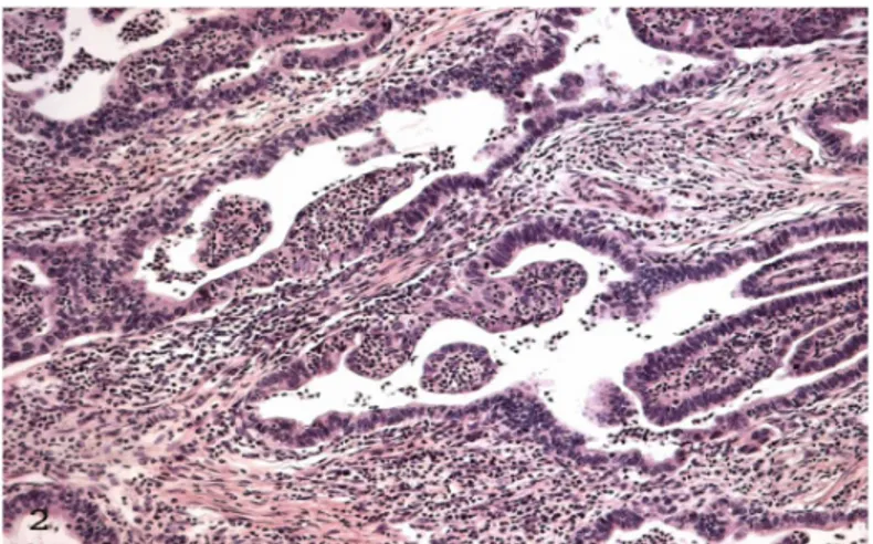
Distribution of TANs is multifaceted. TANs may be found at the invasive front of tumor (peritumoral location) (Figure 1a) or in the center of tumor (Figure 1b) [42-46]. Intratumoral TAN distribution shows heterogeneity, being found as a single massive infiltrate in the tumor stroma or as a series of multifocal aggregates scattered throughout the tumor stroma. In some cases numerous neutrophils may be found within the tumor epithelium (Figure 2) [47]. It has been pointed out that peritumoral TANs are mainly found at the early stage of tumor develop-ment, while intratumoral TANs are seen at later stages [48].
4.1 TANs and tumor necrosis
Mechanisms responsible for neutrophil infiltration in the tumors may be manifold. It is a common experience of pathologists to observe neutrophils in the tumor stroma using light microscopy, and, to date, this phenomenon is interpreted as secondary to tumor necrosis. Consequently, areas of tumor necrosis are not taken into consideration during a TAN count. However, the role of tumor necrosis in the recruitment of TANs is not clear. Innate immune cells may differentially react to necrotic cells in terms of chemotaxis e.g. macrophages may show more rapid and sustained attraction toward necrotic cells rather than dendritic cells and neutrophils[49]. Moreover, it is possible to observe large areas of tumor necrosis without neutrophil infiltration (Figure 3).
4.2 Classification of cell death.
A number of factors such as hypoxia, starvation or acidosis can determine tumor tissue necrosis. However, the classic notions of apoptosis and necrosis, based on morphologic criteria, have recently been modified. The latest recommendations of the Nomenclature Committee on Cell Death suggest two mutually exclusive types of cell death: accidental and regulated cell death [50-51]. Accidental cell death is an uncontrollable, instantaneous, passive type of cell death that occurs after severe tissue damage due to extreme environmental conditions (e.g. high temperature, elevated pressure, trauma). Morphologically, it is characterized by cellular swelling and consequent collapse of the plasma membrane thereby causing spillage of intracellular contents and inflammatory reaction [51]. Other morphologic changes include generalized swelling of cytoplasm and organelles, nuclear membrane dilatation, nuclear chromatin condensation into small and irregular patches [52]. By contrast, regulated cell death is an active program of cell death that can be modulated by pharmacological agents or genetic interventions both in physiological and pathological conditions. It occurs during chronic or mild exogenous perturbations of cell microenvironment following failure of compensative mechanisms (e.g. autophagy) [50-51]. The term programmed cell death refers to regulated cell death occurring in physiological conditions (e.g. embryonic development, adult tissue homeostasis). A classic example of programmed cell death is apoptosis, which affects single cells or small clusters of cells [51]. Morphologic characteristics of apoptosis include reduction of cell volume (pyknosis), chromatin condensation, nuclear fragmentation (karyorrhexis), plasma membrane integrity until final steps of the process, formation of apoptotic bodies, phagocytosis of apoptotic bodies by macrophages or by adjacent cells (so-called efferocytosis), with no inflammation in most cases [51].
4.3 Subtypes of regulated cell death
Recent studies have however suggested that regulated cell deaths include several subtypes such as necroptosis, ferroptosis, pyroptosis, parthanatos, and mitochondrial permeability
transition–driven regulated necrosis, but not apoptosis [50]. These types of regulated cell death are not mutually exclusive and are not governed by a single molecular pathway, but are characterized by a complex interplay and cross-talk between them [50]. Moreover, they manifest a partial or total necrotic morphology [51]. To date, necroptosis is the best studied form of regulated cell death and is now recognized as an important drug-sensible contributor to tissue injury in many pathologic conditions including ischemia-reperfusion damage, acute inflammatory reactions, and tumor necrosis [50-53]. It is characterized by cellular swelling, rapid membrane permeabilization and concomitant release of damage-associated molecular patterns (DAMPs) into the extracellular space. According to recent studies, DAMPs determine neutrophil infiltration in the tumor stroma [54]. Moreover, neutrophils stimulated by dying cells exert NO/H2O2–based cytotoxicity against residual live cells [50-51]. Ferroptosis is a recently described subtype of regulated cell death due to accumulation of lethal lipid ROS produced through iron-dependent lipid peroxidation [55]. Morphologically, it does not show chromatin condensation, characteristically found in apoptosis, nor loss of plasma membrane integrity, as observed in accidental necrosis [50-51]. Instead, it is characterized by mitochondrial shrinkage and increased mitochondrial membrane density [51,55]. These recent revolutionary modifications in the classification of cell death have disclosed new fields of research. It is not surprising that there are a few updated studies focused on the role of regulated cell death in the recruitment of TANs.
4.4 Other Mechanisms of Neutrophil Recruitment in Tumor Stroma
Several literature data suggest that tumor necrosis is not the sole factor responsible for TANs. Immunohistochemical studies have demonstrated that the type of mucin overexpressed by tumor cells is highly correlated with TANs. For example, TANs are prominent in pancreatic and gastric carcinomas with MUC1 overexpression (Figure 4) [56-57] and scarce or absent in mucinous carcinomas that overexpress intestinal mucins MUC2 [58]. These data suggest that tumor histologic type may be correlated with TANs. Accordingly, our previous cancer registry study, undertaken to determine the incidence and clinicopathologic features of neutrophil-rich gastric carcinoma in the Messina province (South Italy), revealed an inverse correlation between TANs and mucinous subtype of gastric cancer, classified according to the WHO classification [57]. Mucinous histologic types are recognized morphologically for the presence of extensive extracellular accumulation of mucins that show a strong MUC2 immunoreactivity [58]. Tumor cells discharge MUC2 into mucin lakes that invade tissues and inhibit the host inflammatory reaction [59]. These data explain the scarce neutrophil infiltration in gastric mucinous carcinomas observed in our study (Figure 5). In addition to immunohistochemical studies, biomolecular research disclosed the importance of oncogenic activation in the induction of TANs. CXC chemokines [e.g., interleukin-8 (IL-8)], produced by oncogene mutations in cancer cells, may evoke neutrophil infiltration in tumor stroma [60]. One oncogene that has
been strongly linked to the recruitment of TANs is the Ras oncogene. Ji et al. [60] showed a link between the presence of activated K-ras mutations and macrophage and neutrophil predominant inflammation in both murine and human lung tumors. IL8 has been shown to be a transcriptional target for Ras signaling and expression is required for tumor-associated inflammation and angiogenesis in a human tumor xenograft model [61].
Other experimental studies showed that engineered tumors to release interleukins or chemokines in their microenvironment evoke a massive neutrophil infiltration that, often in collaboration with CD8+ T lymphocytes, leads to the rejection of engineered tumor cells and the establishment of significant immunity against wild-type parental tumors [62]. Taken together, literature data suggest that TANs have manifold recruitment causes including tumor necrosis, MUC1 overexpression, and oncogene activation leading to direct cytokine production by tumor cells.
5. Ultrastructural Studies of TANs in Gastric Carcinomas
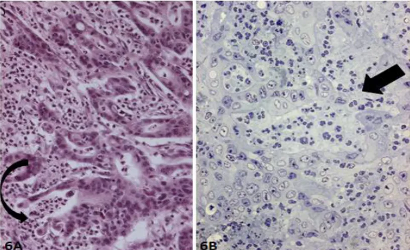
Our ultrastructural investigations, performed in gastric carcinomas, reiterated the dual face of TANs. At light microscopy, well differentiated gastric adenocarcinomas show intraglandular migration of neutrophils associated to necrotic phenomena of variable extent ranging from single adenocarcinoma cell death to segmental disruption of the epithelial layer of a gland (Figures 6a, 6b) up to glandular and stromal necrosis [63-65]. At electron microscopy, we see a spatial relationship between neutrophil intraglandular migration and regulated adenocarcinoma cell death. In Figure 7, one neutrophil is seen in intimate contact with one or a few severely injured adenocarcinoma cells showing increased electron density, loss of microvilli, marked dilatation of nuclear envelope, small condensed chromatin particles, and progressive mitochondrial and endoplasmic reticulum swelling. These ultrastructural changes are different from those described in apoptosis where there is characteristic chromatin condensation and separation of euchromatin and heterochromatin and formation of apoptotic bodies. They are also different from that described in accidental cell death where there is plasma membrane disruption and cytoplasmic organelle swelling. They manifest partial necrotic ultrastructural features, and therefore are compatible with regulated cell death (Figure 8). These ultrastructural observations in vivo are similar to that reported in in vitro studies where neutrophil cytotoxicity requires physical contact between neutrophils and tumor cells [66]. It remains to be investigated whether regulated cell death is triggered by infiltrating neutrophils or alternatively may be responsible for neutrophil infiltration. This question could be resolved through experimental studies of developing disruptions, as opposed to morphologic static observations of disruptions described above.
6. Emperipolesis and Efferocytosis
Humble, in the 1950s [67], coined the term emperipolesis to describe penetration of lymphocytes in other living cells, both in physiological and pathological settings. During emperipolesis, the migrating cell remains viable and can exit without morphological and physiological abnormalities. Efferocytosis is a term used to describe phagocytosis of apoptotic cells, occurring in embryonic development, organogenesis, tissue repair, atrophy, and inflammation [68-69]. Removal of apoptotic cells is necessary to avoid their disintegration in the tissues via a process known as secondary necrosis which leads to uncontrolled leakage of the dying cells and subsequent chronic inflammatory reaction. Macrophages and dendritic cells are the main cells involved in apoptotic cell removal, including apoptotic neutrophils [68-69]. However, also “neighboring” cells such as epithelial cells, endothelial cells, and fibroblasts, may engulf apoptotic cells [69]. In our previous study, we provided morphologic evidence of apoptotic neutrophil efferocytosis by foveolar epithelial cells in chronic active H. pylori gastritis [70].
7. Entosis and Xeno-Cannibalism
In cytological or histological samples of tumors, pathologists can detect cells with cannibalistic properties. This phenomenon may be suggested by the presence of one or more cytoplasmic vacuoles, possibly containing dying cells, that push the nucleus to the periphery giving it the shape of a crescent moon. Classically, the term cannibalism was used to describe the engulfment of tumor cells by other tumor cells. Recently Overholtzer [71] coined the term entosis to describe a process similar to cannibalism and frequently found in human and experimental tumors, whereby cells become internalized into neighboring cells, forming what are called ‘cell-in-cell’ structures. In his studies, he demonstrated that internalized cells initially appear healthy and viable [71]. Over a brief period, some internalized cells are able to escape, but most cells die through a form of cell death which is distinct from apoptosis as dying cells are negative for cleaved caspase-3, and do not exhibit condensed or fragmented nuclei. Instead, LAMP1, a lysosomal membrane protein, localizes around dying cells and acidification occurs at the earliest stages of death, suggesting lysosomal involvement [71]. Recent reports have shown tumor cell phagocytosis of normal cells (xeno-cannibalism) such as neutrophils, lymphocytes, and erythrocytes [72-75]. These new observations imply that cannibal tumor cells do not distinguish between normal and sibling neoplastic cells.
In literature, there is some confusion about the terms emperipolesis, entosis, cannibalism and xeno-cannibalism [76-77]. In our opinion, they must be used appropriately remembering the neoplastic or non-neoplastic context in which they occur. The main distinctive characteristics of emperipolesis, efferocytosis, entosis, and xeno-cannibalism are summarized in Tables 1a and 1b. Their light microscopic identification in sections stained with hematoxylin-eosin (H&E) sometimes requires ancillary techniques such as caspase
immunohistochemistry, electron microscopy, and TUNEL assay which is based on enzymatic incorporation of labelled nucleotides at sites where DNA fragmentation has occurred. For example, we recently provided light microscope, immunohistochemical and ultrastructural evidence of neutrophil xeno-cannibalism by tumor cells in high grade gastric carcinomas as well as in micropapillary gastric carcinomas (Figure 9) [72,78-79]. At the light microscopic level, intra/interepithelial neutrophils showed apoptotic changes such as pyknotic nuclei and cell shrinkage. TUNEL staining documented apoptotic neutrophils in cytoplasmic vacuoles of tumor cells. Electron microscope, a fundamental tool not only to identify apoptosis, but also to discern inter- or intraepithelial neutrophil localization, confirmed the presence of large heterophagosomal vacuoles containing apoptotic neutrophils. Various phases of apoptotic changes were documented in these neutrophils. Ultrastructural signs of early apoptosis included nuclear chromatin separation into dense and electron lucent areas, rounded nuclear profiles, preservation of cytoplasmic granules, and maintenance of cell membrane integrity. Late apoptotic morphology was characterized by cell shrinkage, tightly packed cytoplasmic granules, and uniform collapsed nucleus (Figure 10). Secondary degeneration of apoptotic neutrophils in the phagocytic vacuoles of tumor cells included cellular swelling, electron-lucent cytoplasm, vacuolization and indiscernible cell membrane.
The phenomenon of neutrophil xeno-cannibalism by tumor cells may have a series of pathobiological consequences. It has been suggested that the presence of cannibalized cells may interfere with cell mitosis, leading to non-genetic polyploidy [80]. Phagocytosis of neutrophils by tumor cells may constitute a sort of “feeding” activity. Tumor stroma contains malformed microvasculature that contributes to tumor hypoxia, acidosis, and increased interstitial fluid pressures [81]. Thus, independently of microvasculature, tumor cells cannibalizing neutrophils find nutrients useful for their survival. The phenomenon of neutrophil xeno-cannibalism by tumor cells represents an example of protumor activity of TANs, particularly frequent in high grade gastric carcinomas including micropapillary carcinomas.
8. Prognostic Impact of TANs
The prognostic significance of TANs remains controversial. In our previous study on gastric carcinomas, TANs were morphologically identified by H &E stain and manually counted [82]. The multivariable analysis of possible interaction effects of the clinicopathological factors with TANs revealed that female patients with a moderate or extensive amount of TANs had about a 39% reduction in their risk of mortality, whereas no correlation with outcome of male patients was found [82]. A possible explanation for the interaction between TANs and female patients is that sexual dimorphism exists in the immune response [83]. Both humoral and cell-mediated immunity are more active in females than in males, and steroid gonadal hormones may play an important role in regulating this response [84]. These observations suggest the possibility of an inflammatory (neutrophil) and gender-dependent host natural
cytoxicity in the microenvironment of gastric carcinomas. Subsequently, other studies using immunohistochemical stainings (CD66b, myeloperoxidase and CD15) to identify TANs showed discrepant results. Negative (renal [85], hepatic [45,86], colorectal [87], and gastric carcinomas [88-89]), and positive (colorectal carcinomas [47, 90-94]) correlation with patient prognosis have been described in recent publications. These conflicting results may be due to different methods used in quantifying TANs. In several reviews, shift from morphological to immunohistochemical markers was suggested, including CD15, myeloperoxidase and CD66b [95-96]. Nevertheless, both morphological and immunohistochemical methods have their advantages and disadvantages. Galdiero et al. [93] showed that antibodies against myeloperoxidase were not specific for neutrophils, as also monocytes and immature macrophages stained. CD66b is expressed both on human neutrophils and eosinophils and is recognized as a granulocyte “activation marker” [97]. CD15 is expressed on the surface of leukocytes, mainly in neutrophils, eosinophils, and part of monocytes, and do not reflect the activation status of neutrophils [98]. In addition, CD15 has been demonstrated to be expressed occasionally on tumor cells [93].Therefore, CD15 and CD66b are not specific markers for neutrophils, and light microscope analysis of H&E stained sections remains fundamental to distinguish neutrophils from other inflammatory cells. Given that the different markers used to identify TANs (CD66b, CD15 and cell morphology by H&E stain) may explain contradictory results in prognostic studies of TANs, it is desirable that an optimal combination of markers (i.e. morphology with immunohistochemistry for CD66b) be used in future studies.
Recent studies concerning the role of TANs in human colorectal carcinomas merit further discussion. Berry et al. [91] using morphology and manual counting of TANs in colorectal carcinomas, obtained similar results to ours performed on gastric carcinomas. In particular, they show that in women there was a trend towards better overall survival in all patients with high TAN counts suggesting a potential for different roles for TANs in men and women with colorectal carcinoma [91]. Moreover, higher TAN counts in Stage II patients are associated with a nearly 3-fold increase in overall survival compared to patients with low TAN [91]. The favorable prognostic significance of TANs in colorectal carcinomas is confirmed in two recent studies using CD66b immunohistochemistry as a neutrophil marker. Wikberg et al. [47] have studied the prognostic role of infiltrating neutrophils at different intratumoral subsites including the invasive front, the center of the tumor, and in the tumor epithelium of colorectal carcinomas. Expression of the neutrophil marker CD66b was assessed by immunohistochemistry in 448 archival human tumor tissue samples from patients surgically resected for colorectal carcinomas. They found that high infiltration of CD66b-positive cells in the tumor front is a favorable prognostic factor in stages I-II colon cancers [47]. Galdiero et al [93] used disease-specific and disease-free survival as their endpoints and found that the positive prognostic effect for high TAN counts extended to patients with all stages of disease. TAN density dramatically decreases in Stage IV patients as compared to Stage I-III. They
showed that prognostic significance of TANs can be influenced not only by clinical stage but also 5-fluorouracil (5-FU)-based chemotherapy. In particular, higher TAN density was associated with better response to 5-FU-based chemotherapy [93]. Thus, assessment of TAN infiltration may not only be useful for prognostic informations, but may also have important therapeutic implications, in particular for identifying patients likely to benefit from 5-FU-based chemotherapy. I would like to take the opportunity to stress the favorable prognostic
significance of TANs in gastrointestinal cancer [82,91-94,100] and its evaluation using rigorous
quantitative methodology.
9. Conclusions
Understanding the role of TANs in gastric carcinomas remains incomplete, but studies continue to accumulate. What is certain is that neutrophils are not accidentally present in the tumor stroma, rather they play an active role in tumor growth. Previous studies have suggested positive or negative correlation with patient prognosis. In part, these conflicting results may be due to different methods (morphology vs immunohistochemistry) in quantifying TANs. At the same time, experimental studies reveal antitumoral (N1 phenotype) and protumoral (N2 phenotype) functions of TANs. These data should not necessarily be interpreted as demonstration of two distinct subpopulations of TANs. It is also possible that protumoral and antitumoral neutrophils are two extremes of a spectrum of a sole functional state. Based on recent literature and our own data, this neutrophil functional plasticity correlates strictly with clinicopathological parameters such as gender, tumor stage, intra/peritumoral localization of TANs, and response to chemotherapy. In Table 2, we summarize studies concerning the role of TANs in correlation with clinicopathological parameters. These data suggest that, in particular conditions,TANs represent an antitumoral mechanism that should to be documented in routine pathology and promoted therapeutically. It would also be desirable for pathologists to be involved more and more in determining the type of TAN infiltrate, thereby providing diagnostic and prognostic information as well as suggesting appropriate immune-chemotherapies for each patient.
EMPERIPOLESIS EFFEROCYTOSIS
Migration of a cell in the cytoplasm of another
Engulfment of apoptotic cells by macrophages, dendritic cells or adjacent epithelial/mesenchymal
cells During emperipolesis
migrating and host cell remain viable
Removal of apoptotic cell commonly occurs in non-neoplastic tissue
Table 1a: Cell-in-cell structure in both physiological and pathological (non-neoplastic) conditions
ENTOSIS XENO-CANNIBALISM
Engulfment of a tumor cell in the cytoplasm of another tumor cell
Phagocytosis of an inflammatory cell by a tumor cell
During entosis engulfed cell is degraded by lysosomal enzymes
During cannibalism engulfed inflammatory cell shows apoptosis
Table 2: Relationship between clinicopathological factors and bipolar prognostic role of TAN.
11. Figures
Figure 1
A: TANs are mainly seen at the front of neoplastic tissue. H&E X 100
Figure 3: Large areas of tumor necrosis without neutrophil infiltration. H &E x 100
Figure 4: Strong MUC1 immunoreactivity in the cytoplasm of tumor cells. Note the presence of numerous TANs. X
200
Figure 6A: Neutrophil transepithelial migration associated with focal neoplastic gland disruption (curved arrow). H&E
X 10 B: Semi-thin section showing numerous TANs and break in continuity of adenocarcinoma gland (arrow). Giemsa X 200
Figure 7: Neutrophil in contact with an adenocarcinoma cell showing chromatin condensation, loss of microvilli,
enlarged mitochondria, and dilatation of nuclear envelope (arrow). Note some adjacent tumor cells containing autophagic vacuoles in their cytoplasm (curved arrows). X 8000
Figure 8: Neutrophils are near adenocarcinoma cell showing convoluted nucleus, marked chromatin condensation,
dilatation of nuclear envelope. X 8 000
Figure 9: Micropapillary carcinoma of the stomach. Tumor cells exhibit xeno-cannibalism of neutrophils. H&E X
Figure 10: Tumor cell xeno-cannibalism of a neutrophil showing late apoptotic changes. X 4 000
12. References
1. Torre LA, Bray F, Siegel RL, Ferlay J, Lortet-Tieulent J, Jemal A. Global cancer statistics, 2012. CA Cancer J Clin. 2015; 65: 87–108
2. DeSantis CE, Lin CC, Mariotto AB, Siegel RL, Stein KD, Kramer JL, Alteri R, Robbins AS, Jemal A. Cancer treatment and survivorship statistics, 2014. CA Cancer J Clin. 2014; 64: 252–271.
3. Sobin LH, Compton CC. TNM seventh edition: what’s new, what’s changed. Cancer. 2010; 116: 5336-9
4. Rugge M, Genta RM, Di Mario F, El-Omar EM, El-Serag HB, Fassan M, Hunt RH,Kuipers EJ, Malfertheiner P, Sugano K, Graham DY. Gastric Cancer as Preventable Disease. Clin Gastroenterol Hepatol. 2017; 15: 1833-1843. 5. Cavaleiro-Pinto, M., Peleteiro, B., Lunet, N. et al. Helicobacter pylori infection and gastric cardia cancer: systematic review and meta-analysis. Cancer Causes Control. 2011; 22: 375–387
6. WHO. Schistosomes, Liver Flukes and Helicobacter pylori: IARC Working Group on the Evaluation of Carcinogenic Risks to Humans. IARC Monographs on the Evaluation of Carcinogenic Risks to Humans. IARC Scientific Pub. No. 61, pp. 218-220. Lyon, France: IARC, 1994.
7. Lauren P. The two histological main types of gastric carcinoma: Diffuse and so-called intestinal-type carcinoma. An attempt at a histo-clinical classification. Acta Pathol Microbiol Scand. 1965; 64: 31-49.
8. Bosman, F.T., Carneiro, F., Hruban, R.H. et al. World Health Organization classification of tumours of the digestive system. IARC Press, Lyon; 2010
9. Goseki N, Takizawa T, Koike M. Differences in the mode of the extension of gastric cancer classified by histological type: new histological classification of gastric carcinoma. Gut. 1992; 33: 606-12.
10. Cancer Genome Atlas Research N. Comprehensive molecular characterization of gastric adenocarcinoma. Nature. 2014; 513: 202–209
11. Jácome AA, Coutinho AK, Lima EM, Andrade AC, Dos Santos JS. Personalized medicine in gastric cancer: Where are we and where are we going? World J Gastroenterol. 2016; 22: 1160-71.
12. Tan P, Yeoh KG. Genetics and Molecular Pathogenesis of Gastric Adenocarcinoma. Gastroenterology. 2015;149: 1153-1162..
13. Kruger P, Saffarzadeh M, Weber AN, Rieber N, Radsak M, von Bernuth H, Benarafa C, Roos D, Skokowa J, Hartl D. Neutrophils: Between host defence, immune modulation, and tissue injury. PLoSPathog. 2015 Mar 12; 11(3): e1004651.
14. Nauseef WM, Borregaard N. Neutrophils at work. Nat Immunol. 2014l; 15: 602-11.
2013; 13: 159-75.
16. Mascarella MA, Mannard E, Daniela Silva Wurzba S, Zeitouni A. Neutrophil-to-lymphocyte ratio in head and neck cancer prognosis: A systematic review and meta-analysis. Head Neck. 2018 Jan 22.
17. Minami S, Ogata Y, Ihara S, Yamamoto S, Komuta K. Neutrophil-to-Lymphocyte Ratio Predicts Overall Survival of Advanced Non-Small Cell Lung Cancer Harboring Mutant Epidermal Growth Factor Receptor. World J Oncol. 2017; 8: 180-187.
18. Cao LL, Lu J, Lin JX, Zheng CH, Li P, Xie JW, Wang JB, Chen QY, Lin M, Tu RH, Huang CM. Nomogram based on tumor-associated neutrophil-to-lymphocyte ratio to predict survival of patients with gastric neuroendocrine neoplasms. World J Gastroenterol. 2017;23: 8376-8386. .
19. Liu D, Huang Y, Li L, Song J, Zhang L, Li W. High neutrophil-to-lymphocyte ratios confer poor prognoses in patients with small cell lung cancer. BMC Cancer. 2017;17:882.
20. Liu X, Qu JK, Zhang J, Yan Y, Zhao XX, Wang JZ, Qu HY, Liu L, Wang JS, DuanXY. Prognostic role of pretreatment neutrophil to lymphocyte ratio in breast cancer patients: A meta-analysis. Medicine (Baltimore). 2017; 96: e8101. 21. Song Y, Yang Y, Gao P, Chen X, Yu D, Xu Y, Zhao J, Wang Z. The preoperative neutrophil to lymphocyte ratio is a superior indicator of prognosis compared with other inflammatory biomarkers in resectable colorectal cancer. BMC Cancer. 2017; 17: 744.
22. Tan YG, Eu EWC, Huang HH, Lau WKO. High neutrophil-to-lymphocyte ratio predicts worse overall survival in patients with advanced/metastatic urothelial bladder cancer. Int J Urol. 2017 Nov 1. [Epub ahead of print]
23. Lieto E, Galizia G, Auricchio A, Cardella F, Mabilia A, Basile N, Del Sorbo G,Castellano P, Romano C, Orditura M, Napolitano V. Preoperative Neutrophil to Lymphocyte Ratio and Lymphocyte to Monocyte Ratio are Prognostic Factors in Gastric Cancers Undergoing Surgery. J Gastrointest Surg. 2017; 21: 1764-1774.
24. Wang SC, Chou JF, Strong VE, Brennan MF, Capanu M, Coit DG. Pretreatment Neutrophil to Lymphocyte Ratio Independently Predicts Disease-specific Survival in Resectable Gastroesophageal Junction and Gastric Adenocarcinoma. Ann Surg.2016; 263: 292-7
25. Hu ZD, Huang YL, Qin BD, Tang QQ, Yang M, Ma N, Fu HT, Wei TT, Zhong RQ.Prognostic value of neutrophil to lymphocyte ratio for gastric cancer. Ann Transl Med. 2015; 3: 50.
26. Huang QT, Zhou L, Zeng WJ, Ma QQ, Wang W, Zhong M, Yu YH. Prognostic Significance of Neutrophil-to-Lymphocyte Ratio in Ovarian Cancer: A Systematic Review and Meta-Analysis of Observational Studies. Cell Physiol Biochem.2017; 41: 2411-2418.
27. Zhou Q, Hong L, Zuo MZ, He Z. Prognostic significance of neutrophil to lymphocyte ratio in ovarian cancer: evidence from 4,910 patients. Oncotarget. 2017; 8: 68938-68949
28. Li MX, Liu XM, Zhang XF, Zhang JF, Wang WL, Zhu Y, Dong J, Cheng JW, Liu ZW,Ma L, Lv Y. Prognostic role of neutrophil-to-lymphocyte ratio in colorectal cancer: a systematic review and meta-analysis. Int J Cancer. 2014; 134: 2403-13.
29. Gomez D, Farid S, Malik HZ, Young AL, Toogood GJ, Lodge JP, Prasad KR. Preoperative neutrophil-to-lymphocyte ratio as a prognostic predictor after curative resection for hepatocellular carcinoma. World J Surg. 2008; 32: 1757-62. 30. Ozyalvacli G, Yesil C, Kargi E, Kizildag B, Kilitci A, Yilmaz F. Diagnostic and prognostic importance of the neutrophil lymphocyte ratio in breast cancer. Asian Pac J Cancer Prev. 2014; 15: 10363-6.
31. Templeton AJ, McNamara MG, Šeruga B, Vera-Badillo FE, Aneja P, Ocaña A,Leibowitz-Amit R, Sonpavde G, Knox JJ, Tran B, Tannock IF, Amir E. Prognostic role of neutrophil-to-lymphocyte ratio in solid tumors: a systematic review and meta-analysis. J Natl Cancer Inst. 2014; 106(6)
32. Fridlender ZG, Sun J, Kim S, Kapoor V, Cheng G, Ling L, Worthen GS, Albelda SM. Polarization of tumor-associated neutrophil phenotype by TGF-beta: “N1”versus “N2” TAN. Cancer Cell. 2009; 16: 183-94.
33. Mantovani A. The yin-yang of tumor-associated neutrophils. Cancer Cell. 2009; 16: 173-4.
34. Fridlender ZG, Albelda SM. Tumor-associated neutrophils: friend or foe? Carcinogenesis. 2012; 33: 949-55. 35. Shaul ME, Fridlender ZG. Neutrophils as active regulators of the immune systemin the tumor microenvironment. J Leukoc Biol. 2017; 102: 343-349.
36. Shaul ME, Levy L, Sun J, Mishalian I, Singhal S, Kapoor V, Horng W, FridlenderG, Albelda SM, Fridlender ZG. Tumor-associated neutrophils display a distinct N1 profile following TGFβ modulation: A transcriptomics analysis of pro- vs.antitumor TANs. Oncoimmunology. 2016; 5(11): e1232221.
37. Granot Z, Fridlender ZG. Plasticity beyond cancer cells and the “immunosuppressive switch”. Cancer Res. 2015; 75: 4441-5.
38. Fridlender ZG, Albelda SM, Granot Z. Promoting metastasis: neutrophils and T cells join forces. Cell Res. 2015; 25: 765-6
39. Eruslanov EB, Singhal S, Albelda SM. Mouse versus Human Neutrophils in Cancer:A Major Knowledge Gap. Trends Cancer. 2017; 3: 149-160.
40. Galdiero MR, Varricchi G, Loffredo S, Mantovani A, Marone G. Roles of neutrophils in cancer growth and progression. J Leukoc Biol. 2017 Dec 15.
41. Eruslanov EB. Phenotype and function of tumor-associated neutrophils and their subsets in early-stage human lung cancer. Cancer Immunol Immunother. 2017; 66: 997-1006.
42. Eruslanov EB, Bhojnagarwala PS, Quatromoni JG, Stephen TL, et al.. Tumor-associated neutrophils stimulate T cell responses in early-stage human lung cancer. J Clin Invest. 2014; 124: 5466-80.
43. Coffelt SB, Wellenstein MD, de Visser KE. Neutrophils in cancer: neutral no more. Nat Rev Cancer. 2016; 16: 431-46.
44. He M, Peng A, Huang XZ, Shi DC, Wang JC, Zhao Q, Lin H, Kuang DM, Ke PF, Lao XM. Peritumoral stromal neutrophils are essential for c-Met-elicited metastasisin human hepatocellular carcinoma. Oncoimmunology. 2016; 5: e1219828.
45. Kuang DM, Zhao Q, Wu Y, Peng C, Wang J, Xu Z, Yin XY, Zheng L. Peritumoral neutrophils link inflammatory response to disease progression by fostering angiogenesis in hepatocellular carcinoma. J Hepatol. 2011; 54: 948-55. 46. Li YW, Qiu SJ, Fan J, Zhou J, Gao Q, Xiao YS, Xu YF. Intratumoralneutrophils:a poor prognostic factor for hepatocellular carcinoma following resection. J Hepatol. 2011; 54: 497-505.
47. Wikberg ML, Ling A, Li X, Öberg Å, Edin S, Palmqvist R. Neutrophil infiltration is a favorable prognostic factor in early stages of colon cancer. Hum Pathol. 2017; 68: 193-202.
48. Mishalian I, Bayuh R, Levy L, Zolotarov L, Michaeli J, Fridlender ZG. Tumor-associated neutrophils (TAN) develop pro-tumorigenic properties during tumor progression. Cancer Immunol Immunother. 2013; 62: 1745-56.
49. Dehne N, Mora J, Namgaladze D, Weigert A, Brüne B. Cancer cell and macrophage cross-talk in the tumor microenvironment. Curr Opin Pharmacol. 2017; 35: 12-19.
50. Galluzzi L, Vitale I, Aaronson SA, Abrams JM, Adam D, Agostinis P, Alnemri ES, et al. Molecular mechanisms of cell death: recommendations of the Nomenclature Committee on Cell Death 2018. Cell Death Differ. 2018 Jan 23. 51. Galluzzi L, Bravo-San Pedro JM, Vitale I, Aaronson SA, Abrams JM, Adam D, et al. Essential versus accessory
aspects of cell death: recommendations of the NCCD 2015. Cell Death Differ. 2015; 22: 58-73.
52. Skenderi F, Vranic S, Damjanov I. Regulated cell death in diagnostic histopathology. Int J Dev Biol. 2015; 59: 149-58
53. Wang T, Jin Y, Yang W, Zhang L, Jin X, Liu X, He Y, Li X. Necroptosis in cancer: An angel or a demon? Tumour Biol. 2017 Jun; 39(6)
54. Wang X, Yousefi S, Simon HU. Necroptosis and neutrophil-associated disorders. Cell Death Dis. 2018 Jan 25; 9(2): 111.
55. Yu H, Guo P, Xie X, Wang Y, Chen G. Ferroptosis, a new form of cell death, and its relationships with tumourous diseases. J Cell Mol Med. 2017; 21: 648-657.
56. Reid MD, Basturk O, Thirabanjasak D, Hruban RH, Klimstra DS, Bagci P, AltinelD, Adsay V. Tumor-infiltrating neutrophils in pancreatic neoplasia. Mod Pathol. 2011; 24: 1612-9.
57. Ieni A, Branca G, Parisi A, Fedele F, Irato E, Venuti A, Caruso RA. Neutrophil-rich gastric carcinoma in the integrated cancer registry of eastern Sicily, Italy. Anticancer Res. 2015; 35: 487-92..
58. Baldus SE, Zirbes TK, Engel S, Hanisch FG, Mönig SP, Lorenzen J, Glossmann J, Fromm S, Thiele J, Pichlmaier H, Dienes HP. Correlation of the immunohistochemical reactivity of mucin peptide cores MUC1 and MUC2 with the histopathological subtype and prognosis of gastric carcinomas. Int J Cancer. 1998;79: 133-8.
59. Yang XF, Yang L, Mao XY, Wu DY, Zhang SM, Xin Y. Pathobiological behavior and molecular mechanism of signet ring cell carcinoma and mucinous adenocarcinoma of the stomach: a comparative study. World J Gastroenterol. 2004; 10: 750-4.
60. Ji H, Houghton AM, Mariani TJ, Perera S, Kim CB, Padera R, Tonon G, McNamaraK, Marconcini LA, Hezel A, El-Bardeesy N, Bronson RT, Sugarbaker D, Maser RS,Shapiro SD, Wong KK. K-ras activation generates an inflammatory response in lung tumors. Oncogene. 2006; 25:2105-12.
61. Waugh DJ, Wilson C. The interleukin-8 pathway in cancer. Clin Cancer Res. 2008; 14: 6735-41
62. Di Carlo E, Forni G, Lollini P, Colombo MP, Modesti A, Musiani P. The intriguing role of polymorphonuclear neutrophils in antitumor reactions. Blood. 2001; 97(2): 339-45.
63. Caruso RA, Speciale G, Inferrera C. Neutrophil interaction with tumour cells in small early gastric cancer: ultrastructural observations. Histol Histopathol. 1994; 9: 295-303.
64. Caruso RA, Bonanno A, Finocchiaro G, et al. Ultrastructural observations on inflammatory angiogenesis in gastric carcinomas with massive neutrophil infiltration. Ultrastruct Pathol. 2009; 33: 1-5.
65. Caruso RA, Fedele F, Rigoli L, Branca G, Bonanno A, Quattrocchi E, Finocchiaro G, Venuti A. Apoptotic-like tumor cells and apoptotic neutrophils in mitochondrion-rich gastric adenocarcinomas: a comparative study with light and electron microscopy between these two forms of cell death. Rare Tumors. 2013; 5: 68-71.
66. Granot Z, Jablonska J. Distinct Functions of Neutrophil in Cancer and Its Regulation. Mediators Inflamm. 2015;2015: 701067.
67. Humble, J. G., Jayne, W. H. W., and Pulvertaft, R. J. V., Brit..1. Haematol., 1956, 2, 283.
68. deCathelineau AM, Henson PM. The final step in programmed cell death phagocytes carry apoptotic cells to the grave. Essays Biochem. 2003; 39: 105-17
69. Henson PM. Cell Removal: Efferocytosis.Annu Rev Cell Dev Biol. 2017 6; 33: 127-144.
active Helicobacter pylori gastritis. Virchows Arch. 2012; 461: 489-94.
71. Overholtzer M, Mailleux AA, Mouneimne G, Normand G, Schnitt SJ, King RW, CibasES, Brugge JS. A nonapoptotic cell death process, entosis, that occurs by cell-in-cell invasion. Cell. 2007; 131: 966-79.
72. Caruso RA, Muda AO, Bersiga A, Rigoli L, Inferrera C. Morphological evidence of neutrophil-tumor cell phagocytosis (cannibalism) in human gastric adenocarcinomas. Ultrastruct Pathol. 2002 ;26: 315-21.
73. Caruso RA, Fedele F, Finocchiaro G, Arena G, Venuti A. Neutrophil-tumor cell phagocytosis (cannibalism) in human tumors: an update and literature review. Exp Oncol. 2012; 34: 306-11.
74. Lugini L, Matarrese P, Tinari A, Lozupone F, Federici C, Iessi E, Gentile M, Luciani F, Parmiani G, Rivoltini L, Malorni W, Fais S. Cannibalism of live lymphocytes by human metastatic but not primary melanoma cells. Cancer Res. 2006; 66: 3629-38.
75. Khayyata S, Basturk O, Adsay NV. Invasive micropapillary carcinomas of the ampullo-pancreatobiliary region and their association with tumor-infiltrating neutrophils. Mod Pathol. 2005; 18: 1504-11.
76. Qian Y, Shi Y. Natural killer cells go inside: entosis versus cannibalism. Cell Res. 2009; 19: 1320-1.
77. Mailleux AA, Overholtzer M, Brugge JS. Entosis, a cell death process related to cell cannibalism between tumor cells. Med Sci (Paris). 2008; 24: 246-8
78. Barresi V, Branca G, Ieni A, Rigoli L, Tuccari G, Caruso RA. Phagocytosis (cannibalism) of apoptotic neutrophils by tumor cells in gastric micropapillary carcinomas. World J Gastroenterol. 2015; 21: 5548-54.
79. Caruso RA, Rigoli L, Parisi A, Fedele F,et al.. Neutrophil-rich gastric carcinomas: light and electron microscopic study of 9 cases with particular reference to neutrophil apoptosis. Ultrastruct Pathol. 2013; 37: 164-70.
80. Krajcovic M, Overholtzer M. Mechanisms of ploidy increase in human cancers: a new role for cell cannibalism. Cancer Res. 2012; 72: 1596-601.
81. Matarrese P, Ciarlo L, Tinari A, Piacentini M, Malorni W. Xeno-cannibalism as an exacerbation of self-cannibalism: a possible fruitful survival strategy for cancer cells. Curr Pharm Des. 2008; 14: 245-52.
82. Caruso RA, Bellocco R, Pagano M, Bertoli G, Rigoli L, Inferrera C. Prognostic value of intratumoral neutrophils in advanced gastric carcinoma in a high-risk area in northern Italy. Mod Pathol. 2002; 15: 831-7.
83. Spitzer JA, Zhang P. Gender differences in neutrophil function and cytokine-induced neutrophil chemoattractant generation in endotoxic rats. Inflammation. 1996; 20: 485-98.
84. Da Silva JA. Sex hormones and glucocorticoids: interactions with the immune system. Ann N Y Acad Sci. 1999; 876: 102-17
85. Jensen HK, Donskov F, Marcussen N, Nordsmark M, Lundbeck F, von der MH. Presence of intratumoral neutrophils is an independent prognostic factor in localized renal cell carcinoma. J Clin Oncol. 2009; 27: 4709–17.
86. Margetts J, Ogle LF, Chan SL, Chan AWH, Chan KCA, Jamieson D, Willoughby CE, Mann DA, Wilson CL, Manas DM, Yeo W, Reeves HL. Neutrophils: driving progression and poor prognosis in hepatocellular carcinoma? Br J Cancer. 2018; 118: 248-257.
87. Rao HL, Chen JW, Li M, Xiao YB, Fu J, Zeng YX, Cai MY, Xie D. Increased intratumoral neutrophil in colorectal carcinomas correlates closely with malignant phenotype and predicts patients’ adverse prognosis. PLoS One. 2012; 7: e30806.
88. Hu P, Pang Z, Shen H, Wang G, Sun H, Du J. Tumor-infiltrating neutrophils predict poor outcome in adenocarcinoma of the esophagogastric junction. Tumour Biol. 2015; 36: 2965-71.
89. Zhao JJ, Pan K, Wang W, Chen JG, Wu YH, Lv L, et al. The prognostic value of tumor-infiltrating neutrophils in gastric adenocarcinoma after resection. PLoS One. 2012; 7: e33655.
90. Uehara K, Nakanishi Y, Shimoda T, Taniguchi H, Akasu T, Moriya Y. Clinicopathological significance of microscopic abscess formation at the invasive margin of advanced low rectal cancer. Br J Surg. 2007; 94: 239-43.
91. Berry RS, Xiong MJ, Greenbaum A, Mortaji P, Nofchissey RA, Schultz F, Martinez C, Luo L, Morris KT, Hanson JA. High levels of tumor-associated neutrophils are associated with improved overall survival in patients with stage II colorectal cancer. PLoS One. 2017 Dec 6; 12: e0188799.
92. Governa V, Trella E, Mele V, Tornillo L, Amicarella F, Cremonesi E, MuraroMG,et al. The Interplay Between Neutrophils and CD8(+) T Cells Improves Survival in Human Colorectal Cancer. Clin Cancer Res. 2017; 23: 3847-3858.
93. Galdiero MR, Bianchi P, Grizzi F, Di Caro G, Basso G, Ponzetta A, Bonavita E, et al. Occurrence and significance of tumor-associated neutrophils in patients with colorectal cancer. Int J Cancer.2016;139: 446-56.
94. Zhou G, Peng K, Song Y, Yang W, Shu W, Yu T, Yu L, Lin M, Wei Q, Chen C, Yin L, Cong Y, Liu Z. CD177+ neutrophils suppress epithelial cell tumourigenesis in colitis-associated cancer and predict good prognosis in colorectal cancer.Carcinogenesis. 2017 Dec 8. [Epub ahead of print]
95. Hurt B, Schulick R, Edil B, El Kasmi KC, Barnett C Jr. Cancer-promoting mechanisms of tumor-associated neutrophils. Am J Surg. 2017; 214: 938-944.
96. Donskov F. Immunomonitoring and prognostic relevance of neutrophils in clinical trials. Semin Cancer Biol. 2013; 23: 200-7.
97. Torsteinsdottir I, Arvidson NG, Hallgren R, Hakansson L. Enhanced expression of integrins and CD66b on peripheral blood neutrophils and eosinophils in patients with rheumatoid arthritis, and the effect of glucocorticoids. Scand J Immunol. 1999; 50: 433–9.
98. Buchwalow I, Atiakshin D, Samoilova V, Boecker W, Tiemann M. Identification of autofluorescent cells in human angioimmunoblastic T-cell lymphoma. Histochem Cell Biol. 2018;149:169-177.
99. Chen CL, Wang Y, Huang CY, Zhou ZQ, et al. . IL-17 induces antitumor immunity by promoting beneficial neutrophil recruitment and activation in esophageal squamous cellcarcinoma. Oncoimmunology. 2017; 7(1): e1373234.