© 2016 Francesco Molinaro et al published by De Gruyter Open
This work is licensed under the Creative Commons Attribution-NonCommercial-NoDerivs 3.0 License. DOI 10.1515/med-2016-0037
received January 27, 2016; accepted March 7, 2015
Abstract: Congenital defects of the sternum are rare and due to a failure of midline development and fusion of the sternal bones. Surgical correction of a sternal cleft should be preferred during infancy for functional reasons. Chest wall reconstruction represented a complex problem in the last decades.
We report our successful outcome of sternal reconstruc-tion in a rare case of PHACES syndrome, in which the patient was submitted to reconstruction of the sternum and complete closure of the thoracic defect by the employ of an extracellular matrix XCM Biologic tissue matrix. We promote the use of extracellular matrix in surgi-cal reconstruction of chest defects for its maneuverabil-ity, plasticmaneuverabil-ity, tolerability and the possibility of growing with the children’s chest getting a good compliance and optimal cosmetic results.
Keywords: Phaces syndrome, Sternal reconstruction, Midline development defects.
Open Med. 2016; 11:196-199
*Corresponding author: Alfredo Garzi, University Salerno, Salerno, ITALY, E-mail: [email protected]
Francesco Molinaro, Division of Pediatric Surgery, Department of Medical, Surgical and Neurological Sciences, University of Siena, Italy
Elisa Cerchia, Anna Lavinia Bulotta, Mario Messina Department of Medical, Surgical and Neurological Science. Pediatric Surgery Unit , University of Siena, Italy
Vincenzo Giuseppe Di Crescenzo, Department of Medicine and Surgery, Thoracic Surgery Unit , University of Salerno, Italy Luca Luzzi, Giuseppe Gotti, Thoracic Surgery Unit, University Hospi-tal of Siena, Siena, IHospi-taly
Case Report
Open Access
Francesco Molinaro, Alfredo Garzi*, Elisa Cerchia, Vincenzo Giuseppe Di Crescenzo, Luca
Luzzi, Anna Lavinia Bulotta, Giuseppe Gotti, Mario Messina
Sternal reconstruction by extracellular matrix: a
rare case of phaces syndrome
1 Introduction
Congenital defects of the sternum are rare and due to a failure of midline development and fusion of the sternal bones. The sternal cleft is more common in females and in association with other anomalies like posterior fossa mal-formations, hemangiomas, arterial anomalies, cardiac defects, eye anomalies and supraumbilical midline raphe better known as PHACES syndrome (OMIM 606519) [1]. Surgical correction of a sternal cleft should be preferred during infancy for functional (respiratory impairment and potentially dangerous injuries to the mediastinal organs) and mechanical (more elasticity and thoracic compli-ance) reasons [2]. Chest wall reconstruction represented a complex problem in the last decades due to the difficulty to choose the best surgical approach for closing the defect without compromising the stability and the following evolution of the thoracic wall. We report our successful outcome of sternal reconstruction by bioprosthetic mesh (extracellular matrix) in a rare case of PHACES syndrome.
2 Case report
A 4 year-old girl was referred to our hospital with a congen-ital defect of the anterior chest wall. Her antenatal history was unremarkable and family history revealed parents as cousins of first grade, no traumas or surgical operations were referred. At delivery, the birth weight was 2600 g and no episode of respiratory distress was reported. At clin-ical examination she presented a wide gap in the upper part of anterior wall chest with intermittent herniation of cardiac structures during crying, while its pulsations were easily seen at rest, and also it was evident a paradoxical excursion of the thorax during respiratory acts (Figure 1). Cutaneous examination revealed an erythematous plaque as left sided facial hemangiomas extending to neck (Figure 2 a-b) and a cutaneous raphe from sternal defect to umbilicus (Figure. 1). Ophthalmologic, routine blood
Brought to you by | Universita degli Studi die Siena Authenticated
Sternal reconstruction by extracellular matrix 197 samples, genetic analysis, thyroid profile and cerebral
cranial magnetic resonance were normal. Cardiovascular evaluation revealed a small atrial septal defect hemody-namically stable.
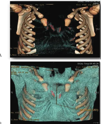
Chest-X ray and computed tomographic scan (CT) with three-dimensional reconstruction showed the presence of a congenital diastasis of sternal manubrium (37 mm)
with agenesis of the corpus sternum, with V-shaped car-tilages (24 mm cranio-caudal diameter) in whose concav-ity the right heart ventricle was made superficial (Figure 3 a-b). After an accurate surgical planning, the patient was submitted to reconstruction of the sternum and complete closure of the thoracic defect by the employ of an extracel-lular matrix XCM Biologic tissue matrix®. The skin overly-ing the sternal defect was incised along the midline from the ideal manubriosternal joint to the ideal xiphisternum separating the subcutaneous tissues from the underly-ing fused pericardium. Then the pericardium was mobi-lized all along the edges and sutured in midline without tension. The second layer was provided by an extracellular matrix previously modeled and then anchored in double layers as a sandwich, at the medial margins of the bilat-eral ribs with multiple intermittent not absorbable suture (Figure 4). The pectoral muscles were reapproximated to the midline and drainage was positioned before closure the skin that was removed on the third day. The post-op-erative recovery was regular even if there was an episode of bradycardia during extubation treated with adminis-tration of atropine and epinephrine with rapid cardio-re-spiratory stabilization. The patient was discharged on the 8th postoperative day with a good cosmetic and functional Figure 1: Sternal cleft in the upper part of anterior wall chest
associ-ated with supra umbilicus raphe.
Figure 2 a-b: Left sided facial hemangiomas extending to neck.
Figure 3 a-b: Three-dimensional reconstruction of CT images showed the presence of a congenital diastasis of sternal manubrium (37 mm) with agenesis of the corpus sternum, with V-shaped cartilages (24 mm cranio-caudal diameter).
A A
B B
Brought to you by | Universita degli Studi die Siena Authenticated
198 Francesco Molinaro et al
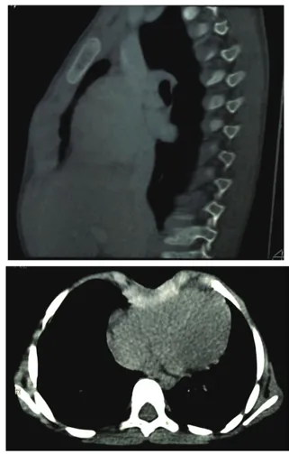
result (Figure 5). At the follow up after 1 year the patient had a normal looking chest wall with absence of abnor-mally thoracic excursion and a good integration of the extracellular matrix with surrounding tissues as shown to chest X-ray and CT results (Figure 6 a-b).
Ethical approval: The research related to human use has been complied with all the relevant national regula-tions, institutional policies and in accordance the tenets of the Helsinki Declaration, and has been approved by the authors’ institutional review board or equivalent committee.
Informed consent: Informed consent has been obtained from all individuals included in this study.
3 Discussion
Sternal cleft is a rare congenital anomaly caused by failed midline development and fusion of the mesodermal lateral plates at about 8 weeks of intrauterine life [3]. It is commonly classified in two types: complete, when the sternum is split from the manubrium to the xiphoid, and incomplete (inferior and superior), when a chondral bar bridges the midline [4]. In about 10% of patients with sternal cleft is reported an associated supra umbilical raphe and cardiac anomalies in about 30% [5]. As in our case, the sternal defect can also be a part of a complex clin-ical condition such as PHACES syndrome. This is a neu-rocutaneous syndrome of which most common features include posterior fossa malformations, hemangiomas, arterial anomalies, cardiac defects, eye anomalies and sternal clefting or superumbilical raphe [6]. The first case of syndrome was described by Pascual-Castroviejo in 1978 [7], while Frieden et al. used the acronym PHACE in 1996,
Figure 6 a-b: At the follow up after 1 year the patient had a normal looking chest wall with good integration of the extracellular matrix with surrounding tissues as shown to chest X-ray and CT images. A
B Figure 4: Extracellular matrix fixed in double layers at the medial margins of the bilateral ribs with multiple intermittent not absorba-ble suture during surgical reconstruction of chest defect.
Figure 5: Cosmetic result after surgical reconstruction of anterior wall chest.
Brought to you by | Universita degli Studi die Siena Authenticated
Sternal reconstruction by extracellular matrix 199 later modified in PHACES by Boulinguez et al. [8]. The
aeti-ology is unknown and there is predominance in females [9], rising the hypothesis of dominant X-linked condition. Diagnostic criteria are the presence of hemangioma and the coexistence of at least one among associated anoma-lies; 70 % of affected children have only one of extracuta-neous manifestation [1]. Only 44 previously reported cases of sternal cleft associated with hemangiomas and supra umbilical raphe has been described in literature [10]. The possible cardiac and cerebrovascular anomalies associ-ated must be carefully searched and excluded before any surgical procedures for avoiding major complications. In literature a surgical correction of sternal defect is highly recommendable and should performed as soon as possi-ble during infancy for an higher thoracic compliance at this age [11]. The choice of best surgical approach is con-troversial and many techniques have been described in literature for reconstructing sternum including primary approximation, sliding or rotating chondrotomies and muscle flaps preferably performed when the flexibil-ity of the chest wall is maximal and compression of the underlying structures is minimal [12]. In some cases the cleft is wide and the sternal remnants hypoplastic, so a primary repair could not be possible. A valid alternative is the employment of prosthetic implantable materials, but there is not consensus about the type to use. Le Roux and Shama reported the ideal characteristics of prosthetic material: rigidity to abolish paradoxical movement with a minimal compression of thoracic organs, inertness to allow in-growth of fibrous tissue and decrease the like-lihood of infection, malleability and radiolucency [13]. In our case, we have chosen to use XCM Biologic tissue matrix®, a sterile non-cross-linked 3-D matrix derived from porcine dermis which is composed of cells and extra-cellular matrix (combination of proteins, proteoglycans, glycosaminoglycans and other biological materials) sub-mitting to procedures of sterilization, decellularization and inactivation of viruses. The result is a strong bio-logic implant providing a structure that can be infiltrated by body’s cells for a better integration with surrounding tissues and gives a good resistance and durability [14]. The advantage of decellularized dermis is that is gradu-ally revascularized and remodeled into autologous tissue while maintaining its structural integrity. Even if more research is needed to elucidate the indications, contrain-dications and outcomes of these materials we promote the use of extracellular matrix in surgical reconstruction of chest defects for its maneuverability, plasticity, tolerabil-ity and the possibiltolerabil-ity of growing with the children’s chest getting a good compliance and optimal cosmetic results.
Conflict of interest statement: Authors state no conflict of interest.
References
[1] Metry D.W., Haggstrom A.N., Drolet B.A., Baselga E., Chamlin S., Garzon M., et al. A prospective study of PHACE syndrome, in infantile hemangiomas: demographic features, clinical findings, and complications, Am J Med Genet A 140 2006; 975-986
[2] Shalak L., Kaddoura I., Obeid M., Hashem H., Haidar R., Bitar F.F. Complete cleft sternum and congenital heart disease : review of the literature. Pediatr Int 2002 ;44:314-316 [3] Jose R.M., De Campos L., Filomeno T.B., et al. Repair of
congenital sternal cleft in infants and adolescent. Ann Thorac Surg 1998;66:1151-1153
[4] Shamberger R.C., Welch K.J. Sternal defects. Pediatr Surg Int 1990; 5:156-164
[5] Metry D.W., Dowd C.F., Barkovich A.J., Frieden I. The many faces of PHACE syndrome. J Ped 2001; 139:117-123
[6] Frieden I.J., Reese V., Cohen D. PHACE syndrome: the association of posterior fossa brain malformations,
hemangiomas, arterial anomalies, coarctation of the aorta and cardiac defects, and eye abnormalities. Arch Dermatol 1996; 132:307-311
[7] Pascual-Castrovejo I. Vascular and nonvascular intracranial malformation associated with external capillary hemangiomas. Neuroradiology 1978; 16:82-84
[8] Boulinguez S., Teillac-Hamel D., Bedane C., Bennaceur S., De Prost Y. Cervicofacial hemangioma and a minor sternal malformation: inclusion in PHACES syndrome? Pediatr Dermatol 1998; 15:119-121
[9] Levin J.H., KAler S.G. Non-random maternal X-chromosome inactivation associated with PHACES. Clin Genet 2007; 72:345-350
[10] Gorlin R.J., Kantaputra P., Aughton D.J., Mulliken J.B. Marked female predilection in some syndromes associated with facial hemangiomas. Am J Med Genet 1994; 52:130-135
[11] Acastello E., Majluf R., Garrido P., Barbosa L.M., Peredo A. Sternal cleft: a surgical opportunity. J Pediatr Surg 2003; 178-183
[12] Snyder B.J., Robbins R.C., Ramos D. Primary repair of complete sternal cleft with pectoralis major muscle flaps. Ann Thorac Surg 1996; 61(3):983-984
[13] Watanabe A., Watanabe T., Obama T., et al. New material for reconstruction of the anterior chest wall, including the sternum. J Thorac Cardiovasc Surg 2003; 126:1212-1214 [14] Badylak S.F., The extracellular matrix as a scaffold for tissue
reconstruction. Seminars in Cell ℓ Developmental Biology. 2002 Oct; 13(5):377-383
Brought to you by | Universita degli Studi die Siena Authenticated