University of Catania
Ph.D. IN NEUROBIOLOGY XXVIII CYCLE
December 2016
Parkinson Disease subtype:
morphostructural and neurobiological correlates
Ph.D. Coordinator: Prof. R. Avola Dr. Alfonso Rubino
Tutor: Prof. Giuseppe Meco University of Rome “Sapienza”
INDEX
1. Background………..pag 1 2. Experimental setting I – Genetic architecture of MAPTGene Region
in Parkinson Disease Subtypes……...………...………pag 3 2.1 Introduction…………...……….…….pag 3 2.2 Methods……….……..pag 4 2.3 Results………….……….……..pag 6 2.4 Discussion.………..………..pag 7
3. Clinical setting: Intercepting Parkinson disease nonmotor subtypes:
a proof-of-principle study in a clinical setting…...………..pag 16 3.1 Background ………..……….pag 16 3.2 Methods………….………..…………...………pag 18 3.3 Results………....pag 19 3.4 Discussion………..pag 24
4. Experimental setting II: DAT Methylation in Parkinson Disease: Correlation with peripheral Dopamine Transporter gene expression
and central activity………pag 27 4.1THE ROLE OF DAT1 IN PARKINSON'S DISEASE……….pag 28 4.2 Methods………...pag 32 4.4 Results……….……pag 34 4.4 Discussion………...pag 36
5. General view……….………..…...………pag 39 6. References……….………..……..pag 41
Background
The widely used term of Parkinson’s Disease (PD) refers to a progressive motor syndrome, classically associated with neuronal loss in the substantia nigra and inclusions containing the synaptic protein α synuclein (α syn) in the cell bodies and processes of surviving neurons (known as Lewy bodies and Lewy neurites, respectively) in this reg ion. Ho wever, the current view of malady reconsider PD as a co mplex clin icopathological entity, where the “proteiform” mu ltio rgan involvement results from convergent multifactorial b io molecular substrates (Di Battista, 2014). It follows that, fro m a clin ical point of v iew, PD presents as an extremely heterogeneous disorder with a wide range of sympto ms and scarcely predictable progression trajectories . Therefore, it beco me necessary subtyping PD in order to better define the pathogenic process, the clin ical progression and the response to treatment. In th is term, the National Institutes of Health delineated subtype identification as one of the top 3 clinical research priorities in PD (Sieber 2014). The research efforts on this topic has been directed into the study that empirically classify PD fro m clinical observation and those that categorize PD without aprioristic assumptions, using a data-driven approach. (Marras 2016)
Approaches to clinical subtyping in Parkinson’s disease. Subtyping approaches can be divided into those that are empirically derived, from clinicalobservation, and data-driven based on co-occurrence of clinical features with no a priori hypothesis about the way clinical features will cluster together.(Marras 2016)
One of the earliest empirical approach for subtyping PD was based on motor phenotype, categorizing the disease into (1) tremor do minant (TD) and (2) non tremor do minant (NTD) (lately specified as PIGD, postural instability and gait disorder) (Jankovic 1990). Because its unidimensional nature and easily application, this classification has influenced several research focusing on PD subtype, including neuroimaging, genetics and clinical studies. Although numerous
reports converge to define TD subtype as a more benign form (Figure, Thenganatt 2013), the biological basis that underlie the difference between the two phenotype remain tendentially incomplete.
Considering that PD subtypes likely represent the resulting from variable contributions of a number of simultaneous pathological processes, the contribute of genetic become determinant for understanding the pathological basis that impact clinical variability.
Experime ntal setting: Genetic Architecture of MAPT Gene Region in Parkinson Disease
Subtypes.
Introduction
The microtubule-associated protein tau (MAPT) is a phosphorylated protein primarily expressed in the brain, where it assists in stabilisation of the cytoskeleton and axonal transport in neurons. The human gene encoding MAPT lies on chromosome 17q21, within a ~900 kb ancestral geno mic inversion that generates a ~1.8 megabase (Mb) region of lin kage disequilibriu m (LD) defined by two extended haplotypes, referred to as H1 and H2 (Baker et al., 1999). In contrast to H2, the H1 haplotype is evolutionarily dynamic and contains a number of variation composed of single nucleotide polymorphisms (SNP) highly correlated with each other (Pittman et al., 2005).
Do minantly inherited mutations in MAPT were formerly associated with forms of frontotemporal dementia and parkinsonism lin ked to chromosome 17, first providing evidence of a link between tau dysfunction and neurodegeneration (Hutton et al., 1998; Spillantini et al., 1998). Beside dominantly inherited diseases, the most common H1 haplotype has been linked not only to tauopathies such as Alzheimer’s disease (AD), progressive supranuclear palsy (PSP) and corticobasal degeneration (CBD) (Myers et al., 2005; Pittman et al., 2006) but also to the most common synucleinopathy such as Parkinson disease (PD), provid ing mostly positive albeit partially conflicting results (see Zabetian et al., 2007 for summary). However, the impact of H1 haplotype as a risk factor for PD has been confirmed in GWA S studies (Simon-Sanchez et al.,2009; Edwards et al.,2010) and meta-analysis by the PDGene foru m (http://www.pdgene.org/) (Lill et al., 2012).
Given the convergent data indicating a ro le of MAPT locus in PD, recent lines of research have moved to refine the potential effect of H1 haplotype into phenotypic traits of the malady (Huang et al. 2015; Davis et al., 2016; Wang et al., 2016), such as the progression of cognitive deficits (Williams-Gray et al., 2009; Seto-Salvia et al., 2011) and motor clinical subtype (Di Battista et al., 2014).
Moreover, taking into account that the true allele risk associated with neurodegenerative disorders could reside at any position within an approximately 900kb region, that includes genes other than MAPT, the H1 haplotype has been partitioned into several H1-specific subhaplotypes in order to more precisely map the disease-associated region. Currently fine-mapping studies of the MAPT H1/H2 clades have identified specific subhaplotypes associated with AD (Myers et al., 2005) and PSP (Pittman et al., 2005), wh ile mostly conflicting results have been described with PD (Fung et al., 2006; Vandrovcova et al., 2009; Seto-Salvia et al., 2011).
Therefore, we sought to analyze the genetic architecture of MAPT in a cohort of PD patients where we had formerly observed an association between H1 homo zygosity and non -tremor dominant PD subtype, and whether specific variants of the H1 clade were linked with clinical phenotypes.
Methods
Subjects
A total of 197 unrelated control subjects (mean age: 68.2 ± 18.8, 67% male) and 181 unrelated sporadic PD patients (mean age: 70.9 ± 8.4 years, 56% male, mean age of diagnosis: 60.7 years) were consecutively recruited fro m March 2011 to July 2012 for the present study. This study cohort has been formerly described in a published study (Di Battista et al., 2014) that analyzed PD motor subtypes in H1 homo zygote vs. H2 carriers, no control subjects were enrolled for our previous report. All patients were of Eu ropean ancestry and originated fro m the reg ions of Central and Southern Italy. Patients were selected from the Parkinson outpatient centre of the Sapienza Un iversity of Ro me and fulfilled the UK Brain Ban k criteria fo r PD. Patients with signs of atypical parkinsonism, doubtful response to dopaminergic replacement therapy, dementia (Min i-Mental State Examination score < 24) or unreliable clin ical data (d isease duration ≤ 3 years, less than 3 clinical assessments) were not included.
Clinical assessment
For the assignment of clinical subtype, the patients underwent at least three clin ical assessments by two expert neurologists in movement d isorders during the study period, moreover, clinical notes were reviewed to obtain retrospective data on clinical onset. The UPDRS III performed in the last visit is reported. The patients were classified into two clinical subtypes, by applying the criteria published in a previous study (Selikhova et al., 2009):
(a) tremor do minant (TD), i.e. patients with tremo r as the only motor sign at onset or tremor as the pro minent motor symptom according to the UPDRS part III;
(b) non-tremor do minant (NTD), i.e. patients with predominant rigid ity and bradykinesia but no tremor or only mild tremo r at rest. Only patients with at least three years of d isease duration were enrolled for the study in order to obtain a reasonable depiction of the clinical subtype.
Clinicians were blinded of patients MAPT background during the study examinations.
The patients’ clinical and demographic data are shown in Table 1. The controls were selected among blood donors. Written informed consent to the study was obtained fro m all the PD p atients and control subjects. The study was approved by the local ethics committee of Sapienza University of Rome.
Genetic analyses
DNA fro m peripheral b lood was isolated using standard procedures. MAPT haplotype was determined by testing for the presence of a 238bp deletion between exons 9 and 10 (del-In9), wh ich is characteristic of the H2 haplotype. The del-In9 polymorphis m was amplified by polymerase chain reaction (PCR) and separated on 6% native polyacrylamide gel and then visualized after ethidiu m bro mide staining by UV transillu mination. The amp lification reaction was set to a volume
2
-1 U of Pro mega Taq DNA poly merase, 30 amp lification cycles were performed. PCR conditions for del-In9 were: 94°C for 30sec, 63°C fo r 30sec and 72°C for 30sec. The forward primer was 5’-GTTTCCA CTGTTTCCA GA GTTCC and the reverse primer was 5’-TTTTACAATCTCA GCCCCTA GC. H1 yields a 574bp product and H2 a 336bp product under these conditions.
The SNPs rs1467967, rs242557, rs3785883, rs2471738, rs7521 were detected by means of the restriction frag ment length polymorphism PCR (PCR-RFLP) method. To amplify by PCR the MAPT haplotype SNPs of interest, oligonucleotide primer pairs were designed using Primer3 software (http://frodo.wi.mit.edu/cgi-bin/primer3/primer3_www.cgi
corresponding restriction endonuclease: rs1467967 (DraI); rs242557 (ApaHI); rs3785883 (Bsa HI); rs2471738 (BstEII); rs7521 (PstI
the temperature ind icated by the manufacturer. The sequences of primers for PCR reactions, restriction enzy mes used for RFLPs and size of frag ments are shown in Table 2. For the SNPs rs1467967, rs242557 and rs2471738 a multiplex
for 30sec. PCR conditions for the rs3785883 were: 94°C for 30sec, 54°C for 30sec and 72°C for 30sec. The SNP rs7521 was amplified using the following parameters: 94°C for 30sec, 62°C for 30sec and 72°C for 30sec.
Genotyping accuracy was confirmed randomly by DNA sequencing.
Statistical analysis
We assessed each SNP for Hardy-Weinberg equilibriu m in cases and control subjects. For all markers, Fisher's exact test was used to test for allele frequencies association between cases (PD, NTD -PD, and TD-PD) and controls. P-values were considered significant at P < 0.05.
The program FAMHAP Ver.19 (http://famhap.meb.uni-bonn.de/) (Hero ld and Becker, 2009) was used to reconstruct H1 subhaplotypes and calculate haplotypes frequencies. For co mp arison reasons, subhaplotypes with frequencies <5%
were included in the analysis only if observed at a higher frequency (>5%) in one of the other groups. We first performed a global likelihood ratio test to assess whether the overall subhaplotype frequency distribution differed between cases and control subjects. In instances in wh ich the overall distribution significantly d iffered we examined the effect of each individual subhaplotype compared with all others. We calculated pairwise LD (measured as D’) bet ween subhaplotypes in cases and control subjects, and created a graphic representations of the data using Haploview 4.1 (https://www.broadinstitute.org/scientific-co mmun
ity/science/programs/medical-and-population-genetics/haploview/downloads) (Barrett et a l., 2005). A Bonferroni correction was used to take into account multiple testing.
Results
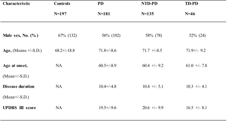
Following the classification procedure, we found 46 Tremor Do minant (TD-PD) and 135 Non Tremor Do minant (NTD-PD) PD patients. Patient groups (NTD-PD vs. TD-(NTD-PD) were co mparable with regard to age, age at onset and disease duration. A statistically significant difference in motor performance assessed with UPDRS part III at last visit was observed between the two PD groups (NTD-PD 20.6±9.9 vs. TD-PD 16.5±8.1; p<0.05) (Table 1).
Six MAPT htSNPs were tested for association in all patients and control group. Allele frequencies for all SNPs were in Hardy-Weinberg equilibriu m, except for rs2471738, fo r which a marg inally significant deviation was seen in cases (p=0.02) but not in control subjects (p=0.9).
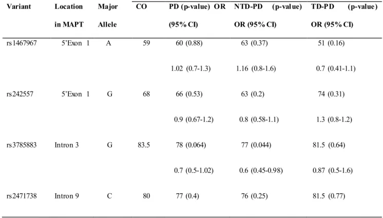
To test the relation between H1-SNPs and PD risk, we first performed a single-locus analysis of the MAPT genetic variants. Results are shown in Table 3. A significant overrepresentation of the H1 allele in the entire PD group (comp rising both NTD-PD and TD- 2 =9.9; OR, 1.7; 95% CI, 1.2-2.4; p=0.002) was detected. The association was greater in the PD subgroup of NTD patients compared with controls (82 vs. 2 =13.6; OR, 2.03; 95% CI, 1.4-3; p=0.0003) and remained significant after correction for multiple testing (pcorrect=0.008), while no statistically significant difference was disclosed between PD subgroup of TD patients and
2 =0.17; OR, 1.1; 95% CI, 0.7-1.9; p=0.7).
Among the other SNPs, only the rs3785883 was marg inally statistical significant in the NTD -PD subgroup compared 2 =4.3; OR, 1.5; 95% CI, 1.02-2.3; p=0.044), but this difference d id not remain significant after statistical correction considering the number of SNPs analyzed.
To clarify the association found between PD and the MAPT H1 variation and to assess whether any of the H1 subclades previously described (Myers et al., 2005; Pittman et al., 2005) could be influencing P D risk or motor phenotypes, we performed a haplotype association study comparing MAPT subhaplotype frequencies between the
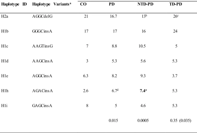
different group of patients and controls. On the H1 background, 20 subhaplotypes were identified with a frequency of ≥1% and only those with frequencies >5% in at least one of the groups in study were considered for the analysis. A total of seven subhaplotypes were selected (Table 4). Significant overall d ifferences in selected subhaplotype frequencies (defined by the tagging SNPs rs1467967, rs242557, rs3785883, rs2471738, del-in9, rs7521) were found between PD patients and controls (p=0.014), between NTD-PD and controls (p=0.0005) and between TD-PD vs. NTD-PD (p=0.035), while no statistically significant difference was detected between controls and TD-PD patients (p=0.35).
The MAPT H2a haplotype was significantly underrepresented in the NTD-PD subgroup compared with controls (p=0.024; OR, 0.6) and with TD-PD patients (p=0.018; OR, 0.5), suggesting a protective effect.
Detailed analyses showed a significant difference in the frequency of the H1h subhaplotype in the PD group (p=0.013; OR, 2.6) co mpared with controls. However, the difference was greater in the subgroup of NTD-PD patients (p=0.007; OR, 2.9). After correction for the number o f haplotypes analyzed only this last difference remained statistically significant.
The pairwise linkage disequilibriu m analysis (LD) for all SNPs was performed in the control and PD groups. The LD plot showed in the Fig 1, indicates in PD patients, that alleles at rs242557, rs3785883, rs2471738 and rs7521 are in strong LD with the del-in9 (marker 5), but are not in strong linkage with each others, indicating that these markers are H1 specific.
Discussion
Analyzing the role of MAPT locus in neurodegenerative disorders, such as PD, represents an effort to elucidate the interaction between genetics and functional disease outcomes. Although several studies including ours have found an association between the H1 haplotype and PD, the functional role of this variant still remains to be identified.
This paper aimed at refining the MAPT role in PD by examining the architecture of the entire gene in order to determine its possible associations with PD, and PD motor phenotypes, in a cohort of patients of Italian ancestry. Our results are consistent with the growing body of evidence that supports the MAPT H1 haplotype as a risk factor for sporadic PD. Moreover, we observed a peculiar risk distribution of MAPT haplotype, and H1 subhaplotype, according to PD motor phenotype. Indeed, wh ile NTD-PD subgroup showed a statistically significant overrepresentation of H1 clade, no differences were observed between TD-PD subgroup and control subjects. Among the other SNPs analyzed, the rs3785883 poly morphis m was no minally significant, exclusively in the NTD-PD subgroup compared with controls. This finding is consistent with a prev ious study where a moderate association at SNP rs3785883 was also found in a
Greek cohort of PD patients (Fung et al., 2006). With regard to MAPT subhaplotype, we found that H1h was associated with PD and this association remained statistically significant after correction for multiple testing when NTD -PD subgroup was considered. To our knowledge this is the first study comprehensively assessing MAPT locus in PD according to clinical motor subtype and the strong points of this study are the regular motor clin ical assessments for all patients included in the cohort.
The results of our study potentially raise two orders of consideration: the first report of a significant PD risk of the H1 haplotype in a PD subgroup, namely NTD-PD and the original finding of an association between the H1h subhaplotype and the same PD clinical subtype, in a cohort of Caucasian European ancestry.
At present, phenotype-genotype association studies that analyze the role of MAPT haplotypes on PD are mostly focused on cognitive profiling. These researches overall indicate an involvement of H1 haplotype on specific cognitive domains such as memory and visuo-spatial functions (Williams-Gray et al., 2009). According to these studies, MAPT variation influences cognition and the function of specific brain circuitry even in early phases of PD (No mbela et al., 2014) and even in healthy control subjects (Winder-Rhodes et al., 2015). Although the relationship between MAPT haplotype and cognitive functions remains to be determined since no specific regional degeneration or neurochemical alterations have been provided, the effect seems to be detectable even in relative small number of subjects.
Unlike many other quantitative phenotypic traits, there are now ev idences suggesting that clinical motor phenotype of PD may not represent a mere semiological matter. Indeed, while the
TD-PD patients could be considered a subgroup with a benign clinical course and a slower process of degeneration at least for the most part of the d isease course (Selikhova et al., 2009; Eggers et al., 2012; Deuschl 2013; Selikhova et al., 2013), NTD-PD are likely mo re prone to develop a series of motor and non motor co mp laints, inherent to the spreading of the degenerative process (Rosenberg-Katz et al., 2013; Zhang et al., 2013; Herman et al., 2014; Solla et al., 2015).
Interestingly, the contribution of MAPT gene in motor impairment has been described in a large co mmun ity-based cohort of neurologically healthy aging individuals (Shulman et al., 2014), where an association between H1 haplotype and mild parkinsonian signs, especially bradykinesia, has been observed without evidence of PD hallmark at pathological assessment. The authors speculated that neuroanatomical dysfunction of cortico -nigro-striatal pathways, different fro m those classically observed in PD, may contribute to the development of parkinsonian signs. Therefore, given the prevailing view o f H1 haplotype as a genetic risk factor fo r neurodegeneration, we can speculate that H1 background may partake to the exp ression of a PD clinical motor subtype associated with increased functional disability.
On the other hand, given the overall underrepresentation of H2 haplotype in a range of neurodegenerative disorders and assuming that this haplotype is associated with more efficient brain function, our observation of a significant overrepresentation of H2 in TD-PD may indicate a protective role of the H2 haplotype. So me studies
assessed the role of MAPT variants in gene expression (Hutton et al., 1998; Spillantin i et al., 1998; Caffrey et al., 2006; Pittman et al., 2006). So me authors reported that the disease risk conferred by MAPT variants could be related to a higher total or 4R tau levels. Additionally, a protective effect of MAPT H2 -haplotype due to an increase espression in N-terminal exon-containing MAPT transcripts has been speculated. …….
Although several lines of evidence, including basic researches, pathological findings and genotype-phenotype association support the role of MAPT haplotypes in PD, the exact mechanistic model of this lin k remains to be determined. Moreover, the pathological findings related to MAPT background in neurodegenerative disorders are partially conflicting. Indeed, while some studies conducted on PSP hu man brain indicate that H1 haplotype does not affect the pathological or b iochemical phenotypes (Liu et al., 2001), other found an higher expression of 4R-tau fro m the H1 haplotype compared to H2. Other authors observed that the MAPT H1 haplotype enhances the overall α-synuclein deposition type pathology in dementia with Lewy Body (Colo m-Cadena et al., 2013) and Alzheimer Disease (Wider et al., 2012).
Nevertheless, no comprehensive assessments of α-synuclein burden or Alzheimer-like pathology according to MAPT haplotype are currently available in PD; therefore, systematic and well-powered analysis exp loring the functional outcomes of MAPT region variations remain mandatory.
Moreover, we found that a specific subhaplotype, the H1h variant, was overrepresented in our PD population, more significantly in NTD-PD patients.
A number of studies have been performed for association of MAPT subhaplotypes variability and PD with inconsistent results (Fung et al., 2006; Vandrovcova et al., 2009; Seto-Salv ia et al., 2011). Moreover, other studies analyzed just few of the SNPs described to characterize the H1 specific subhaplotypes (Fidani et al., 2006; Winkler et al., 2007; Das et al., 2009; Refenes et al., 2009; Huang et al., 2015; Wang et al., 2016).
Given the concern that H1h variants may be driven by the genetic architecture related to ethnicity (Fung et al., 2006), it is worth considering that a b iological link between such variant and neurodegenerative process has been formerly reported. Indeed, it has been found in Alzheimer’s disease (AD) that the A -allele of the rs3785883 SNP is associated with increased cerebrospinal flu id (CSF) tau levels and tau mRNA exp ression (Kauwe et al., 2008). Moreover, the authors observed that the MAPT genetic variation defined as H1h subhaplotype showed significant elevation of CSF tau compared with the H2 haplotype. The contribution of the A -allele of the rs3785883 to tau exp ressions in AD has been subsequently confirmed in a larger series (Allen et al., 2014). Therefore, our findings may be congruent with, and complementary to these reports considering that NTD-PD patients showed significantly higher levels of CSF tau protein and tau/beta index if compared to TD-PD (Jellinger 2012; Prikrylova et al., 2012).
If confirmed, this result may indicate that a specific H1 subhaplotype increases the risk of developing a NTD-PD disease, at least in populations of South European ancestry.
Our study supports the hypothesis that genetic variability in the MAPT region is involved in PD susceptibility and may contribute to PD phenotypic expression, confirming that large-scale evaluation in different populations could be relevant to understand the role of population-specific heterogeneity.
Given the findings that H1 may act synergistically with other gene variants in determining risk for PD, future researches should concern with gene–gene interactions to provide critical insights into mechanisms of disease susceptibility. Nevertheless, the genetic architecture of MAPT in determining PD phenotypic expression as well as the possible functional effect of H1h subhaplotype deserves attention
Figure 1. Lin kage disequilibriu m (LD) between the MAPT H1 genotyped SNPs in our PD group. The relative position of the MAPT H1 tagging Single-Nucleotide Po ly morphisms, SNPs, is shown (top). Within each diamond the pairwise standardized coefficient of LD (D′ values x 100) are presented.
Table 1. Demographic and clinical characteristics of patients and controls.
Characteristic Controls N=197 PD N=181 NTD-PD N=135 TD-PD N=46
Male sex, No. (% ) 67% (132) 56% (102) 58% (78) 52% (24)
Age, (Means +/-S.D.) 68.2+/-18.8 71.8+/-8.6 71.7 +/-8.5 71.9+/- 9.2 Age at onset, (Mean+/-S.D.) NA 60.5+/-8.9 60.4 +/- 9.2 61.0 +/- 7.8 Disease duration (Mean+/-S.D.) NA 10.4+/-4.8 10.4 +/- 5.1 10.3 +/- 4.1
UPDRS III score NA 19.5+/-9.6 20.6 +/- 9.9 16.5 +/- 8.1
Table 2 Primer sequences and restriction enzymes used for detection of the investigated polymorphisms.
Gene Polymorphism (alleles)
PCR primers (5’- 3’) Enzyme (fragment bp)
Rev: GGCTCCACCCTTCA GTTTTGGA
rs242557 (A/G) Fwr: CTTGATGATGCATGGA CCTCTC
Rev: TTGACAGTACCCACGACA CGTG
ApaHI (211/ 139, 72)
rs3785883 (A/G) Fwr: CCATCACCTTGTCA GAAACTC
Rev: AGCCATGTGGTA GCCTCA G
BsaHI (277/ 164, 113)
rs2471738 (T/C) Fwr: CTCTCTGGACCCTCATCCACC
Rev: GAGAACCGAATGA GGA CTGGAA
BstEII (170/ 104, 66)
rs7521 (G/A) Fwr: ACCTCTGTGCCA CCTCTCAC
Rev: AGGTGAGGCTCTA GGCCA GT
PstI (231/ 160, 71)
Fwr: forward primer; Rev: reverse primer.
Table 3. MAPT single SNPs association results.
Major Allele Frequency Variant Location in MAPT Major Allele CO PD (p-value) OR (95% CI) NTD-PD (p-value) OR (95% CI) TD-PD (p-value) OR (95% CI) rs1467967 5’Exon 1 A 59 60 (0.88) 1.02 (0.7-1.3) 63 (0.37) 1.16 (0.8-1.6) 51 (0.16) 0.7 (0.41-1.1) rs242557 5’Exon 1 G 68 66 (0.53) 0.9 (0.67-1.2) 63 (0.2) 0.8 (0.58-1.1) 74 (0.31) 1.3 (0.8-1.2) rs3785883 Intron 3 G 83.5 78 (0.064) 0.7 (0.5-1.02) 77 (0.044) 0.6 (0.45-0.98) 81.5 (0.64) 0.87 (0.5-1.6) rs2471738 Intron 9 C 80 77 (0.4) 76 (0.25) 81.5 (0.77)
0.87 (0.6-1.2) 0.8 (0.5-1.2) 1.12 (0.6-2) del-in9 Intron 9 H1 69.5 79.5 (0.002) 1.7 (1.2-2.4) 82 (0.0003) 2.03 (1.4-3) 72 (0.7) 1.1 (0.7-1.8) rs7521 3’exon 14 G 55 53 (0.6) 0.92 (0.7-1.2) 52 (0.47) 0.89 (0.6-1.2) 55 (1) 1.03 (0-6-1.6)
Abbreviations: CO, controls; PD Parkinson Disease; PD-NTD, Parkinson Disease Non Tremor Do minant; PD-TD, Parkinson Disease Tremor Do minant; OR, odds ratio; 95% CI, Confidence Interval. Boldface values indicate significant results after Bonferroni correction for multiple testing (pcorrect=0.008).
Table 4. MAPT haplotype association results.
Haplotype Frequency
Haplotype ID Haplotype Variantsa CO PD NTD-PD TD-PD
H2a AGGCdelG 21 16.7 13b 26c H1b GGGCinsA 17 17 16 24 H1c AAGTinsG 7 8.8 10.5 5 H1d AAGCinsA 3 5.3 5.6 5.3 H1e AGGCinsA 6.3 8.2 9.3 3.7 H1h AGACinsA 2.6 6.7d 7.4e 5.3 H1i GAGCinsA 8 5 4.6 5.3 0.015 0.0005 0.35 (0.035)
Abbreviations: CO, controls; PD, Parkinson Disease; PD-NTD, Parkinson Disease Non Tremor Do minant; PD-TD, Parkinson Disease Tremor Do minant; MAPT, microtubule-associated protein tau. OR, odds ratio; 95% CI, Confidence Interval.
aAlleles for the SNPs defining the haplotype are given in the 5’ to 3’ order as follows: rs1467967, rs242557, rs3785883, rs2471738, del-In9, and rs7521.
b0.024, OR 0.6; 95%CI 0.4-0.9: NTD-PD vs. CO. c0.018, OR 0.5; 95%CI 0.28-0.87: NTD-PD vs. TD-PD. d0.013, OR 2.6; 95%CI 1.2-5.5: PD vs. CO.
e0.007, OR 2.9; 95% CI 1.3-6.3; NTD-PD vs .CO. (Boldface value indicate significant result after Bonferroni
correction for multiple testing, pcorrect = 0.00714).
Nu mbers on the last line indicate p values that result from g lobal haplotype frequency comparison between each group of patients and controls, while the p value in brackets results from co mparison between TD-PD and NTD-PD patients.
Clinical setting : Intercepting Parkinson disease nonmotor subtypes: a proof-of-principle study in a clinical setting.
Background
Parkinson’s disease (PD) is a progressive neurodegenerative disorder, with proteiform clinical spectrum and scarcely predictable evolution (Di Battista, 2014). A great effort has been engaged to define homogenous groups in order to intercept a pathophysiological coherence and prognostic trajectories of the disease. To this purpose, the identification of non-motor domains remains a priority for evaluation and managing of PD patients. First, non-motor symptoms (NMS) are integral to the disease and complementary to the diagnostic procedure. Second, identifying NMS subtypes may have a possible prognostic value. Th ird, fro m a research point of view, NMS are significant determinants of etiopathological studies. Finally, and most important, the burden of NMS highly affects the health -related quality of life of PD patients. Different models of PD non-motor subtyping have been recently proposed in literature. So me authors have postulated that certain symptoms tend to aggregate in specific clusters following an anatomo -clin ical correlation model (Marras 2016). According to this v iew, the occurrence of NMS clusters may be exp lained by the presence of different stages which underlie the pathological process observed in PD patients. Based on this “anatomo -clinical model” Chaudhuri and Marras (2016) have identified three main subtypes of nonmotor profile: a brainstem subtype characterized by the prevalence of sleep and autonomic dysfunctions, a limb ic variant with depression, fatigue and weight loss, and, a cognitive subtype, with a particular predo minance of cholinerg ic dysfunctions such as memory impairment, apathy and anxiety.
a. a brainstem subtype characterized by a prevalence of sleep dysfunction and autonomic control, b. a limbic variant with depression and fatigue ad weight loss
c. a cognitive subtype, with a particular predominance of cholinergic dysfunctions as memory impairment, apathy and anxiety.
( Marras and Chaudhuri, 2016)
Using another approach, other groups investigated the correlation between motor phenotypes and the overall burden of NMS. However, the results of these studies based on empirical “motor model” of NMS profile are partially conflicting (Kadastik-Eerme, 2016). Khoo et al found that a greater number of NMS is associated with the PIGD phenotype in newly diagnosed nondemented PD patients (2013). Constipation, autonomic and sensory symptoms were found prevalent in the non-tremor do minant (NTD) subtype by Muller et coll. (2011). In contrast, other authors did not find a higher overall burden of NMS in the akinetic -rigid phenotype compared with tremor dominant (TD) indiv iduals within an incident PD cohort.
Another aspect of great interest is the potential contribution of the genetic background to the prevalence and progression of NMS in PD; to date, previous researches have mostly focused on cognitive decline, which is one of the most disabling non-motor co mplication of latest stages of PD. Convergent data suggest a role for MAPT genotype in Parkinson’s disease dementia (PDD) (Robakis 2016). However, the possible association of MAPT genotype (in particular H1/H1 ho mo zygous vs H2 carriers) to non-motor phenotypes has not yet been investigated; this finding could support a “genetic model” of NMS subtypes.
Whatever the NMS model (“anato mo-clinical”, “motor” or “genetic”), it should be based on its relevance in common clinical practice.
Our study aims to investigate whether in a sample of PD patients a genetic model according to the genotype MAPT distribution of NMS subtypes can be argued; moreover, we proposed to conduct a cluster analysis on NMS pattern of our cohort.
Methods
Subjects
We included in our cohort PD patients consecutively afferent to the outpat ients Parkinson’s Disease Unit of Sapien za University of Ro me. All subjects fulfilled the UK Brain Bank criteria for idiopathic PD (ref). Indiv iduals were excluded if they had signs of atypical parkinsonism, dementia and/or doubtful response to dopaminergic replacement therapy. The study protocol was approved by the XXX Hospital Ethics Co mmittee and all patients gave their informed written consent.
Patients assessment
We collected demographic data (age, gender and education), neurological history (age a t onset of PD, duration of disease and specific treatments); the levodopa equivalent daily dose (LEDD) was also calculated.
All patients underwent a clinical and a neuropsychological examination and a genetic analysis.
Clin ical evaluation. Three neurologists with expertise in movement disorders (MD, A R, XX) investigated PD patients using motor scales, such as Unified Parkinson’s Disease Rating Scale (UPDRS) part III and V (Hoen and Yahr Scale [H&Y]), freezing of gait questionnaire (FOGq) and non motor scales, such as Non Motor Symptoms Scale (NMSS) to assess frequency and severity of a wide range of NMS (ref NMSS+ ref NOSTRO A RTICOLO!!), Autonomic Scale for Outcomes in Parkinson’s disease-Motor (SCOPA -Aut) for evaluate dysautonomic dysfunctions and Epworth Sleepiness Scale (EPSS) for sleep disturbances evaluation. Three main motor phenotypes have been considered: tremor-do minant subtype (TD), non-tremor dominant subtype (NTD) and postural gait and instability disorder (PIGD) subtype
Genetic analyses.
MAPT haplotype was determined in this cohort as described above.
Data analysis
Descriptive statistics were used for the characterization of the sample, by means of the univariate ANOVA test for continuous variables and the Pearson’s χ2 test for categorical variables.
To explore currently proposed NMS clusters of PD (ref), we performed a correlation analysis by means of Pearson’s correlation coefficient (r) of the following NMS: dysautonomic sympto ms (total score of SCOPA aut) with respect to sleep disorders and to fatigue (sleep/fatigue do main of NMSS) in order to investigate a “brainstem variant”, depression (total score of BDI) with respect to anxiety and apathy (subscores of NPI) to investigate a “limbic variant”, cognitive functions (total score of MOCA) with respect to anxiety and apathy (subscores of NPI) to investigate a “cognitive variant”.
We explored the relationship among the different NMSS domains with other measures for the same construct (or other related constructs) and motor scores, respectively,
In order to reduce data originating fro m raw scores of several NMS scales (investigating some overlapping sympto ms), we perfo rmed a factor analysis with varimax rotation; we have excluded scores with a poor dispersion of the values. We performed then a hierarchical cluster analysis by using the factors identified as parameters.
Standard values for acceptability and reliability were established based on previous studies (ref). Statistical analyses were performed by means of the Statistical Package for the Social Sciences (SPSS 23).
Results
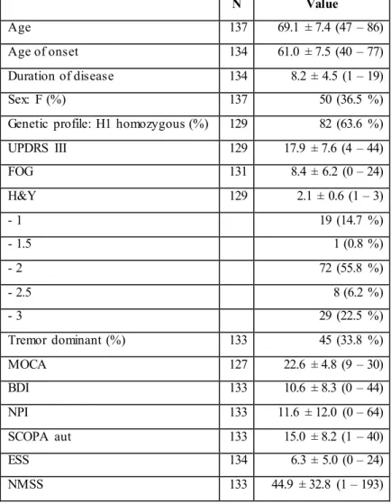
One hundred and thirty-seven patients with PD were included in the study. Table 1 shows demographic, clinical characteristics and scores distribution of motor and non-motor scales of the whole sample.
Ho mozygous and heterozygous H1 PD patients did not differ with respect to age of onset, duration of disease, severity of motor sympto ms investigated by UPDRS part III and H&Y scale, motor phenotypes, total score of all the scales used to assess NMS and the subitems of NMSS.
Considering motor phenotypes, we did not found any significant difference between motor and non -motor scales, except for a lower total score of MOCA (F 13.317, p < 0.001) and a higher FOG score (F 8.908, p< 0.001) in PIGD group with respect to other phenotypes (TD and NTD).
In the attempt to verify the anatomo-clin ical model proposed by Chaudhuri et al. , we have observed a significant positive relation between sleep/fatigue domain of NMSS and dysautonomic symptoms (R = 0.452, p< 0.001) and depression (R = 0.620, p < 0.001), and between cognitive functions and apathy (R = – 0.289, p = 0.001).
We have checked this last finding by mean o f an univariate ANOVA test using total score of MoCA as a continuous variable and the subscore for apathy of NPI as a factor: th is further analysis confirmed previous results (F = 3.546, p
= .017).
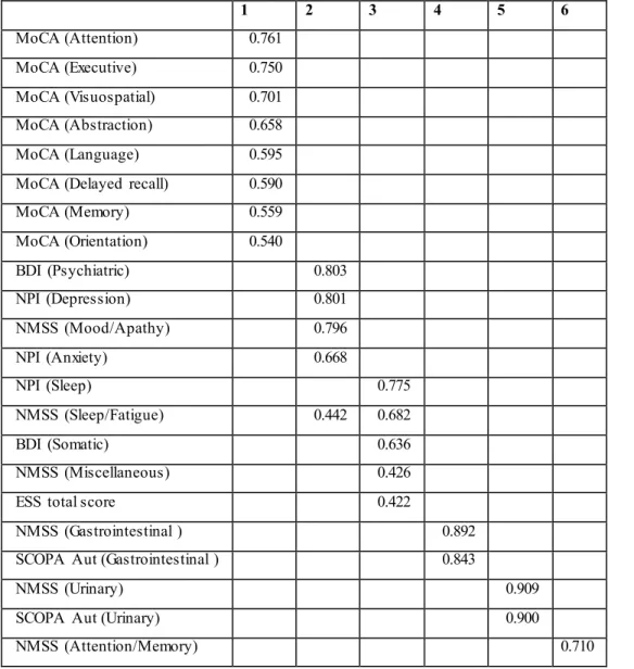
Factor analysis has identified six factors, as shown in table 2, which can be su mmarized into: 1= cognitive functions 2= mood d isorders 3= sleep dysfunctions 4= gastrointestinal disorders 5 = uro logical disorders 6= concern of their own cognitive impairment. No statistically significant differences has been found between these six factors and aplotype. Through hierarchical cluster analysis using the 6 above-mentioned factors as parameters, we sought to identify cluster of at least 10 people; we have explored the possibility of generating fro m 3 to 20 clusters. Only the choice to divide the samp le in 10 clusters follo wed 3 groups of 83 (cluster 1), 13 (cluster 4) and 11(cluste r 7), subjects; the other 7 clusters consisted in a small number of individuals.
No statistically significant differences in aplotype has been found in the 10 clusters explored.
The three main clusters were similar in terms of age, sex, d isease duration and cognitive functions (assessed by MoCA total score),
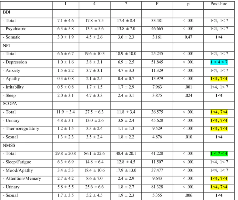
Statistically significant differences in NMS scales (total and subitems scores) among the three main clusters are reported in table 3. Overall, cluster 1, which is the most numerous group, showed a less severe motor, neuropsychiatric and functional impairment; cluster 4 had a greater motor and non-motor impairment and cluster 7 was found to be more depressed with respect to the other main clusters.
We performed then a sensitivity analysis with non parametric tests (Kruskal Wallis) given the s mall number of subjects in cluster 4 and 7 with respect to cluster ; differences encountered in BDI and NPI scores between the groups 4 and 7 were lost; significant statistically differences remain in memo ry / attention and urological subitems of NMSS, where cluster 7 achieved intermed iate scores between cluster 1 and 4. Therefore, cluster 7 seems to be more concerned about his own difficulties of memo ry and concentration than cluster 4, wh ile no significant diffe rences have been found in real cognitive performances at Mo CA. Moreover, total score of MoCA does not correlate with the score of memo ry / subitem attention of the NMS.
Table 1 Demographic data and assessment of motor and non-motor symptoms in patients with Parkinson’s disease. N Value Age 137 69.1 ± 7.4 (47 – 86) Age of onset 134 61.0 ± 7.5 (40 – 77) Duration of disease 134 8.2 ± 4.5 (1 – 19) Sex: F (%) 137 50 (36.5 %)
Genetic profile: H1 homozygous (%) 129 82 (63.6 %) UPDRS III 129 17.9 ± 7.6 (4 – 44) FOG 131 8.4 ± 6.2 (0 – 24) H&Y 129 2.1 ± 0.6 (1 – 3) - 1 19 (14.7 %) - 1.5 1 (0.8 %) - 2 72 (55.8 %) - 2.5 8 (6.2 %) - 3 29 (22.5 %) Tremor dominant (%) 133 45 (33.8 %) MOCA 127 22.6 ± 4.8 (9 – 30) BDI 133 10.6 ± 8.3 (0 – 44) NPI 133 11.6 ± 12.0 (0 – 64) SCOPA aut 133 15.0 ± 8.2 (1 – 40) ESS 134 6.3 ± 5.0 (0 – 24) NMSS 133 44.9 ± 32.8 (1 – 193)
Data are expressed as mean ± standard deviation (range) except where indicated
UPDRS III, Unified Parkinson’s Disease Rating Scale part III; FOG, Freezing Of Gate scale; H & Y, Hoen & Yahr scale; MOCA, Montreal Cognitive Assessment; BDI, Beck Depression Inventory; NPI, Neuropsychiatric Inventory ; , SCOPA-Aut, Autonomic Scale for Outcomes in Parkinson’s disease-Motor; ESS, Epworth Sleepiness Scale; NMSS, Non Motor Symptoms Scale.
Table 2 Factor analysis with varimax rotation of raw scores of different non-motor scales used to assess non-motor symptoms. 1 2 3 4 5 6 MoCA (Attention) 0.761 MoCA (Executive) 0.750 MoCA (Visuospatial) 0.701 MoCA (Abstraction) 0.658 MoCA (Language) 0.595
MoCA (Delayed recall) 0.590
MoCA (Memory) 0.559 MoCA (Orientation) 0.540 BDI (Psychiatric) 0.803 NPI (Depression) 0.801 NMSS (Mood/Apathy) 0.796 NPI (Anxiety) 0.668 NPI (Sleep) 0.775 NMSS (Sleep/Fatigue) 0.442 0.682 BDI (Somatic) 0.636 NMSS (Miscellaneous) 0.426
ESS total score 0.422
NMSS (Gastrointestinal ) 0.892
SCOPA Aut (Gastrointestinal ) 0.843
NMSS (Urinary) 0.909
SCOPA Aut (Urinary) 0.900
NMSS (Attention/Memory) 0.710
1= cognitive functions 2= mood disorders 3= sleep dysfunctions 4= gastrointestinal disorders 5 =
urological disorders 6= concern of their own cognitive impairment.
MOCA, Montreal Cognitive Assessment; BDI, Beck Depression Inventory; NPI, Neuropsychiatric Inventory ; , SCOPA-Aut, Autonomic Scale for Outcomes in Parkinson’s disease-Motor; ESS, Epworth Sleepiness Scale; NMSS, Non Motor Symptoms Scale.
Table 3 Comparative statistics for different non-motor symptoms scales (total and subitems scores)
among the three main clusters
1 4 7 F p Post-hoc BDI - Total 7.1 ± 4.6 17.8 ± 7.5 17.4 ± 8.4 33.481 < .001 1<4, 1< 7 - Psychiatric 6.3 ± 5.8 13.3 ± 5.6 13.8 ± 7.0 46.665 < .001 1<4, 1< 7 - Somatic 3.0 ± 1.9 4.5 ± 2.6 3.6 ± 2.3 3.161 0.47 1<4 NPI - Total 6.6 ± 6.7 19.6 ± 10.3 18.9 ± 10.0 25.235 < .001 1<4, 1< 7 - Depression 1.0 ± 1.6 3.8 ± 3.1 6.9 ± 2.5 51.845 < .001 1 < 4 < 7 - Anxiety 1.5 ± 2.2 3.7 ± 3.1 4.7 ± 3.3 11.329 < .001 1<4, 1< 7 - Apathy 0.3 ± 0.8 2.1 ± 2.5 0.4 ± 0.7 13.979 < .001 1<4, 7<4 - Irritability 0.5 ± 0.8 1.7 ± 1.5 1.7 ± 2.9 7.963 .001 1<4, 1< 7 - Sleep 2.0 ± 3.1 4.7 ± 3.3 2.4 ± 3.1 3.875 .024 1<4 SCOPA - Total 11.9 ± 3.4 27.5 ± 6.3 11.8 ± 3.4 36.575 < .001 1<4, 7<4 - Urinary 4.8 ± 3.1 13.0 ± 2.6 3.8 ± 2.4 45.628 < .001 1<4, 7<4 - Thermoregulatory 1.2 ± 1.5 3.3 ± 2.4 1.1 ± 1.3 9.529 < .001 1<4, 7<4 - Sexual 1.3 ± 2.3 3.5 ± 2.4 1.8 ± 2.2 4.876 .010 1<4 NMSS - Total 29.8 ± 20.8 86.1 ± 22.6 48.4 ± 20.1 41.228 < .001 1 < 7 < 4 - Sleep/Fatigue 6.3 ± 6.9 14.8 ± 6.4 12.8 ± 4.5 11.507 < .001 1<4, 1< 7 - Mood/Apathy 3.4 ± 5.3 18.4 ± 10.6 17.9 ± 13.0 37.477 < .001 1<4, 1< 7 - Attention/Memory 2.7 ± 4.2 8.6 ± 7.0 2.4 ± 2.9 9.643 < .001 1<4, 7<4 - Urinary 5.8 ± 5.5 25.6 ± 6.6 1.8 ± 2.7 81.328 < .001 1<4, 7<4 - Sexual 1.7 ± 3.5 5.2 ± 4.5 1.9 ± 2.3 5.355 .006 1<4
Discussion.
In our cohort of incident unselected PD patients, we were not able to intercept some of the clusters of PD nonmotor subtypes recently proposed in literature. None of the models tested in our cohort exh ibit statistical evidence of a cluster coherence within the nonmotor symptoms spectrum.
Overall, these results likely confirm the complex pathophysiology of PD where no single bio logical mechanism is the solely determinant.
Although the discrete clusters described in literature are not recognizable, in our population some sympto ms of the anatomical model tend to correlate; indeed, we observed a significant correlation between fatigue/excessive sleepiness and depression or cognitive functions and apathy. It is worth considering that these associations have been frequently reported in literature, possibly reflecting a phenotypic trait rather than the expression of pathogenicity in the anato mical model.
Moreover, the lack of association between MAPT haplotypes and a specific profile in the whole spectrum of nonmotor symptoms may suggest that MAPT background partake to the “cortical side” of PD. Indeed, MAPT H1 haplotype, and in some cases specific subhaplotypes, have been associated with dementia, motor subtypes and recently with cortical load of Lewy body, which are cortical pertinent aspects.
When we analyzed the correlation between motor phenotype (Tremor dominant, mixed forms, and PIGD patients) and the nonmotor profile we were not able to find any significant correlation. In our cohort no specific nonmotor profile was found in association with the empirical model of PD motor syndromes. Although PD motor phenotypes probably present different biological basis and are relevant in terms of prognostic value, it is possible to speculate that the route of progression of motor d isability follows degenerative trajectories that are only partial superimposable to those of nonmotor phenomenology. Furthermore, the motor subtypes attribution may not take into acco unt for the instability of these subtype, especially when patients are classified fo llo wing an adequate observational period. Finally, it is worth to consider that after the initial and intermed iate stages of the disease duration the majority of cases tend to converge in a more complex motor syndrome.
The cluster analysis performed in our cohort identified three main groups of patients, of different nu merosity, that share a similar nonmotor symptoms profile:
group number 1, the vast majority of patients, that express a moderate burden of all nonmotor symptoms,
group number 4, patients with severe dysautonomic sympto ms, apathy, excessive sleepiness, urologic and sexual dysfunctions;
and finally group number 7, with a prominent depressive phenotype despite a relative benign form of PD.
The three groups are comparab le in terms of age, disease duration and age at onset, patients have also similar d isease motor impairment.
Given that these nonmotor phenotypic traits tend to aggregate in patients with similar disease duration, we may assume that they clinically reflect the dysfunction of the neurotrasmettitorial networks involved beside the ineluctable dopamine depletion.
In our “network-based model”, patients of group 1, which are the majo rity of PD patients and represent he main phenotypic PD trait, have a classical dopamine related syndrome or are in a state of the malady were dopaminergic depletion still underlies for the majority of symptoms.
In group 4, the presence of prominent autonomic sympto ms, apath y, sleep dysfunctions, urologic and sexual dysfunctions may be the expression of a cholinergic and autonomic systems failure attendant to the dopaminergic syndrome. This minority of patients display also display a statistically significant presence of FOG, wh ich can be considered a straddling cognitive-motor sympto m. Most of these symptoms are potentially treatable and the identification of autonomic, cognitive or FOG related may yield to a better treatment.
Patients in phenotypic trait 7 present an overall intermediate burden of nonmotor sympto ms but a higher degree of depression, fatigue and sleep disturbances. Such phenotypic trait may be ascribable, according to our network based model, to a louder monoaminerg ic depletion. Indeed, findings from clinical, neuro imaging, and animal studies shows that dopaminerg ic system is the major contributor to the pathophysiology of these symptoms; nonetheless, the degeneration of noradrenergic and serotonergic projection systems also has an impact on psychiatric sympto ms of PD. Whether depressive symptoms are at risk for subsequent cognitive collapse or motor worsening remains a matter of debate, with current available studies showing partially conflicting results.
In conclusion, the overall results of our study suggest that nonmotor subtyping may not be feasible in a real life clinical setting and the vast majority of patients with intermediate stages of disease do not correspond to a specific cluster or a clin ico-pathological subsyndrome of PD. However, searching for autonomic-cognitive dysfunctions (trait number 4) and depression (trait number 7) may allow to intercept phenotypic traits of the disease with potentially treatable complaints that juxtapose motor impairment.
We are aware o f the relative small nu merosity of our cohort, however, the lack of stringent selection criteria and the incident quality of the cohort probably resemble a real life ambulatory population of PD patients. Furthermo re, our patients were assessed with a co mprehensive battery of scales and questionnaire to examine the whole spectrum of nonmotor symptoms.
Given the potential relevance of nonmotor subtypes, a possible perspective is to standardize a clin ical and instrumental work-up to define uniformly nonmotor PD subtypes.
Experimental setting II: DAT Methylation in Parkinson Disease: Correlation with peripheral Dopamine Transporter geneexpression and central activity
The role of epigenetic mechanisms in the function and homeostasis of the central nervous system and their participation in the neurological d isorders remain an intriguing feature. The epigenetic phenomenon has been defined as “the study of changes in gene function that are mitotically and/or meiotically heritable and that does not entail a cha nge in DNA sequence”. DNA methylation is the best-understood epigenetic modification modulating transcriptional plasticity. It refers to binding a methyl group to a Cp G islands in the genomic sequence. Methyl groups bound to the genomic sequence reduce the DNA binding capacity for transcription factors. However, in some cases methyl groups are ab le to enhance transcription factors attachment to promoter regions.
The correlation between DNA methylation and neurodegenerative diseases, and in particular in PD , was pointed out by some studies. (table, Haoyang Lu et al. 2013)
SNCA
SNCA (intron1 and promoter)
Hypermethylation Decreased expression of SNCA
Hypomethylation Overexpression of SNCA
Inflammatory cytokines
TNF-α Hypomethylation
Increased risk of apoptosis in dopaminergic neurons
Clock genes CRY1 NPAS2 Devoid of
methylation
Disorder of circadian rhythms
Telomere Subtelomeric region Constant methylation Telomere shortening
Other genes PARK16/1q32 GPNMB STX1B Methylation alteration PD risk
Cytochrome P45 2E1 Hypomethylation Increased PD susceptibility
Although these findings indicate the potential ro le of DNA methylation in the neurodegenerative diseases, the mechanisms remain inadequately characterized.
PD is a comp lex multiorgan clin icopathological entity, typically characterized by progressive degeneration of dopaminergic nigro-striatal pathways.
Considering the prominent role o f dopaminergic system dysregulation in Parkinson Disease, the epigenetic derangement of the dopamine metabolism determinants deserves a peculiar attention. In this term, Dopamine Transporter (DAT1) exerts a pro minent ro le in the process of dopaminerg ic striatal signalling. Given the evidence that mo lecular DAT imaging provides in v ivo a measure of dopamine terminal depletion in PD, we sought to explore the potential influence of DAT gene epigenetic modification on central and peripheral expression.
THE ROLE OF DAT1 IN PARKINSON'S DISEASE
The Dopamine Active Transporter-1 (DAT1), also known as SLC6A3, is a membrane-spanning protein that re-uptakes Dopamine (DA) from the synaptic cleft into the presynaptic neurons (Uhl, 2003).
This process is driven by the ion-gradient created by the Na+/K+-ATPase, two Na+ ions and one Cl- ion are transported with the substrate (Torres, 2003). The result of this process is the decrease of DA concentration in the synaptic cleft and and increase in the presynaptic neuron. DAT-KO mice were found to have a 300-fold increase in time of DA in the striatal extracellular space, a 5-fold increase of extracellular DA concentration and a 95% decrease in striatal intracellular DA concentration (Torres, 2003). As every monoamine transporter, DAT is expressed almost only in cells that contain its cognate neutrotransimetter. In DAT's case, the highest concentrations of this protein are found in the cell bodies of neurons of the Ventral Tegmental Area (VTA) and pars compacta of the Substantia Nigra (SNpc) of the mesencephalic brain steam.
Psychostimulants that enhance locomotor activities interact with DAT. Cocaine and Amphetamine act has competitive inhibitors of DA uptake (Uhl, 2003; Torres 3003).
DAT has also been found to be a transporter for 1 -methyl-4-phenylpyridinium ion (MPP+), a product of the extracellular metabolisation of N-methyl-4-phenyl-1,2,5,6-tetrahydropyridine (MTPT). MPP+ is Parkinsonism inducing neurotoxin that causes death specifically in Dopaminergic neurons.
Kitayama et al studied the correlation between MPP+ toxicity and MPP+ tra nsport in cell-lines expressing wild-type and mutant DAT1 genes. They discovered not only that cell-lines insensitive to high concentrations of MPP+ became susceptible when expressing DAT, but also that the sensibility to MPP+'s toxicity was parallel to an increase in DA
uptake velocity. Analyzing MPP+ toxicity among the different DAT mutants, Kitayama and colleagues concluded that the toxicity was directly correlated to the affinity of the transporter (Km) for MPP+, rather than the velocity of uptake.(Kitayama 1998)
It has also been suggested that DAT interacts with alpha-synucleine, increasing the activity of the transporter (Uhl,2003).
The human DAT/SLC6A3 gene is located in the chromosome 5p15.3 and is divided in 15 exons. In the 3' untranslated region of the gene there is a Variable Number of Tandem Repeat (VNTR) of approximately 40 bp. The VNTR copy number can be between three and eleven, with the most frequent VNTR copy number being nine (9R) and ten (10R) (Vanderbergh, 1998). At least fifty Single Nucleotide Polymorphisms (SNPs) have also been discovered in the 5' regions of the gene and, in vivo , two haplotypes were found that distinguish different levels of [11 C] cocaine binding (T-841C/rs2652511/C-839T and A11821G/rs2937639/G11736A) (Drgon,2006).
Moreover, the 10R VNTR was associated with a higher gene expression in vitro (Fuke, 2006). It has been, therefore, argued that the polymorphism in 3' and 5' could influence the gene expression and DAT availability in the Striatum. Van de Giessen and coll. measured striatal DAT availability using 123I-(2-b-carbomethoxy-3-b(4-iodophenyl)-tropane) (123I-b-CIT). High affinity for DAT-SPECT in healthy subjects that expressed the 40 bp VNTR and specific SNPs in the 5' end of the gene. Further studies observed that the 9R allele repeat was associated with a higher striatal DAT concentration compared to the 10R homozygotes; on the other hand, in this report did not emerge any association bewteen the 5' SNPs and DAT availability. (Drgon,2006)”Haplotype analysis revealed that the identified effect of the 9R allele seems to be caused mainly by a subgroup of 9R carriers, the T-A-9R (rs2652511– rs2937639–VNTR) ” (van de Giessen, 2009).
A meta-analysis, reviewing studies on striatal availability of DAT through SPECT both in healthy controls and in neuro-psychiatric patients, moderated previous results. The author observed that while there was an increase in DAT availability in certain haplotypes, this was not statistically significant and that the majority of the previous studies were underpowered, concluding that while there might be a correlation between VNTR and 5' polymorphisms and DAT availability in the striatum, this needs to be further investigated (Costa, 2011).
The selectivity of the expression of DAT coupled with the death of DA neurons that strongly express DAT has led to the hypothesis of DAT's involvement in the pathogenesis of PD. If we co mpare the DAT concentrations in three areas of the brain that contain dopaminergic neurons: arcuate nulceus of the hypotha lamus, ventral tegmental area, Substantia Nigra pars compacta, we can see that it correlates with the degree of neuronal loss in PD “(SNpc > VTA > arcuate nucleus)” (Guzey, 2012; Uhl, 2003).
Consequently, polymorphisms in DAT/SLC6A3 might be involved in the pathogenesis of PD. Several studies have been conducted in order to observe a correlation between the 3' VNTR and the 5' SNPs and PD (Arai,1995; Le Couteur, 1997;Kim, 2000;Morino, 2000).
Le Couteur and colleagues, in a case-control study, found that the 11R allele was present in circa the 2,5% of patients and only 0,5% of the controls. This allele was therefore associated with a ten-fold increase in risk of developing PD between the two populations and could explain up to 5% of cases (Le Couteur, 1997). Kim and colleagues, in a similar study in a Korean population, partially confirmed previous results (2,5 fold increased risk) (Kim, 2000). Neither of these studies attempted to explain the involvement of DAT in the pathogenesis of PD.
Morino et al investigated the role of a specific SNP (exon 9 1215 A/G) in a PD Japanese coorte. The report suggested that the 1215 A/G SNP was associated with a decrease in susceptibility to PD (Morino, 2000). However further studies failed to replicate the results (Liu 2001,Kimura 2002).
In 2014 Zhai and colleagues reviewed 18 studies on 3' VNTR and 11 on SNP. The meta -analysis proved conclusively only that the 10R allele conferred protection against PD only in East -Asian populations but not in Caucasians. Other results were found to be the product of either statistically biased or underpowered studies. The review also analysed the combined effects of the four SNPs (rs6347, rs3756450, rs2652510 and rs2550956) in the SLC6A3 gene locus on risk for PD. The results showed that only rs2652510 genotype GG is associated with a higher PD risk in all population (Zhai,2014).
Several approaches confirmed that the dopaminergic neurons loss correlate decrease in DAT concentration in PD. Harrington et al (1996) and Counihan & Penney (1998) have examined DAT mRNA expression in both healthy controls and in PD patients in the Ventral Mesencephalon from post mortem samples. Both reports underline increase DAT mRNA expression in the SNpc of healthy donors if compared to PD patients. The reserchers detected also a marked decrease in DAT mRNA concentrations in the surviving neurons in PD patients, especially in the SNpc. Results that are consistent with the scientific literature on the matter. The marked reduction of DAT could be ascribable either 1) a compensatory mechanism for diminshed DA concentration in the synaptic cleft or 2) might just be the expression of a general decrease of overall activity in a dying cell (which parallels the decrease in level of transcription of TH, Tyrosine Hydroxylase, the rate limiting enzyme for the synthesis of DA) (Harrington,1996;Counihan & Penney,1998). The laboratory findings are supported by neuroimaging reports. SPECT with [123-I]beta-CIT and [123-I]FP-CIT, radioligands that selectively bind the dopamine transporter, are routinely used for detecting the carrier-protein decrease in vivo, corroborating the diagnosis of PD when non dopamine-deficient syndromes are considered ( i.e. dystonic and severe essential tremors), or for early PD detection (Innis,1993;Booij,1997, Brooks 2010).
The study of DAT, its polymorphisms and its involvement in the pathogenesis of PD has led to hypothesise its contribution also for PD treatment and, in particular, for the common disabling motor complication of this as levodopa-induced dyskinesia (LID).
A PET study suggested that LID development was associated with low levels of DAT expression in pre-synaptic terminals. These levels were significantly lower than DAT expression levels in patients with Motor Fluctuations (MF), suggesting an association between dyskinesias and DAT expression. Moreover the study suggested that the known risk factors for LID (age, disease duration, UPRDS) are “effect modifiers […] of a main determinating factor, that is, the downregulation of putaminal DAT”(Troiano,2009). A subsequent retrospective cohort study, that enrolled 127-drug naïve de novo patients with PD and conducted 18F-FP-CIT PET scanning, monitored the PD progression for a mean follow-up period of 3.4 years. The study demonstrated that patients with low baseline DAT levels have a significantly higher risk of developing LID, confirming the hypothesis that pre-synaptic dopaminergic denervation in the putamen, but not in the caudate, is crucial in the development of LID (Jin Yong Hong, 2014;Troiano 2009).
Much effort has been made in investigating the extent to which the various polymorphisms (VNTR and SNP) of DAT influence the response to levodopa and LID time latency (Altmann 2016; Kaplan, 2014; Contin 2004).
M. Contin et al. (2004) examined through [123I]-FP-CIT SPECT a cohort of PD patients that expressed 9R and 10R homozygotes VNTR and not only it did not find differences in DAT expression in the striatum, it did not also find that these polymorphisms influenced the response to levodopa nor levodop a-linked dyskinesias.
Another retrospective study tried to identify genetic factors associated with LID. The study analysed 14 SNP and VNTR in SLC6A3. After statistical analysis it was proved a significant association between the intronic SNP rs393795 and a higher latency to LID onset (time ratio = 4,96) (Kaplan,2014).
A recent paper addressed the issue of the high inter-individual fluctuation of the response to levodopa, trying to determine how much of it was ascribable to “biological, pharmacological and genetic factors” (Altmann,2014). The study analyzed the 3' UTR VNTR and tried to assess the response to levodopa of the various genotypes and found the minimum levodopa dosage at which every allele responds. The subset of patients with 9R/9R VNTR genotype needed less levodopa than any other allele. This seems consistent with previous studies that showed that the age -related DAT expression decreased was higher in 9R/9R. It was therefore hypothesised that, expressing lower levels of DAT, these patients would have higher DA concentration in the synaptic cleft and need less levodopa. The authors concluded that their paper has to be considered a first attempt and not a definitive guide towards a “pharmacogenetic algorithm for levodopa dose prediction” (Altmann,2014).
Methods
Subjects. Un related sporadic PD patients are consecutively recruited fro m the Parkinson outpatient centre of the Sapienza University of Ro me and fulfilled the UK Brain Bank criteria fo r PD. Inclusion criteria included: 1) stable dopaminergic treatment 2) Mini Mental State Examination (MMSE) score >26; 3) no dementia according to the Movement Disorder Society (MDS) clinical d iagnostic criteria (Emre et al, 2007) using an extensive neuropsychological battery; and 4) suitability for DAT-SCAN and MIBG. Exclusion criteria included: 1) h istory of neurological d iseases other than idiopathic PD; 2) unclear h istory of chronic dopaminerg ic treatment responsiveness; 3) presence of major non stabilized medical illnesses (i.e. non stabilized diabetes, obstructive pulmonary disease or asthma, hematologic/oncologic disorders, vitamin B12 o r fo late deficiency, pern icious anemia, clinically significant and unstable active gastrointestinal, renal, hepatic, endocrine or cardiovascular disorders and recently t reated hypothyroidism); 4) known or suspected history of alcoholism, drug dependence and abuse, head trauma and mental disorders (apart from mood or an xiety disorders) according to the DSM -IV TR criteria; and 5) presence of vascular brain lesions, brain tumor and/or marked cortical and subcortical atrophy on MRI scan.
The sample’s clinical records were reviewed and patients were assessed by neurologists who are experts in movement disorders; the patients’ clinical and demographic data are shown in Tab le belo w. The variables collected included: demographic data, age at onset, disease duration, familiarity for PD, total levodopa equivalent dose (LED) and UPDRS III at the time of last visit. The patients were finally classified in two clinical subtypes, by usin g criteria published in previous studies (Selikhova et al. 2009):
(a) tremor do minant (TD), i.e. patients with tremo r as the only motor sign at onset or tremor as the pro minent motor symptom according to the UPDRS part III
(b) non-tremor do minant (NTD), i.e. patients with predominant rigid ity and bradykinesia but no tremor or only mild tremor at rest.
Genotyping
10 ml of blood samples in EDTA were taken from the participants and used for DNA and RNA extractions.
Pyrosequencing has been used as a standard technique to detect amount of methylation using bisulfite converted DNA. Details of pyrosequencing assay (hsl_SLC6A 3_01PM Pyro mark Cp G assay (PM00022064)) including primer sequences are provided at dealer website (www.q iagen.com). After DNA extraction, 0.5 μg of DNA fro m each sample was treated with bisulfite, using a DNA methylation kit (Zy mo Research, Orange, CA, USA). Bisulfite treated DNA was amplified by PyroMark PCR Kit (Qiagen) in according to the manufacturer’s protocol. PCR conditions were as
follows: 95°C for 15 minutes, follo wed by 45 cycles of 94°C for 30s, 56°C for 30s, 72°C for 30s, and, finally, 72°C for 10 minutes. PCR products were verified by agarose electrophoresis. Pyrosequencing methylation analysis was conducted using the PyroMark Q24 (Qiagen). The level of methylation was analysed using PyroMark Q24 Software (Qiagen), wh ich calculates the methylation percentage (mC/(mC+C)) for each Cp G site, allowing quantitative comparisons (mC is methylated cytosine, C is unmethylated cytosine).
Imaging study
Patients underwent two sequential nuclear medicine investigations: 123I-FP-CIT and 123I-MIBG. 123I-FP-CIT execution: after thyroid blocking with Lugol solution, an intravenous injection o f 185 MBq of 123I-FP-CIT will be performed and brain SPECT images will be obtained 3 hours after inject ion. Semiquantitative analysis was performed by selecting three consecutive slices with the highest striatal uptake. Reg ions of interest of a fixed size was bilaterally drawn over the striatu m (caudate nucleus and putamen); the occip ital cortex will be used as the reference reg ion and Striatu m/occipital lobe ratio (R/L) will be calcu lated. 123I-MIBG execution: after thyroid b locking with Lugol solution, at 20 and 240 minutes after 185 MBq of MIBG ad min istration, planar and SPECT cardiac images will be obtained. Planar MIBG images will be analy zed by means of heart to mediastinu m ratio obtained drawing region of interest (ROI), to achieve semi-quantitative parameters relative to tracer distribution, early HM and late HM will be obtained. The planar SPECT cardiac imaging will be acquired at 15 and 255 minutes after tracer ad ministration and LSS will be obtained by MIBG SPECT myocardial receptorial images.