Università degli Studi della Calabria
DOTTORATO DI RICERCA IN BIOLOGIA ANIMALE
CICLO XXIV
Settore disciplinare BIO/09
Gene expression patterns and stress response
in the copepod Calanus helgolandicus
Dott.ssa Chiara Lauritano
COORDINATORE CO-TUTOR
Prof.ssa Maria Carmela Cerra Dott.ssa Adrianna Ianora
TUTOR Dott. Gabriele Procaccini
Dott.ssa Margherita Branno
The PhD project was performed at:
TABLE OF CONTENTS
Abstract
7Chapter 1: Introduction
91.1 Gene expression patterns and stress response in marine organisms 10
1.2 Copepods 15
1.3 Diatoms 20
1.4 Dinoflagellates 24
1.5 THESIS AIM 27
Chapter 2: First molecular evidence of diatom effects in the copepod
Calanus helgolandicus on microtubule subunits
292.1 Brief introduction 30
2.2 Methods 32
2.2.1 Collection of copepods 32
2.2.2 RNA Extraction and quantification 35
2.2.3 cDNA synthesis 35
2.2.4 Primer design 36
2.2.5 Reverse transcription-quantitative polymerase chain reaction (RT-qPCR) 39 2.3 Results 39
2.3.1 Validation of best reference genes for RT-qPCR 39
2.3.2 Expression level of genes of interest (GOI) 44
Chapter 3: Molecular Evidence of the Toxic Effects of Diatom Diets on
Gene Expression Patterns in Copepods.
483.1 Brief introduction 49
3.2 Methods 52
3.2.1 Microalgae culture 52
3.2.2 Copepod feeding experiments 53
3.2.3 RNA extraction and cDNA synthesis 54
3.2.4 Primer design 54
3.2.5 Reverse transcription-quantitative polymerase chain reaction(RT-qPCR)55 3.3 Results 57
3.4 Discussion 62
Chapter 4: Gene expression patterns in Atlantic and Mediterranean
Calanus helgolandicus populations feeding on the same diatom diet.
67 4.1 Brief introduction 684.2 Methods 69
4.2.1 Copepod sampling and feeding experiments 69
4.2.2 DNA extraction and population identification 70
4.2.3 RNA extraction and cDNA synthesis 71
4.2.4 Reverse transcription-quantitative polymerase chain reaction (RT-qPCR) 72 4.2.5 Egg viability 72
4.3 Results 73
4.3.1 Population identification 73
4.3.2 Expression level of genes of interest (GOI) 74
4.3.2.2 Atlantic Calanus helgolandicus population 77
4.3.3 Egg viability 80
4.4 Discussion 81
Chapter 5: Calanus helgolandicus expression levels during natural
diatom blooms in the North Adriatic Sea.
855.1 Brief introduction 86
5.2 Methods 87
5.2.1 Copepod sampling 87
5.2.2 Egg viability 89
5.2.3 RNA extraction and cDNA synthesis 90
5.2.4 Reverse transcription-quantitative polymerase chain reaction (RT-qPCR) 90 5.3 Results 91
5.3.1 2010: Egg viability and Gene expression analyses 91
5.3.2 2011: Egg viability and Gene expression analyses 94
5.4 Discussion 98
Chapter 6: Gene expression patterns in Calanus helgolandicus exposed
to the toxic dinoflagellate Karenia brevis.
1016.1 Brief introduction 102
6.2 Methods 103
6.2.1 Copepod sampling and feeding experiments 103
6.2.2 RNA extraction and cDNA synthesis 104
6.2.3 Reverse transcription-quantitative polymerase chain reaction (RT-qPCR)105 6.2.4 Microscopy 106
6.3.1 Validation of best reference genes for RT-qPCR 106
6.3.2 Expression level of genes of interest (GOI) 111
6.3.3 Microscopy 113
6.4 Discussion 114
Chapter 7: Conclusions
117APPENDIX 1 :
RNA and DNA extraction protocols125
APPENDIX 2 :
Reverse transcription -quantitative polymerase chain reaction (RT-qPCR) details125
APPENDIX 3 :
Solutions 127APPENDIX 4 :
Abbreviations 128REFERENCES
132Abstract
Diatoms and dinoflagellates are dominant photosynthetic organisms in the world‟s oceans and are considered essential in the transfer of energy through marine food chains. However, these unicellular organisms produce secondary metabolites such as products deriving from the oxidation of fatty acids collectively termed oxylipins (including polyunsaturated aldehydes or PUAs; by diatoms) or potent neurotoxins (brevetoxins; by dinoflagellates). It is often assumed that harmful algae toxins are grazing deterrents to discourage zooplankton grazers from eating these algae. Some laboratory studies have suggested that some toxic algae are either not eaten by various grazers or that grazers ingesting toxic algae suffer adverse effects such as reduced feeding rates, diminished reproductive success, behavioral modification or increased mortality (Cohen et al., 2007, Kubanek et al., 2007; Prince et al., 2006).
The aim of this thesis was to study in the copepod Calanus helgolandicus the effects of toxic diets at the molecular level. Expression level analyses by the sensible technique reverse transcription-quantitative polymerase chain reaction (RT-qPCR) allowed the study of specific genes of interest (GOI) which are known to have a primary role in generic stress responses, defense systems (e.g. aldehyde, free fatty acid and free radical detoxification) or apoptosis regulation in other organisms, from humans to marine organisms (Bouraoui, et al., 2009; Einsporn, et al., 2009; Hasselberg, et al., 2004; Kim, et al., 2008; Olsvik, et al., 2009; Salazar-Medina, et al., 2010; Snyder, 2000; Vasiliou, et al., 2004; Wan, et al., 2011). The GOI analyzed were two heat shock proteins (HSP70 and HSP40), six Aldehyde dehydrogenases (ALDH2, ALDH3, ALDH6, ALDH7, ALDH8, ALDH9), Cytochrome P450-4 (CYP4), Catalase (CAT), Superoxide Dismutase (SOD), Glutathione S-Transferase (GST), Glutathione Synthase (GSH-S), Inhibitor of Apoptosis Protein (IAP), Cell Cycle and Apoptosis Regulatory 1
Protein (CARP), Cellular Apoptosis Susceptibility Protein (CAS), actin (ACT) and Alpha and Beta tubulins (ATUB and BTUB). These GOI were analyzed in various experimental conditions: copepods exposed to algae which produce or do not produce toxic metabolites, including dinoflagellates (Prorocentrum minimum, Rhodomonas
baltica or Karenia brevis) and diatoms (Chaetoceros socialis and Skeletonema marinoi), during field or laboratory experiments. In addition, the effect of the oxylipin
producing diatom Skeletonema marinoi has been tested on two different C.
helgolandicus populations: the Mediterranean population collected in the Adriatic Sea
and the Atlantic population collected in the English Channel.
According to the results obtained, expression levels of the specific GOI changed depending on the tested algae, times of exposure, copepod population analyzed and field/laboratory experiments. Gene expression level patterns in the different experimental conditions tested may help to understand the copepod response to stressful conditions. The identification of new genes, for example using cDNA libraries or new generation sequencing, and the application of new tools, such as functional proteomic approaches, may allow for a more comprehensive overview of how copepods respond to specific stressors in the laboratory, but also to predict the response under natural environmental conditions and the effects of these responses on higher trophic levels.
Chapter 1: Introduction
In part in press as:
Lauritano Chiara, Procaccini Gabriele and Ianora Adrianna. (2011) Gene expression patterns and stress response in marine copepods. Marine environmental research. In press
1.1 Gene expression patterns and stress response in marine organisms
Aquatic organisms are constantly exposed to environmental stimuli and natural and/or dissolved anthropogenic variables/compounds, including both physical (e.g. cold, heat and osmotic condition) and chemical (e.g. endocrine disruptor chemicals and hydrocarbons) stressors. Organisms may react to these stressors by activating a series of cellular defense systems that can be summarized as “first and second lines of defense” (Kozlowsky-Suzuki, et al., 2009; Luckenbach, Epel, 2008). A synopsis of the most important enzymes and proteins involved in defense systems and stress responses is reported in table 1.1. The first line of defense is a multixenobiotic resistance system (MXR), also known as multidrug resistance system (Sarkadi, et al., 2006), involved in the efflux of a large number of structurally and functionally diverse, moderately hydrophobic compounds, including anthropogenic pollutants and natural toxins (Bard,
et al., 2002; Kozlowsky-Suzuki, et al., 2009). MXR are plasma membrane proteins (e.g.
P-glycoprotein, P-gp), members of the adenosine triphosphate (ATP)-binding cassette (ABC) super family of energy-dependent efflux protein pumps, that act as active efflux pumps leading to lower intracellular accumulation of xenobiotic substrates (Kozlowsky-Suzuki, et al., 2009; Luckenbach, Epel, 2008). MXRs have been detected in a plethora of marine organisms including sponges, mussels, oysters, crabs, worms, sea stars, clams and fishes (Bard, 2000; Epel, et al., 2006; Faria, et al., 2011; Minier, et al., 2008; Roepke, et al., 2006; Sauerborn, et al., 2004) exposed to different stress conditions: e.g. reef coral Montastraea franksi exposed to copper (Venn, et al., 2009) and fish
Mugilogobius abei exposed to hydrocarbons, pesticides and heavy metals (He, et al.,
not clear if such compounds are powerful MXR inhibitors or if inhibition is a result of saturation of MXR proteins by numerous substrates (Smital, et al., 2004). In copepods, MXR proteins have only been studied in the sea lice Lepeophtheirus salmonis (L.
salmonis) exposed to emamectin benzoate (EMB), the most effective drug administered
to salmon against L. salmonis infestation (Tribble, et al., 2007).
The “second line of defense”, via metabolic enzymes, essentially consists in two phases, catalyzed by Phase I and Phase II enzymes, the purpose of which is to facilitate elimination of compounds from the body (Kozlowsky-Suzuki, et al., 2009). Phase I reactions can involve oxidation, reduction, hydrolysis, hydration and dehalogenation of these compounds. The most common reaction is to oxidize them by converting a C-H bond to a C-OH, which is the reaction site for successive possible detoxification reactions. Phase I enzymes are mainly the cytochrome P450 (CYP450) supergene enzyme family that generally constitute the first enzymatic defense against foreign compounds. Phase II reactions generally follow those of Phase I and consist in conjugation reactions that render the substrate water-soluble thereby facilitating elimination from the cell. Examples of conjugation enzymes are glutathione S-transferase, glucuronyl transferase and sulphotransferase which transfer a polar compound (glutathione, glucuronic acid and sulphate, respectively) to the product obtained from phase I reactions. Both lines of defense have been found in many organisms, from prokaryotes to eukaryotes (van Straalen, Roelofs, 2006), even if the isoform number of each enzyme differs between species. Cytochrome P450 enzyme and glutathione S-transferase alterations in marine organisms have been found after exposure to many typical pollutants that are continuously released into the environment: polycyclic aromatic hydrocarbons, heavy metals (e.g. Pb and Cu) and endocrine disruptor chemicals (e.g. benzo[a]pyrene and alkylphenols) (Bouraoui, et al., 2009;
Einsporn, et al., 2009; Hasselberg, et al., 2004; Salazar-Medina, et al., 2010; Snyder, 2000; Wan, et al., 2011).
Acronime and gene name Gene function
First line of defense
Multixenobiotic resistance system (MXR) (i.e. P-gp)
Efflux of compounds, including anthropogenic pollutants and natural toxins Second line of defense
Phase I:
Cytochrome P450 (CYP450) enzymes The most common reaction is to oxidize compounds by converting a C-H bond to a C-OH, which is the reaction site for successive possible detoxification reactions
Phase II:
Glutathione S-transferases (GSTs) The transfer of a polar compound, glutathione, to the product obtained from phase I reactions.
Glucuronyl transferase The transfer of glucuronic acid to the product obtained from phase I reactions. Sulphotransferase The transfer of sulphate to the product
obtained from phase I reactions. Free radical detoxification
Superoxide dismutase Superoxide radicals (*O2-) are dismutated into hydrogen peroxide (H2O2)
Catalase H2O2 is further converted into water and
divalent oxygen (O2)
Glutathione peroxidase H2O2 is further converted into water and
divalent oxygen (O2)
Glutathione, carotenes (vit A), ascorbic acid (vit C) and tocopherols (vit E)
Antioxidant molecules
Heat shock proteins (HSPs) Molecular chaperones that can be involved in protein folding and unfolding, and degradation of mis-folded or aggregated proteins
Aldehyde deydrogenases (ALDHs) ALDHs catalyze the oxidation of endogenous and exogenous aldehydes into their corresponding carboxylic acids.
Table 1.1. Synopsis of the most important enzymes and proteins involved in defence systems and stress response.
An important source of stress for most organisms is the increase of reactive or toxic intermediates and free radicals such as superoxide anion, hydroxyl and nitric oxide radicals, and peroxynitrite. In low quantities, these are rapidly converted to less reactive forms, but, when present in abnormally high quantities, these free radicals can be very damaging to DNA, RNA and proteins. To detoxify these deleterious molecules, cells possess their own free radical detoxification enzymes such as catalase, superoxide dismutase and glutathione peroxidase, and scavenger molecules such as glutathione, carotenes (vit A), ascorbic acid (vit C) and tocopherols (vit E). Superoxide radicals (*O2-) are dismutated into hydrogen peroxide (H2O2) by superoxide dismutase (SOD). H2O2 is further converted into water and divalent oxygen (O2), benign molecules, by catalase (CAT) and glutathione peroxidase, while the scavenger molecules act as cellular antioxidants preventing damage by reactive oxygen species (ROS). Proteins damaged by ROS can be denatured or form cross-links. Their refolding is driven by heat shock proteins (HSPs) that are activated in response to various environmental stress factors such as heat, hypoxia, UV radiation, and chemical exposure (Bierkens, 2000; Feder, Hofmann, 1999; Tartarotti, Torres, 2009). HSPs are molecular chaperones that can be involved in protein folding and unfolding, and degradation of mis-folded or aggregated proteins (Sorensen, et al., 2003). Temperature and salinity variations, EDC and heavy metal exposure have been found to induce significant changes in HSP activities in various marine organisms in laboratory and field conditions (Franzellitti, et
al., 2010; Monari, et al., 2011; Ribecco, et al., 2011; Roccheri, et al., 2004) and, in
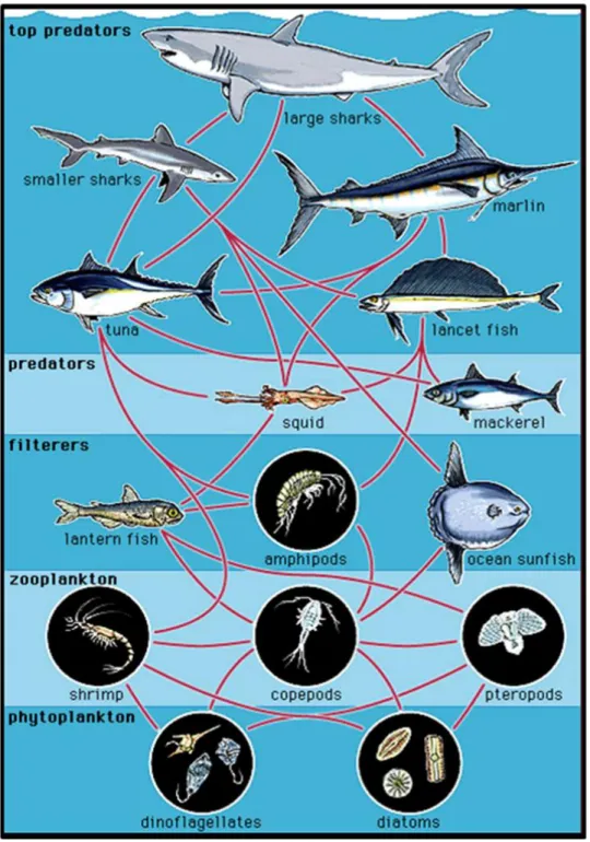
some cases, the effects were reversible (Roccheri, et al., 2004). Catalase and superoxide dismutase have been analyzed in marine invertebrates mainly after heavy metal stress exposure (Buffet, et al., 2011; Main, et al., 2010). Few studies have been performed on stress responses in copepods, an important class of crustaceans that is critical in the
transfer of energy from primary producers to higher trophic levels in marine food web (Figure 1).
1.2 Copepods
Copepods are a group of small crustaceans that are critical components of the world‟s freshwater and marine ecosystems. Their name “copepod” derives from the greek word “kope”, meaning “oar”, and “podos”, meaning “foot” (Mauchline, 1998). In fact, copepods are characterized by paddle-shaped appendages that function as filters for collecting food from the environment, even though many species are also raptorial. Copepods, with more than 11,500 species (Humes, 1994), show a variety of shapes and sizes (Figure 1.2). The smallest adults measure <0.1 mm (e.g. Sphaeronellopsis
monothrix, a parasite of ostracods) and the largest measure up to 23 cm (Penella balaenopterae, whale parasites).
Copepods act as grazers of photoautotrophs and microheterotrophs, as prey for diverse invertebrates and vertebrates sustaining important fisheries, as extremely sensitive indicators of environmental change and as carriers of carbon between trophic levels. Some species are also parasites of economically important fish species or vectors of human disease (e.g. cholera).
The biology of copepods is rather well known (Mauchline, 1998) with several studies addressing mechanisms of diapause, developmental biology, and host-pathogen interactions. Despite their central importance in sustaining the carbon cycle and important fisheries, there are currently few publically available sequences and this lack of available genomic resources for copepods has proven a major barrier to the wider application of molecular methods in copepod research.
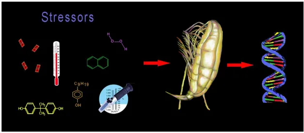
Few studies report gene expression patterns induced in copepods exposed to physical (temperature and salinity) and chemical (heavy metals, endocrine disruptor chemicals, H2O2, hydrocarbons, diatom toxins, and other toxicants) stressors. A recent review on copepod stress response to these stressors has just been published by Lauritano et al. (Lauritano, et al., 2011c) (Figure 1.3).
Figure 1.3. This graphical abstract (Lauritano et al., 2011c) shows some of the stressors tested on copepods to evaluate expression level changes of specific genes.
This paper reviews the literature on the defense systems, including detoxification enzymes and proteins (e.g. glutathione S-transferases, heat shock proteins, superoxide dismutase and catalase), studied in copepods at the molecular level including some of the results reported in this thesis. The data indicate high inter- and intra-species variability in copepod response, depending on the type of stressor tested, the concentration and exposure time, and the enzyme isoform studied.
Members of the genus Calanus are among the largest copepods and are of great importance in the diet of the juvenile stages of some economically important fish (such as cod, haddock, herring and mackerel) (Gaard, Reinert, 2002; Gislason, Astthorsson,
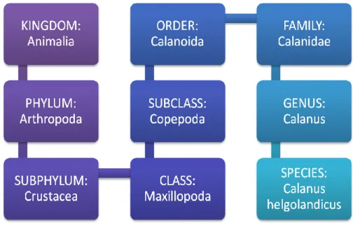
2002; Ringuette, et al., 2002). The model species I have worked on during the course of my PhD thesis is the copepod Calanus helgolandicus (C. helgolandicus) (Figure 1.4, 1.5) which is a widespread epipelagic copepod species whose geographical range extends from the temperate Atlantic Ocean to the northern Mediterranean Sea (Bonnet,
et al., 2005).
Figure 1.4. Calanus helgolandicus scientific classification.
At a European scale, especially in the North Sea, C. helgolandicus has progressively become more abundant and widely distributed, while its congeneric species Calanus finmarchicus, adapted to live in colder waters, has migrated northwards. These shifts in distribution provide a strong stimulus for further studies on
C. helgolandicus. Moreover, if climate warming continues, C. helgolandicus will
probably expand geographically. C. finmarchicus/helgolandicus ratio is currently being used by the European Environmental Agency as a climate change impact indicator (Bonnet, et al., 2005). C. helgolandicus typically lives in warmer waters, between 5 and
28°C, and with a salinity range between 32-39 ppt (Bonnet, et al., 2005; Unal, et al., 2006).
Figure 1.5. Calanus helgolandicus adult female.
In the Mediterranean Sea, C. helgolandicus commonly occurs in the North Adriatic Sea where it represents the main diet of the juvenile stages of some economically important fish species and plays a key role in the food web affecting the biological productivity in this marine ecosystem. In addition, it occurs in concomitance with a winter phytoplankton bloom which represents a fundamental food source for copepods. Induced chemical defenses in response to grazing have been shown for several bloom-forming algae (i.e. diatoms and dinoflagellates). Some of these produce
defensive metabolites that have been reported to have multiple simultaneous functions including antipredator, allelopathy, antibacterial, and cell to cell signaling (e.g. diatom polyunsaturated aldehydes PUAs). In the successive two sections I will briefly introduce two important phytoplankton groups: diatoms and dinoflagellates.
1.3 Diatoms
Diatoms are eukaryotic unicellular plants that constitute one of the major components of marine phytoplankton, comprising up to 40% of annual productivity at sea (Falkowski, 1994) and representing 25% of global carbon-fixation (Nelson, et al., 1995). Copepods feed willingly on diatoms and have evolved strong teeth-like structures to break the strong silica wall of diatom cells. There are more than 200 genera of living diatoms, with about 100.000 species (Figure 1.6).
Diatoms have traditionally been considered a prefered food for zooplankton grazers such as copepods and for the transfer of organic carbon to higher trophic levels sustaining important fisheries (Mauchline, 1998) (Figure 1.1).
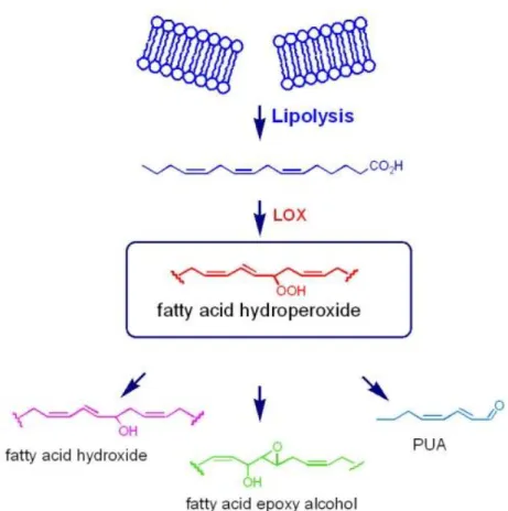
However, numerous studies have shown that these unicellular plants at times produce secondary metabolites with toxic effects on reproductive processes in crustacean copepods (Fontana, et al., 2007b; Ianora, et al., 2004; Miralto, et al., 1999) and cladocerans (Carotenuto, et al., 2005), echinoderm sea urchins (Romano, et al., 2010) and sea stars (Caldwell, 2009; Guenther, et al., 2009), polychaete worms (Caldwell, et al., 2002; Simon, et al., 2010), and ascidians (Tosti, et al., 2003). Diatom metabolites, collectively termed oxylipins, are the end-products of a lipoxygenase/hydroperoxide lyase metabolic pathway (Cutignano, et al., 2006;
d'Ippolito, et al., 2004; d'Ippolito, et al., 2009; Fontana, et al., 2007a; Pohnert, 2000) initiated by damage to algal cells, as occurs through grazing by predators. Cell damage activates lipase enzymes, which liberate polyunsaturated fatty acids (PUFAs) from cell membranes that are immediately oxidized and cleaved within seconds to form polyunsaturated aldehydes (PUAs) and a plethora of other metabolites collectively termed oxylipins (Figure 1.7).
Figure 1.7. Biosynthetic sketch for the synthesis of oxylipins in marine diatoms (Fontana et al., 2007).
Oxylipins, and PUAs in particular, have important biological and biochemical properties including the disruption of gametogenesis, gamete functionality, fertilization, embryonic mitosis, and larval fitness and competence (Caldwell, 2009). Although the effects of such toxins are less catastrophic than those inducing poisoning and death of predators, they are none-the-less insidious inducing abortions, birth defects and reduced larval survivorship (Ianora, et al., 2004; Miralto, et al., 1999) (Figure 1.8).
Figure 1.8. Effects of diet on C. helgolandicus offspring fitness. a, b, After feeding on Skeletonema marinoi, such nauplii were strongly deformed. c, Nauplii generated from females fed the control diet Prorocentrum minimum appeared normal. Scale bar, 90 m (from Ianora et al., 2004).
Such antiproliferative compounds may discourage herbivory by sabotaging future generations of grazers, thereby allowing diatom blooms to persist when grazing pressure would otherwise have caused them to crash.
The specific type and quantity of oxylipins produced differs between diatom species and strains due to a variety of precursor PUFAs and enzymes with variable effects on grazers (Ianora, Miralto, 2010). Oxylipin production also differs depending on the physiological status of the same diatom culture due to e. g. different nutrient regimes (Ribalet, et al., 2007; Ribalet, et al., 2009) or various growth stages (d'Ippolito,
et al., 2009; Ribalet, et al., 2007; Vidoudez, Pohnert, 2008). Similar wound-activated
compounds are also found in terrestrial plants where they play a pivotal role in defense because of their antibacterial, wound healing and antiproliferative activity (Andreou, et
1.4 Dinoflagellates
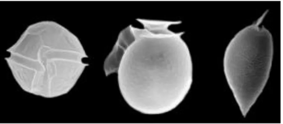
Dinoflagellates are unicellular protists which exhibit a great diversity of forms (Figure 1.9). Their name derives from the greek dinos "whirling" and latin flagellum "whip, scourge".
Figure 1.9. Three dinoflagellate structures from Stazione Zoologica Anton Dohrn website (www.szn.it).
Many are photosynthetic, manufacturing their own food using the energy from sunlight, some species are capable of producing their own light through bioluminescence, while other dinoflagellates are parasites on fish or on other protists. Dinoflagellates are the largest group of marine eukaryotes aside from the diatoms and primary producers making them an important part of the aquatic food web (Figure 1.1). Most are marine but they are also common in fresh water habitats as well.
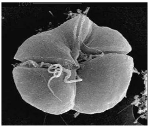
Many phytoplankton species are known to form harmful algal blooms (HABs), including: dinoflagellates, diatoms, haptophytes and raphidophytes. The species within these different groups show variable abundance/biomass and toxicity patterns. The most dramatic effect of some species (e.g. the dinoflagellate Karenia brevis, figure 1.10) is to
cause "blooms" in such great numbers that the water may appear golden or red, producing a "red tide" (Figure 1.11) (Landsberg, 2002; Van Dolah, et al., 2009).
Many algae produce secondary metabolites which can adversely affect other organisms. In general, the major producers of toxins in the marine environment are the dinoflagellates and diatoms. Dinoflagellates can produce neurotoxins (e.g. brevetoxin A, the most potent neurotoxin secreted by the dinoflagellate Karenia brevis) which affect muscle function in susceptible organisms (Landsberg, 2002; Nicolaou, et al., 1998). Humans may also be affected by eating fish or shellfish containing the toxins. Gastrointestinal, neurosensory, neurocerebellar, neuromuscular, systemic and general CNS effects are the common deseases induced by dinoflagellate toxins, also known as neurotoxic shellfish poisoning (NSP) (Watkins, et al., 2008).
Figure 1.10. Scanning electron micrograph of Karenia brevis. Micrograph: Florida Marine Research Institute ( http://www.bioone.org/doi/abs/10.1641/0006-3568%282003%29053%5B0918%3AHABBPN%5D2.0.CO%3B2).
Figure 1.11. Red algal bloom at Leigh, near Cape Rodney. Photo by Miriam Godfrey (from http://serc.carleton.edu/microbelife/topics/redtide/general.html).
1.5 Thesis aim
The object of this thesis was to investigate the defense systems and stress reponses in adult copepods exposed to toxic diatom and dinoflagellate diets using a molecular approach.
mRNA expression patterns induced by feeding on diatoms or dinoflagellates were examined by using Reverse Transcription-quantitative Polymerase Chain Reaction (RT-qPCR). mRNA expression changes are an important early signal compared to other physiological parameters and/or phenotype changes.
1. The first part of the thesis was a set up for gene expression studies in this copepod species. In order to have enough animals for the molecular experiments, a cultivating breeding system was set up with researchers at the Stazione Zoologica Anton Dohrn where I conducted my PhD work (Carotenuto, Esposito, Pisano, Lauritano et al., 2011, submitted). Then I selected and screened a list of putative reference genes (RGs), necessary for the RT-qPCR analyses, by using three different softwares (BestKeeper, NormFinder and GeNorm). Once the best RGs were obtained, I analyzed, for the first time, the effects of the ingestion of the toxic oxylipin-producing diatom Skeletonema
marinoi by the calanoid copepod Calanus helgolandicus by evaluating
expression levels of specific genes of interest, the microtubule subunits alpha and beta tubulins, after 2 days of feeding (Lauritano et al., 2011a).
2. To further investigate gene expression patterns induced by S. marinoi on defence systems in C. helgolandicus, the second part of the thesis focused on analyzing a
series of genes involved in generic stress response, aldehyde detoxification and apoptosis regulation in this copepod species (Lauritano et al., 2011b).
3. A third part of the thesis was concentrated on analyzing gene expression changes with time-series experiments for two different C. helgolandicus populations (North Adriatic Sea and North Atlantic Ocean) fed the same toxic diatom diet (Lauritano et al., in preparation). The experiments were in part carried out at the Station Biologique de Roscoff and were financed by the EU FP7 ASSEMBLE project. The data provided interesting results on common or population-specific responses in expression patterns in copepods.
4. Then I examined the effect of a Skeletonema marinoi-dominated spring bloom in the Northern Adriatic Sea in 2010 and 2011. Field zooplankton samples were also collected during the oceanographic cruise ENVADRI (Lauritano et al., in preparation)(Bastianini, et al., 2011).
5. Finally, I analysed gene expression changes when C. helgolandicus was exposed to the toxic dinoflagellate Karenia brevis compared to when C. helgolandicus was exposed to toxic diatom diets (Lauritano et al., in preparation).
In order to better examine each topic, I will separately present and discuss each one in separate chapters reporting a brief specific introduction, methods, results and
Chapter 2:
First molecular evidence of diatom effects in
the copepod Calanus helgolandicus on
microtubule subunits
Published as:
Lauritano Chiara, Borra Marco, Carotenuto Ylenia, Biffali Elio, Miralto Antonio, Procaccini Gabriele and Ianora Adrianna. (2011)
First molecular evidence of diatom effects in the copepod Calanus helgolandicus. Journal of Experimental Marine Biology and Ecology, 404, 79-86
2.1. Brief introduction
Buttino and co-workers (Buttino, et al., 1999) were the first to show that water-soluble extracts of the diatom Thalassiosira rotula induced aberrations in embryonic tubulin organization leading to cell blockage and the absence of spindle formation in the sea urchin Paracentrotus lividus, but the molecules responsible for these effects were unknown at the time. Miralto et al. (Miralto, et al., 1999) later isolated the PUAs 2-trans-4-cis-7-cis-decatrienal, 7-cis-decatrienal and 2-trans-4-trans-decadienal and showed that they arrested embryonic development of copepod and sea urchin embryos in a dose-dependent manner, and also had antiproliferative and apoptotic effects on human carcinoma cells. Hansen et al. (Hansen, et al., 2004) studied the effects of decadienal on the sea urchin Sphaerechinus granularis and showed that this PUA inhibited cyclin B/Cdk1 kinase activity and DNA replication. Staining of alpha-tubulin subunits showed that tubulin polymerization was disrupted and aberrations were induced in mitotic spindles (Hansen, et al., 2004).
Tubulins are proteins that are the building blocks of microtubules (MTs), one of the active components of the cytoskeleton. MTs play an important role in many cellular functions including development and maintenance of cell shape, growth, signalling, protein movement, intracellular vesicle transport, organization and positioning of membranous organelles, and segregating replicated chromosomes into daughter cells during mitosis and meiosis (Calligaris, et al., 2010; Harrison, et al., 2009; Jordan Mary Ann, 2004; Nogales, et al., 1998).
MTs consist in - and -tubulin monomers which constantly switch between a state of polymerization and depolymerization (Calligaris, et al., 2010; Nogales, et al.,
1998), the ratio of which can be altered by many natural products and drugs, especially anti-cancer compounds targeting MTs (Calligaris, et al., 2010; Harrison, et al., 2009; Jordan Mary Ann, 2004; Sashidhara, et al., 2009). Compounds that bind to tubulin modify the formation and function of MTs and can affect the proper functioning and formation of the mitotic spindle. If MT function is altered and spindle dynamics is compromised, a mitotic block or the slowing down in cell cycle progression occurs at the metaphase-anaphase transition, eventually leading to apoptosis (Jordan Mary Ann, 2004).
The aim of this chapter was to evaluate, for the first time, the effects of ingestion of the oxylipin-producing diatom Skeletonema marinoi (SKE) by the copepod Calanus
helgolandicus on - and -tubulin gene expression levels using the reverse transcription-quantitative real time polymerase chain reaction (RT-qPCR). Previous studies have already shown that a diet of S. marinoi which contains high levels of PUAs and other oxylipins (same strain as in this study) reduces egg hatching success and female survival in this copepod species (Fontana, et al., 2007b; Ianora, et al., 2004), with a concomitant appearance of apoptosis in both copepod embryos and female tissues (Buttino, et al., 2008), but there are no studies on gene expression analyses in the copepod C. helgolandicus. We also investigated tubulin gene expression levels in C.
helgolandicus feeding on the non-oxylipin producing flagellate Rhodomonas baltica
(RHO) and dinoflagellate Prorocentrum minimum (PRO) currently being used to rear C.
helgolandicus in our laboratory. P. minimum was considered the control diet since
several previous studies have shown that C. helgolandicus reproduces and grows well on this diet (Ianora, et al., 2004). Although the biology of C. helgolandicus species is rather well known (Mauchline, 1998), very little is known about its genome, except for the gene sequences of cytochrome oxidase subunit I, antennapedia proteins 1 and 2,
cytochrome b, and 16S, 18S and 28S ribosomal RNA as reported in the public database PubMed. Since there are few studies on gene expression analysis via RT-qPCR in copepods (Frost, Nilsen, 2003; Hansen, et al., 2008a; Hansen, et al., 2008b; Hansen, et
al., 2009; Hansen, et al., 2011; Kvamme, et al., 2004; Lee, et al., 2008; Lee, et al.,
2007; Seo, et al., 2006a; Skern-Mauritzen, et al., 2009; Tarrant, et al., 2008), and none of these focus on C. helgolandicus, here we also describe the first evaluation of reference genes as internal controls for RT-qPCR analyses in C. helgolandicus. It is important to note that the analyses of potential reference genes were conducted according to the MIQE (Minimum Information for Publication of Quantitative Real-Time PCR Experiments) suggestions and checklist (Bustin, et al., 2009; Bustin, et al., 2010).
2.2 Methods
2.2.1 Collection of copepods
Calanus helgolandicus specimens were collected in the North Adriatic Sea from
March to April 2009 and transported to Naples where they were placed in a 500 L re-circulating copepod breeding system (Buttino, et al., 2011) maintained at 20°C, 38 ppm salinity, 12 h:12 h light:dark cycle, and a mixed diet of the flagellates Rhodomonas
baltica, Prorocentrum minimum and Isochrysis galbana.
Adult female C. helgolandicus specimens were sampled from the culturing tanks, isolated under a Leica stereomicroscope and transferred to 10 L beakers filled with 0.22 m filtered sea water at 20°C. Beakers containing from 5 to 40 adult females were fed either unialgal diets of the control non-oxylipin producing dinoflagellate P.
Skeletonema marinoi (SKE) (45.000-60.000 cells/ml) and the non-oxylipin producing
flagellate R. baltica (RHO) (7500-8000 cells/ml). The strains belong to the Stazione Zoologica Anton Dohrn culture collection. R. baltica and P. minimum were cultured in 2-l glass jars with 0.22 µm-filtered FSW enriched with k medium at 20oC and on a 12:12 hr dark:light cycle. The diatom S. marinoi was cultured under the same experimental conditions but with F2 medium.
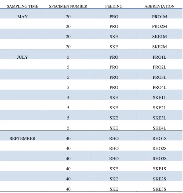
Copepods were collected from the culturing tank on three different occasions (Table 2.1). The first sampling was conducted in May 2009 when two replicates of 20 adult C. helgolandicus were fed PRO (replicates designated as PRO1M and PRO2M, respectively) and another two were fed SKE (replicates designated as SKE1M and SKE2M, respectively). The second collection was in July 2009 when four groups of 5 animals were fed PRO (replicates designated as PRO1L, PRO2L, PRO3L and PRO4L, respectively) and another four were fed SKE (replicates designated as SKE1L, SKE2L, SKE3L and SKE4L, respectively). The third sampling was in September when a third algae was introduced, RHO. Three groups of 40 specimens were fed RHO (replicates designated as RHO1S, RHO2S and RHO3S, respectively) and three were fed SKE (replicates designated as SKE1S, SKE2S and SKE3S, respectively).
During each sampling, animals were fed for two days ad libitum on either one of the three algal diets and were then transferred to filtered sea water (FSW) for 24h to eliminate any algal residues in the gut. After this, each replicate was washed in 50 l of FSW and carefully transferred to 500 l Trizol Reagent (Invitrogen), frozen directly in liquid nitrogen and stored at -80°C for few weeks until RNA extraction. To study the extent to which the selected genes were differentially expressed in our experimental conditions, we extracted RNA and retro-transcribed it in Complementary DNA (cDNA)
(double-stranded DNA version of an mRNA molecule), which was used as the template for our molecular analyses.
SAMPLING TIME SPECIMEN NUMBER FEEDING ABBREVIATION
MAY 20 PRO PRO1M
20 PRO PRO2M
20 SKE SKE1M
20 SKE SKE2M
JULY 5 PRO PRO1L
5 PRO PRO2L 5 PRO PRO3L 5 PRO PRO4L 5 SKE SKE1L 5 SKE SKE2L 5 SKE SKE3L 5 SKE SKE4L
SEPTEMBER 40 RHO RHO1S
40 RHO RHO2S
40 RHO RHO3S
40 SKE SKE1S
40 SKE SKE2S
40 SKE SKE3S
Table 2.1. Copepod collections including information on sampling month, number of specimens fed and frozen for RNA extraction, algae used for feeding experiments and abbreviation of the treated groups.
2.2.2 RNA extraction and quantification
Total RNA was extracted using a modified Trizol manufacturer‟s protocol (Invitrogen) (For more details see Appendix 1). To remove hypothetically contaminating DNA, each sample was treated with DNaseI (Invitrogen) according to the instruction manual. RNA quantity was assured by Nano-Drop (ND-1000 UV-Vis spectrophotometer; NanoDrop Technologies) monitoring the absorbance at 260 nm; purity was determined by monitoring the 260/280 nm and 260/230 nm ratios using the same instrument. All samples were free from protein and organic solvents used during RNA extraction. RNA quality was also evaluated by gel electrophoresis showing minimal degradation of RNA, which was almost completely intact, with sharp 18S and 28S ribosomal bands.
2.2.3 cDNA synthesis
The amount of RNA used for the reverse transcription steps was always 1 g. This amount of RNA was converted into cDNA with the ProScript First Strand cDNA Synthesis Kit (New England Biolabs), following the manifacturer‟s instructions, and using the GeneAmp PCR System 9700 (Perkin Elmer).The first reaction was carried out in 16 l final volume with 1 g of RNA, 50M of dt23 VN Primer, 15 M of Random Primer 9, 2.5 M of dNTP Mix and H2O. The mix was first denatured by heating at 70°C for 5 min. A second mix with 1% RT buffer, 10 units/l of RNase Inhibitor and 25 units/l of M-MuLV was added to reach a final volume of 20 l, and the total reaction mix was incubated at 42°C for 90 min and at 95°C for 5 min. RNase H (2units/l) was added to eliminate non-converted (or potential contaminant) RNA by incubating at 37°C for 20 min, followed by 5 min at 95°C to stop the reaction. To
evaluate the efficiency of cDNA synthesis, a PCR was carried out with primers of a constitutive gene, S20. The reaction was carried out on the GeneAmp PCR System 9700 (Perkin Elmer) in 20 l final volume with 2 l 10 PCR reaction buffer Roche, 2 l 0.1% BSA, 2 l 102mM dNTP, 0.8l 5U/l Taq Roche, 1l 20 pmol/l of each oligo, template cDNA and nuclease-free water to 20l. The PCR program consisted of a denaturation step at 95°C for 3 min, 40 cycles at 95°C for 30 sec, 60°C for 1 min and 72°C for 30 sec, and a final extension step at 72°C for 7 min.
2.2.4 Primer design
To perform RT-qPCR in this copepod species we designed specific primers to isolate and amplify selected genes. Primers for hypothetical Reference Genes (RGs) and Genes of Interest (GOI) were designed considering the alignment of conserved domains in different arthropod species such as Calanus finmarchicus, Calanus californicus,
Tigriopus japonicus, Homarus americanus, Drosophila melanogaster and Anopheles gambiae. Alignments were performed with Clustal W (Clustal) and BioEdit (BioEdit).
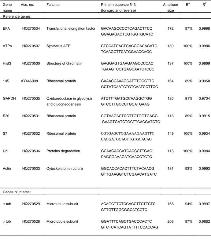
Gene Runner, V3.05 (Hasting Software) was used to predict primer melting temperature (Tm) and check if primers formed dimers, hairpin, bulge and internal loops. The primers for 18S were designed from the known sequence (Accession Number: AY446908) using Primer3 software, v. 0.4.0. RGs belonging to different functional classes were selected in order to reduce the possibility that they might be co-regulated (e.g. S20, S7 and S18). The selected RGs were: -actin (ACT), elongation factor 1(EFA), glyceraldehyde-3-phosphate dehydrogenase (GAPDH), ribosomal units (18S, S7, S20), adenosine-3-phosphate synthase (ATPs), histone 3 (HIST). The GOI were the two microtubule subunits: -tubulin (tub) and -tubulin (tub). Table 2.2 lists gene functions, primers‟ sequences, amplicon size, correlation coefficient (R2
(Ea). Primers were synthesized commercially by Primm Labs. Primers were designed to amplify cDNA regions ranging from 100 to 200 bp in size, in order to facilitate cross-comparison of assays and assure equal PCR efficiencies. PCR conditions were optimized on a GeneAmp PCR System 9700 (Perkin Elmer). Reactions were carried out in 20 l volume with 2 l of 10 PCR reaction buffer Roche, 2 l of 0.1% BSA, 2 l of 10 2mM dNTP, 0.8l of 5U/l Taq Roche, 1l of 20 pmol/l for each oligo, template cDNA and nuclease-free water to 20l. The PCR program consisted of a denaturation step at 95°C for 3 min, 40 cycles at 95°C for 30 sec, 60°C for 1 min and 72°C for 30 sec, and a final extension step at 72°C for 7 min. Amplified PCR products were analyzed by 1.5% agarose gel electrophoresis in 1X TBE buffer. In order to verify the correct assignment of amplicons to target genes, the resulting bands were excised from the gel and extracted according to the QIAquick Gel Extraction Kit protocol (QIAGEN) and sequence analyzed. Sequence reactions were obtained by BigDye Terminator Cycle
Sequencing technology (Applied Biosystems) and purified using the Agencourt CleanSEQ Dye terminator removal Kit (Agencourt Bioscience Corporation) in
automation by the robotic station Biomek FX (Beckman Coulter). Products were analysed on the Automated Capillary Electrophoresis Sequencer 3730 DNA Analyzer (Applied Biosystems). The identity of each sequence was confirmed using the bioinformatics tool BLAST (Basic local alignment search tool) (BLAST). The sequences are deposited in GenBank under the Accession Numbers shown in Table 2.2.
Gene Acc. no. Function Primer sequence 5'-3' Amplicon Ea R2
name (forward and reverse) size
Reference genes
EFA HQ270534 Translational elongation factor GACAAGCCCCTCAGACTTCC 172 97% 0.9998 GGAGAGACTCGTGGTGCATC
ATPs HQ270507 Synthesis ATP CTCCATCACTGACGGACAGATC 150 100% 0.9986 TCAAGCTTCATGGAACCAGC
Hist3 HQ270530 Structure of chromatin GAGGAGTGAAGAAGCCCCAC 137 100% 0.9969 TGAAGTCCTGAGCAATCTCCC
18S AY446908 Ribosomal protein GAAACCAAAGCATTTGGGTTC 164 89% 0.9958 GCTATCAATCTGTCAATCCTTCC
GAPDH HQ270535 Oxidoreductase in glycolysis ATCTTTGATGCCAAGGCTGG 126 91% 0.9704 and gluconeogenesis GTCCTTGCCCTGCATGAAG
S20 HQ270531 Ribosomal protein CGTAAGACTCCTTGTGGTGAGG 113 89% 0.9915 GAAGTGATCTGCTTCACGATCTC
S7 HQ270532 Ribosomal protein CGTGAGCTGGAAAAGAAGTTC 149 100% 0.9934 CAGGATGGAGTTGTGGACAG
Ubi HQ270536 Proteins degradation GCAAGACCATCACCCTTGAG 113 100% 0.9984 CAGCGAAAGATCAACCTCTG
Actin HQ270533 Cytoskeleton structure GGCACCACACTTTCTACAACG 131 93% 0.9993 GTTGAAGGTCTCGAACATGATC
Genes of interest
tub HQ270529 Microtubule subunit ACAGCTTCTCCACCTTCTTCTC 168 94% 0.9997
GTTGTTGGCGGCATCCTC
tub HQ270528 Microtubule subunit GGATTTCAGCTGACCCACTC 206 97% 0.9862
GTCTCATCAGTATTTTCCACCAG
a
= Oligo efficiency
Table 2.2. PubMed accession numbers for candidate reference genes (RG) and genes of interest (GOI) for Calanus helgolandicus qPCR assays. Putative RGs were elongation factor 1(EFA), adenosine 3-phosphate synthase (ATPs), histone 3 (HIST), glyceraldehyde-3-phosphate dehydrogenase (GAPDH), ribosomal units (18S, S7, S20), ubiquitin (UBI), -actin (ACT) and GOIs were - and - tubulins. Gene functions, primer sequences, amplicon sizes, oligo efficiencies (Ea) and correlation factors (R2) are also given.
2.2.5 Reverse transcription-Quantitative real time polymerase chain reaction (RT-qPCR)
The fluorescent dye SYBR GREEN was used to value expression levels of the selected genes by RT-qPCR. This dye shows maximal fluorescence only with double-strand DNA and was used to detect our amplicons (For details see APPENDIX 2).
Three different algorithms were utilized to identify the best reference genes in our experimental design: BestKeeper (Pfaffl, et al., 2004), geNorm (Vandesompele, et
al., 2002) and NormFinder (Andersen, et al., 2004).
To study the expression of each target gene relative to the most stable RGs, we used REST tool (Relative expression software tool) (Pfaffl, et al., 2002). This tool used a mathematical model based on the PCR efficiencies and the mean crossing point deviation between the sample and the control group. The advantage of REST is that this software tool tests the group differences for significance with the Pair-Wise Fixed Reallocation Randomization Test (Pfaffl, et al., 2002).
Statistical analysis was performed using GraphPad Prim statistic software, V4.00 (GraphPad Software).
2.3 Results
2.3.1 Validation of best reference genes for RT-qPCR
Raw Ct data of potential RGs are reported in Figure 2.1. According to the mathematical approach of BestKeeper, RG expression stability considers the standard deviation of the Ct values (Table 2.3). Hence the most stable RGs have a standard deviation (SD) lower than 1 and these were S20, S7, GAPDH, HIST and UBI.
BestKeeper analysis (Pfaffl, et al., 2004) indicated S20 as the most stable gene, followed by S7 and GAPDH.
GeNorm analysis (Vandesompele, et al., 2002) confirmed the results of BestKeeper, showing that the two most stable genes, with the lowest expression stability (M), were S20 and S7 (Figure 2.2). Pair-wise variation was subsequently calculated to evaluate the effect of adding another RG to those already analyzed. Below the cut-off value of 0.15 the inclusion of additional RGs was not required. According to our results (Figure 2.3), the two best RGs, S20 and S7, were sufficient for the analysis.
According to the statistical approach of NormFinder, our best candidate reference genes, with the lowest stability values, were S20, S7 and GAPDH, as reported in figure 2.4. The rank pattern was the same as for Bestkeeper and geNorm analyses as summarized in table 2.4.
Although the three approaches agreed that the best RGs were S20 and S7, we decided to also use the third best RG, GAPDH, because the two ribosomal proteins S20 and S7, belonging to the same functional class, might be co-regulated. Moreover three RGs are suggested as a minimum significant number for reliable assessment of gene expression using qPCR (Bustin, et al., 2010).
Figure 2.1. Ct value profile.The Ct values (ordinates) obtained for all candidate reference genes (RG) during all samplings (abscissa) for normalization in C. helgolandicus qPCR assays. Each curve represents the degree of stability of Ct values for each RG.
Figure 2.2. Gene expression stability M of candidate RGs calculated by geNorm. The stepwise exclusion of genes that are more variable among samples using the geNorm program. Data points represent the average expression stability values of remaining RGs.
Figure 2.3. Determination of the optimal number of control genes for normalization. Pair-wise variation to evaluate the effect of adding another RG to those already analyzed. The inclusion of additional RGs was not required below the cut-off value of 0.15. The two best RGs, S20 and S7, were sufficient for this analysis, but the third best RG, GAPDH, was also used because the two ribosomal proteins S20 and S7, belonging to the same functional class, might be co-regulated.
Figure 2.4. Determination of the most stable RG using NormFinder. The NormFinder algorithm ranks the data set of candidate normalization genes according to their expression stability in a given experimental design. Red bars represent the stability values of our candidate genes.
6.57 4.96 3.05 8.82 20.59 5.85 3.87 2.89 2.22 CV 1.41 0.94 0.67 2.1 3.29 1.11 0.76 0.68 0.42 SD 24.01 20.67 23.9 31.06 24.38 22.67 22.67 25.55 19.65 max 19.15 17.31 20.09 20.83 11.26 16.49 18.29 22.47 17.5 min 21.41 18.94 22.02 23.81 15.98 18.92 19.55 23.64 18.74 AR 21.35 18.91 22 23.66 15.52 18.86 19.52 23.62 18.73 GM 18 18 18 18 18 18 18 18 18 n ACT UBI S7 ATPS 18S EFA HIST3 GAPDH S20
Table 2.3. BestKeeper results. Descriptive statistics of the derived crossing points for each putative RG, where n = number of samples; GM = geometric mean; AR = arithmetic mean; min and max = extreme values of the crossing points (CP); SD = standard deviation of CP; CV = coefficient of variance.
18S 18S 18S 9 ATPs ATPs ATPs 8 EFA HIST ACT 7 HIST EFA EFA 6 UBI UBI UBI 5 ACT ACT HIST 4 GAPDH GAPDH GAPDH 3 S7 S7 S7 2 S20 S20 S20 1 NormFinder GeNorm BestKeeper Rank
Table 2.4. Stability of putative reference genes in Calanus helgolandicus. Candidate RGs are ranked from most to least stable using the three different softwares described in the text. Three best reference genes used for the analysis were S20, S7 and GAPDH.
2.3.2 Expression level of genes of interest (GOI)
To investigate if microtubules in Calanus helgolandicus were affected by diets of SKE and RHO, we analyzed the relative expression levels of the two MT subunits: - and -tubulin (Figure 2.5). The analyses were carried out on all samples using the three best RGs (S20, S7 and GAPDH) to normalize our data and considering copepods fed PRO as the control group using the Relative expression software tool (REST tool) (Pfaffl, et al., 2002).
Figure 2.5 shows that the expression levels of our GOI in copepods fed on the oxylipin-producing diatom SKE were significantly lower than on the flagellate RHO (students‟t-test, p<0.0001, for both genes). According to REST analysis, -tubulin was about 2-fold down-regulated while -tubulin was about 3-fold down-regulated in SKE treated samples. As expected, - and -tubulin gene expression in copepods fed RHO compared to copepods fed the control diet PRO did not show significantly different expression levels.
The behaviour of GOI was confirmed by the replicates of the experiments (listed in table1) with a standard deviation (SD) of 0.194 and 0.052 for -tubulin in SKE and RHO conditions, respectively, and 0.080 and 0.018 for -tubulin for SKE and RHO, respectively (Figure 2.5).
Figure 2.5. Relative target gene expression in copepods fed on Skeletonema
marinoi and Rhodomonas baltica. The figure shows alpha- and beta-tubulin (A TUB
and B TUB, respectively) gene expression levels (y-axis, Mean SD) in copepods fed on the test diatom Skeletonema marinoi (SKE) and the flagellate Rhodomonas baltica (RHO), respectively. The data are normalized with our three best reference genes, S20, S7 and GAPDH, using the dinoflagellate Prorocentrum minimum as a control alga.
2.4 Discussion
Previous studies have shown that when C. helgolandicus is fed on S. marinoi, egg hatching success is impaired after 3 days of maternal feeding, with the production of apoptotic teratogenic nauplii that do not develop to adulthood (Buttino, et al., 2004). Our results indicate that a diet of S. marinoi affects - and - tubulin gene expression levels in adult females after only two days of feeding by possibly reducing MT subunits and microtubule filament formation, with the cascading effect of altering pronuclear migration, DNA replication and mitotic events. Alternatively, PUAs and other oxylipins may affect protein and organelle transport along microtubules, impairing cellular
homeostasis. Ingestion of S. marinoi induced a pronounced down regulation of tubulin genes, with about a 3-fold down-regulation of -tubulin and 2-fold down-regulation of-tubulin. By contrast, diets of the dinoflagellate P. minimum and the flagellate R.
baltica induced no significant up- or down-regulation of these genes.
The best RGs for C. helgolandicus fed on the diatom S. marinoi, the dinoflagellate
P. minimum and the flagellate R. baltica were S20, S7 and GAPDH. These results
confirm the stability of the ribosomal protein S20 as reported for the salmon louse L.
salmonis (Frost, Nilsen, 2003) even if this species belongs to a different copepod order.
Glyceraldehyde-3-Phosphate Dehydrogenase (GAPDH) which is widely used in gene expression studies (Hansen, et al., 2008a; Spinsanti, et al., 2006; Toegel, et al., 2007) has been shown to be up- or down-regulated in various conditions (Bustin, 2000). In our analysis GAPDH scored third. Gene expression profiles of Elongation factor (EFA), selected as RG in C. finmarchicus (Hansen, et al., 2008a; Hansen, et al., 2008b; Hansen, et al., 2009) and L. salmonis (Kvamme, et al., 2004; Skern-Mauritzen, et al., 2009), and of -actin (ACT), used as RG in Tigriopus japonicus (Lee, et al., 2008; Lee,
et al., 2007; Seo, et al., 2006a), were less stable in our test conditions and were
therefore considered invalid for this analysis. Histone 3 (IST), Ubiquitin (UBI), ATP synthase (ATPs) and ribosomal protein 18S gene expression levels were less stable as well. In general, our results confirm that reference genes are not universal for a given species but may change depending on experimental conditions.
Interestingly, - and - tubulin subunits have also often been selected as putative RGs to normalize qPCR data (Andersen, et al., 2004; Carvalho, et al.; Heckmann, et al., 2006; Ransbotyn, Reusch, 2006; Sirakov, et al., 2009), but in our experiments their expression levels were evidently affected by the oxylipin-producing diatom SKE.
Several studies have shown that the functioning of tubulin is affected by exposure to toxic compounds (Chavez, et al., 2010; Lee, et al., 2009), but these results have usually been obtained in humans. For example, exogenous exposure to another aldehyde, 4-hydroxy-2(E)-nonenal (HNE), in human monocytic THP-1 cells induced HNE-tubulin alpha-1B chain adducts leading to inhibition of tubulin polymerization (Chavez, et al., 2010). Prenatal exposure to cocaine causes cyto-architectural alterations in the developing neocortex inducing impairments in fetal brain development and RT-qPCR analyses (Lee, et al., 2009) and the down-regulation of cytoskeleton-related genes such as alpha 3d tubulin and alpha 8 tubulin in neural and/or A2B5+ progenitor cells.
Our results support, for the first time at the molecular level, previous findings by Buttino et al. (Buttino, et al., 1999) and Hansen et al. (Hansen, et al., 2004), that PUAs inhibit tubulin-polymerization affecting proper spindle formation and cell division. Future studies on the biological effects of these molecules should address whether the functioning of other important proteins are altered as well. Our results may help explain why recruitment processes in copepods during diatom blooms may be compromised with possible effects on cohort size of the next generation.
Chapter 3:
Molecular Evidence of the Toxic Effects of
Diatom Diets on Gene Expression Patterns in
Copepods
Published as:
Lauritano Chiara, Borra Marco, Carotenuto Ylenia, Biffali Elio, Miralto Antonio, Procaccini Gabriele and Ianora Adrianna. (2011)
Molecular Evidence of the Toxic Effects of Diatom Diets on Gene Expression Patterns in Copepods. PLoS One, 6; 10
3.1 Brief Introduction
The aim of the present chapter was to further explore the toxic effects of diatoms on copepod females at the gene level under two different experimental conditions: when females were fed for 2 days (2d) on S. marinoi compared to when they received a diet of another diatom Chaetoceros socialis (C. socialis) that does not produce PUAs and synthesizes only low levels of other oxylipins (Fontana, et al., 2007b), and was thus, in theory, “less toxic” for copepods.
In addition to the previously investigated alpha and beta tubulins, here we analyzed the effects of these two diatom diets on the expression levels of genes which are known to have a primary role in generic stress responses, defense systems (e.g. aldehyde, free fatty acid and free radical detoxification) or apoptosis regulation in other organisms, from humans to marine organisms (Bouraoui, et al., 2009; Einsporn, et al., 2009; Hasselberg, et al., 2004; Kim, et al., 2008; Olsvik, et al., 2009; Salazar-Medina,
et al., 2010; Snyder, 2000; Vasiliou, et al., 2004; Wan, et al., 2011) (Figure 1). We
expected an activation of enzymes and proteins involved in stress responses (e.g heat shock proteins, phase I and phase II enzymes), but, in particular, we hypothesized expression level increases of enzymes that could detoxify and/or metabolize toxic diatom PUAs.
To study the generic stress response of C. helgolandicus to diatom toxicity, we analyzed the heat shock protein families 40 and 70 (HSP40 and HSP70, respectively). HSPs are highly conserved proteins that are activated in response to various environmental stress factors (Bierkens, 2000; Feder, Hofmann, 1999). HSP70 can be involved in the tolerance of hyperthermia, ischemia/hypoxia, resistance to hydrogen
peroxide, escape from drug-induced cell cycle arrest, tolerance to ultraviolet radiation and apoptosis (Feder, Hofmann, 1999) whereas HSP40 is often co-localized with HSP70 and plays a role in regulating the ATPase activity of HSP70 (Fan, et al., 2003).
Since oxylipins induce LPO and an increase in free fatty acids (Fink, et al., 2006), we also analyzed the microsomal cytochrome P450 family 4 monooxygenases (CYP4) that generally catalyze the ω-hydroxylation of fatty acids, arachidonic acid and derivatives such as leukotrines and prostanoids (Hsu, et al., 2007). Cytochrome P450 (CYP) enzymes, especially members of the CYP1, CYP2, CYP3, and CYP4 families are generally involved in oxidative modification (known as Phase I reaction) of chemicals into more hydrophilic metabolites to enhance their elimination or inactivation (Goldstone, et al., 2006).
Diatom oxylipins are also known to induce an increase in free radicals, such as reactive oxygen species (ROS) (Fontana, et al., 2007b) which induce oxidative stress and damage to DNA, RNA, proteins, lipids and carbohydrates. We therefore analyzed enzymes involved in ROS detoxification, such as catalase (CAT) and superoxide dismutase (SOD) (Bigot, et al., 2010) and antioxidant activity, such as glutathione synthase (GSH-S) and glutathione S-transferase (GST). Glutathione is an important cell scavenger molecule which facilitates dis-activation of radical compounds. GST is involved in phase II detoxification reactions, catalyzing the nucleophilic attack of glutathione on electrophilic substrates, decreasing their reactivity with cellular macromolecules, facilitating dissolution of the complex glutathione-substrate in the aqueous cellular and extracellular media and, consequently, its elimination from the body (Sheehan, et al., 2001).
We also analyzed aldehyde dehydrogenases (ALDHs) which constitute a superfamily of enzymes that catalyze the oxidation of endogenous and exogenous aldehydes into their corresponding carboxylic acids. These enzymes are generally involved in many processes including amino acid catabolism, neurotransmitter metabolism, xenobiotic and drug biotransformation, protection from osmotic stress and detoxification reactions (Vasiliou, et al., 2004; Yoshida, et al., 1998). We selected several aldehyde dehydrogenase isoforms (ALDH2, ALDH3, ALDH6, ALDH7, ALDH8 and ALDH9) that are mainly involved in aldehyde detoxification due to lipid peroxidation (LPO) (Brocker, et al., 2010; Marchitti, et al., 2008; Vasiliou, et al., 2004). Previous studies have shown that PUAs and oxylipins also induce apoptosis and teratogenesis in the offspring of female copepods that have fed on diatoms for 5d (Ianora, et al., 2004). We therefore determined the transcription level of a protein belonging to the Inhibitor of apoptosis family (IAP), the cell cycle and apoptosis regulatory 1 protein (CARP) and the cellular apoptosis susceptibility protein (CAS). IAP levels increase in certain tumors probably contributing to resistance to apoptosis (Lederman, et al., 2008), CAS and CARP are also both involved in apoptosis (Brinkmann, 1998; Kim, et al., 2008; Tai, et al., 2010): CARP is a novel cell growth regulator (Rishi, et al., 2006) and CAS is necessary in the mitotic spindle checkpoint that ensures genomic stability during cell division (Tai, et al., 2010). In addition, we also analyzed microtubule subunits (alpha and beta tubulins), necessary for mitotic spindle formation. Microtubules (MTs) have many other cellular functions including development and maintenance of cell shape, growth, signaling, protein movement, intracellular vesicle transport and organization and positioning of membranous organelles (Calligaris, et al., 2010; Harrison, et al., 2009; Jordan Mary Ann, 2004; Nogales, et al., 1998).
Figure 3.1. Putative systems affected by diatom metabolites in the copepod
Calanus helgolandicus. A synopsis of the defense and detoxification systems and
generic stress response (with selected genes in parenthesis) studied in C. helgolandicus females exposed to different diatom diets (Skeletonema marinoi and Chaetoceros
socialis). Selected genes were two HSPs (HSP70 and HSP40), six Aldehyde
dehydrogenases (ALDH2, ALDH3, ALDH6, ALDH7, ALDH8, ALDH9), Cytochrome P450-4 (CYP4), Catalase (CAT), Superoxide Dismutase (SOD), Glutathione S-Transferase (GST), Glutathione Synthase (GSH-S), Inhibitor of Apoptosis Protein (IAP), Cell Cycle and Apoptosis Regulatory 1 Protein (CARP), Cellular Apoptosis Susceptibility Protein (CAS) and Alpha and Beta tubulins (ATUB and BTUB).
3.2 Methods
3.2.1 Microalgae culture
The planktonic diatoms Skeletonema marinoi (SMFE6; Adriatic Sea isolate FE6) and Chaetoceros socialis (CSFE17) were cultured as described in (Gerecht, et al., 2011) and harvested during the stationary growth phase. Both species are part of the culture collection at the SZN. SMFE6 produces the PUAs 2-trans-4-cis-hepta-2,4-dienal as the
dominant compound with smaller quantities of trans-4-cis-octa-2,4-dienal and 2-trans-4-cis-octa- 2,4,7-trienal as well as a number of other products deriving from the oxidation of fatty acids including 9S-hydroxy-hexadecatrienoic acid, 11,9-hydroxy-epoxy-hexadienoic acid, 9S-hydroxy-hexatetraenoic acid, 5R- and 15S-hydroxy-eicosapentaenoic acids and 13,14S-hydroxy-epoxy-eicosatetraenoic acid, as described in (Fontana, et al., 2007b; Gerecht, et al., 2011). CSFE17 produces 9S-hydroxy-eicosapentaenoic acid, 9S-hydroperoxy-9S-hydroxy-eicosapentaenoic acid and 7,8-hydroxy-epoxy-eicosatetraenoic acid but not PUAs (Fontana, et al., 2007b).
3.2.2 Copepod Feeding Experiments
Calanus helgolandicus specimens were collected in the North Adriatic Sea and
transported to Naples where they were placed in a 500 L re-circulating copepod breeding system (Buttino, et al., 2011). C. helgolandicus adult females were isolated under a Leica stereomicroscope, transferred to 1000 ml bottles (about 15-20 copepods/bottle) filled with 0.22 µm filtered sea water (FSW) at 20°C and fed either unialgal diets of the control flagellate Rhodomonas baltica (7500-8000 cells/ml) (which does not produce any oxylipins) or the test diatoms S. marinoi (45.000-60.000 cells/ml) and C. socialis (48.000-55.000 cells/ml) for two days (2d). After 2d, copepods were transferred to clean bottles with FSW for 24h to eliminate any algal residues in the gut. For each diet, triplicate samples of 5 animals each were carefully transferred to 500 µl Trizol Reagent (Invitrogen), frozen directly in liquid nitrogen and stored at -80°C until RNA extraction.